Transdermal vs. Oral Estradiol: A Comprehensive Pharmacokinetic and Clinical Analysis for Drug Development
This article provides a systematic review of the pharmacokinetic profiles of transdermal and oral estradiol, crucial for researchers and drug development professionals.
Transdermal vs. Oral Estradiol: A Comprehensive Pharmacokinetic and Clinical Analysis for Drug Development
Abstract
This article provides a systematic review of the pharmacokinetic profiles of transdermal and oral estradiol, crucial for researchers and drug development professionals. It explores the foundational principles of absorption and metabolism, including the significant first-pass effect associated with oral administration and its bypass via transdermal routes. The content details methodological approaches for evaluating drug delivery, from in vitro permeation tests to novel formulations like microneedles. It further addresses critical challenges in clinical application, such as substantial interindividual variation in absorption, and offers strategies for therapy optimization. Finally, the article presents a comparative analysis of the clinical implications of each route, covering efficacy, safety profiles, and effects on biomarkers, synthesizing evidence to inform future therapeutic development and personalized treatment strategies.
Absorption and Metabolism: Foundational PK Principles of Estradiol Delivery
The pharmacokinetic profile of estradiol, a primary estrogen hormone, is critically defined by its route of administration. The oral route, while convenient, subjects estradiol to extensive first-pass metabolism, resulting in characteristically low systemic bioavailability and disproportionately high conversion to estrone. This metabolic fate distinguishes oral from non-oral administration routes, such as transdermal, vaginal, or sublingual, which bypass pre-systemic elimination. Understanding these distinct pathways is fundamental for drug development professionals and researchers designing hormone therapeutics with optimized efficacy and safety profiles. This technical guide examines the mechanistic basis, quantitative outcomes, and experimental evidence defining the oral route's unique pharmacokinetics.
Core Pharmacokinetic Principles and First-Pass Metabolism
Defining First-Pass Metabolism
First-pass metabolism, or pre-systemic elimination, refers to the extensive intestinal and hepatic metabolism of a drug following oral administration, before it reaches systemic circulation. For estradiol, this process dramatically reduces the amount of intact hormone available for biological activity and transforms its metabolic profile [1] [2].
Anatomical and Metabolic Pathway
Orally administered estradiol follows a specific pathway:
- Absorption: Estradiol is absorbed from the gastrointestinal tract.
- Portal Circulation: The absorbed drug enters the portal vein and is transported directly to the liver.
- Hepatic Metabolism: Liver enzymes subject estradiol to extensive phase I and phase II metabolism, primarily hydroxylation, sulfation, and glucuronidation [1].
- Systemic Availability: Only a small fraction of unchanged estradiol exits the liver into the systemic circulation.
This pathway contrasts sharply with non-oral routes. Transdermal, sublingual, and vaginal administration allow estradiol to diffuse directly into the capillary network, entering the systemic circulation directly and bypassing the initial portal and hepatic metabolism [1] [3] [2]. This fundamental difference underpins the profound disparities in bioavailability and metabolic ratios observed between routes.
Quantitative Pharmacokinetic Profile by Administration Route
The impact of first-pass metabolism is quantitatively demonstrated by comparing key pharmacokinetic parameters across different administration routes.
Table 1: Comparative Pharmacokinetics of Estradiol by Route of Administration
| Route of Administration | Bioavailability | E2:E1 Ratio | Key Metabolites | Elimination Half-Life |
|---|---|---|---|---|
| Oral | ~5% (range 0.1-12%) [1] | ~1:5 to >1:20 [1] [4] | Estrone, Estrone Sulfate, Glucuronides [1] | 13-20 hours [1] |
| Sublingual | ~10% (animal models); relative bioavailability 2-5x oral [1] [5] | ~3:1 [1] | Less extensive conjugation [5] | 8-18 hours [1] |
| Transdermal (Gel) | ~20x higher than oral [2] | ~1:1 [4] | Minimal first-pass metabolites [4] | ~37 hours [1] |
| Vaginal | High (bypasses first-pass) [2] | ~5:1 [1] | Minimal first-pass metabolites [3] | Data specific to route |
| Intramuscular | ~100% [1] | ~2:1 (as Estradiol Valerate) [1] | Ester cleavage products [2] | 4-10 days (varies by ester) [1] |
Table 2: Representative Serum Level Changes After a Single Dose (Adapted from Wiki Data [1])
| Route | Dose (mg) | Time Measured | Δ Estradiol (E2) (pg/mL) | Δ Estrone (E1) (pg/mL) | Resulting E2:E1 Ratio |
|---|---|---|---|---|---|
| Oral | 2 | 3 hours | +40 | +250 | 0.16 |
| Sublingual | 0.5 | 1 hour | +250 | +85 | 3.0 |
| Vaginal Cream | 0.5 | 3 hours | +830 | +150 | 5.0 |
| Transdermal Gel | 3 | 12 hours | +45-279 | +31-230 | ~1.0 |
The data in these tables highlight the oral route's defining characteristics: low absolute bioavailability, a low estradiol-to-estrone (E2:E1) ratio, and a complex metabolite profile dominated by estrone and its conjugates.
Detailed Experimental Protocols for Key Findings
The quantitative data summarized above are derived from rigorous clinical and preclinical studies. The following protocols detail the methodologies used to establish these foundational pharmacokinetic parameters.
Protocol: Establishing Oral Bioavailability and Estrone Ratio
Objective: To determine the absolute bioavailability of oral micronized estradiol and characterize its conversion to estrone in postmenopausal women.
Methodology (as derived from cited studies [1] [4] [2]):
- Study Design: Open-label, single-dose, crossover or parallel-group study.
- Subjects: Postmenopausal women with confirmed low endogenous estrogen levels.
- Intervention:
- Test Group (Oral): Administration of a single dose of micronized estradiol (e.g., 2 mg) with water after an overnight fast.
- Control Group (IV): Administration of a reference dose of estradiol via intravenous injection (e.g., 0.3 mg) to establish 100% bioavailability.
- Sample Collection: Serial blood samples are collected pre-dose and at multiple time points post-dose (e.g., 0.5, 1, 1.5, 2, 3, 4, 6, 8, 12, 18, 24, 36, and 48 hours).
- Bioanalysis: Serum is separated and analyzed using a validated method (e.g., radioimmunoassay (RIA) or liquid chromatography-tandem mass spectrometry (LC-MS/MS)) for concentrations of:
- 17β-Estradiol (E2)
- Estrone (E1)
- Pharmacokinetic Analysis: Non-compartmental analysis is performed to calculate for both E2 and E1:
- AUC(_{0-\infty}): Area under the concentration-time curve from zero to infinity.
- C~max~: Maximum observed concentration.
- t~max~: Time to reach C~max~.
- t~1/2~: Elimination half-life.
- Absolute Bioavailability (F): For E2, calculated as (AUC~oral~ / Dose~oral~) / (AUC~IV~ / Dose~IV~) × 100%.
- Metabolic Ratio: The E2:E1 ratio is calculated from AUC values.
Protocol: Comparing Sublingual vs. Oral Pharmacokinetics
Objective: To evaluate the relative bioavailability and pharmacokinetic profile of sublingual estradiol compared to oral administration.
Methodology (as derived from cited studies [1] [5]):
- Study Design: Randomized, crossover study with a washout period between treatments.
- Subjects: Postmenopausal women or another hypogonadal population.
- Interventions: Each subject receives both treatments in random order:
- Treatment A (Sublingual): A single dose of micronized estradiol tablet (e.g., 0.5 mg or 1 mg) placed under the tongue until fully dissolved. Subjects are instructed not to swallow saliva during this period.
- Treatment B (Oral): The same dose of micronized estradiol tablet swallowed whole with water.
- Sample Collection: Frequent early time points are critical. Blood samples are drawn pre-dose and at 0.25, 0.5, 0.75, 1, 1.5, 2, 3, 4, 6, 8, 12, and 24 hours post-dose.
- Bioanalysis: As in Protocol 4.1, serum is analyzed for E2 and E1 concentrations.
- Pharmacokinetic Analysis: Parameters are calculated as above. Key comparisons include:
- Relative Bioavailability: (AUC~sublingual~ / AUC~oral~) for the same dose.
- Peak Concentration (C~max~) and Timing (t~max~): To capture the rapid absorption and sharp peak of the sublingual route.
- Fluctuation Index: To quantify the greater peak-trough variation with sublingual dosing due to its shorter half-life.
Metabolic Pathway Visualization
The following diagram illustrates the divergent metabolic fates of estradiol based on its route of administration, highlighting the central role of first-pass metabolism.
Experimental Workflow for Pharmacokinetic Characterization
The logical flow of a comprehensive study to characterize the pharmacokinetics of a novel estradiol formulation is outlined below.
The Scientist's Toolkit: Key Research Reagents and Materials
Table 3: Essential Reagents and Materials for Estradiol PK Studies
| Item | Function/Application | Critical Notes |
|---|---|---|
| Micronized Estradiol API | Active Pharmaceutical Ingredient for formulation. | Particle size (< 20 μm) is critical for oral absorption [1]. |
| Validated LC-MS/MS Assay | Gold-standard for specific, sensitive quantification of E2, E1, and conjugates in biological matrices. | Essential for distinguishing structurally similar analytes at low concentrations [6]. |
| Stable Isotope-Labeled E2/E1 | Internal standards for mass spectrometry. | Corrects for matrix effects and recovery losses, ensuring quantitative accuracy. |
| Estradiol and Estrone Immunoassays | Higher-throughput, cost-effective alternative for clinical screening. | May have cross-reactivity; less specific than LC-MS/MS [6]. |
| Specialized Vehicle Formulations | For non-oral delivery (e.g., transdermal gels, patches, sublingual solutions). | Ensure consistent and reproducible delivery, avoiding crystallization. |
| In Vitro Liver Models | Preliminary metabolism studies (e.g., hepatocytes, microsomes). | Predicts first-pass metabolism and major metabolic pathways pre-clinically. |
The oral administration of estradiol is defined by its inescapable pharmacokinetic signature: low systemic bioavailability of the parent compound and a metabolic profile dominated by estrone. This is a direct consequence of extensive first-pass metabolism. In contrast, transdermal, sublingual, and vaginal routes bypass this initial metabolism, resulting in higher delivery of intact estradiol and a more physiological E2:E1 ratio. These differences are not merely academic; they have direct implications for therapeutic efficacy, safety profiles (particularly regarding hepatic protein synthesis and thrombotic risk), and individual variability in response. Future drug development must continue to leverage these pharmacokinetic principles to design next-generation hormone therapies that provide precise, predictable, and personalized treatment outcomes.
The route of administration fundamentally determines the pharmacokinetic profile of estradiol, primarily by dictating its exposure to pre-systemic hepatic metabolism. Oral administration subjects estradiol to extensive first-pass metabolism, resulting in non-physiological hormone ratios and altered metabolic effects. This technical review delineates the mechanistic basis for how transdermal delivery systems—including gels and patches—bypass this hepatic first-pass effect, facilitating a more physiological estradiol-to-estrone (E2:E1) ratio and a superior safety profile, particularly regarding cardiovascular risk. Framed within a broader thesis on estradiol pharmacokinetics, this analysis synthesizes current evidence to guide researchers and drug development professionals in optimizing gender-affirming and menopausal hormone therapies.
The clinical efficacy and safety profile of estradiol therapy are intrinsically linked to its pharmacokinetics, which are predominantly governed by the route of administration. The central challenge in oral estradiol delivery is the first-pass effect, where the drug is metabolized in the gut and liver before reaching the systemic circulation [1]. This process extensively converts estradiol into estrone and its conjugates, leading to a supraphysiological estrone burden and a suboptimal E2:E1 ratio [7]. This imbalance has been implicated in undesirable estrogenic effects, including those on hepatic protein synthesis and thrombosis pathways [8] [7].
Transdermal drug delivery systems (TDDS) represent a paradigm shift in hormone therapy by bypassing the hepatic first-pass metabolism. Since their first approval in 1979, TDDS have evolved to deliver drugs like estradiol systemically through the skin [9]. This route offers a direct pathway to the bloodstream, facilitating a more physiological hormone profile and mitigating the hepatic-mediated risks associated with oral therapy. This whitepaper explores the pharmacokinetic evidence underpinning this advantage, providing a detailed technical guide for scientific and development audiences.
Metabolic Pathways: Oral vs. Transdermal Estradiol
The following diagram illustrates the fundamental pharmacokinetic divergence between oral and transdermal estradiol administration, highlighting the key processes of first-pass metabolism and systemic delivery.
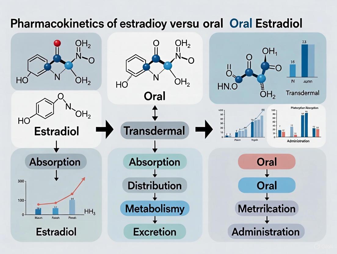
Diagram 1: Metabolic Pathways of Oral vs. Transdermal Estradiol. Oral administration leads to extensive first-pass metabolism in the liver, producing high levels of estrone (E1). Transdermal delivery bypasses this, allowing direct estradiol (E2) absorption and a more physiological E2:E1 ratio.
The Oral Route and First-Pass Metabolism
- Absorption and Bioavailability: Oral estradiol has very low systemic bioavailability, typically ranging from 2% to 10% [1] [7]. Even with micronization to enhance absorption, the drug is subject to profound first-pass metabolism.
- Metabolic Consequences: In the liver, estradiol is rapidly converted via hydroxylation, sulfation, and glucuronidation into metabolites, primarily estrone (E1) and estrone sulfate (E1-S) [1]. This results in a dramatic shift in the E2:E1 ratio. As shown in Table 1, oral administration produces an E2:E1 ratio of approximately 0.10 to 0.16, far from the premenopausal physiologic ratio that approaches unity [1] [10].
- Hepatic Effects: The high concentration of estradiol in the liver stimulates the synthesis of various proteins, including sex hormone-binding globulin (SHBG), thyroid-binding globulin (TBG), and proteins involved in the coagulation cascade. This underlies the increased risk of venous thromboembolism (VTE) and other cardiovascular complications associated with oral estrogen therapy [8].
The Transdermal Route and Hepatic Bypass
- Absorption and Bioavailability: Transdermal systems deliver estradiol directly through the stratum corneum into the systemic circulation. This bypasses intestinal and hepatic first-pass metabolism, leading to a higher effective bioavailability and lower required doses [11] [1].
- Metabolic Advantages: By avoiding first-pass metabolism, transdermal delivery results in a serum E2:E1 ratio that is close to 1, mimicking the natural balance observed in premenopausal women [1] [10]. This more physiological profile is believed to be central to its improved safety.
- Clinical Safety Implications: The avoidance of high hepatic estrogen exposure is linked to a reduced risk of cardiovascular adverse effects, including more favorable lipid profiles, lower systolic and diastolic blood pressure, and a decreased incidence of VTE compared to oral therapy [8].
Quantitative Pharmacokinetic Comparison
The pharmacokinetic differences between administration routes are quantifiable across key parameters, as summarized in the table below.
Table 1: Comparative Pharmacokinetics of Estradiol Administration Routes [8] [1] [7]
| Parameter | Oral | Transdermal Gel | Transdermal Patch | Vaginal Cream |
|---|---|---|---|---|
| Bioavailability | 5% (0.1-12%) | High (Bypasses Liver) | High (Bypasses Liver) | High (Primarily Local) |
| E2:E1 Ratio | 0.10 - 0.16 | ~1.0 | ~1.0 | ~5.0 |
| Half-Life | 13-20 hours | ~37 hours | Varies by patch | N/A |
| Peak E2 (Dose Example) | +50 pg/mL (4 mg) | +45-1310 pg/mL (3 mg) | Relatively Stable | +800 pg/mL (1 mg) |
| Peak E1 (Dose Example) | +500 pg/mL (4 mg) | +31-500 pg/mL (3 mg) | Relatively Stable | +150 pg/mL (1 mg) |
| First-Pass Metabolism | Extensive | Avoided | Avoided | Avoided |
| Impact on Hepatic Synthesis | Significant Increase | Minimal to No Effect | Minimal to No Effect | Minimal |
Analysis of Tabulated Data
- Fluctuation in Levels: Transdermal gels can show significant intersubject variability in absorbed amounts, sometimes with peak-trough fluctuations comparable to oral tablets, while patches aim for more stable delivery, though levels may decline towards the end of the wearing period [10].
- Bioequivalence: Different transdermal formulations are often not bioequivalent; for instance, the bioavailability of a 1.5 mg gel was 109% that of a 50 μg/24h patch, indicating significant formulation differences that preclude simple dose substitution [10].
- Implications for Drug Development: The data underscores that the choice of delivery system (e.g., gel vs. patch) is not merely a matter of patient convenience but a critical determinant of the drug's pharmacokinetic and safety profile. Development strategies must prioritize achieving target E2:E1 ratios and minimizing hepatic exposure.
Experimental Protocols for Pharmacokinetic Assessment
Robust experimental methodologies are essential for characterizing the pharmacokinetics of transdermal estradiol formulations. The following protocol is synthesized from key studies.
In Vivo Pharmacokinetic Study Design for Transdermal Formulations
Objective: To compare the absorption, bioavailability, and steady-state pharmacokinetics of transdermal estradiol (gel and patch) against oral estradiol valerate in a postmenopausal or hypogonadal animal model or human cohort.
Materials and Reagents:
- Test Formulations: Transdermal estradiol gel (e.g., 1.5 mg/day), matrix-type transdermal patch (e.g., 50 μg/24h), oral estradiol valerate tablet (e.g., 2 mg).
- Subjects: Postmenopausal women or an appropriate animal model (e.g., ovariectomized Sprague-Dawley rats).
- Key Equipment: LC-MS/MS system for high-sensitivity steroid hormone assay, pharmacokinetic analysis software (e.g., WinNonlin).
Methodology:
- Study Design: A randomized, open-label, crossover design is optimal, with adequate washout periods (e.g., 1-2 weeks) between treatments to eliminate carryover effects [10].
- Dosing and Sampling:
- Administer a single dose followed by repeated dosing to steady state (typically 14-18 days).
- Collect serial venous blood samples at predetermined time points. For a single dose: pre-dose, 0.5, 1, 2, 4, 5, 6, 8, 12, 18, 24, and 36/48/72 hours post-dose. At steady-state, sample over a full dosing interval [10].
- Sample Analysis: Quantify serum concentrations of estradiol (E2) and estrone (E1) using a validated, highly specific method like radioimmunoassay (RIA) or preferably LC-MS/MS [10].
- Data Analysis:
- Calculate pharmacokinetic parameters: AUC (Area Under the Curve, total exposure), C~max~ (maximum concentration), T~max~ (time to C~max~), and t~1/2~ (elimination half-life).
- Determine the E2:E1 ratio at various time points and as an AUC ratio.
- Calculate relative bioavailability (F~rel~) of transdermal formulations using the oral route as a reference:
F_rel = (AUC_transdermal / Dose_transdermal) / (AUC_oral / Dose_oral)[10].
This workflow is depicted in the following diagram.
Diagram 2: Experimental PK Study Workflow. The key phases of a comparative pharmacokinetic study, from subject allocation to final calculation of relative bioavailability.
Ex Vivo Permeation Studies
Objective: To evaluate the release characteristics and skin permeation of estradiol from a newly developed transdermal patch.
Materials and Reagents:
- Franz Diffusion Cell: A standard apparatus for permeation studies.
- Membrane: Excised animal or human skin (e.g., dermatomed porcine ear skin).
- Receptor Medium: Phosphate-buffered saline (PBS) or another physiologically compatible buffer.
- Test Formulation: Fabricated matrix-type transdermal patches.
Methodology:
- Mount the skin membrane between the donor and receptor compartments of the Franz cell.
- Apply the test patch to the skin surface in the donor compartment.
- At predetermined time intervals, withdraw samples from the receptor medium and replace with fresh medium to maintain sink conditions.
- Analyze the samples using HPLC or UV-Vis spectroscopy to determine the cumulative amount of estradiol permeated.
- Calculate key parameters: Steady-state flux (J~ss~), Permeability coefficient (K~p~), and Lag time [12].
The Scientist's Toolkit: Essential Research Reagents and Materials
Table 2: Key Reagents and Materials for Transdermal Estradiol Research
| Item | Function & Application in Research |
|---|---|
| Micronized Estradiol | The active pharmaceutical ingredient (API) with reduced particle size to enhance dissolution and absorption for both oral and some topical formulations [1]. |
| Polymer Matrices (MC, SA, CS) | Polymers like Methylcellulose (MC), Sodium Alginate (SA), and Chitosan (CS) are used to fabricate the monolithic film of matrix-type transdermal patches, controlling drug release [12]. |
| Penetration Enhancers | Chemicals (e.g., certain alcohols, glycols) incorporated into formulations to temporarily alter the stratum corneum's barrier properties and improve drug flux. |
| Franz Diffusion Cell | The standard apparatus for ex vivo permeation studies, used to evaluate the release and penetration kinetics of a drug formulation through a biological membrane [12]. |
| LC-MS/MS | Liquid Chromatography with Tandem Mass Spectrometry is the gold-standard analytical technique for the specific, sensitive, and simultaneous quantification of estradiol, estrone, and their metabolites in biological samples. |
| Validated Hormone Assay | Immunoassays (e.g., RIA) for high-throughput clinical testing of serum hormone levels, though with potential for cross-reactivity compared to LC-MS/MS [10]. |
The evidence is conclusive: transdermal estradiol delivery offers a pharmacokinetically superior and safer profile than oral administration by bypassing hepatic first-pass metabolism. The resultant physiological E2:E1 ratio is a key biomarker for effective and tolerable feminizing hormone therapy and menopausal replacement. Future research should prioritize the development of next-generation transdermal systems with enhanced permeation and more consistent delivery profiles. Long-term, prospective studies are still needed to fully quantify the reduction in cardiovascular and thrombotic risk in transgender and gender-diverse populations using transdermal GAHT. For drug development professionals, the focus must remain on designing formulations that optimize pharmacokinetics to achieve desired clinical outcomes with the highest possible safety margin.
The pharmacokinetic profile of estradiol, a primary endogenous estrogen, is profoundly influenced by its route of administration. Understanding these differences is critical for drug development, therapeutic efficacy, and safety profiling. This whitepaper examines the core pharmacokinetic parameters—systemic bioavailability and protein binding—that differentiate oral and transdermal estradiol administration, contextualized within the broader framework of estrogen pharmacology. Research demonstrates that administration route significantly impacts first-pass metabolism, metabolic ratios, and ultimately, the therapeutic window of estradiol formulations [1] [13]. These variations are not merely pharmacokinetic curiosities; they translate directly to clinically significant differences in side effect profiles, particularly regarding thrombotic risk and hepatic impact [13] [4]. The following sections provide a detailed analysis of these parameters, supported by quantitative data and experimental methodologies relevant to pharmaceutical research and development.
Comparative Pharmacokinetics of Administration Routes
Fundamental Pharmacokinetic Differences
The route of administration dictates the pathway estradiol takes to enter systemic circulation, fundamentally altering its pharmacokinetic fate.
Oral Administration: When administered orally, estradiol undergoes extensive first-pass metabolism in the gut and liver [1] [13]. This process results in a significantly reduced systemic bioavailability, reported to be approximately 2-10% [7] [2]. The majority of an oral dose is metabolized into estrone (E1) and its conjugates (estrone sulfate), leading to an unfavorable estrone-to-estradiol ratio (E1:E2) that can reach 5:1 or higher [1] [4]. This means circulating levels of the less potent estrone exceed those of the biologically active estradiol, creating a non-physiological hormone profile. The terminal elimination half-life for oral estradiol is typically reported to be between 13 to 20 hours [1].
Transdermal Administration: Transdermal delivery (patches, gels) bypasses first-pass metabolism [1] [13]. Estradiol is absorbed directly into the systemic circulation, resulting in a much higher effective bioavailability—approximately 20 times higher than the oral route according to some analyses [2]. This route produces a physiological E2:E1 ratio close to 1:1, mirroring the natural state in premenopausal women [4] [10]. The half-life for transdermal estradiol is longer; for instance, transdermal gel has a reported half-life of about 37 hours [1].
Table 1: Core Pharmacokinetic Parameter Comparison by Route
| Parameter | Oral Estradiol | Transdermal Estradiol |
|---|---|---|
| Systemic Bioavailability | 2–10% [7] [2] | ~20x higher than oral [2] |
| First-Pass Metabolism | Extensive [1] [13] | Bypassed [1] [13] |
| E2:E1 Ratio | ~0.1–0.16 (E1 > E2) [1] | ~0.4–1.0 (Near 1:1) [1] [10] |
| Elimination Half-Life | 13–20 hours [1] | ~37 hours (gel) [1] |
| Key Metabolic Consequence | High estrone sulfate pool; pronounced hepatic effects [4] | Physiological metabolite profile; minimized hepatic exposure [13] [4] |
Protein Binding and Distribution
Regardless of the administration route, estradiol is highly protein-bound in the circulation. Approximately 98% of circulating estradiol is bound to plasma proteins [1] [2]. This binding is divided between:
- Albumin (~60%): Low-affinity, high-capacity binding.
- Sex Hormone-Binding Globulin (SHBG) (~38%): High-affinity, low-capacity binding [1] [14]. Only the free fraction (~2%) is considered biologically active and able to diffuse into tissues and bind to estrogen receptors [1]. It is important to note that oral estradiol has been shown to increase the hepatic synthesis of SHBG, which can alter the free fraction over time—an effect that is minimized with transdermal administration [13] [4].
The differences in bioavailability and metabolism manifest as distinct serum concentration profiles for estradiol and its metabolites. The following table consolidates key pharmacokinetic data from various study formulations.
Table 2: Detailed Pharmacokinetic Parameters by Formulation and Dose
| Route | Formulation & Dose | ΔE2 (pg/mL) | ΔE1 (pg/mL) | E2:E1 Ratio | Tmax | Key Findings |
|---|---|---|---|---|---|---|
| Oral | 2 mg Micronized Tablet | +40 | +250 | 0.16 | 3–6 h [1] | High E1 exposure, significant first-pass. |
| Sublingual | 0.5 mg Tablet | +250 | +85 | ~3.0 | ~1 h [1] | High E2, rapid absorption, favorable ratio. |
| Transdermal | 50 μg/day Patch | Steady-state levels ~40-50 pg/mL [14] | Similar to E2 [10] | ~1.0 | Varies by patch type [14] | Stable levels, physiological ratio. |
| Transdermal Gel | 1.5 mg daily | +45–279 (steady-state) | +31–230 | ~1.0 | 4–5 h post-application [10] | Bioavailability 109% vs. patch [10]. |
| Vaginal Cream | 0.5 mg dose | +830 | +150 | 5.0 | 3 h [1] | Very high local absorption, favorable ratio. |
| IM Injection | 5 mg Estradiol Valerate | 667 (Cmax) | 324 (Cmax) | 2.1 | 2.2–2.7 days [1] | Very long half-life, depot effect. |
Experimental Protocols and Methodologies
To ensure the reliability and reproducibility of pharmacokinetic data, rigorous standardized protocols are employed in clinical trials. The following details a typical study design for evaluating estradiol formulations.
Standard Bioequivalence Study Design
A recent Phase I study provides a robust model for a comparative pharmacokinetic trial [15].
- Study Population: The study enrolled healthy postmenopausal female volunteers (aged 45-65). Key inclusion criteria were natural menopause for >12 months, endometrial thickness <5 mm, follicle-stimulating hormone (FSH) >40 IU/L, and estradiol <110 pmol/L. Subjects were required to have a Body Mass Index (BMI) between 18–28 kg/m² [15].
- Study Design: A randomized, open-label, single-dose, two-period crossover design was used. Participants were randomly assigned to a treatment sequence (either Test-Reference or Reference-Test) with a 7-day washout period between doses to prevent carryover effects [15].
- Dosing and Conditions: The study was conducted under both fasting and fed conditions. In the fasting arm, subjects received a 1 mg estradiol valerate tablet after a minimum 10-hour overnight fast. In the fed arm, the same dose was administered within 30 minutes after consuming a high-fat, high-calorie breakfast [15].
- Blood Sampling: Intensive blood sampling was performed to characterize the full concentration-time profile. In the fasting study, 24 samples were collected from pre-dose up to 72 hours post-dose. The fed study included 25 samples over the same period, with more frequent early time points to capture potential differences in absorption kinetics [15].
- Bioanalytical Methods: Plasma concentrations of total estrone, estradiol, and unconjugated estrone were quantified using a validated liquid chromatography-tandem mass spectrometry (LC-MS/MS) method, which is considered the gold standard for sensitivity and specificity in hormone assays [15].
- Statistical Analysis: Bioequivalence was determined by calculating the 90% confidence intervals for the geometric mean ratios of C~max~, AUC~0-t~, and AUC~0-∞~. The standard bioequivalence range of 80–125% was used [15].
Transdermal Patch Comparison Protocol
Another study compared the bioavailability of two matrix transdermal delivery systems, Menorest and Climara [14].
- Design: A single-center, open, randomized, comparative cross-over study in 20 healthy postmenopausal women.
- Treatment: Two 14-day treatment periods separated by a 4-week washout. Patches were applied according to manufacturer instructions (Menorest: 3-4 day wear; Climara: 7-day wear), both with a nominal delivery rate of 50 μg/24 h.
- Sampling: Plasma estradiol levels were monitored during the second week of each treatment to assess steady-state pharmacokinetics. Parameters included AUC, C~max~, C~min~, C~average~, and fluctuation index [14].
Diagram 1: Estradiol Absorption and Distribution Pathways
Metabolic Pathways and Hepatic Handling
The divergent effects of oral and transdermal estradiol are largely attributable to differences in hepatic handling and metabolic pathway saturation.
Metabolic Fate of Estradiol
Estradiol is metabolized in the liver primarily through hydroxylation, sulfation, and glucuronidation [1] [13]. The major metabolites include estrone (E1), estrone sulfate (E1S), estrone glucuronide, and estradiol glucuronide [1]. Estrone sulfate, in particular, serves as a significant circulating reservoir for the formation of active estrogens. Oral administration leads to saturation of these metabolic pathways in the liver due to the high concentration of the drug delivered via the portal vein, which is the root cause of its pronounced hepatic effects [4].
Diagram 2: Estradiol Metabolism and Elimination Pathways
The Scientist's Toolkit: Research Reagent Solutions
Table 3: Essential Reagents and Materials for Estradiol Pharmacokinetic Research
| Reagent / Material | Function & Application in Research |
|---|---|
| LC-MS/MS Systems | Gold-standard for quantifying plasma concentrations of estradiol, estrone, and their metabolites with high sensitivity and specificity [15]. |
| Validated Bioanalytical Assays | Essential for obtaining reliable pharmacokinetic parameters (AUC, C~max~, T~max~, t~1/2~); requires pre-study validation for precision and accuracy [15]. |
| Matrix Transdermal Delivery Systems | Research-grade patches and gels for studying controlled dermal absorption and bioavailability without reservoir systems [14]. |
| Micronized Estradiol Formulations | Critical for oral administration studies; micronization increases surface area and improves dissolution and absorption [1]. |
| Estradiol Valerate API | The pro-drug form used in many oral and injectable formulations; must be characterized for ester content and purity [15]. |
| Stable Isotope-Labeled Estradiol | Internal standards (e.g., ¹³C or ²H-labeled) used in mass spectrometry to improve quantification accuracy and correct for recovery variations [15]. |
| SHBG and Albumin | Key binding proteins used in in vitro assays to determine free fraction and protein-binding kinetics [1] [2]. |
| CYP450 Enzymes (e.g., CYP3A4) | Hepatic enzymes for in vitro metabolism studies; CYP3A4 is responsible for ~95% of estradiol valerate metabolism [15]. |
The route of administration is a decisive factor in the pharmacokinetics of estradiol, primarily governing its systemic bioavailability and metabolic profile. Oral administration, characterized by low bioavailability and high first-pass metabolism, results in an unphysiological estrone-dominant profile and increased hepatic exposure. In contrast, transdermal delivery bypasses first-pass effects, yields a physiological estradiol-to-estrone ratio, and minimizes hepatic stimulation. These pharmacokinetic distinctions underpin the differentiated clinical safety profiles, particularly concerning thrombotic risk. For researchers and drug development professionals, these insights are paramount for designing novel formulations, optimizing therapeutic efficacy, and mitigating adverse effects in future hormone therapy products.
This whitepaper provides a comprehensive analysis of the distinct metabolic pathways and elimination characteristics of oral versus transdermal estradiol formulations. Through systematic evaluation of pharmacokinetic data, we demonstrate that the oral administration route is characterized by extensive first-pass metabolism, low bioavailability (2-10%), and non-physiological estrogen ratios, while transdermal delivery bypasses hepatic first-pass effects, provides more consistent serum levels, and achieves estradiol-to-estrone ratios approximating natural physiology. These differences have profound implications for drug development, therapeutic efficacy, and safety profiling in hormone replacement therapy and other clinical applications.
Estradiol (17β-estradiol) is the primary endogenous estrogen in humans, exhibiting complex pharmacokinetics that vary dramatically with route of administration. The fundamental distinction between oral and transdermal delivery systems lies in their interaction with first-pass metabolism - a critical determinant of bioavailability, metabolic profile, and eventual pharmacological effects [1] [7]. Oral estradiol undergoes extensive hepatic and intestinal metabolism before reaching systemic circulation, resulting in significantly altered estrogen profiles characterized by disproportionately elevated estrone levels [16] [17]. In contrast, transdermal delivery facilitates direct absorption into systemic circulation via the stratum corneum, bypassing initial hepatic metabolism and providing more consistent estradiol levels with ratios of estradiol to estrone that approximate unity [16] [11]. This technical analysis examines the elimination characteristics of both routes, with implications for research and drug development.
Metabolic Pathways of Estradiol
Primary Metabolic Routes
Estradiol undergoes complex metabolism primarily via hydroxylation, sulfation, and glucuronidation pathways [1]. The liver serves as the principal site of metabolism for orally administered estradiol, while transdermally delivered estradiol undergoes more distributed metabolic processing. The metabolic fate of estradiol involves conversion to various metabolites with differing estrogenic activities:
- Estrone (E1): The primary oxidative metabolite formed via 17β-hydroxysteroid dehydrogenase
- Estrone Sulfate (E1S): A major circulating storage form with minimal estrogenic activity
- Estriol (E3): A terminal metabolite formed through 16α-hydroxylation
- Catechol Estrogens: Formed via 2- or 4-hydroxylation, with potential for further methylation
Route-Dependent Metabolic Variation
The administration route significantly influences the metabolic fate of estradiol. Oral administration results in pronounced first-pass metabolism, with up to 90% of absorbed estradiol converted to estrone and its conjugates during initial liver passage [7]. This creates a non-physiological estrogen profile characterized by estrone predominance. Transdermal administration bypasses this initial metabolic processing, resulting in a metabolic profile closer to premenopausal physiology with balanced estradiol-to-estrone ratios [16] [17].
Figure 1: Metabolic Pathway Divergence Between Oral and Transdermal Estradiol Administration
Elimination Characteristics and Half-Life Profiles
Route-Specific Elimination Parameters
The elimination of estradiol exhibits significant variation between administration routes, influenced by absorption characteristics, protein binding, and metabolic clearance. The following table summarizes key pharmacokinetic parameters across delivery methods:
Table 1: Comparative Pharmacokinetic Parameters of Estradiol by Route of Administration
| Route | Bioavailability | Elimination Half-Life | Tmax | Protein Binding | E2:E1 Ratio |
|---|---|---|---|---|---|
| Oral | 2-10% [1] [7] | 13-20 hours [1] | 6-8 hours [18] [19] | ~98% (SHBG 38%, Albumin 60%) [1] | 0.10-0.16 [16] [17] |
| Transdermal (Gel) | ~20x oral [2] | ~37 hours [1] | 12-20 hours [1] | ~98% [1] | ~1.0 [16] [17] |
| Transdermal (Patch) | Avoids first-pass [11] | Similar to gel [20] | Varies by system [21] | ~98% [1] | ~1.0 [16] |
| Sublingual | ~10% [1] | 8-18 hours [1] | ~1 hour [19] | ~98% [1] | ~1.1 [19] |
Half-Life Variations and Clinical Implications
The elimination half-life of estradiol varies substantially by route, reflecting differences in absorption kinetics and release mechanisms. Transdermal gels demonstrate an extended half-life of approximately 37 hours compared to 13-20 hours for oral administration [1]. This prolonged half-life contributes to more stable serum concentrations and reduced peak-trough fluctuations. Transdermal patches maintain consistent delivery over multiple days, though specific patch technologies exhibit different release profiles. Matrix-type patches may show declining delivery after 12-30 hours in some systems, while reservoir-type patches maintain more consistent release [21]. These variations in elimination kinetics directly impact dosing frequency requirements and steady-state concentration achievement.
Experimental Protocols for Pharmacokinetic Assessment
Comparative Bioavailability Study Design
To evaluate the pharmacokinetic differences between oral and transdermal estradiol, researchers have employed standardized clinical trial methodologies:
Figure 2: Standardized Protocol for Comparative Estradiol Pharmacokinetic Studies
Key Methodological Considerations:
Population Selection: Studies typically enroll 24-32 healthy postmenopausal women with confirmed hypoestrogenic status (estradiol <20 pg/mL, FSH >50 IU/L) to establish consistent baseline conditions [20] [18].
Crossover Design: Randomized, open-label, multiple-crossover designs minimize interindividual variability and allow direct comparison of formulations in the same subjects [16] [20].
Dosing Protocols: Common comparative doses include oral micronized estradiol (2mg) or estradiol valerate (equivalent to 2mg estradiol) versus transdermal systems delivering 50μg/24 hours [16] [20].
Sample Collection: Blood samples are typically collected at baseline and at multiple timepoints post-administration (e.g., 1, 2, 3, 4, 6, 8, 12, 24, 48, 72, 96 hours) to fully characterize absorption and elimination profiles [18] [19].
Analytical Methods: Modern studies employ liquid chromatography with tandem mass spectrometry (LC-MS/MS) for specific estradiol quantification, while earlier studies used specific direct radioimmunoassays (RIA) with extraction and chromatographic separation [21] [19].
Steady-State and Dose Proportionality Assessment
For comprehensive elimination characterization, steady-state evaluations are essential:
Extended Application Protocol:
- Transdermal systems are applied continuously for 12-18 days with pharmacokinetic assessment during the final application period (days 15-18) [20] [21]
- Serial blood sampling over the entire application period (typically 3-4 days per patch) captures peak, trough, and fluctuation parameters
- Multiple crossover periods with different dosage strengths (0.025, 0.05, 0.1 mg/day) establish dose proportionality [16]
Quantitative Data Comparison: Oral vs. Transdermal Estradiol
Systemic Exposure and Fluctuation Parameters
The following table compiles critical quantitative parameters from comparative pharmacokinetic studies:
Table 2: Quantitative Pharmacokinetic Data from Comparative Studies
| Parameter | Oral Estradiol (2mg) | Transdermal Patch (50μg/day) | Transdermal Gel (1mg/day) | Sublingual (1mg) |
|---|---|---|---|---|
| Baseline E2 (pg/mL) | <15 [1] | <15 [1] | <15 [1] | <15 [1] |
| Peak E2 (Cmax, pg/mL) | 35-50 [19] | 42-46 [21] | 45-279 [1] | 144-450 [1] [19] |
| Time to Peak (Tmax) | 6-8 hours [18] | 12 hours [21] | 12-20 hours [1] | ~1 hour [19] |
| AUC (h·pg/mL) | 1,015 (E2, 48h) [18] | Comparable to gel [20] | Similar to patch [20] | 1.8x oral (0-8h) [19] |
| Estrone Cmax (pg/mL) | 174-250 [16] [18] | ~42 [16] | 31-230 [1] | 24-160 [1] [19] |
| Fluctuation Index | High [20] | Moderate [21] | Low [20] | Very High [19] |
Metabolic Ratios and Accumulation Potential
A critical differentiator between administration routes is their impact on estrogen metabolite profiles:
- Estradiol:Estrone Ratio: Oral administration produces ratios of 0.10-0.16, reflecting estrone predominance, while transdermal delivery achieves ratios approximating 1.0 [16] [17]
- Metabolite Accumulation: Oral estrogen administration shows signs of metabolite retention after only three doses, while transdermal delivery demonstrates no accumulation of estradiol or its conjugates over 3 weeks of continuous application [16]
- Interindividual Variability: Both transdermal gel and patch systems demonstrate considerable variability in absorption, with coefficients of variation around 30-39% for AUC and Cmax [20]
The Scientist's Toolkit: Essential Research Reagents and Materials
Table 3: Key Research Reagents for Estradiol Pharmacokinetic Studies
| Reagent/Material | Specifications | Research Application |
|---|---|---|
| LC-MS/MS System | High-sensitivity platform with lower limit of quantification <5 pg/mL | Gold standard for specific estradiol quantification in serum/plasma [19] |
| Specific RIA Kits | Antibodies with <1% cross-reactivity to estrone and estriol | Validated alternative when LC-MS/MS unavailable; requires extraction and separation [21] |
| Estradiol Standards | Certified reference materials for E2, E1, E1S | Calibration curve preparation and method validation |
| Transdermal Systems | Matrix-type patches (50μg/24h), Reservoir patches, Gels (0.06%) | Comparative delivery system evaluation [20] [21] |
| Oral Formulations | Micronized estradiol (1-2mg), Estradiol valerate (1-2mg) | Reference comparator for bioavailability studies [16] [18] |
| SPE Cartridges | C18 or mixed-phase for steroid extraction | Sample preparation and clean-up prior to analysis |
| Chromatographic Columns | Reverse-phase C18 with 2.1-4.6mm internal diameter | Analytic separation for LC-MS/MS and HPLC methods |
The elimination characteristics of estradiol are fundamentally governed by administration route, with oral delivery subject to extensive first-pass metabolism and transdermal systems providing more physiologic profiles. These differences extend beyond pharmacokinetics to influence therapeutic efficacy, safety profiles, and individual variability. For drug development professionals, these findings highlight the importance of:
- Route-Specific Dosing Strategies: Development programs must account for profound bioavailability differences between routes
- Therapeutic Individualization: Considerable interindividual variability in transdermal absorption necessitates flexible dosing options
- Metabolic Impact Assessment: First-pass hepatic effects of oral estradiol produce distinct impacts on hepatic protein synthesis and metabolic parameters
- Formulation Optimization: Patch technologies demonstrate varying release profiles requiring careful pharmacokinetic characterization
Future research directions should include long-term comparative studies on bone density preservation, cardiovascular risk reduction, and comprehensive metabolic impact to fully elucidate the clinical implications of these pharmacokinetic differences.
From Bench to Bedside: Methodologies in Delivery System Design and Evaluation
In Vitro Permeation Test (IVPT) Validation for Transdermal Formulation Assessment
In Vitro Permeation Testing (IVPT) has emerged as a critical methodology in the development and evaluation of transdermal drug delivery systems, providing essential data on drug permeation kinetics and formulation performance. Using Franz diffusion cells and human skin, IVPT measures transdermal permeated amounts, flux rates, and layer distribution to predict in vivo performance [22]. For estradiol formulations used in menopausal hormone therapy, IVPT offers particularly valuable insights by enabling direct comparison of permeation profiles between transdermal and oral delivery routes, thus supporting the broader pharmacokinetic thesis that transdermal administration avoids hepatic first-pass metabolism and provides more stable plasma levels [23] [14].
Regulatory agencies worldwide now recognize IVPT as a vital tool for demonstrating bioequivalence for generic topical products and optimizing new formulations [24] [25]. The European Medicines Agency (EMA) has recently formalized a stepwise approach in its 2024 guideline that can exempt certain topical products from clinical studies when sufficient IVPT data is available [24]. Similarly, the U.S. Food and Drug Administration (FDA) provides detailed guidance on IVPT implementation, with upcoming workshops in April 2025 focusing on hands-on training for regulatory compliance [26]. This whitepaper provides a comprehensive technical guide to IVPT validation, with specific application to transdermal estradiol formulations within the context of comparative pharmacokinetic research.
Critical IVPT Validation Parameters
Core Validation Components
A robust IVPT validation protocol must demonstrate that the method consistently produces reliable and meaningful permeation data. Key validation parameters with their acceptance criteria are summarized in Table 1 below.
Table 1: Essential IVPT Validation Parameters and Acceptance Criteria
| Validation Parameter | Experimental Approach | Acceptance Criteria | Reference Application |
|---|---|---|---|
| Apparatus Qualification | Capacity, orifice diameter, temperature, stir speed measurements [27] | Capacity: 12 ± 0.6 mL; Orifice: 15 ± 0.75 mm; Skin surface: 32 ± 1°C; Stir speed: 600 ± 60 rpm [27] | Ensures Franz cell system operates within specified parameters before IVPT initiation |
| Sink Condition Maintenance | Solubility testing of drug in receptor medium [27] | Drug solubility ≥10× highest measured concentration in samples [27] | Confirmed estradiol solubility of 217.54 µg/mL vs. ~1 µg/mL sample concentration |
| Analytical Method Stability | Bench-top (24h RT) and long-term (14d -80°C) stability testing [27] | Mean concentration deviation ≤±15% from nominal [27] | Estradiol demonstrated stability under both conditions (95.45-101.57% of nominal) |
| Dilution Integrity | Accuracy/precision of 5× to 300× dilutions with blank matrix [27] | Accuracy and precision within ±20% nominal concentrations [27] | Acceptable up to 200× dilution (82.76-103.50% accuracy); 300× dilution rejected |
| Method Sensitivity | Flux measurement with varying drug doses (3, 10, 30 mg/cm²) [27] | Significant flux increase with increasing dose (p<0.05) [27] | Successfully distinguished different estradiol formulation doses |
| Method Selectivity | Permeation comparison between formulations with known delivery differences [27] | Significant discrimination in flux profiles (p<0.05) [27] | Differentiated commercial vs. compounded estradiol formulations |
Mass Balance Considerations
Complete mass balance determination is essential for IVPT validity, particularly under the EMA guideline which requires reporting of both skin deposition and permeated amounts [25]. Mass balance involves quantifying the active pharmaceutical ingredient (API) in all compartments: receptor fluid, skin layers (stratum corneum, viable epidermis, dermis), application site residue, and any system losses. Recovery rates of 100%±15% are generally acceptable, with deviations potentially indicating API instability, inadequate extraction methods, or non-specific binding [25]. For estradiol, which exhibits significant skin retention in some formulations, complete mass balance is particularly crucial for accurate interpretation of permeation data [27].
Experimental Methodology
Skin Preparation and Integrity Assessment
Human skin, typically dermatomed to 400-500 μm thickness from abdominal or breast reduction surgeries, represents the most relevant membrane for IVPT [25]. Skin should be used within 24 hours if refrigerated or within months if properly frozen at -20°C or below [23]. Membrane integrity verification is critical and can be assessed through:
- Electrical resistance measurement: Values >15 kΩ typically indicate intact barrier function [22]
- Transepidermal water loss (TEWL): Established baseline values for human skin
- Reference compound permeation: Using compounds with known permeability
Table 2: Research Reagent Solutions for IVPT Studies
| Reagent/Category | Specific Examples | Function/Application | Technical Considerations |
|---|---|---|---|
| Skin Membranes | Human cadaver skin (dermatomed to 400-500μm) [25] | Biologically relevant permeation barrier | Should be integrity tested; source (abdomen, thigh, breast) affects permeability [23] |
| Artificial Membranes | Synthetic membranes for IVRT [22] | Quality control and formulation screening | Selected based on API physicochemical properties |
| Receptor Media | PBS with preservatives, PEG400, surfactants [27] | Maintains sink conditions and API stability | Must not affect skin integrity; estradiol solubility >10× sample concentration [27] |
| Analytical Instruments | Franz diffusion cells [25], UPLC [27], UV-Vis Spectrophotometer [28] | Permeation measurement and sample analysis | Apparatus qualification required before studies [27] |
| Reference Standards | Color reference solutions (for visual examination) [28] | Product stability assessment | Quantitative spectrophotometric methods preferred over subjective visual examination [28] |
| Heating Apparatus | Heating lamps, temperature controllers [29] | Simulating elevated temperature conditions | Critical for evaluating heat effects on TDS; skin surface temperature control vital [29] |
IVPT Experimental Protocol
A standardized IVPT protocol for estradiol formulations includes these critical steps:
- Apparatus Setup: Franz diffusion cells with maintained skin surface temperature of 32±1°C [27] and receptor medium stirring at 600±60 rpm [27]
- Membrane Mounting: Skin sections carefully positioned with stratum corneum facing donor compartment
- Integrity Verification: Pre-study barrier function assessment
- Formulation Application: Uniform application of test formulations (commercial EstroGel vs. compounded formulations) at doses ranging from 3-30 mg/cm² [27]
- Sample Collection: Automated or manual sampling from receptor compartment at predetermined intervals (e.g., 0.5, 1, 2, 4, 6, 8, 12, 16, 24h) with receptor medium replacement
- Sample Analysis: Ultra Performance Liquid Chromatography (UPLC) or validated analytical method for quantifying estradiol concentrations [27]
- Termination Phase: At study end (typically 24-72h), residual drug quantification from skin layers (tape stripping for stratum corneum, extraction from viable skin) and donor compartment
Diagram 1: IVPT Experimental Workflow (IVPT Method Validation)
Evaluating Environmental Factors
Temperature significantly impacts transdermal permeation, with elevated conditions potentially altering drug delivery. A systematic approach to heat effect evaluation includes:
- Temperature Control: Maintaining skin surface at 32±1°C for baseline and 42°C for elevated temperature conditions [29]
- Heating Protocols: Various regimens including sustained heat (0-72h), short exposures (1h at different timepoints), and multiple applications [29]
- Activation Energy Determination: Using Arrhenius relationship to predict temperature effects on permeation
For fentanyl TDS, increasing temperature from 32°C to 42°C resulted in approximately 2-fold flux increase, consistent with the activation energy determined for the molecule [29]. Similar principles apply to estradiol formulations, though the magnitude of enhancement varies with formulation characteristics.
Regulatory Framework and Equivalence Assessment
EMA Stepwise Approach
The EMA's 2024 guideline introduces a structured framework for demonstrating therapeutic equivalence of topical products [24]. The stepwise approach, illustrated in Diagram 2, begins with comprehensive qualitative and quantitative composition comparison, followed by physicochemical characterization, in vitro performance testing (IVRT/IVPT), and if needed, clinical studies [24].
Diagram 2: EMA Equivalence Decision Framework (EMA Equivalence Pathway)
IVPT in Bioequivalence Assessment
For transdermal estradiol formulations, IVPT serves as a critical tool in bioequivalence assessment, potentially replacing clinical endpoint studies. The FDA recommends IVPT for supporting demonstrations of bioequivalence for generic topical drug products [26]. Successful IVPT bioequivalence studies require:
- Adequate Study Power: Typically 18-24 skin sections from a minimum of 6 donors [25]
- Statistical Analysis: Comparison of AUC and Jmax between test and reference formulations using ANOVA
- Acceptance Criteria: 90% confidence intervals for geometric mean ratios falling within 0.80-1.25 for both parameters
Application to Estradiol Transdermal Formulations
Comparative Permeation Profiles
IVPT studies enable direct comparison of estradiol permeation from different formulation types. Research demonstrates significant differences between commercial and compounded formulations:
- Commercial EstroGel: Exhibits rapid absorption peak within 0.5h followed by decline [27]
- Compounded Formulations: Show slower, steady increase with peak flux at 6h and sustained absorption over 16h [27]
- Anhydrous vs. Aqueous Bases: Anhydrous formulations demonstrate higher release rates compared to aqueous bases [27]
These differences in permeation profiles directly impact therapeutic performance, with steady absorption profiles potentially offering clinical advantages for hormone replacement therapy by minimizing peak-trough fluctuations [27].
Correlation with Pharmacokinetic Data
IVPT data for estradiol formulations shows strong correlation with in vivo pharmacokinetic parameters. Studies comparing matrix transdermal delivery systems (Menorest and Climara) demonstrated similar bioavailability despite different application schedules (3-4 days vs. 7 days) [14]. However, the rate of absorption differed significantly, with Menorest showing shorter Tmax, highlighting how IVPT can predict absorption rate differences that may impact therapeutic performance [14].
The permeation of estradiol from gel formulations (Sandrena Gel and Oestrogel) measured in IVPT studies equated to 18.2±3.5% and 17.4±4.8% of applied doses respectively over 24h, demonstrating similar extent of permeation despite different estradiol concentrations (0.1% vs. 0.06%) [23]. These findings support the bioequivalence of the products at their recommended dose levels.
Comprehensive IVPT validation represents a cornerstone in the development and regulatory approval of transdermal estradiol formulations. Through rigorous attention to apparatus qualification, method validation parameters, membrane integrity, and mass balance requirements, researchers can generate robust, predictive data that supports the pharmacokinetic thesis of transdermal delivery superiority over oral administration for estradiol. The evolving regulatory landscape, particularly the EMA's 2024 guideline and FDA's ongoing workshops, emphasizes the growing importance of standardized IVPT methodologies in demonstrating product equivalence and performance. As transdermal hormone therapy continues to evolve, validated IVPT protocols will remain essential tools for optimizing formulation design and ensuring therapeutic efficacy and consistency.
The administration of estradiol via the skin represents a significant advancement in hormone delivery, fundamentally designed to circumvent the first-pass hepatic metabolism associated with oral administration. This avoidance leads to more stable serum hormone levels and a distinct adverse effect profile [8] [30]. Transdermal delivery systems (TDDS) provide a controlled release of drugs through the skin into systemic circulation, offering advantages including improved bioavailability, reduced dosing frequency, and enhanced patient compliance [30] [31]. Available in several forms—such as gels, patches, and increasingly sophisticated compounded bases—these systems hinge on their ability to overcome the formidable barrier function of the skin's outermost layer, the stratum corneum [32] [33]. This whitepaper provides an in-depth technical analysis of current advanced transdermal systems for estradiol, focusing on their design, the experimental methodologies for evaluating their performance, and their position within the broader pharmacokinetic framework of estradiol research.
Fundamental Principles of Transdermal Drug Delivery
Skin Structure and Permeation Pathways
The skin is a multi-layered organ, with the stratum corneum forming the primary rate-limiting barrier for most drug molecules [30] [33]. This outermost layer of the epidermis, approximately 10-15 μm thick, consists of dead keratinocytes (corneocytes) embedded in a lipid-rich matrix, creating a highly cohesive and relatively impermeable membrane [30]. Below the stratum corneum lies the viable epidermis and the dermis, which contains microcirculation that ultimately absorbs the drug for systemic distribution [30].
Drugs can permeate the skin via three primary pathways, visualized in the diagram below:
Skin Permeation Pathways
- The Intercellular Route: This pathway involves the diffusion of molecules, primarily lipophilic drugs, through the continuous lipid matrix surrounding the corneocytes. The interdigitating nature of the cells creates a tortuous pathway, making it challenging for drug permeation [32] [30].
- The Transcellular Route: This route involves the direct passage of molecules, typically hydrophilic drugs, through the keratinocytes themselves. This requires the drug to sequentially partition into and out of the cells' hydrated keratin structure [32] [33].
- The Appendageal Route: This pathway bypasses the stratum corneum via hair follicles and sweat glands. While these appendages occupy a small fraction of the skin's surface area (approximately 0.1%), they provide a crucial shunt for larger molecules, such as proteins, peptides, and nanocarriers [32] [30].
Kinetics of Transdermal Delivery
The permeation of a drug across the skin is governed by Fick's laws of diffusion. The steady-state flux ((J_{ss})), which is the rate of drug permeation per unit area, is described by the equation:
[ J{ss} = \frac{D \cdot K \cdot Cd}{L} ]
Where:
- (D) is the diffusion coefficient of the drug in the skin
- (K) is the partition coefficient of the drug between the skin and the vehicle
- (C_d) is the concentration of the drug in the donor vehicle
- (L) is the overall thickness of the skin membrane [30]
A critical factor for successful TDDS is maintaining a concentration gradient where (Cd) is substantially greater than the drug concentration in the receptor compartment ((Cr)). This ensures a constant rate of drug permeation, as shown in the simplified equation (dQ/dt = P \cdot Cd), where (P) is the permeability coefficient [30]. The cumulative amount of drug permeating ((Q)) a skin surface area ((A)) over time ((t)) after a lag time ((tL)) is given by:
[ Q = P \cdot A \cdot Cd (t - tL) ]
Advanced Transdermal Formulations for Estradiol
Commercially Available Gels and Patches
Commercially available transdermal estradiol products, such as ESTROGel, are absorptive hydroalcoholic gels designed to provide systemic estradiol replacement. These formulations are engineered to deliver a controlled release of the hormone, with one pump of ESTROGel typically delivering 1.25 g of gel containing 0.75 mg of estradiol [34]. Transdermal patches, on the other hand, are typically categorized as either reservoir or drug-in-adhesive (DIA) systems. The DIA type consists of a matrix polymer (e.g., a polyacrylate adhesive like Duro-Tak 387-2510) in which the drug is directly dissolved or suspended. The formulation is cast onto a backing liner, and solvents are evaporated to form a solid adhesive layer [35].
Permeation-Enhancing Compounded Bases
Customized formulations prepared by compounding pharmacies offer an alternative to commercial products. These are often designed to meet individual patient needs and can utilize proprietary aqueous or anhydrous permeation-enhancing bases. A recent in vitro study directly compared the performance of such compounded formulations with ESTROGel [34]. The key finding was a significant difference in the permeation profile: while the commercial gel showed a rapid peak and decline in estradiol flux, the compounded bases facilitated a slower, more steady absorption, which is a desired characteristic for stable hormone replacement therapy [34].
Table 1: In Vitro Permeation Profiles of Commercial vs. Compounded Estradiol Formulations
| Formulation Type | Peak Flux Time (h) | Peak Flux Value (ng/cm²/h) | Absorption Profile Characteristics |
|---|---|---|---|
| ESTROGel (Commercial) | 0.5 | >70 | Rapid increase to peak, followed by a rapid decline within 4 h, then a slower decline up to 16 h. |
| Compounded Anhydrous Base (E2) | ~6 | Data Not Specified | Slow and steady increase to peak flux, with steady absorption maintained within 16 h. |
| Compounded Aqueous Base (E2) | ~6 | Data Not Specified | Slow and steady increase to peak flux, with steady absorption maintained within 16 h. |
Chemical Permeation Enhancement
The use of chemical permeation enhancers is a common strategy to increase the skin's permeability to drugs. Dimethyl sulfoxide (DMSO) is a well-known amphiphilic enhancer that interacts with the lipid bilayers of the stratum corneum, increasing membrane fluidity and, at higher concentrations, forming aqueous pores [35]. Incorporating DMSO into a DIA patch for estradiol has been shown to be technically feasible. When patches were dried at 35-40°C, they retained significant DMSO (≤10 mg/patch), which resulted in a 4-fold increase in estradiol skin permeation ((J{ss} = 4.12 \, \mu g/cm^{-2}h^{-1})) compared to a DMSO-negative control ((J{ss} = 1.1 \pm 0.2 \, \mu g/cm^{-2}h^{-1})) [35]. An additional benefit was that DMSO inhibited estradiol recrystallization in the patch matrix, thereby improving physical stability [35].
Table 2: Permeation Enhancement Strategies and Their Mechanisms
| Technology/Enhancer | Mechanism of Action | Key Advantage | Example Formulation |
|---|---|---|---|
| Chemical Enhancer (DMSO) | Interacts with skin lipids, increasing fluidity and creating pores [35]. | Significant flux increase (4-fold for estradiol); inhibits drug recrystallization [35]. | DIA patch with Duro-Tak and 10 mg DMSO/patch. |
| Microneedles (MNs) | Creates microscopic conduits through the stratum corneum [32] [31]. | Enables delivery of macromolecules; minimally invasive. | Solid, hollow, dissolving, and coated MNs [32]. |
| Iontophoresis | Uses a small electric current to drive charged molecules [32] [33]. | Active control over delivery rate; suitable for ionic drugs. | Integrated patch system with electrodes and power source. |
| Sonophoresis | Uses ultrasound energy to disrupt the lipid structure of the stratum corneum [32] [33]. | Can be applied for a wide range of molecule sizes. | Handheld device used with a standard gel formulation. |
Experimental Protocols for Formulation Evaluation
In Vitro Permeation Test (IVPT)
The IVPT is the cornerstone experimental method for evaluating the percutaneous absorption of transdermal formulations. The following protocol, adapted from a recent study on compounded estradiol formulations, outlines the key steps [34].
IVPT Experimental Workflow
- Skin Membrane Preparation: Use excised human or porcine skin. Porcine ears are a suitable model and can be obtained from an abattoir. The skin should be dermatomed to a consistent thickness (e.g., 500-700 μm) and stored appropriately before use [34] [35].
- IVPT Apparatus Setup: Use Franz-type diffusion cells. The receptor chamber is filled with a suitable receptor medium, such as a phosphate buffer saline (PBS) often with a surfactant like sodium lauryl sulphate (SLS) to maintain sink conditions. The apparatus must be maintained at a constant temperature (e.g., 32°C) to mimic skin surface temperature [34]. The skin membrane is mounted between the donor and receptor compartments.
- Formulation Application: Apply a finite, precise dose of the test formulation (e.g., 3-30 mg/cm²) to the surface of the skin in the donor compartment [34].
- Sample Collection & Analysis: At predetermined time intervals, aliquot samples from the receptor medium and replace with fresh medium to maintain sink conditions. Analyze the drug concentration in the samples using a validated analytical method, such as Ultra-Performance Liquid Chromatography (UPLC) [34].
- Data Calculation: The cumulative amount of drug permeated per unit area (Q) is plotted against time. The steady-state flux ((J{ss})) is determined from the slope of the linear portion of the curve. The lag time ((tL)) is obtained by extrapolating this linear portion to the time axis [30] [34].
Critical IVPT Validation Steps
For reliable results, the IVPT method must be rigorously validated [34]:
- Apparatus Qualification: Ensure the diffusion cells and equipment meet predefined acceptance criteria for integrity and performance.
- Sink Condition Verification: Confirm that the solubility of the drug in the receptor medium is at least 10 times higher than the highest measured concentration to ensure continuous diffusion.
- Method Robustness: Test the method's resilience to minor perturbations, such as variations in skin surface temperature (e.g., 30°C to 34°C). The method should also demonstrate selectivity by distinguishing the permeation profiles of different formulations (e.g., commercial gel vs. compounded bases) [34].
The Scientist's Toolkit: Essential Research Reagents and Materials
Table 3: Key Reagents and Materials for Transdermal Formulation Research
| Reagent/Material | Function/Application | Example & Notes |
|---|---|---|
| Acrylate Polymer Adhesive | Matrix for Drug-in-Adhesive (DIA) patches; controls drug release and ensures skin adhesion. | Duro-Tak 387-2510 (Henkel) – supplied as a viscous gel in ethyl acetate/hexane [35]. |
| Chemical Permeation Enhancer | Increases skin permeability by disrupting stratum corneum lipids. | Dimethyl Sulfoxide (DMSO) – amphiphilic properties; enhances both hydrophilic and hydrophobic drugs [35]. |
| Backing & Release Liners | Provides occlusive backing and protects the adhesive/drug layer before application. | 3M Scotchpak – drug-impermeable and occlusive [35]. |
| Skin Membrane Model | A biophysically relevant model for in vitro permeation testing. | Excised porcine ear skin (from local abattoir) or human skin [34] [35]. |
| Receptor Medium Additive | Maintains sink conditions in the receptor phase by increasing drug solubility. | Sodium Lauryl Sulphate (SLS) in PBS buffer [34] [35]. |
| Analytical Standard | Reference material for quantifying drug concentration and validating analytical methods. | β-Estradiol (E2) USP standard; used for HPLC/UPLC calibration [34] [35]. |
Advanced transdermal systems for estradiol, including gels, patches, and permeation-enhancing compounded bases, offer a sophisticated means of drug delivery with distinct pharmacokinetic benefits over oral administration. The core challenge in their development lies in overcoming the skin's barrier function, a goal achieved through careful formulation design involving chemical enhancers like DMSO and innovative vehicle bases. Robust experimental protocols, particularly the IVPT, are critical for evaluating and validating the performance of these systems. As research continues to advance—driven by innovations in microneedles, nanocarriers, and personalized compounded formulations—transdermal delivery is poised to offer even more effective and tailored therapeutic options for hormone replacement therapy.
Long-acting microneedle (MN) systems represent a transformative advancement in transdermal drug delivery, directly addressing key pharmacokinetic challenges associated with conventional administration routes. These micron-scale devices, typically measuring 0.1–1 mm in length, are designed to painlessly bypass the stratum corneum—the skin's primary barrier—creating microscopic channels for controlled drug delivery into the dermal layers [36] [37]. For estradiol therapy, this platform offers distinct pharmacokinetic advantages over oral administration, including avoidance of first-pass hepatic metabolism, which significantly reduces metabolite formation and liver exposure [36] [38]. The sustained release profiles achievable with MN systems enable maintenance of steady plasma concentrations, minimizing the peak-trough fluctuations characteristic of oral dosing that often lead to adverse effects [31].
The evolution of microneedle technology spans multiple generations, beginning with simple patch-based systems and progressing to the current integrated platforms that combine sensing and delivery capabilities [36]. As a physical enhancement method, MNs greatly expand the spectrum of drugs suitable for transdermal delivery, including macromolecules, peptides, and proteins that typically demonstrate poor oral bioavailability [36]. For hormone therapies like estradiol, this enables precise control over release kinetics that can be tailored to individual patient needs, representing a significant step toward personalized medicine in endocrine disorders [37] [38].
Table 1: Comparative Pharmacokinetic Profiles of Estradiol Delivery Platforms
| Parameter | Oral Estradiol | Conventional Transdermal Patch | Microneedle Systems |
|---|---|---|---|
| Bioavailability | Low (~5%) due to first-pass metabolism | Moderate to high | High (bypasses hepatic metabolism) |
| Tmax (Time to peak concentration) | 4-6 hours | 12-24 hours | Adjustable (2-24 hours based on design) |
| Plasma Half-life | 12-20 hours | Variable | Extended via sustained release |
| Cmax Fluctuations | Significant | Moderate | Minimal with optimized systems |
| Dosing Frequency | Multiple times daily | Once or twice weekly | Weekly to monthly (long-acting) |
| Metabolite Profile | High estrone/estradiol ratio | Favorable | Favorable, similar to transdermal |
Types of Long-Acting Microneedle Systems
System Architectures and Release Mechanisms
Microneedle platforms for sustained release encompass several distinct architectural designs, each with unique drug release mechanisms and kinetic profiles. Dissolving microneedles fabricated from biodegradable polymers such as polyvinylpyrrolidone (PVP), polyvinyl alcohol (PVA), and poly-lactic-coglycolic-acid (PLGA) encapsulate the active pharmaceutical ingredient within the needle matrix itself [36] [38]. Following insertion, the polymer hydrates and dissolves in the skin's interstitial fluid, releasing the drug payload through a combination of diffusion and polymer erosion mechanisms. The release kinetics can be precisely tuned by modifying polymer composition, molecular weight, and cross-linking density [38]. For estradiol delivery, this enables sustained release profiles ranging from days to several weeks, significantly reducing dosing frequency compared to oral formulations.
Core-shell microneedles represent a more sophisticated architecture featuring a protective outer layer surrounding a drug-loaded core [31]. This design provides temporal control over drug release, potentially delaying initial release while maintaining long-term delivery. The shell composition and thickness can be engineered to degrade at specific timepoints, enabling complex release profiles including pulsatile or multi-phasic kinetics [31]. Hydrogel-forming microneedles swell upon contact with interstitial fluid, creating a porous network through which drugs diffuse at rates controlled by hydrogel cross-linking density and composition [38]. These systems are particularly suitable for week-long estradiol delivery, as demonstrated in recent preclinical studies [31].
Coated microneedles provide an alternative approach where the drug is applied as a thin coating on the surface of solid microneedles [38]. While typically used for more rapid delivery, sophisticated multilayer coatings incorporating rate-controlling polymers can extend release over several days. The coating thickness, composition, and application method directly influence both the drug loading capacity and release kinetics [38].
Table 2: Microneedle System Architectures for Sustained Drug Delivery
| MN Type | Materials | Release Mechanism | Release Duration | Drug Loading Capacity |
|---|---|---|---|---|
| Dissolving | PVP, PVA, PLGA, PLA, Hyaluronic Acid | Polymer dissolution and diffusion | 1-7 days | Low to moderate (~10-25% by weight) |
| Core-Shell | PLGA shell, variety of cores | Delayed and sustained release via shell degradation | 1-4 weeks | Moderate |
| Hydrogel-forming | PVA, Poly(methyl vinyl ether-alt-maleic anhydride) | Swelling-controlled diffusion | 1-2 weeks | Moderate to high |
| Coated | Stainless steel, Titanium, Silicon with polymer coatings | Surface dissolution and diffusion | 1-3 days | Low (limited by surface area) |
| Hollow | Silicon, Metals with microfluidic channels | Passive or active controlled infusion | Hours to days (externally controlled) | High |
Advanced Fabrication Techniques
The manufacturing of long-acting microneedle systems employs sophisticated fabrication techniques that enable precise control over microstructure and drug distribution. Micro-molding represents the most established approach, utilizing master templates created through photolithography or laser ablation to produce MN arrays with consistent geometry and sharpness [36]. This method is particularly suitable for dissolving MNs, where drug-polymer solutions are cast into molds and dried under controlled conditions. For more complex architectures like core-shell systems, advanced 3D printing techniques including micro-stereolithography and two-photon polymerization enable layer-by-layer construction with micron-scale resolution [39] [40]. These additive manufacturing approaches facilitate unprecedented control over internal structure, potentially allowing multiple drugs or release modifiers to be incorporated within a single MN array.
Layer-by-layer (LbL) assembly has emerged as a powerful technique for creating nanoscale coatings with precise composition and thickness control [39]. By alternately depositing oppositely charged polymers and drug molecules, LbL assembly can create complex release profiles through controlled degradation of the multilayered structure. This approach is particularly valuable for maintaining the stability of sensitive molecules like peptides or protein-based therapeutics during storage and delivery [39].
Recent innovations in continuous manufacturing address the challenge of scaling up MN production while maintaining quality control. Micro-injection molding and hot embossing enable high-throughput production of polymeric MNs, with integrated analytical technology providing real-time monitoring of critical quality attributes [36]. These advances in fabrication technology are essential for translating long-acting MN systems from laboratory prototypes to commercially viable pharmaceutical products.
Experimental Protocols for MN Development and Evaluation
Formulation Development and Characterization
The development of estradiol-loaded MN systems begins with comprehensive pre-formulation studies to identify compatible polymer carriers and excipients. A standard protocol involves preparing polymer solutions (e.g., 20-30% w/w PVA or PVP) in aqueous or hydroalcoholic solvents, followed by the incorporation of estradiol (0.5-5% w/w) with optional penetration enhancers such as terpenes or fatty acids [41]. The mixtures are homogenized (10,000-15,000 rpm for 5-10 minutes) and degassed under vacuum to remove air bubbles before proceeding to fabrication.
For dissolving MN fabrication using the micro-molding technique, the drug-polymer solution is carefully applied to polydimethylsiloxane (PDMS) molds under negative pressure (approximately 5-10 kPa) to ensure complete cavity filling [36]. The filled molds are centrifuged (3000-5000 ×g for 10-20 minutes) to concentrate the formulation at the needle tips, then dried under controlled conditions (20-25°C, 40-60% RH for 24-48 hours). The resulting MN arrays are demolded and visually inspected for completeness using scanning electron microscopy (SEM) to verify tip sharpness and structural integrity [36].
Critical quality attributes are quantified through standardized assays: drug content uniformity is assessed by dissolving individual MNs (n=10) in suitable solvent with HPLC-UV analysis at 280 nm; mechanical strength is evaluated using compression testing with a texture analyzer to determine failure force; and insertion capability is verified in porcine ear skin using a standardized applicator (5-20 N force) with trypan blue staining to visualize microconduits [36] [38]. For estradiol-specific formulations, skin permeation studies are conducted using Franz diffusion cells with dermatomed human or porcine skin, collecting receptor samples at predetermined intervals over 24-168 hours for HPLC analysis to establish release kinetics [41].
In Vivo Pharmacokinetic Evaluation
Robust pharmacokinetic assessment of long-acting estradiol MN systems requires carefully controlled animal studies. A standardized protocol involves administering estradiol MNs to ovariectomized rodent models (Sprague-Dawley rats, 200-250 g) to simulate postmenopausal conditions [42]. The MN patches (containing 50-200 μg estradiol) are applied to the dorsal region after gentle skin cleaning, with secure attachment using medical-grade adhesive tapes. Control groups receive conventional transdermal patches (e.g., 50 μg/day) or oral suspensions (1 mg/kg) for comparative pharmacokinetics.
Blood sampling (200-300 μL) is performed via jugular vein cannulation at predetermined intervals: 0, 1, 2, 4, 8, 12, 24, 48, 72, 96, 120, 144, and 168 hours post-administration [42]. Plasma is separated by centrifugation (5000 ×g for 10 minutes at 4°C) and stored at -80°C until analysis. Estradiol concentrations are quantified using validated LC-MS/MS methods with a lower limit of quantification of 1-5 pg/mL, enabling precise characterization of the absorption and elimination phases [42].
Pharmacokinetic parameters are calculated using non-compartmental analysis: area under the curve (AUC0-t) via the linear trapezoidal method, maximum concentration (Cmax), time to Cmax (Tmax), and elimination half-life (t1/2) [42]. For comparative assessment relative to oral administration, the absolute bioavailability (F) is calculated using dose-normalized AUC ratios. Additionally, the fluctuation index (FI) is determined as (Cmax - Cmin)/Cavg to quantify steady-state stability, with lower values indicating smoother plasma profiles characteristic of ideal sustained-release systems [42].
Integrated Biosensing and Closed-Loop Systems
The convergence of microneedle technology with biosensing platforms represents a groundbreaking advancement in therapeutic monitoring, enabling real-time pharmacokinetic assessment and personalized dosing. Recent innovations include microneedle-based continuous biomarker monitoring (MCBM) systems that integrate biosensors within MN arrays for simultaneous measurement of drug concentrations and relevant biomarkers in dermal interstitial fluid (ISF) [39]. These systems utilize electrochemical sensing principles, with working electrodes functionalized with specific enzymes or antibodies to detect target analytes. For diabetes management, researchers have demonstrated a dual-sensor MN system capable of monitoring both glucose and metformin concentrations continuously, providing rich pharmacokinetic-pharmacodynamic data to optimize dosing regimens [39].
Interstitial fluid serves as an excellent medium for therapeutic drug monitoring, as it contains virtually all the biomarkers found in blood but in a cleaner matrix that requires minimal processing [43]. The development of self-powered MN patches has further advanced continuous monitoring capabilities by incorporating osmotic pressure-driven microfluidic systems that passively wick ISF into collection reservoirs for subsequent analysis [43]. These fully passive systems can collect biomarker samples over periods ranging from 15 minutes to 24 hours without external power sources, enabling comprehensive pharmacokinetic profiling [43].
The integration of MN-based biosensing with controlled-release systems creates opportunities for closed-loop therapeutic platforms that automatically adjust drug delivery based on real-time physiological measurements. While still primarily in research stages, these systems employ feedback algorithms that process sensor data to modulate drug release from reservoirs through mechanisms including thermally activated membranes, electro-responsive polymers, or miniature piezoelectric pumps [39]. For estradiol therapy, such systems could potentially monitor multiple hormones and metabolites simultaneously, enabling precise hormonal balance maintenance with minimal clinician intervention.
The Scientist's Toolkit: Essential Research Reagents and Materials
Table 3: Key Research Reagents and Materials for MN Development
| Category | Specific Examples | Function/Application | Notes |
|---|---|---|---|
| Polymers | PVP, PVA, PLGA, PLA, Hyaluronic acid, Carboxymethyl cellulose | Matrix for dissolving and hydrogel MNs | Molecular weight and viscosity critical for mechanical properties |
| Silicon & Metals | Silicon wafers, Stainless steel, Titanium | Substrates for solid, hollow, and coated MNs | Precision etching and micromachining required |
| Cross-linkers | Glutaraldehyde, Genipin, EDC/NHS | Stabilize hydrogel matrices and control degradation | Cytotoxicity considerations important |
| Molding Materials | Polydimethylsiloxane (PDMS), PMMA | Replica molding templates | Low surface energy for easy demolding |
| Penetration Enhancers | Terpenes (menthol, limonene), Fatty acids, Sulfoxides | Increase skin permeability for enhanced delivery | Compatibility with MN materials must be verified |
| Stabilizers | Trehalose, Mannitol, Sucrose | Maintain protein/peptide stability during fabrication | Particularly important for biologics |
| Biosensing Components | Glucose oxidase, Horseradish peroxidase, Prussian Blue, AuNPs | Enzyme-based detection and signal amplification | Require immobilization strategies on MN surfaces |
| Characterization Reagents | Trypan blue, MTT, Evans Blue, Fluorescent dyes (Rhodamine B) | Visualization of microconduits and cytotoxicity assessment | Staining techniques for insertion efficiency |
Translational Challenges and Future Perspectives
The translation of long-acting microneedle systems from research laboratories to clinical practice faces several significant challenges that require multidisciplinary solutions. Manufacturing scalability remains a primary hurdle, as the precision engineering required for MN fabrication demands specialized equipment and stringent quality control measures [36]. Current Good Manufacturing Practice (cGMP) compliance for MN-based products necessitates advanced manufacturing technologies such as continuous micro-molding and automated quality verification systems to ensure batch-to-batch consistency [31]. Sterilization considerations present another complex challenge, as conventional methods like gamma irradiation or autoclaving may compromise polymer integrity or drug stability, necessitating the development of alternative aseptic processing techniques [36].
The regulatory pathway for combination products integrating drug delivery and sensing capabilities requires careful coordination between relevant agencies, with demonstration of both safety and reliability under real-world conditions [31]. Stability testing must account for potential changes in mechanical properties and drug release profiles under various storage conditions, particularly for hydrogel-based systems susceptible to humidity effects [36]. Additionally, human factors engineering plays a crucial role in ensuring patient-friendly applicator designs that provide consistent insertion across diverse skin types and patient populations [40].
Future developments in long-acting MN systems are likely to focus on personalized medicine applications through integration with artificial intelligence and machine learning algorithms that optimize release kinetics based on individual patient characteristics [38]. Biodegradable and bioresorbable systems represent another frontier, eliminating device removal requirements and enhancing patient compliance [37]. The convergence of MN technology with telemedicine platforms creates opportunities for remote therapeutic monitoring, potentially revolutionizing management of chronic conditions requiring hormone replacement therapy [39]. As these technologies mature, long-acting microneedle systems are poised to transform standard of care across numerous therapeutic areas, offering unprecedented control over drug delivery with enhanced patient experience.
The precise quantification of steroid hormones is a cornerstone of endocrine research and drug development, particularly in studies comparing the pharmacokinetics of different estrogen administration routes. Research into the pharmacokinetics of transdermal versus oral estradiol (E2) relies on analytical techniques that can provide not only precise concentration measurements but also functional activity data. Liquid chromatography-tandem mass spectrometry (LC-MS/MS) has emerged as the gold standard for specific hormone quantification due to its high specificity and sensitivity, while bioassays provide complementary data on the functional biological activity of hormones within living systems. The combination of these techniques offers a comprehensive analytical framework for understanding the complex metabolic and functional differences between hormone formulations, which is critical for optimizing hormone replacement therapies and developing new pharmaceutical agents.
Technical Foundations of LC-MS/MS for Hormone Analysis
Principles and Methodological Advantages
LC-MS/MS combines the physical separation capabilities of liquid chromatography with the exceptional mass analysis capabilities of tandem mass spectrometry, creating a powerful platform for steroid hormone quantification. This technique offers significant advantages over traditional immunoassays, including superior specificity, the ability to multiplex analytes, and enhanced sensitivity particularly at lower concentration ranges [44]. The specificity of LC-MS/MS derives from its ability to separate compounds chromatographically and then identify them based on their unique mass-to-charge ratios and fragmentation patterns, virtually eliminating cross-reactivity issues that plague immunoassay methods [45].
The technical workflow for LC-MS/MS hormone analysis typically involves sample preparation through liquid-liquid extraction or solid-phase extraction, chromatographic separation, and mass spectrometric detection with multiple reaction monitoring (MRM). This approach allows researchers to simultaneously quantify multiple steroid hormones in a single analytical run, with typical lower limits of quantification ranging from 0.003-10 ng/mL for serum samples [45]. The capability to measure complete steroid profiles rather than single hormones provides a more comprehensive understanding of endocrine status and metabolic pathways.
Critical Methodological Considerations
Several methodological factors must be optimized for reliable LC-MS/MS hormone quantification. Sample preparation is particularly crucial, with protein precipitation combined with solid-phase extraction often employed to reduce matrix effects and concentrate analytes [44]. For complex matrices like breast cancer tissue, additional purification steps such as Sephadex LH-20 chromatography may be necessary to remove lipid impurities [45].
Method validation must establish linearity, precision, accuracy, and sensitivity parameters. Well-validated LC-MS/MS methods demonstrate strong linearity (R² > 0.992), high sensitivity with limits of detection ranging from 0.05-0.5 ng/mL, and excellent precision with coefficient of variations typically below 15% [44]. Recovery rates should fall between 91.8%-110.7% for serum and 76%-110% for tissue samples to ensure accurate quantification [44] [45].
Fundamentals of Bioassays in Hormone Research
Conceptual Framework and Historical Context
Bioassays represent a fundamentally different approach to hormone analysis, focusing on measuring functional activity rather than mere concentration. A bioassay is defined as "a set of reagents that produces a detectable signal, allowing a biological process to be quantified" or alternatively as "analysis to quantify the biological activity or activities of one or more components by determining its capacity for producing an expected biological activity on a culture of living cells or on test organisms" [46]. This functional perspective is particularly valuable in hormone research where post-translational modifications, protein binding, and cellular context can significantly influence biological activity.
The history of bioassays dates back to the late 19th century when Paul Ehrlich established the foundation for standardization through the reactions of living matter [47]. His work on diphtheria antitoxin established principles that would later be applied to hormone research. Throughout the 20th century, bioassays evolved from whole-animal models to increasingly sophisticated cellular systems, reflecting advances in cell culture techniques and molecular biology [46].
Classification and Implementation Approaches
Bioassays can be classified according to various criteria, including the biological system employed and the type of response measured. The major categories include:
- In vivo bioassays: Utilize whole living organisms to estimate potency from dose-response curves
- Ex vivo bioassays: Employ cells or tissues from human or animal donors cultivated in laboratory settings
- In vitro bioassays: Quantify biological activity using cultured cell lines, which may be derived from tumors, engineered cell lines, or non-transformed cell lines [46]
Additionally, bioassays can be characterized as direct or indirect, and as producing either quantal (binary) or quantitative (continuous) responses [47]. In hormone research, common applications include receptor activation assays, cell proliferation assays, and gene expression reporter assays, each providing different insights into hormonal activity.
Table 1: Comparison of LC-MS/MS and Bioassay Approaches for Hormone Analysis
| Parameter | LC-MS/MS | Bioassay |
|---|---|---|
| Primary Measurement | Analytic concentration | Functional biological activity |
| Specificity | Based on mass and fragmentation pattern | Based on biological response in specific system |
| Sensitivity | Excellent (picogram to nanogram range) | Variable depending on assay system |
| Throughput | High for multiplexed analyses | Generally lower due to biological complexity |
| Biological Relevance | Provides concentration data only | Direct measurement of functional effect |
| Standardization | Well-defined reference materials | Biological standards required |
| Sample Requirements | Compatible with various matrices | Limited by viability of biological system |
Application in Transdermal vs. Oral Estradiol Research
Pharmacokinetic Profiling Using LC-MS/MS
Research comparing transdermal and oral estradiol administration has extensively utilized LC-MS/MS to characterize fundamental pharmacokinetic differences. A pivotal study examining girls with Turner syndrome demonstrated that transdermal E2 administration resulted in E2 concentrations of 38 ± 13 pg/mL (low-dose) and 114 ± 31 pg/mL (high-dose), while oral administration yielded 18 ± 2.1 pg/mL (low-dose) and 46 ± 15 pg/mL (high-dose) [48]. More significantly, oral administration produced disproportionately high estrone (E1) concentrations compared to transdermal delivery, creating non-physiological E2:E1 ratios.
The metabolic pathways differ substantially between administration routes, as illustrated below:
This differential metabolism has profound implications for both physiological response and clinical outcomes. Transdermal administration bypasses first-pass hepatic metabolism, resulting in more stable serum levels and a E2:E1 ratio approximating 1, similar to the physiological ratio observed in premenopausal women [17]. In contrast, oral administration produces E2:E1 ratios closer to 5:1, creating a non-physiological hormone environment that may influence therapeutic efficacy and side effect profiles [7].
Functional Assessment Through Bioassays
Bioassays provide critical functional data that complement the concentration measurements obtained through LC-MS/MS. In the Turner syndrome study, researchers employed a recombinant cell bioassay using transformed yeast expressing the estrogen receptor to measure total bioactive estrogens [48]. This approach revealed that the high-dose transdermal group exhibited bioestrogen activity closest to normal premenopausal levels, despite similar LC-MS/MS measured E2 concentrations in the high-dose oral and transdermal groups.
The integration of bioassay data with pharmacokinetic measurements provides a more comprehensive understanding of hormone activity. Research has demonstrated that transdermal E2 achieves greater suppression of luteinizing hormone (LH) and follicle-stimulating hormone (FSH) at lower doses compared to oral administration, suggesting enhanced biological activity despite similar circulating concentrations [48]. This disconnect between concentration and effect highlights the importance of functional assessment in fully characterizing hormone formulations.
Experimental Protocols and Workflows
Comprehensive LC-MS/MS Protocol for Steroid Hormone Quantification
A robust LC-MS/MS method for simultaneous quantification of multiple steroid hormones involves the following key steps:
Sample Preparation:
- Serum/Plasma: Utilize 250 μL of sample with addition of deuterated internal standards for each target analyte
- Liquid-liquid extraction using hexane/methyl tert-butyl ether (3:1 v/v) mixture
- Vigorous mixing for 10 minutes followed by 30-minute incubation at room temperature
- Centrifugation at 3000 rpm for 10 minutes with collection of organic phase
- Tissue samples require additional homogenization and purification through Sephadex LH-20 chromatography to remove lipid interference [45]
LC-MS/MS Analysis:
- Chromatographic separation using reverse-phase C18 column with gradient elution
- Mobile phases typically consisting of water or ammonium buffers and organic modifiers such as methanol or acetonitrile
- Mass spectrometric detection in multiple reaction monitoring (MRM) mode
- Specific mass transitions for each steroid hormone and corresponding internal standards
- Typical run times of 8-15 minutes per sample to ensure adequate separation [45]
Quality Assurance:
- Implementation of 8-point calibration curves with concentrations spanning physiological ranges
- Inclusion of three quality control levels in each analytical batch
- Assessment of accuracy (98%-126%), intra-assay CV (<15%), and inter-assay CV (<11%) [45]
Bioassay Protocol for Estrogenic Activity
A representative bioassay for measuring estrogenic activity involves the following workflow:
Cell System Preparation:
- Utilize engineered yeast or mammalian cells stably expressing human estrogen receptor
- Maintain cells in appropriate culture media with selection antibiotics to preserve receptor expression
- Harvest cells during logarithmic growth phase for optimal responsiveness [48] [46]
Exposure and Response Measurement:
- Serum samples from subjects receiving transdermal or oral E2 are added to cell culture system
- Incubation for predetermined time periods (typically 16-24 hours)
- Measurement of response using reporter systems (e.g., luciferase, β-galactosidase)
- Generation of standard curve using reference E2 preparations [48] [46]
Data Analysis:
- Calculation of relative potency compared to standard preparations
- Determination of effective concentration (EC50) values
- Statistical comparison of bioactivity between treatment groups [46]
The following diagram illustrates the integrated experimental workflow:
Comparative Data Analysis
Quantitative Comparison of Administration Routes
Table 2: Pharmacokinetic Parameters of Oral vs. Transdermal Estradiol [48] [17]
| Parameter | Low-Dose Oral (0.5 mg) | Low-Dose Transdermal (0.0375 mg) | High-Dose Oral (2.0 mg) | High-Dose Transdermal (0.075 mg) | Normal Premenopausal Women |
|---|---|---|---|---|---|
| E2 (pg/mL) | 18 ± 2.1 | 38 ± 13 | 46 ± 15 | 114 ± 31 | 96 ± 11 |
| E1 (pg/mL) | Much higher than transdermal | Lower concentrations | Much higher than transdermal | Lower concentrations | 70 ± 7 |
| E2:E1 Ratio | ~0.1-0.2 | ~0.7-1.0 | ~0.1-0.2 | ~0.7-1.0 | ~1.0-1.4 |
| Bioestrogen Activity | Lower than normal | Closer to normal | Lower than normal | Closest to normal | Reference |
| Gonadotropin Suppression | Moderate | Significant with lower doses | Significant | Significant | Natural regulation |
Analytical Performance Metrics
Table 3: Performance Characteristics of Analytical Techniques [44] [45]
| Performance Measure | LC-MS/MS | Bioassay |
|---|---|---|
| Lower Limit of Quantification | 0.003-10 ng/mL (serum) | Variable by system |
| Linearity (R²) | >0.992 | Variable |
| Intra-assay CV | <15% | Typically 10-20% |
| Inter-assay CV | <11% | Typically 15-25% |
| Accuracy/Recovery | 91.8%-110.7% | Compared to biological standards |
| Multiplexing Capacity | High (10+ analytes) | Limited |
| Throughput | 50-100 samples/day | 20-50 samples/day |
Essential Research Reagent Solutions
Table 4: Key Reagents and Materials for Hormone Quantification Studies
| Reagent/Material | Specification | Application and Importance |
|---|---|---|
| Deuterated Internal Standards | d4-E2, d4-E1, d7-A4, d3-T, d9-P4 | Essential for stable isotope dilution MS quantification, correcting for matrix effects and recovery variations [45] |
| Chromatography Columns | Reverse-phase C18 (2.1 × 50-100 mm, 1.7-2.6 μm) | High-resolution separation of steroid hormones prior to MS detection [44] |
| Extraction Solvents | HPLC-grade methanol, hexane, methyl tert-butyl ether | Efficient extraction of steroid hormones from biological matrices with minimal interference [45] |
| Recombinant Cell Lines | Engineered yeast or mammalian cells expressing estrogen receptor | Bioassay systems for measuring functional estrogenic activity [48] [46] |
| Quality Control Materials | Charcoal-stripped serum spiked with known steroid concentrations | Method validation and ongoing quality assurance for both LC-MS/MS and bioassays [45] |
| Reference Standards | USP-grade steroid hormones for calibration | Establishment of standard curves for quantitative analysis [46] |
The integration of LC-MS/MS and bioassay techniques provides a powerful synergistic approach for comprehensive hormone analysis in pharmacokinetic studies comparing transdermal and oral estradiol. LC-MS/MS delivers precise, specific quantification of hormone concentrations and metabolic profiles, revealing fundamental differences between administration routes including the more physiological E2:E1 ratio achieved with transdermal delivery. Bioassays complement these data by measuring functional activity, demonstrating that hormone concentration does not always directly correlate with biological effect. Together, these techniques enable researchers to develop a complete picture of hormone pharmacokinetics and pharmacodynamics, supporting the optimization of hormone replacement therapies and advancing our understanding of endocrine function. The continued refinement of these analytical approaches will further enhance our ability to personalize hormone therapies and develop improved pharmaceutical formulations.
Addressing Variability and Enhancing Efficacy in Clinical Practice
Transdermal estradiol is a cornerstone of menopausal hormone therapy (HT), yet a significant subset of patients experience subtherapeutic outcomes due to poor drug absorption. This whitepaper synthesizes current research to delineate the scope of interindividual variation in transdermal estradiol pharmacokinetics, explore its underlying mechanisms, and provide evidence-based protocols for identifying and managing poor absorbers. Within the broader thesis of transdermal versus oral estradiol research, we highlight how non-oral routes present unique absorption challenges that necessitate a departure from one-size-fits-all dosing paradigms. We present quantitative data from real-world cohorts demonstrating that up to 25% of women using the highest licensed transdermal doses exhibit subtherapeutic estradiol levels. This guide provides researchers and drug development professionals with advanced methodologies for pharmacokinetic assessment, including standardized serum monitoring protocols and contamination control measures. The imperative for dose customization is clear; leveraging these strategies is essential to ensure therapeutic efficacy and unlock the full clinical potential of transdermal estradiol formulations.
The therapeutic goal of menopausal hormone therapy is to achieve and maintain serum estradiol concentrations within a defined therapeutic window, typically 220-550 pmol/L (60-150 pg/mL), for optimal relief of vasomotor symptoms and prevention of bone resorption [6]. Transdermal 17β-estradiol has emerged as the preferred delivery route for many patients due to its avoidance of first-pass hepatic metabolism, resulting in a more physiological estradiol-to-estrone ratio and a lower risk of thromboembolic events compared to oral formulations [48] [16]. However, the assumption of predictable, dose-dependent serum concentrations underpinning licensed dosing regimens is fundamentally challenged by significant interindividual variation in drug absorption and bioavailability.
Early, small-scale pharmacokinetic studies (n=11 to 50) suggested a linear correlation between transdermal dose and mean plasma estradiol concentration [6]. These studies were instrumental in establishing initial dosing guidelines but failed to capture the extensive variability observed in heterogeneous, real-world populations. Contemporary, large-scale analyses reveal that mean estradiol levels can be misleading, masking up to ten-fold differences in serum concentrations between individuals using the same nominal dose of patch or gel [6]. This variability necessitates a paradigm shift from fixed-dose to personalized dosing strategies, particularly for the substantial minority of patients who are "poor absorbers"—individuals who fail to achieve therapeutic estradiol levels despite using the highest licensed doses.
Quantitative Evidence of Interindividual Variation
Real-World Data on Absorption Variability
A recent large-scale, cross-sectional analysis of a real-world clinic cohort provides the most compelling contemporary evidence of the absorption variability problem. The study, which analyzed serum samples from 1,508 perimenopausal and postmenopausal women using transdermal estradiol, revealed a strikingly wide range of serum concentrations [6] [49].
Table 1: Summary of Key Findings from a Real-World Cohort Study (n=1,508) [6] [49]
| Parameter | Finding | Clinical Significance |
|---|---|---|
| Median Serum Estradiol | 355.26 pmol/L | Within the therapeutic range (220-550 pmol/L) |
| Interquartile Range (IQR) | 198.44 - 646.15 pmol/L | Indicates 50% of values span subtherapeutic to supraphysiological |
| Overall Reference Interval (2.5th - 97.5th percentile) | 54.62 - 2,050.55 pmol/L | Demonstrates >37-fold difference between lowest and highest values |
| Prevalence of Poor Absorption | 24.84% (1 in 4) | Proportion with subtherapeutic levels (<200 pmol/L) on highest licensed dose |
| Factors Linked to Greater Variance | Younger age; Gel formulation | P = 0.002 for both |
| Factors Linked to Low Levels | Older age (≥50 y); Patch use | Higher odds ratio for subtherapeutic levels |
This data underscores that achieving the therapeutic target is not guaranteed with standard dosing. The finding that a quarter of women on the highest licensed dose remain subtherapeutic confirms that poor absorption is a prevalent clinical challenge, not a rare anomaly [6] [49].
Formulation-Specific Pharmacokinetics
The variation in serum estradiol profiles is influenced by the specific transdermal formulation used. Understanding the pharmacokinetic differences between gels, matrix patches, and reservoir patches is crucial for interpreting serum levels and guiding formulation choice.
Table 2: Pharmacokinetic Profile of Different Transdermal Estradiol Formulations
| Formulation | Bioavailability & Key PK Metrics | Absorption Profile & Variability |
|---|---|---|
| Transdermal Gel | Relative bioavailability ~61% vs. oral tablet [50]. Peak concentration 4-5 hours post-application [50]. | Shows greater interindividual variance compared to patches (P=0.002) [6]. Significant residue remains on skin, posing transfer risk [51]. |
| Matrix Patch | Designed for more stable delivery. One study showed coefficient of variation (CV) for plasma E2 of 29-41% [52]. | Incorporates absorption enhancers (e.g., lauric acid). Identifies a specific subset of poor absorbers distinct from reservoir patches [52]. |
| Reservoir Patch | Provides systemic exposure similar to new matrix patches [52]. | Higher fluctuation in E2 levels during wear time (CV up to 84%) [52]. Different pattern of poor absorbers identified [52]. |
The gel's significant skin residue and higher variability highlight the importance of controlling for application technique in research and clinical practice. The distinct populations of poor absorbers between patch types suggest that absorption is influenced by a complex interaction between skin biology and formulation excipients.
Mechanistic Insights and Research Workflows
Conceptual Framework of Interindividual Variation
The journey from transdermal application to systemic effect is a multi-stage process susceptible to variation at every point. The following diagram maps the key sources of this variability and their interrelationships, from application to clinical outcome.
This framework illustrates that the primary drivers of variability for transdermal estradiol are linked to absorption at the skin level, with individual metabolic differences playing a lesser role compared to oral administration, which is subject to significant first-pass metabolism.
Experimental Protocol for Identifying Poor Absorbers
Robust identification of poor absorbers in a research setting requires a standardized protocol that controls for known confounding variables. The following workflow details a systematic approach for assessing absorption status in study participants.
Detailed Protocol Steps:
- Participant Screening: Enroll perimenopausal or postmenopausal women using a specific transdermal estradiol formulation at a stable, documented dose for a minimum of 3 months to ensure steady-state pharmacokinetics [6]. Key exclusion criteria should include use of other estrogen-containing products, severe obesity (e.g., BMI >36 kg/m²), and conditions affecting skin integrity at the application site [48].
- Standardized Formulation: Record the exact formulation (gel, matrix patch, reservoir patch), dose, and application schedule. For gels, note the application site (e.g., arms, thighs) [51].
- Contamination Control: To prevent artificially elevated measurements, particularly with gels, instruct participants to avoid any physical contact with the application site for at least 60 minutes before blood sampling. Research has shown that skin estradiol levels remain high and transferable for the first 30 minutes, dropping significantly only after 60 minutes [51].
- Serum Sample Collection: Collect venous blood samples. While trough levels are often most informative, some protocols may involve serial sampling over 24 hours post-application to determine AUC (Area Under the Curve), C~max~ (peak concentration), and C~min~ (trough concentration) [50] [53].
- LC-MS/MS Analysis: Analyze serum samples using high-sensitivity liquid chromatography-tandem mass spectrometry (LC-MS/MS). This method offers superior accuracy and specificity compared to conventional immunoassays, with a quantitation limit as low as 2.5 pg/mL and intra-assay coefficients of variation below 20% [48]. This is critical for reliable detection at the lower end of the postmenopausal range.
- Data Interpretation: Classify absorption status based on the measured serum estradiol level. The therapeutic range for symptom relief and bone protection is 220-550 pmol/L (~60-150 pg/mL) [6]. Participants with levels persistently below 200 pmol/L (~55 pg/mL) despite using an adequate licensed dose are classified as "poor absorbers."
The Scientist's Toolkit: Essential Research Reagents and Materials
Table 3: Key Reagents and Materials for Transdermal Estradiol Pharmacokinetic Research
| Item | Specification/Function | Research Application & Rationale |
|---|---|---|
| Transdermal Formulations | Gels (e.g., 0.06%), Matrix Patches, Reservoir Patches. | Comparative PK studies; investigating formulation-specific absorption profiles and interindividual variation [6] [52]. |
| LC-MS/MS System | Liquid Chromatography-Tandem Mass Spectrometry. | Gold-standard for accurate, specific quantification of serum 17β-estradiol and estrone (E1) at low concentrations [48] [53]. |
| High-Performance Liquid Chromatography (HPLC) | Used with tandem mass spectrometry (MS/MS). | Detection and quantification of estradiol from skin swab samples to assess skin residue and transfer potential [51]. |
| Standardized Estradiol Assay | e.g., Atelica IM Enhanced Estradiol (eE2) assay. | Automated, calibrated immunoassay for high-throughput clinical studies; requires rigorous quality control [6]. |
| Skin Swab/Sampling Kits | Sterile cotton swabs, dry specimen containers. | Collection of skin surface estradiol residue at timed intervals post-application to study absorption kinetics and contamination risk [51]. |
Management Strategies: Guiding Dose Customization
For the identified poor absorber, clinical management requires thoughtful dose customization, which may involve off-label prescribing. The guiding principle is that the "lowest effective dose" must be personalized, and for a significant minority, this will be an off-label dose [6].
Evidence for Dose Escalation: The rationale for increasing the dose beyond licensed limits is supported by the observation that many "poor absorbers" who achieve normal estradiol levels on high off-label doses are unlikely to be at greater risk than "good absorbers" on standard doses, as their overall systemic exposure is similar [6]. The goal is normalization of serum estradiol, not merely dose escalation.
Formulation Switching: Given that poor absorption may be formulation-specific, switching from a gel to a patch or vice versa, or between different patch technologies (matrix vs. reservoir), can be an effective strategy [52]. A patient who is a poor absorber with one formulation may achieve therapeutic levels with another.
Progestogen Co-treatment: Current guidelines caution against automatically increasing progestogen dose when using high off-label estrogen doses. The progestogen dose should be tailored to provide adequate endometrial protection based on the patient's actual serum estradiol level, not the nominal estrogen dose. This avoids unnecessary progestogen side effects, which can compromise adherence [6].
The identification and management of poor absorbers of transdermal estradiol represent a critical frontier in personalized menopausal medicine. Robust evidence confirms that interindividual variation is not a minor confounding factor but a central pharmacokinetic reality, affecting approximately one in four women using the highest licensed doses. Effective management hinges on recognizing this variability, employing standardized serum monitoring to identify subtherapeutic levels, and implementing customized dosing strategies that may include off-label doses or formulation switching.
For researchers and drug developers, these findings highlight several urgent priorities: the need for larger, more diverse pharmacokinetic studies in real-world populations; the development of novel transdermal formulations with improved absorption consistency and reduced transfer risk; and the establishment of long-term safety data for women requiring off-label dosing to achieve therapeutic efficacy. Embracing dose customization is not merely a clinical imperative but a necessary step to ensure that all women can reliably access the benefits of hormone therapy.
Achieving therapeutic serum estradiol levels represents a significant challenge in hormone therapy due to substantial interindividual variation in pharmacokinetics. This technical guide synthesizes current evidence on dose-response relationships for transdermal and oral estradiol formulations, providing researchers and drug development professionals with strategic frameworks for titration protocol development. Contemporary real-world evidence demonstrates that approximately 25% of patients using the highest licensed transdermal doses still exhibit subtherapeutic levels (<200 pmol/L), underscoring the critical need for personalized dosing approaches [6]. This review integrates quantitative pharmacokinetic data, experimental methodologies for serum monitoring, and visualization tools to advance the development of precision hormone therapeutics.
The fundamental principle underlying dose customization is the recognition that fixed-dose estrogen regimens produce highly variable serum concentrations between individuals. While early pharmacokinetic studies suggested predictable dose-response relationships, these investigations were typically limited by small sample sizes of 11-50 participants [6]. Contemporary large-scale analyses reveal substantial interindividual variation, with up to 10-fold differences in estradiol levels between patients using the same transdermal dose [6]. This variability stems from multiple factors including absorption differences, metabolic capacity, body composition, and application site physiology.
The therapeutic window for estradiol concentration is established at 220-550 pmol/L (60-150 pg/mL) for relief of menopausal symptoms and prevention of bone resorption [6]. While levels of 220 pmol/L relieve vasomotor symptoms in approximately 50% of women, complete elimination typically requires concentrations approaching 400 pmol/L [6]. The clinical challenge lies in achieving these target ranges consistently across diverse patient populations, necessitating sophisticated titration strategies based on pharmacokinetic principles and individual patient factors.
Comparative Pharmacokinetics: Transdermal Versus Oral Estradiol
Fundamental Pharmacokinetic Differences
Table 1: Route-Specific Pharmacokinetic Parameters of Estradiol Formulations
| Parameter | Transdermal Estradiol | Oral Micronized Estradiol | Oral Conjugated Estrogens |
|---|---|---|---|
| Bioavailability | ~20x higher than oral due to avoidance of first-pass metabolism [54] | ~5% (range 0.1-12%) due to extensive first-pass metabolism [1] | Similar first-pass effect as oral estradiol |
| Estradiol:Estrone Ratio | Approximately 1.0 (physiologic) [16] [17] | Approximately 0.15-0.16 (non-physiologic) [1] | Not specifically reported in sources |
| Protein Binding | ~98% (Albumin: 60%, SHBG: 38%, Free: 2%) [1] | ~98% (Albumin: 60%, SHBG: 38%, Free: 2%) [1] | Not specifically reported in sources |
| Elimination Half-Life | Transdermal gel: ~37 hours [1] | 13-20 hours [1] | Not specifically reported in sources |
| Primary Metabolites | Estrone, estrone sulfate, estrone glucuronide, estradiol glucuronide [54] | Estrone, estrone sulfate, estrone glucuronide, estradiol glucuronide [1] | Not specifically reported in sources |
The transdermal route bypasses first-pass hepatic metabolism, resulting in substantially different metabolic profiles compared to oral administration. This fundamental pharmacokinetic difference explains the more physiological estradiol:estrone ratio (approximately 1.0) achieved with transdermal systems compared to the disproportionate increase in estrone seen with oral administration [16] [17]. Transdermal delivery maintains stable serum levels throughout the wear period (3.5-7 days depending on formulation) without the peak-trough fluctuations characteristic of oral dosing [55] [54].
Dose-Response Relationships Across Formulations
Table 2: Serum Estradiol Levels by Formulation and Dose
| Formulation | Dose | Mean Serum Estradiol (pmol/L) | Mean Serum Estradiol (pg/mL) | Interindividual Variability |
|---|---|---|---|---|
| Transdermal Patch [54] | 0.025 mg/day | 22 pg/mL | 80.5 pmol/L | Considerable (RSD* ~50%) |
| 0.05 mg/day | 41 pg/mL | 150.5 pmol/L | Considerable (RSD ~50%) | |
| 0.1 mg/day | 87 pg/mL | 319.5 pmol/L | Considerable (RSD ~50%) | |
| Transdermal Gel [6] | Licensed doses (median) | 355.3 pmol/L | 96.8 pg/mL | Wide range (54.6-2050.6 pmol/L) |
| Oral Estradiol [56] | 1 mg/day | 65.8 pg/mL | 241.5 pmol/L | Not specified |
| 2 mg/day | 107.6 pg/mL | 394.9 pmol/L | Not specified | |
| Oral Conjugated Estrogens [56] | 0.45 mg/day | 60.1 pg/mL | 220.6 pmol/L | Not specified |
| 0.625 mg/day | 76.8 pg/mL | 281.9 pmol/L | Not specified |
*RSD = Relative Standard Deviation
Real-world evidence demonstrates that the relationship between applied dose and serum concentration is not perfectly proportional. A 2024 cross-sectional study of 1,508 perimenopausal and postmenopausal women using transdermal estradiol revealed a median concentration of 355.3 pmol/L with a remarkably wide reference interval (54.6-2050.6 pmol/L) across the population [6]. This substantial variability highlights the limitations of fixed-dosing approaches without therapeutic drug monitoring.
Experimental Protocols for Serum Level Monitoring
Methodology for Pharmacokinetic Assessment
Protocol 1: Standardized Serum Estradiol Measurement in Clinical Research
- Population Selection: Include women with confirmed menopause (absence of spontaneous menstruation for ≥12 months) or perimenopausal status as defined by STRAW criteria. Exclusion criteria should include recent MHT regimen changes (<3 months), bilateral oophorectomy, hepatic/renal impairment, malignancy, and use of medications affecting estrogen metabolism [56].
- Dose Standardization: Categorize transdermal estradiol doses using pump equivalents (PE) for gels or delivery rates (mcg/day) for patches. For oral formulations, document exact estrogen type (estradiol hemihydrate, valerate, or conjugated estrogens) and dose [6] [56].
- Sample Collection: Draw blood samples after ≥3 months of stable dosing to ensure steady-state concentrations. Schedule sampling consistently relative to application time (for transdermal) or administration time (for oral). Morning sampling after overnight fasting is recommended [56].
- Analytical Methodology: Utilize validated immunoassays (e.g., Atelica IM Enhanced Estradiol assay) with appropriate sensitivity ranges (e.g., 40.95-10,410 pmol/L). Implement rigorous quality control including daily calibration and monthly external quality assurance [6]. Centrifuge samples promptly and freeze at -20°C until analysis.
- Statistical Analysis: Calculate descriptive statistics (median, IQR, range) due to typically non-normal distributions of estradiol levels. Define reference intervals using 2.5th-97.5th percentiles with bootstrapped confidence intervals. Use Levene's test to evaluate variance across subgroups [6].
Figure 1: Experimental workflow for serum estradiol monitoring studies.
Protocol for Identifying Poor Absorbers
Protocol 2: Characterization of Subtherapeutic Response
- Definition: Establish criteria for subtherapeutic levels (<200 pmol/L or <60 pg/mL) despite using highest licensed doses [6].
- Assessment Timing: Measure serum estradiol after ≥3 months of consistent use of maximum licensed dose (e.g., 100 mcg/day patch or 4 pumps/day gel) [6].
- Confounding Factors: Document application site (abdomen vs. buttocks), as buttock application produces approximately 17% higher average concentrations [54]. Record potential absorption barriers (skin conditions, excessive sweating).
- Dose Escalation: For confirmed poor absorbers, consider off-label dose escalation with appropriate informed consent and monitoring [6].
Dose Titration Strategies and Clinical Implications
Practical Framework for Titration
Figure 2: Clinical decision pathway for estradiol dose titration.
The titration process requires systematic assessment of both serum levels and clinical response. Current evidence indicates that approximately 25% of patients using the highest licensed transdermal doses (100 mcg/day) still exhibit subtherapeutic levels (<200 pmol/L) and may require off-label dosing [6]. Key considerations for titration include:
- Dose-Response Nonlinearity: Serum estradiol levels do not increase in direct proportion to dose increases. A study of oral estradiol found that doubling the dose from 1mg to 2mg increased levels by only 60% rather than 100% [56].
- Formulation-Specific Considerations: Gel formulations demonstrate greater interindividual variability compared to patches (P=0.002) [6]. Older women (≥50 years) and patch users are more likely to have low levels (OR 1.77 and 1.51, respectively) [6].
- Therapeutic Goals: For symptomatic relief, target levels of 220-550 pmol/L are recommended, with levels >400 pmol/L typically required for complete resolution of vasomotor symptoms [6].
Research Reagent Solutions for Pharmacokinetic Studies
Table 3: Essential Research Materials for Estradiol Dose-Response Investigations
| Reagent/Assay | Specifications | Research Application |
|---|---|---|
| Enhanced Estradiol Immunoassay | Atelica IM Enhanced Estradiol (eE2) assay or equivalent; Detection range: 40.95-10,410 pmol/L; Intra-assay CV: 2.7-7% [6] | Quantitative serum estradiol measurement with appropriate sensitivity for therapeutic range |
| Transdermal Delivery Systems | Estradiol patches (0.025, 0.0375, 0.05, 0.06, 0.075, 0.1 mg/day); Estradiol gels (0.06%: 0.52, 0.75 mg/pump) [54] [1] | Standardized transdermal estradiol delivery for dose-response studies |
| Oral Formulation References | Micronized estradiol tablets (1mg, 2mg); Estradiol valerate (1mg, 2mg); Conjugated estrogens (0.45mg, 0.625mg) [56] [1] | Comparative pharmacokinetic assessments between routes |
| Quality Control Materials | Manufacturer-provided calibration standards; External quality assurance samples [6] | Assay validation and precision monitoring |
| Sample Processing Supplies | Serum separation tubes; -20°C freezing capabilities; Tracked shipping systems for multi-site studies [6] | Sample integrity maintenance throughout analysis workflow |
Dose customization for estradiol therapy requires acknowledgment of substantial interindividual pharmacokinetic variation that cannot be addressed through fixed-dosing paradigms. Contemporary evidence demonstrates that approximately one-quarter of patients fail to achieve therapeutic levels even with maximum licensed doses, necessitating both serum monitoring and consideration of off-label dosing when clinically appropriate [6]. Future research directions should include pharmacogenetic studies to identify predictors of absorption variability, development of novel formulations with improved consistency, and randomized trials comparing fixed-dose versus titrated regimens. The integration of therapeutic drug monitoring with clinical response assessment represents the optimal strategy for ensuring both efficacy and safety in estradiol therapy across diverse patient populations.
Within the broader research on the pharmacokinetics of transdermal versus oral estradiol, optimizing formulations for controlled release and steady-state kinetics represents a critical engineering challenge. The fundamental goal of any controlled release dosage form is to extend, confine, and target drug delivery to diseased tissue with protected interaction, thereby maximizing therapeutic efficacy while minimizing adverse effects [57]. For hormone replacement therapy (HRT) with estradiol, the route of administration—whether oral or transdermal—profoundly influences drug pharmacokinetics, metabolic pathways, and clinical outcomes [58] [1]. This technical guide examines the engineering principles underlying controlled release systems for estradiol, with particular emphasis on achieving optimal steady-state kinetics through formulation design.
The clinical necessity for steady-state kinetics becomes evident when considering the pharmacokinetic profiles of different estradiol formulations. Fluctuations in estrogen levels are considered undesirable because high levels can cause estrogen-dependent side effects, while low levels may compromise therapeutic effects for both acute symptom relief and preventive outcomes [14]. Transdermal delivery systems for estradiol exemplify the application of controlled release principles to maintain prolonged administration at relatively constant dose rates, resulting in stable blood levels of estrogen that contrast sharply with the peaks and troughs characteristic of oral therapy [14]. The following sections provide a comprehensive engineering-focused analysis of controlled release mechanisms, quantitative pharmacokinetic comparisons, experimental methodologies, and material considerations relevant to optimizing estradiol formulations.
Controlled Release Engineering Principles
Fundamental Drug Delivery System Architectures
Controlled release formulations are primarily designed based on physical mechanisms rather than chemical degradation, enzymatic degradation, or prodrug approaches [59]. These systems can be classified according to their fundamental release mechanisms and structural configurations:
Diffusion-Controlled Systems: In these formulations, drug molecules diffuse through a polymeric membrane. Diffusion occurs either through pores in the polymer matrix or by passing directly through polymer chains [59]. These systems are further subdivided into:
- Reservoir Systems: A water-insoluble polymeric membrane encloses a core of drug, with the rate-limiting membrane controlling diffusion into the surrounding environment. This configuration typically delivers the drug at a relatively constant rate [59].
- Matrix Systems: The active component is dispersed homogeneously throughout the polymer matrix to form a homogeneous system. In this design, the release rate normally decreases as a function of time since the active agent must travel increasingly longer distances to be released [59].
Dissolution-Controlled Systems: These products control the drug dissolution rate using slowly soluble polymers or microencapsulation techniques [59]. They can be configured as:
- Encapsulated Dissolution Systems: Individual drug particles or granules are coated with a slow-dissolving polymer membrane, with drug release governed by the thickness and dissolution rate of the polymer coating [59].
- Matrix Dissolution Systems: The drug is compressed with a slow-dissolving carrier, with drug release occurring as the polymer matrix dissolves through an erosion-controlled mechanism [59].
Osmotic Pump Systems: These utilize osmosis—the natural movement of water into a solution through a semipermeable membrane—to achieve zero-order release kinetics [59]. The rate of drug release is determined by the constant flow of water across a semipermeable membrane into a reservoir containing an osmotic agent (osmogen). The constant release rate is unaffected by the gastrointestinal tract environment, relying solely on water passage into the dosage form [59].
Swelling-Controlled Release Systems: These systems are particularly suitable for prolonging the release of highly soluble drugs. They utilize hydrophilic gums of natural origin, semisynthetic, or synthetic materials that swell upon hydration, controlling drug release through a combination of diffusion and erosion mechanisms [59].
Application to Estradiol Delivery Systems
The principles of controlled release engineering find direct application in estradiol delivery systems, particularly in transdermal patches. Matrix-type transdermal patches represent a specific implementation of monolithic matrix systems, where estradiol is homogenously dispersed throughout a polymer adhesive layer. In contrast, reservoir-type patches implement a distinct reservoir system where estradiol is contained in a separate compartment surrounded by a rate-controlling membrane [52].
Comparative pharmacokinetic studies between these two patch architectures reveal significant differences in performance. Matrix patches demonstrate more stable delivery, with coefficients of variation of plasma estradiol concentrations (12-72 hours) of 29-41%, compared to 63% for reservoir patches [52]. This enhanced stability directly results from the engineered diffusion characteristics of the matrix design. Furthermore, matrix patches incorporating penetration enhancers like lauric acid achieve both improved estradiol bioavailability and more stable plasma levels compared to reference matrix and reservoir patches [52].
The evolution from reservoir to matrix patches also reflects improvements in material science and biocompatibility. Early reservoir patches required ethanol to facilitate estradiol absorption, which could cause skin irritation, while modern matrix systems exclude this component, resulting in better local tolerability [14]. This demonstrates how formulation optimization must balance pharmacokinetic performance with patient compliance and tolerability.
Quantitative Pharmacokinetic Comparisons
Steady-State Kinetics Across Formulations
The engineering efficacy of controlled release systems for estradiol is quantitatively demonstrated through comparative pharmacokinetic parameters at steady state. The following table summarizes key performance metrics across different formulation approaches:
Table 1: Steady-State Pharmacokinetic Parameters of Estradiol Formulations
| Formulation Type | Dose | Cₐᵥₑᵣₐᵍₑ (pg/mL) | Fluctuation Index | Tₘₐₓ (h) | Elimination Half-life (h) |
|---|---|---|---|---|---|
| Oral Micronized E₂ [1] | 2 mg | ~40 | High (peaks & troughs) | 3-6 | 13-20 |
| Transdermal Matrix Patch [52] | 50 μg/day | 32-35 | Low (CV: 29-41%) | 12-24 | 37 (gel) |
| Transdermal Reservoir Patch [52] | 50 μg/day | 32 | Moderate (CV: 63%) | ~24 | - |
| Menorest Matrix Patch [14] | 50 μg/day | - | Stable, minimal fluctuation | Significantly shorter | - |
| Climara Matrix Patch [14] | 50 μg/day | - | Stable, minimal fluctuation | Longer than Menorest | - |
The data clearly demonstrate that transdermal matrix patches achieve more stable plasma concentrations with lower fluctuation indices compared to both oral administration and reservoir-type patches. This enhanced steady-state performance directly results from the optimized engineering of the matrix system, which provides more consistent controlled release of estradiol.
Impact of Route on Metabolism and Clinical Outcomes
The route of administration fundamentally influences estradiol pharmacokinetics due to differential metabolic processing. Oral estradiol undergoes significant first-pass metabolism in the liver, resulting in extensive conversion to estrone and estrogen conjugates, with oral bioavailability of approximately 5% (range: 0.1-12%) [1]. In contrast, transdermal administration bypasses first-pass metabolism, resulting in more favorable metabolic profiles and clinical outcomes:
Table 2: Clinical Outcomes by Administration Route
| Parameter | Oral Administration | Transdermal Administration |
|---|---|---|
| Bioavailability | 5% (0.1-12%) [1] | Not subject to first-pass metabolism [1] |
| VTE Risk | Significantly higher [58] | Lower risk profile [58] |
| Hepatic Impact | High impact on liver metabolism [1] | Minimal hepatic impact [1] |
| E₂:E₁ Ratio | 0.10-0.16 (unfavorable) [1] | ~1.0 (more physiological) [1] |
| Steady-State Maintenance | Peaks and troughs [14] | Stable, continuous delivery [14] |
The transdermal route demonstrates clear advantages for steady-state kinetics, particularly regarding VTE risk reduction—the clearest and strongest clinical difference between the two administration routes [58]. This risk differential likely relates to the avoidance of first-pass hepatic metabolism, which oral administration cannot circumvent without advanced engineering approaches.
Experimental Methodologies for Formulation Assessment
Clinical Pharmacokinetic Study Designs
Rigorous assessment of controlled release formulations for estradiol requires specifically designed clinical pharmacokinetic studies. The following methodologies represent best practices for formulation comparison:
Randomized Cross-Over Design: A single-center, open, randomized, comparative cross-over study in healthy postmenopausal women allows direct comparison of multiple formulations in the same subjects, reducing inter-individual variability [14]. This typically involves two treatment periods (e.g., 14 days for each formulation type) separated by an appropriate washout period (e.g., 4 weeks) to eliminate carryover effects [14].
Steady-State Assessment: Monitoring plasma estradiol levels during the second week of each treatment period ensures assessment at steady-state conditions, with blood sampling at multiple time points (e.g., 6, 12, 24, 48, and 72 hours after application) to fully characterize the pharmacokinetic profile [52].
Latin-Square Design: For comparing multiple formulations (e.g., prototype patches, industrial counterparts, reference matrix, and reservoir formulations), a Latin-square design with a minimum 4-day wash-out period between treatments effectively controls for sequence effects while enabling comprehensive comparisons [52].
Performance Metrics and Analytical Methods
The evaluation of controlled release system performance requires specific pharmacokinetic parameters and analytical approaches:
Key Pharmacokinetic Parameters:
- AUC (Area Under the Curve): Measures total systemic exposure to estradiol over time
- Cₘₐₓ and Cₘᵢₙ: Peak and trough plasma concentrations, indicating fluctuation magnitude
- Cₐᵥₑᵣₐgₑ: Average plasma concentration during dosing interval
- Tₘₐₓ: Time to reach maximum concentration, indicating absorption rate
- Fluctuation Index: Degree of concentration variation around the average [14]
Bioanalytical Methods:
- Radioimmunoassay: Used for precise measurement of plasma estradiol concentrations [52]
- Free Estradiol Calculation: Determined using established methods accounting for albumin and SHBG concentrations [60]
- Protein Binding Assessment: Measurement of serum albumin and sex-hormone binding globulin (SHBG) concentrations, as binding differences significantly impact free estradiol concentrations despite equivalent dosing [60]
Population Pharmacokinetic Modeling Approaches
Advanced modeling techniques enable more sophisticated formulation optimization:
Model-Informed Precision Dosing (MIPD): Utilizes population pharmacokinetic (PopPK) models to provide quantitative interpretation of drug fate in the body, allowing tailored dosing regimens through Bayesian feedback [61].
Predictive Performance Assessment: Three primary approaches for evaluating PopPK model performance:
- Population Predictions: Forward-looking forecasts based only on patient characteristics and dosing records
- Individual Fitted Predictions: Backward-looking assessments based on fits to historical therapeutic drug monitoring data
- Individual Forecasted Predictions: Forward-looking predictions based on fits to retrospective data but predicting subsequent measurements [61]
Key Evaluation Metrics:
- Bias Assessment: Measured via Mean Percentage Error (MPE), indicating whether predictions systematically under- or overshoot observed data
- Accuracy Assessment: The percentage of predictions within an acceptable range of measured values, incorporating both bias and error magnitude [61]
Research Reagent Solutions Toolkit
Table 3: Essential Research Materials for Controlled Release Formulation Development
| Category | Specific Items | Function in Formulation Development |
|---|---|---|
| Polymer Matrix Materials | Hydroxypropyl methylcellulose (HPMC), Ethyl cellulose (EC), Polyacrylic acid derivatives | Control drug release rate through diffusion and dissolution mechanisms [59] |
| Penetration Enhancers | Lauric acid, Ethanol | Improve skin absorption of estradiol in transdermal systems [52] [14] |
| Osmotic Agents | Sodium chloride, Potassium chloride | Drive osmotic pumping in pump-based delivery systems [59] |
| Bioadhesive Polymers | Cross-linked polyacrylic acid, Chitosan | Enhance residence time at application site through mucoadhesion [59] |
| Analytical Standards | 17β-estradiol, Estrone, Estrone sulfate, Estrone glucuronide | Quantify drug concentrations and metabolite formation in pharmacokinetic studies [52] [60] |
| Binding Proteins | Albumin, Sex-hormone binding globulin (SHBG) | Assess protein binding interactions and free drug concentrations [60] |
The optimization of controlled release formulations for estradiol represents a sophisticated interplay between engineering principles, material science, and clinical pharmacology. Matrix-type transdermal delivery systems demonstrate clear advantages for achieving steady-state kinetics, with more stable plasma concentrations and reduced fluctuation indices compared to both oral administration and reservoir-type patches. The integration of advanced penetration enhancers like lauric acid further optimizes bioavailability while maintaining delivery stability.
The route of administration fundamentally influences estradiol pharmacokinetics and clinical outcomes, with transdermal systems avoiding first-pass metabolism and demonstrating superior safety profiles regarding VTE risk. Future formulation development should focus on innovative mechanisms for maintaining steady-state kinetics across diverse patient populations, including those with special considerations such as end-stage renal disease, where protein binding alterations significantly impact free estradiol concentrations despite equivalent dosing [60]. Through continued refinement of controlled release engineering principles and rigorous pharmacokinetic assessment, optimized estradiol formulations can provide enhanced therapeutic efficacy while minimizing adverse effects across diverse clinical applications.
A significant proportion of patients undergoing transdermal estradiol therapy fail to achieve therapeutic serum concentrations despite using maximum licensed doses, a phenomenon requiring systematic pharmacokinetic investigation. This technical guide analyzes the etiology of subtherapeutic levels and provides evidence-based protocols for utilizing off-label dosing regimens. We present quantitative data from recent large-scale clinical studies demonstrating that approximately 25% of patients using the highest licensed transdermal estradiol doses (100 mcg/day) exhibit serum estradiol levels below 200 pmol/L, with interindividual variability spanning an order of magnitude. Methodological frameworks for identifying "poor absorbers" through therapeutic drug monitoring are detailed, alongside ethical considerations for off-label prescribing. The analysis is contextualized within the broader pharmacokinetic thesis comparing transdermal and oral estradiol administration, particularly the impact of administration route on estradiol-to-estrone ratios and metabolic pathways.
The fundamental pharmacokinetic premise of transdermal estradiol administration bypasses first-pass hepatic metabolism, producing physiological estradiol-to-estrone ratios approximating 1:1 compared to the non-physiological 1:5 ratio characteristic of oral administration [16] [17]. Despite this pharmacokinetic advantage, recent real-world evidence demonstrates substantial interindividual variation in transdermal estradiol absorption, with serum concentrations varying up to tenfold between patients using identical doses [6] [49]. This variability presents a significant clinical challenge in hormone therapy optimization.
Therapeutically effective estradiol concentrations for complete relief of vasomotor symptoms and bone mineral density preservation generally range between 200-550 pmol/L (approximately 60-150 pg/mL) [6] [62]. Levels below 200 pmol/L are considered subtherapeutic for many clinical indications, including osteoporosis prevention [6] [49] [62]. A 2025 cross-sectional study of 1,508 perimenopausal and postmenopausal women revealed that 24.84% of those using the highest licensed transdermal dose (100 mcg/day) exhibited subtherapeutic estradiol concentrations (<200 pmol/L), with older women (≥50 years) and patch users demonstrating increased susceptibility to low levels [6] [49].
Quantitative Analysis of Interindividual Variability
Real-World Population Data
Recent large-scale analyses have quantified the substantial variability in serum estradiol concentrations achieved with transdermal therapy. The table below summarizes key findings from a cross-sectional study of 1,508 women using transdermal estradiol.
Table 1: Serum Estradiol Concentration Variation Across Transdermal Formulations (N=1,508)
| Parameter | Overall Cohort | Gel Users | Patch Users | Highest Licensed Dose Users |
|---|---|---|---|---|
| Median E2 (pmol/L) | 355.26 | 398.44 | 315.18 | 446.35 |
| Interquartile Range (pmol/L) | 198.44-646.15 | 225.60-712.89 | 175.33-585.42 | 245.75-784.92 |
| Reference Interval (pmol/L) | 54.62-2,050.55 | 61.24-2,248.67 | 49.15-1,856.33 | 89.45-2,455.18 |
| % with Subtherapeutic E2 (<200 pmol/L) | 18.7% | 15.2% | 22.3% | 24.84% |
| Variance Association | NA | Higher (P=0.002) | Lower | Trend toward greater variance (P=0.074) |
This dataset demonstrates that variance was significantly greater in gel formulations compared to patches (P=0.002) and showed a non-significant trend toward increased variability at higher doses [6] [49]. The prevalence of subtherapeutic levels despite appropriate dosing indicates that a substantial patient subgroup requires dose individualization beyond licensed limits.
Comparative Pharmacokinetics: Transdermal vs. Oral Administration
Understanding the metabolic fate of different estradiol formulations provides crucial context for troubleshooting subtherapeutic levels. The table below compares key pharmacokinetic parameters across administration routes.
Table 2: Comparative Pharmacokinetics of Estradiol Formulations
| Parameter | Transdermal Estradiol | Oral Micronized Estradiol (2mg) | Oral CEE (1.25mg) | Sublingual Estradiol (1mg) |
|---|---|---|---|---|
| Peak Serum E2 (pg/mL) | Steady-state: ~40-100* | 35 (LC-MS/MS) | Similar to oral E2 | 144 (LC-MS/MS) |
| Time to Peak | Continuous delivery | 8 hours (LC-MS/MS) | 4-8 hours | 1 hour |
| E2:E1 Ratio | ~1:1 | ~0.7:1 (E2-dominated) | ~1:5 (E1-dominated) | 1.1:1.0 |
| AUC (0-8 hours) | Dose-proportional | Reference | Similar to oral E2 | 1.8× higher than oral |
| Accumulation Potential | No accumulation over 3 weeks | Signs of retention after 3 doses | Signs of retention after 3 doses | Unknown |
| First-Pass Metabolism | Avoided | Significant | Significant | Partially avoided |
*Dose-dependent: 0.025mg/day ~25pg/mL; 0.05mg/day ~50pg/mL; 0.1mg/day ~100pg/mL [16] [63] [17]
The non-physiological estrone dominance observed with oral conjugated equine estrogens (CEE) creates a metabolite pattern divergent from premenopausal physiology, while transdermal administration replicates the early follicular phase estradiol profile [16] [17]. Sublingual administration demonstrates unique pharmacokinetics with higher peak concentrations and improved E2:E1 ratios compared to oral administration, though its long-term efficacy and safety profile remain less characterized [63].
Experimental Protocols for Investigating Absorption Issues
Serum Estradiol Monitoring Methodology
Protocol 1: Standardized Serum Collection and Analysis
- Sample Collection: Venous blood samples drawn at consistent time relative to application (trough levels recommended for patches; 4-6 hours post-application for gels)
- Stability Handling: Immediate processing; serum separation within 2 hours; frozen at -20°C if not analyzed immediately
- Analytical Method: Liquid chromatography with tandem mass spectrometry (LC-MS/MS) preferred for specificity over immunoassays
- Quality Control: Regular calibration using standardized controls; participation in external quality assurance programs [6] [63] [49]
Protocol 2: 24-Hour Urinary Estrogen Metabolite Assessment
- Sample Collection: Four urine samples collected within 24-hour period using dried urine collection method
- Analytical Advantage: Integrates fluctuating serum levels; provides average hormone exposure
- Normalization: Creatinine correction for urinary concentration variations
- Therapeutic Threshold: Approximately 0.7-2.4 ng/mg creatinine for bone protection [62]
Contamination and Transfer Risk Assessment
Recent experimental evidence indicates that transdermal gel formulations present contamination risks that may impact efficacy measurements:
Experimental Design:
- Participants: 40 menopausal women using estradiol gel versus 40 controls
- Application: 2.5g gel (1.5mg estradiol) applied to forearm (5cm × 20cm area)
- Sampling: Skin estradiol levels measured at 10, 30, 60, and 120 minutes post-application
- Contact Simulation: Horizontal and vertical rubbing (≥15 times) for ≥30 seconds
- Analysis: High-performance liquid chromatography (HPLC) for estradiol quantification [51]
Key Findings:
- Estradiol levels remained high (193.64±61.17 ng/cm²) at 30 minutes
- Significant reduction occurred by 60 minutes (99.15±37.34 ng/cm²)
- Transfer to contacts occurred at all timepoints but diminished substantially after 60 minutes
- Recommendation: Avoid skin contact for at least 60 minutes post-application [51]
Research Reagent Solutions for Estradiol Pharmacokinetics
Table 3: Essential Research Materials for Estradiol Absorption Studies
| Reagent/Equipment | Specification | Research Application | Key Considerations |
|---|---|---|---|
| LC-MS/MS System | Triple quadrupole mass spectrometer | Gold standard serum/urinary estradiol quantification | Superior specificity vs. immunoassays; detects <5 pg/mL |
| HPLC with UV/FLD | C18 reverse-phase column | Skin estradiol contamination studies | Alternative when MS unavailable; higher quantification limits |
| Transdermal Patches | Matrix-type; 0.025-0.1 mg/day delivery | Controlled dose-response studies | Consistent delivery profile; minimal residue issues |
| Estradiol Gels | 0.06% 17β-estradiol; pump delivery | Absorption variability research | Standardized application; higher absorption variance |
| Dried Urine Kits | 4-spot collection over 24h | Integrated estrogen exposure assessment | Correlates with bone-protective levels [62] |
| Skin Wipe Samplers | Cotton swabs; standardized surface area | Transfer and contamination quantification | HPLC-compatible extraction protocols [51] |
Decision Framework for Off-Label Dosing Implementation
Identification of "Poor Absorbers"
The clinical determination of inadequate absorption requires a systematic approach:
Diagnostic Criteria:
- Persistent vasomotor symptoms despite ≥8 weeks of licensed dose therapy
- Serum estradiol <200 pmol/L despite appropriate transdermal administration
- Exclusion of application technique errors or formulation-specific issues
- Consideration of genetic polymorphisms in absorption or metabolism pathways
Risk Factors for Poor Absorption:
- Age ≥50 years (OR 1.77, 95% CI 1.22-2.62)
- Patch formulations (OR 1.51, 95% CI 1.18-1.95)
- Higher body mass index (trend, not statistically significant)
- Comorbidities affecting skin integrity or blood flow [6] [49]
Ethical and Regulatory Considerations
Off-label prescribing requires adherence to ethical frameworks specifically developed for this purpose:
Ethical Prescribing Guidelines:
- Scientific Justification: Evidence-based decision with documentation of subtherapeutic levels
- Informed Consent: Comprehensive discussion of benefits/risks, including off-label nature
- Therapeutic Purpose: Prescription for patient benefit, not research without oversight
- Monitoring Protocol: Regular assessment of efficacy and safety parameters [64]
Regulatory Compliance:
- Acknowledgment that licensed limits informed by clinical trials, not necessarily safety maxima
- Understanding that "lowest effective dose" may be off-label for poor absorbers
- Documentation of failed response to licensed doses before escalation
- Consideration of regional regulations regarding off-label prescribing [6] [64]
The pharmacokinetic profile of transdermal estradiol—characterized by physiological E2:E1 ratios, avoidance of first-pass metabolism, and dose-proportional serum increments—establishes it as a preferred administration route for hormone therapy. However, the substantial interindividual variability in absorption necessitates a precision medicine approach to dosing. Emerging real-world evidence demonstrates that approximately 25% of patients using maximum licensed doses remain subtherapeutic, justifying controlled off-label dosing protocols.
The research protocols and decision frameworks presented provide methodological rigor for identifying poor absorbers and implementing evidence-based dose escalation. Future investigations should focus on genetic determinants of transdermal absorption variability and long-term safety outcomes in patients requiring off-label dosing to achieve therapeutic targets.
Clinical Validation and Comparative Outcomes of Administration Routes
Within the broader investigation into the pharmacokinetics of transdermal versus oral estradiol (E2), the comparative suppression of gonadotropins—luteinizing hormone (LH) and follicle-stimulating hormone (FSH)—stands as a critical pharmacodynamic (PD) endpoint. The hypothalamic-pituitary-gonadal (HPG) axis is regulated by a classic negative feedback loop, wherein gonadal steroids inhibit the release of Gonadotropin-Releasing Hormone (GnRH) from the hypothalamus and subsequently the secretion of LH and FSH from the pituitary gland [2]. The efficacy of exogenous estrogen formulations is often evaluated by their ability to suppress these gonadotropins, thereby reducing endogenous sex hormone production. This is a cornerstone of treatment in gender-affirming hormone therapy for transfeminine individuals and in the management of hypogonadal states [8] [4]. The route of E2 administration—oral or transdermal—imparts distinct pharmacokinetic (PK) profiles, which in turn drive significant differences in gonadotropin suppression efficacy and safety, independent of the drug's chemical form [48].
Pharmacokinetic Foundations of Pharmacodynamics
The pharmacodynamics of gonadotropin suppression are inextricably linked to the pharmacokinetics of the administered estradiol. The fundamental PK differences between oral and transdermal E2 are summarized in Table 1.
Table 1: Comparative Pharmacokinetics of Oral vs. Transdermal Estradiol
| Pharmacokinetic Parameter | Oral Estradiol | Transdermal Estradiol |
|---|---|---|
| Bioavailability | Low (∼5%), due to extensive first-pass metabolism [1] | High, avoids first-pass metabolism [2] |
| Primary Metabolites | High levels of estrone (E1) and estrone sulfate; E1:E2 ratio ∼5:1 [4] | Estradiol (E2); E1:E2 ratio close to 1:1 [4] |
| Estrogen Profile | Unphysiological, high estrone [4] | Physiological, mimics ovarian secretion [4] |
| Liver Exposure | High, due to first-pass effect [4] | Low, systemic absorption [4] |
| Elimination Half-life | 13-20 hours [1] | ∼37 hours (gel) [1] |
Oral administration subjects E2 to extensive first-pass metabolism in the liver, converting a large proportion into estrone (E1), a weaker estrogen [1] [4]. This results in an unphysiological E1:E2 ratio and disproportionately high hepatic exposure to estrogen. In contrast, transdermal delivery provides non-invasive, continuous systemic absorption of E2, bypassing first-pass metabolism. This yields a more stable, physiological serum E2 level and E1:E2 ratio [48] [4]. These PK differences directly influence the PD effect on the HPG axis, as the liver-driven metabolism of oral E2 does not directly contribute to the systemic bioestrogen concentration available for gonadotropin suppression.
Quantitative Review of Gonadotropin Suppression
Clinical studies directly comparing gonadotropin suppression across E2 routes provide key quantitative insights. A pivotal PK/PD study in hypogonadal girls with Turner syndrome offers high-quality, comparative data [48].
Table 2: Gonadotropin Suppression and Pharmacokinetic Parameters from a Crossover Study in Turner Syndrome [48]
| Parameter | Low-Dose Oral (0.5 mg) | Low-Dose Transdermal (0.0375 mg) | High-Dose Oral (2.0 mg) | High-Dose Transdermal (0.075 mg) | Normal Cycling Controls |
|---|---|---|---|---|---|
| Avg. E2 (pg/mL) | 18 ± 2.1 | 38 ± 13 | 46 ± 15 | 114 ± 31 | 96 ± 11 (Follicular/Luteal) |
| Avg. E1 (pg/mL) | Much higher than transdermal and controls | Lower than oral | Much higher than transdermal and controls | Lower than oral | 70 ± 7 |
| Bioestrogen | Not closest to normal | Not closest to normal | Not closest to normal | Closest to normal | - |
| LH/FSH Suppression | Less suppression than low-dose transdermal | Greater suppression than low-dose oral | Similar suppression to high-dose transdermal | Similar suppression to high-dose oral | - |
This study demonstrated that at low doses, transdermal E2 resulted in greater suppression of LH and FSH than oral E2, despite delivering a substantially lower nominal dose [48]. Furthermore, the transdermal route achieved E2 and bioestrogen concentrations closer to those observed in normally menstruating controls. The high estrone levels produced by oral administration did not appear to contribute proportionally to gonadotropin suppression, as the bioestrogen activity was highest in the high-dose transdermal group.
Experimental Protocols for Key Studies
To ensure reproducibility and critical appraisal, the methodology of the primary study cited [48] is detailed below.
Study Design and Participant Recruitment
- Design: A randomized, 2x2 crossover study with washout period.
- Participants: Ten hypogonadal girls with Turner syndrome (TS), mean age 17.7 ± 0.4 years.
- Controls: Twenty normally menstruating controls.
- Inclusion/Exclusion: TS subjects had completed linear growth and had elevated FSH. Significant obesity (BMI >36 kg/m²) or systemic illness were exclusion criteria.
Intervention and Dosing
- Randomization: Subjects were randomized to two dose groups.
- Low-Dose Sequence: 0.5 mg daily oral E2 vs. 0.0375 mg twice-weekly transdermal patch, each for 2 weeks.
- High-Dose Sequence: 2.0 mg daily oral E2 vs. 0.075 mg twice-weekly transdermal patch, each for 2 weeks.
- Washout: A 2-week washout period separated the two interventions in each sequence.
Pharmacodynamic and Pharmacokinetic Assessment
- Blood Sampling: On day 15 of each intervention, subjects were admitted for 24-hour PK/PD sampling. Blood was drawn at 0, 4, 8, 12, 16, and 24 hours after dosing.
- PD Assays: LH and FSH were measured using standard assays (Luminex, ELISA).
- PK Assays: E2 and E1 concentrations were measured using highly sensitive and specific liquid chromatography tandem mass spectrometry (LC-MS/MS). Total bioactive estrogens were measured using a recombinant cell bioassay.
- Data Analysis: PK parameters (Cmax, Tmax, AUC) were derived using WinNonLin software. A mixed-effects model was used to compare the mean changes in LH and FSH from baseline.
Signaling Pathways and Experimental Workflows
The following diagrams, generated using Graphviz DOT language, illustrate the core biological pathway and the experimental workflow used in the cited research.
Hormonal Regulation of the HPG Axis
Diagram Title: HPG Axis Negative Feedback
PK/PD Crossover Study Workflow
Diagram Title: Crossover Study Design
The Scientist's Toolkit: Research Reagent Solutions
Table 3: Essential Reagents and Assays for Gonadotropin Suppression Studies
| Research Reagent / Tool | Function & Application | Example from Literature |
|---|---|---|
| Liquid Chromatography Tandem Mass Spectrometry (LC-MS/MS) | High-sensitivity and high-specificity quantification of steroid hormones (E2, E1) in serum/plasma, superior to immunoassays. | Mayo Clinic laboratory; Quantification limit of 2.5 pg/mL for E2 [48]. |
| Recombinant Cell Bioassay | Measures total bioactive estrogen levels in plasma, providing a functional readout of estrogenic activity. | Yeast cell assay expressing human estrogen receptor; sensitivity of 0.2 pg/mL [48]. |
| Luminex/xMAP Technology | Multiplex immunoassay platform for simultaneous quantification of multiple protein biomarkers (e.g., LH, FSH) from a single sample. | Used for LH and FSH measurement with intra-assay CV of 6.3-7.2% [48]. |
| WinNonLin Software | Industry-standard software for non-compartmental and compartmental pharmacokinetic/pharmacodynamic data analysis. | Used to derive PK parameters (Cmax, Tmax, AUC) [48]. |
| Transdermal Estradiol Patches | Provides continuous, non-pulsatile delivery of E2, bypassing first-pass metabolism for stable serum levels. | Vivelle (Novartis) patch used in clinical studies [48]. |
| Oral Micronized Estradiol | The bioidentical E2 formulation for oral administration, subject to first-pass metabolism. | Estrace (Bristol-Myers Squibb) used in clinical studies [48]. |
The assessment of gonadotropin suppression serves as a pivotal pharmacodynamic endpoint for comparing estrogen formulations. Evidence consistently demonstrates that the transdermal route of estradiol administration achieves a more physiological pharmacokinetic profile and, on a dose-for-dose basis, can induce superior suppression of LH and FSH compared to the oral route, particularly at lower doses. This enhanced PD efficacy is attributable to the avoidance of first-pass liver metabolism, which allows transdermal E2 to directly and efficiently engage the negative feedback mechanisms of the HPG axis without the dilutional effect of weak estrogen metabolites. These findings are of paramount importance for drug development and clinical practice, underscoring that the route of administration is a critical determinant of both the efficacy and safety profile of estradiol therapeutics.
The route of estrogen administration is a critical determinant of its pharmacokinetic profile and, consequently, of its cardiovascular risk-benefit spectrum. Oral estradiol undergoes extensive first-pass metabolism in the liver, resulting in non-physiologic hormone ratios and profound effects on hepatic protein synthesis [65] [17]. This first-pass effect underlies many of the differential impacts on cardiovascular risk factors, including lipid metabolism, blood pressure regulation, and thrombotic pathways. In contrast, transdermal delivery systems provide steady-state estradiol levels that more closely mimic the premenopausal physiologic state, bypassing hepatic first-pass metabolism and yielding a more favorable estradiol-to-estrone ratio approximating 1 [17]. This technical review examines the mechanistic basis for these route-dependent effects, providing researchers and drug development professionals with a comprehensive analysis of the cardiovascular risk profiles associated with each administration pathway.
Metabolic Pathways: Estrogen Absorption and Distribution
Pharmacokinetic Fundamentals
The differential cardiovascular effects of estrogen formulations originate in their distinct absorption and metabolic pathways. Figure 1 illustrates the key pharmacokinetic differences between oral and transdermal administration that ultimately dictate their risk profiles.
Figure 1. Pharmacokinetic Pathways of Oral vs. Transdermal Estradiol Administration. Oral administration subjects estradiol to extensive first-pass hepatic metabolism, resulting in significant effects on hepatic protein synthesis. Transdermal delivery bypasses this first-pass effect, leading to more physiologic systemic distribution with minimal hepatic impact.
Oral estradiol administration results in significant hepatic exposure via the portal circulation, leading to a metabolic cascade that includes excessive rises in estrone and the creation of a non-physiologic estrone-to-estradiol ratio close to 5 [17]. This first-pass effect stimulates hepatic protein synthesis, increasing production of both beneficial (sex-hormone-binding globulin) and detrimental (clotting factors) proteins [65]. Transdermal systems deliver 17β-estradiol directly to the systemic circulation, achieving steady-state levels typical of the early follicular phase in premenopausal women with an estradiol-to-estrone ratio approximating 1 [17]. This fundamental pharmacokinetic difference explains the divergent effects on cardiovascular risk parameters.
Experimental Protocols for Pharmacokinetic Assessment
Protocol: Comparative Pharmacokinetics of Estradiol Formulations
Objective: To characterize and compare the pharmacokinetic profiles and metabolic effects of oral versus transdermal estradiol administration.
Study Design: Open-label, multiple-crossover study comparing transdermal systems delivering 0.025, 0.05, or 0.1 mg/day with oral dosages of 2 mg micronized 17β-estradiol or 1.25 mg conjugated equine estrogens [17].
Key Methodologies:
- Blood Sampling: Serial measurements of serum estradiol, estrone, and estrone sulfate levels at baseline and during treatment phases.
- Urinary Metabolites: Measurement of estrogen conjugates (sulphates and glucuronides) in 24-hour urine collections.
- Hepatic Impact Assessment: Quantification of sex-hormone-binding globulin (SHBG) and vitamin-D-binding globulin as markers of hepatic protein synthesis stimulation [65].
- Pharmacodynamic Endpoints: Monitoring of follicle-stimulating hormone (FSH) suppression and assessment of climacteric symptom relief.
Analytical Techniques: Radioimmunoassay for hormone level quantification; standardized clinical scales for symptom assessment; conventional laboratory methods for hepatic protein measurement.
Differential Effects on Cardiovascular Risk Factors
Lipid and Lipoprotein Profiles
The route of estrogen administration produces distinct effects on lipid parameters, as summarized in Table 1. These differences stem from the degree of hepatic exposure, with oral administration exerting more pronounced effects on lipid metabolism.
Table 1. Differential Effects of Estrogen Formulations on Lipid Parameters
| Lipid Parameter | Oral Estrogen Impact | Transdermal Estrogen Impact | Clinical Significance |
|---|---|---|---|
| Total Cholesterol | Reduced by 9-18 mg/dL [66] | Reduced by 13.37 mg/dL (with MPA) [67] | Both routes provide beneficial reduction |
| LDL Cholesterol | Reduced by 9-18 mg/dL [66] | Reduced by 12.17-13.09 mg/dL [67] [68] | Both routes provide beneficial reduction |
| Apolipoprotein B | Not specifically reported | Reduced by 7.26 mg/dL (with MPA) [67] | Transdermal may offer additional benefit |
| Lipoprotein(a) | Reduced by 20-30% [66] | No significant reduction [67] | Oral more effective for Lp(a) lowering |
| HDL Cholesterol | Increases initially, then declines [66] | No significant effect [67] [68] | HDL in menopause may lose cardioprotective function [66] |
| Triglycerides | Variable (formulation-dependent) | No significant effect [67] [68] | Transdermal more neutral |
Oral estrogen consistently demonstrates more substantial impacts on lipid metabolism, particularly for lipoprotein(a) [Lp(a)] reduction, achieving 20-30% decreases compared to non-significant changes with transdermal delivery [66] [67]. Both administration routes effectively reduce atherogenic lipids including total cholesterol and LDL cholesterol, though the magnitude differs slightly between formulations. The combination of transdermal 17β-estradiol with norethisterone acetate significantly lowers LDL cholesterol by 13.09 mg/dL and total cholesterol by 12.61 mg/dL [68]. Similarly, transdermal estrogen with medroxyprogesterone acetate (MPA) reduces LDL cholesterol by 12.17 mg/dL and total cholesterol by 13.37 mg/dL [67].
Blood Pressure and Hemodynamic Parameters
Menopause itself is associated with increases in systolic blood pressure (4-7 mm Hg) and diastolic blood pressure (3-5 mm Hg) [66]. The route of estrogen administration differentially impacts this parameter, with oral estrogen demonstrating variable effects while transdermal estrogen shows neutral or potentially beneficial outcomes.
Oral conjugated equine estrogen may promote hypertension more than estradiol formulations, with longer durations and higher doses increasing this risk [69]. When combined with progestins, oral therapy can lead to small increases in systolic blood pressure [66]. In contrast, transdermal estrogen demonstrates neutral or beneficial hemodynamic effects, decreasing diastolic blood pressure by up to 5 mm Hg without adversely affecting systolic pressure [66]. This favorable profile positions transdermal delivery as preferable for women with hypertension or cardiovascular risk factors.
Venous Thromboembolism (VTE) Risk
The most clinically significant difference between administration routes involves thrombotic risk, particularly for venous thromboembolism (VTE). Figure 2 illustrates the molecular mechanisms through which oral estrogen increases thrombotic potential compared to transdermal delivery.
Figure 2. Molecular Mechanisms of Estrogen-Related Thrombotic Risk. Oral estrogen significantly increases thrombotic risk through multiple coagulation pathways, while transdermal delivery demonstrates a neutral profile. Odds ratios (OR) with 95% confidence intervals (CI) are from the Estrogen and Thromboembolism Risk study [70].
The Estrogen and Thromboembolism Risk study, a multicenter case-control investigation, demonstrated a 4.2-fold increased risk of VTE with oral estrogen compared to non-users, while transdermal estrogen showed no increased risk (OR 0.9) [70]. This profound difference stems from the hepatic first-pass effect of oral administration, which upregulates procoagulant factors including factors VII, VIII, and IX while downregulating natural anticoagulants like antithrombin [71] [70]. Transdermal administration bypasses this hepatic stimulation, resulting in minimal impact on coagulation parameters.
Specific prothrombotic changes associated with oral estrogen include increased thrombin activity, downregulation of plasmin activity, and elevated levels of factor VII [69]. Transdermal estrogen combined with norethisterone acetate significantly reduces fibrinogen (WMD: -0.18 g/L) and factor VII (WMD: -9.58) levels, indicating a less prothrombotic profile [68].
Research Reagent Solutions
Table 2 outlines essential research tools for investigating the cardiovascular effects of estrogen formulations, derived from the methodologies cited in the literature.
Table 2. Essential Research Reagents for Estrogen Cardiovascular Studies
| Reagent Category | Specific Examples | Research Application | Functional Role |
|---|---|---|---|
| Estrogen Formulations | Transdermal 17β-estradiol patches (0.025-0.1 mg/day); Oral micronized 17β-estradiol (2 mg); Conjugated equine estrogens (1.25 mg) [17] | Comparative pharmacokinetic and pharmacodynamic studies | Investigate route-dependent metabolic and cardiovascular effects |
| Progestogen Components | Medroxyprogesterone Acetate (MPA); Norethisterone Acetate; Micronized Progesterone [67] [68] | Combination hormone therapy studies | Evaluate modulation of estrogen effects on cardiovascular risk parameters |
| Coagulation Assays | Factor VII, Fibrinogen, Antithrombin activity, Prothrombin activation peptide, Tissue plasminogen activator antigen [68] [70] | Thrombotic risk assessment | Quantify prothrombotic and antifibrinolytic effects of different estrogen routes |
| Lipid Panels | LDL-C, HDL-C, Total Cholesterol, Triglycerides, Apolipoprotein B, Lipoprotein(a) [66] [67] | Cardiovascular risk profiling | Assess atherogenic lipid changes associated with different administration routes |
| Inflammatory Markers | C-reactive protein (CRP), Cytokine panels [70] | Vascular inflammation assessment | Evaluate non-lipid contributions to cardiovascular risk |
The cardiovascular risk profile of menopausal hormone therapy is intrinsically linked to its route of administration, with transdermal delivery offering a more favorable safety profile for several key parameters. Oral estrogen's significant first-pass hepatic metabolism underlies its potent effects on lipid modulation but also drives undesirable increases in thrombotic risk and blood pressure in susceptible individuals. Transdermal systems provide more physiologic hormone delivery with neutral effects on coagulation parameters and blood pressure, making them particularly suitable for women with elevated cardiovascular risk.
For drug development professionals, these findings highlight the importance of administration route selection in optimizing therapeutic index. Future research should focus on elucidating the molecular mechanisms behind the observed clinical differences, particularly the signaling pathways that differentiate hepatic versus peripheral estrogen receptor activation. Additional studies are needed to clarify the long-term cardiovascular outcomes associated with contemporary transdermal formulations and to identify patient subgroups most likely to benefit from specific administration routes.
The pharmacokinetic profiles of estradiol formulations are pivotal in optimizing therapeutic efficacy and safety in specialized endocrine care, particularly for menopause management and gender-affirming hormone therapy (GAHT). Administration route fundamentally determines hormone bioavailability, metabolic pathways, and potential adverse effect profiles. Oral estradiol undergoes extensive first-pass hepatic metabolism, resulting in conversion to estrone and elevated synthesis of hepatic proteins associated with coagulation and lipid metabolism [72] [5]. This metabolic pathway underlies increased risks of venous thromboembolism (VTE) and cardiovascular complications observed with oral administration. In contrast, transdermal and other non-oral routes bypass first-pass metabolism, delivering estradiol directly into systemic circulation, thereby producing more physiological hormone levels and potentially mitigating hepatically-mediated risks [73] [8].
The clinical implications of these pharmacokinetic differences are substantial for both menopause management and feminizing hormone therapy. For menopausal patients, transdermal estradiol offers a favorable risk-benefit profile, especially for those with contraindications to oral therapy, such as migraine with aura, hypertension, or elevated cardiovascular risk [73]. Similarly, in GAHT, route of administration significantly influences cardiovascular risk factors including systolic blood pressure (SBP), diastolic blood pressure (DBP), and lipid profiles (triglycerides, HDL, LDL) [8]. This whitepaper provides a comprehensive technical analysis of the comparative pharmacokinetics, experimental methodologies, and clinical applications of transdermal versus oral estradiol across these specialized care domains.
Comparative Pharmacokinetics of Administration Routes
Quantitative Pharmacokinetic Parameters
Table 1: Comparative Pharmacokinetic Parameters of Estradiol Formulations
| Parameter | Oral Estradiol | Transdermal Patch | Transdermal Gel | Sublingual Estradiol | Vaginal Estradiol |
|---|---|---|---|---|---|
| Time to Peak Concentration (T~max~) | 4-6 hours [5] | Sustained release over application period [72] | Gradual absorption [72] | 1-2 hours [5] | Varies by formulation |
| Peak Concentration (C~max~) for 1 mg | ~35 pg/mL [5] | Achieves steady state [72] | Comparable to patch [72] | ~144 pg/mL [5] | Higher than oral [74] |
| Bioavailability | ~5% (wide variation) [5] | Bypasses first-pass metabolism [73] | Bypasses first-pass metabolism [73] | 2-5x oral [5] | High tissue concentration [74] |
| Elimination Half-Life | 13-20 hours [5] | Continuous delivery [72] | Continuous delivery [72] | Few hours [5] | Not specified |
| Interindividual Variability (CV) | High [72] | ~39% (AUC) [72] | ~35% (AUC) [72] | High [5] | Not specified |
Table 2: Clinically Equivalent Dosing Across Administration Routes
| Therapeutic Level | Oral Estradiol | Oral Estradiol Valerate | Sublingual Estradiol | Sublingual Estradiol Valerate |
|---|---|---|---|---|
| Low Dose | 2 mg/day | 3 mg/day | 0.5-1 mg/day | 0.75-1.5 mg/day |
| Moderate Dose | 4 mg/day | 6 mg/day | 1-2 mg/day | 1.5-3 mg/day |
| High Dose | 8 mg/day | 10 mg/day | 2-4 mg/day | 2.5-5 mg/day |
| Very High Dose | 10 mg/day | 12 mg/day | 2.5-5 mg/day | 3-6 mg/day |
Metabolic Pathway Analysis
Diagram 1: Estradiol metabolic pathways by administration route. Oral administration undergoes significant first-pass metabolism, increasing thrombotic risk, while non-oral routes bypass this pathway, offering superior safety profiles for susceptible patients.
The divergent metabolic pathways illustrated above explain fundamental efficacy and safety differences between administration routes. Oral estradiol's first-pass metabolism not only reduces bioavailability but generates metabolic byproducts that activate thrombotic pathways [73] [72]. Transdermal systems maintain stable estradiol levels with minimal fluctuation, while sublingual administration produces rapid peaks and troughs requiring more frequent dosing [72] [5]. Vaginal administration demonstrates particularly high tissue concentration in endometrial tissue, making it advantageous for fertility treatments involving thin endometrium [74].
Experimental Methodologies in Estradiol Research
Clinical Pharmacokinetic Study Design
Table 3: Key Methodologies in Estradiol Pharmacokinetic Research
| Methodology | Application | Technical Specifications | Outcome Measures |
|---|---|---|---|
| Randomized Crossover Trial | Compare formulation bioavailability [72] | 24 postmenopausal women, 18-day treatment, no washout between periods | Peak estradiol level, AUC, trough concentration, fluctuation index |
| Serum Radioimmunoassay (RIA) | Quantify estradiol concentrations [74] | Hexane/ethyl acetate extraction, tritiated E2 internal standard, duplicate aliquots | Serum E2 levels (pmol/L), tissue E2 concentration |
| Endometrial Tissue Analysis | Assess local hormone concentration [74] | Tissue homogenization, centrifugation, supernatant collection | Endometrial E2 concentration, receptivity markers (LIF, Muc1) |
| Prospective Cohort Design | Long-term cardiovascular safety [8] | Tracking participants over time, baseline and follow-up measurements | Blood pressure, lipid profiles, BMI, BMD changes |
| Retrospective Analysis | Clinical outcomes comparison [74] | 502 cases with thin endometrium, group comparison | Endometrial thickness, clinical pregnancy rate, live birth rate |
Endometrial Receptivity Experimental Workflow
Diagram 2: Endometrial receptivity study protocol evaluating different estradiol regimens in patients with thin endometrium. The combined oral-vaginal administration demonstrated superior endometrial growth without compromising receptivity markers.
The experimental workflow for endometrial receptivity exemplifies sophisticated methodology for evaluating local versus systemic hormone effects. This retrospective analysis of 502 patients with thin endometrium compared three hormone replacement therapy regimens in frozen-thawed embryo transfer cycles [74]. The protocol incorporated precise timing of tissue collection five days after progesterone conversion, with multiple preservation methods enabling comprehensive analysis. Results demonstrated that combined oral-vaginal administration of micronized 17-beta estradiol hemihydrate produced significantly higher serum estradiol levels and endometrial thickness compared to oral-only regimens, without altering expression of key receptivity markers LIF and Muc1 [74].
The Scientist's Toolkit: Essential Research Reagents and Materials
Table 4: Essential Research Materials for Estradiol Pharmacokinetic Studies
| Reagent/Equipment | Specifications | Research Application | Functional Role |
|---|---|---|---|
| Micronized 17-Beta Estradiol Hemihydrate | Pharmaceutical grade (Femoston) [74] | Endometrial receptivity studies | Bioidentical estrogen for HRT regimens |
| Estradiol Valerate Tablets | Progynova (Bayer) [74] | Comparative bioavailability | Synthetic ester of estradiol |
| Radioimmunoassay (RIA) Kit | Tritiated E2 internal standard [74] | Serum and tissue estradiol quantification | High-sensitivity hormone measurement |
| Dual X-ray Absorptiometry (DEXA) | Not specified [75] | Bone mineral density monitoring | Assessment of skeletal health in GAHT |
| Transdermal Matrix-Type Patch | Estradot; 50 μg/24h release [72] | Transdermal pharmacokinetic studies | Continuous estradiol delivery system |
| Transdermal Gel Formulation | EstroGel; 1.0 mg daily dose [72] | Comparative absorption studies | Non-occlusive estradiol delivery |
| qPCR Equipment | Takara reverse transcription kit [74] | Endometrial receptivity marker analysis | Quantification of LIF and Muc1 expression |
| Immunohistochemistry Antibodies | Muc1 (Abcam ab109185), LIF (Proteintech 26757-1-AP) [74] | Endometrial tissue analysis | Protein localization and expression |
Clinical Applications and Safety Profiles
Menopause Management
In menopause hormone therapy (MHT), route of administration significantly influences risk-benefit considerations. Transdermal estradiol formulations (patches and gels) demonstrate comparable efficacy to oral preparations for managing vasomotor symptoms but with superior safety profiles for specific patient populations [73]. Current evidence indicates transdermal estrogen carries lower risks of venous thromboembolism and may be preferable for women with comorbidities such as hypertension, migraine with aura, or elevated cardiovascular risk [73]. The 2024 rapid review from NCBI identified seven systematic reviews and four primary studies addressing this comparison, though noted insufficient evidence regarding cost-effectiveness [73].
Clinical implementation varies significantly by provider specialty and training. A recent analysis of nearly 5,500 women revealed that only 17.1% received treatment for menopause symptoms, with prescribing patterns heavily influenced by provider type [76]. Obstetrician/gynecologists were more likely to prescribe systemic estrogen, while internal medicine and family medicine providers more frequently selected SSRIs [76]. Midwives and nurse practitioners demonstrated higher utilization of systemic estrogen compared to physicians, highlighting educational disparities in menopause management training across specialties [76].
Gender-Affirming Hormone Therapy
In feminizing hormone therapy, administration route selection balances efficacy, safety, and patient preference. Transdermal estradiol offers pharmacological advantages for transgender women and nonbinary individuals, particularly those with cardiovascular risk factors or contraindications to oral therapy [8]. Emerging evidence suggests transdermal administration results in lower systolic and diastolic blood pressure and healthier lipid profiles compared to oral regimens [8].
Current research gaps in GAHT include limited prospective randomized trials comparing administration routes. The 2024 NCBI review identified only one systematic review, three primary studies, and four evidence-based guidelines addressing transdermal versus oral estrogen in this population [8]. None of the primary studies were randomized controlled trials, highlighting the need for more rigorous investigation. Ongoing research, such as the 2025 Wisconsin Public Psychiatry project, aims to address these gaps by examining brain and cardiovascular health before and after initiating GAHT using advanced imaging and physiological assessments [75].
The pharmacokinetic principles governing estradiol absorption and metabolism directly inform clinical decision-making in specialized endocrine care. Transdermal administration systems bypass first-pass hepatic metabolism, offering distinct safety advantages for patients with cardiovascular risk factors or contraindications to oral therapy. Emerging administration routes, including sublingual and vaginal delivery, provide additional options for individualizing therapy based on patient-specific factors and treatment goals.
Significant research gaps remain, particularly regarding long-term cardiovascular outcomes in GAHT and cost-effectiveness analyses of transdermal versus oral formulations. Future investigations should prioritize randomized controlled trials comparing administration routes, standardized outcome measures across studies, and comprehensive economic evaluations to guide healthcare policy and reimbursement decisions. The integration of pharmacological principles with clinical efficacy and safety data will continue to optimize care for individuals receiving estradiol therapy across the lifespan.
This technical analysis provides a comprehensive examination of the long-term health outcomes associated with transdermal versus oral estradiol administration, with particular emphasis on bone mineral density (BMD) preservation in menopausal women. Within the broader context of estrogen pharmacokinetics, we evaluate route-specific impacts on cardiovascular risk factors, thromboembolic safety profiles, and skeletal health mechanisms. The analysis synthesizes current clinical evidence, detailing molecular pathways and methodological approaches for investigating estrogen's osteoprotective effects, and provides standardized experimental protocols for comparative route assessment. Our findings indicate that while both administration routes effectively preserve BMD, transdermal delivery offers distinct advantages in safety profiles and may enable more precise dose customization to achieve therapeutic targets.
The route of estrogen administration fundamentally influences its pharmacokinetic profile and subsequent long-term health outcomes. Menopause, marked by the permanent cessation of menstruation due to ovarian follicle depletion, results in dramatically reduced estrogen levels that precipitate various health consequences, including accelerated bone loss, increased cardiovascular risk, and vasomotor symptoms [73]. Hormone therapy (HT) represents the primary intervention for managing these sequelae, with administration routes primarily categorized as oral or transdermal.
Transdermal estrogen delivery has gained prominence as an alternative to oral administration due to its distinct metabolic pathway. By bypassing first-pass hepatic metabolism, transdermal systems provide more stable serum hormone levels and avoid the creation of potentially thrombogenic metabolites [77]. This route-specific difference carries profound implications for bone mineral density preservation, cardiovascular risk modulation, and overall therapeutic safety. The pharmacokinetic distinctions between these administration routes form the foundation for understanding their differential effects on long-term health outcomes, particularly BMD maintenance in postmenopausal women.
This analysis systematically evaluates the current evidence regarding route-specific outcomes, with emphasis on BMD preservation, cardiovascular effects, and molecular mechanisms. We further provide standardized methodological approaches for comparative investigation and clinical application.
Estrogen Pharmacokinetics: Route-Specific Fundamentals
Metabolic Pathways and Bioavailability
The metabolic fate of estradiol differs substantially based on administration route, profoundly influencing its therapeutic and safety profiles:
Oral Administration: Orally administered estradiol undergoes extensive first-pass metabolism in the liver, where it is converted to estrone and estrone sulfate, resulting in approximately 5% bioavailability. This hepatic passage stimulates increased production of clotting factors (including factors II, VII, IX, X, and fibrinogen), renin substrate, and sex hormone-binding globulin (SHBG) [77]. The resulting high portal vein estrogen concentration creates a prothrombotic milieu while reducing insulin sensitivity.
Transdermal Administration: Transdermal delivery bypasses hepatic first-pass metabolism, directly entering systemic circulation. This route maintains a physiological estradiol-to-estrone ratio (~1:1) and avoids induction of hepatic protein synthesis. The more stable serum levels minimize peak-trough fluctuations and provide a superior metabolic safety profile regarding thrombosis and hypertension risk [77].
Table 1: Pharmacokinetic Comparison of Estradiol Administration Routes
| Parameter | Oral Estradiol | Transdermal Estradiol |
|---|---|---|
| Bioavailability | ~5% (extensive first-pass metabolism) | ~75% of oral dose (direct systemic absorption) |
| Estradiol:Estrone Ratio | 1:5 (non-physiological) | 1:1 (physiological) |
| Impact on Hepatic Proteins | Significantly increased | Minimal to no increase |
| Steady-State Achievement | 5-6 days | 12-14 days |
| Dose-Response Relationship | Predictable between patients | Significant interindividual variation (up to 10-fold) |
Interindividual Variation and Dose Customization
A critical consideration in transdermal estrogen therapy is the substantial interindividual variation in absorption and serum levels. Real-world evidence demonstrates that among perimenopausal and postmenopausal women using the same transdermal estradiol dose, serum concentrations vary widely, with a reference interval of 54.62-2,050.55 pmol/L [6]. This variability stems from differences in skin permeability, subcutaneous fat distribution, and application technique.
Approximately 25% of women using the highest licensed transdermal dose exhibit subtherapeutic estradiol levels (<200 pmol/L), classifying them as "poor absorbers" who may require off-label dosing to achieve therapeutic effects [6]. The optimal plasma estradiol concentration for symptom relief and bone loss prevention is 220-550 pmol/L, with levels around 400 pmol/L required for complete elimination of hot flashes and bone accretion [6]. This variability necessitates a personalized medicine approach with serum monitoring, particularly for women with suboptimal response to standard dosing.
Bone Mineral Density Outcomes: Route-Specific Evidence
Clinical Efficacy for BMD Preservation
Both oral and transdermal estrogen administration effectively preserve bone mineral density in menopausal women, though their efficacy profiles differ slightly:
Transdermal estrogen therapy demonstrates significant BMD improvement across multiple studies. A meta-analysis of nine clinical trials found that transdermal estrogen increased BMD by 3.4% (95% CI: 1.7-5.1) after one year and 3.7% (95% CI: 1.7-5.7) after two years of treatment [78]. This BMD preservation occurred with minimal side effects and excellent tolerability. The magnitude of BMD improvement with transdermal therapy is comparable to oral regimens, with one randomized trial finding no significant difference in lumbar spine BMD after two years of therapy between routes [79].
Oral estrogen therapy similarly demonstrates robust efficacy for BMD preservation. The Women's Health Initiative, despite its limitations in participant age and timing of initiation, established that oral conjugated equine estrogen significantly reduces fracture risk. However, oral administration may offer theoretical advantages for bone metabolism due to its greater impact on hepatic insulin-like growth factor-1 production, which may enhance osteoblastic activity.
Table 2: Bone Mineral Density Outcomes by Administration Route
| Study Design | Oral Estradiol BMD Change | Transdermal Estradiol BMD Change | Duration |
|---|---|---|---|
| Meta-analysis (Abdi et al.) | Not reported | +3.4% (95% CI: 1.7-5.1) | 1 year |
| Meta-analysis (Abdi et al.) | Not reported | +3.7% (95% CI: 1.7-5.7) | 2 years |
| RCT (Çetinkaya et al.) | No significant difference between routes | No significant difference between routes | 2 years |
| Systematic Review | Effective for BMD preservation | Effective for BMD preservation | Varied |
Molecular Mechanisms of Estrogen-Mediated Bone Protection
Estrogen preserves bone mineral density through multiple mechanisms, primarily by inhibiting osteoclast-mediated bone resorption. Recent research has elucidated an additional pathway: enhanced intestinal calcium absorption. Estrogen deficiency in ovariectomized mice reduces expression of duodenal calcium transport proteins (PMCA1b and TRPV6), impairing calcium absorption and contributing to osteoporosis [80]. Estrogen replacement normalizes these proteins and restores calcium absorption.
The ERβ receptor pathway plays a crucial role in this process. Estrogen enhances the expression and functionality of PMCA1b in duodenal mucosal cells via ERβ, promoting duodenal calcium absorption and ameliorating postmenopausal osteoporosis [80]. This pathway represents a previously underappreciated mechanism by which estrogen regulates systemic calcium homeostasis independently of its direct effects on bone remodeling.
Combination Therapies for Enhanced Bone Protection
The combination of MHT with exercise provides superior BMD outcomes compared to either intervention alone. A scoping review of 20 studies determined that structured exercise, particularly resistance training 2-3 days per week at moderate-to-high intensity combined with impact activity, enhances BMD in menopausal women [81]. When combined with MHT, the osteoprotective effects are synergistic, with combined therapy producing greater BMD benefits than either intervention alone.
The mechanotransduction pathway explains this synergy: mechanical loading during exercise stimulates osteocytes to send signals that inhibit osteoclast activity and promote osteoblast function. Estrogen further enhances this process by reducing osteocyte apoptosis and promoting osteoclast apoptosis [81]. This combination represents the most effective strategy for preserving BMD in menopausal women, particularly when initiated early in the menopausal transition.
Cardiovascular and Metabolic Outcomes
Lipid Profiles and Cardiovascular Risk Markers
The route of estrogen administration significantly influences cardiovascular risk factors, particularly lipid metabolism:
Transdermal estrogen combinations demonstrate favorable effects on lipid parameters. A meta-analysis of 14 randomized controlled trials found that transdermal estrogen combined with medroxyprogesterone acetate (MPA) significantly decreased total cholesterol (WMD: -13.37 mg/dL, 95% CI: -21.54 to -5.21), low-density lipoprotein cholesterol (LDL-C) (WMD: -12.17 mg/dL, 95% CI: -23.26 to -1.08), and apolipoprotein B (WMD: -7.26 mg/dL, 95% CI: -11.48 to -3.03) compared to control [67]. No statistically significant effects were observed on triglycerides, HDL-C, lipoprotein(a), or apolipoprotein A1.
Oral estrogen therapy tends to produce different lipid effects, with some studies showing greater triglyceride elevation but potentially more substantial LDL-C reduction. The clinical implications of these route-specific lipid changes remain uncertain, particularly regarding actual cardiovascular event reduction.
Table 3: Cardiovascular Risk Factor Modulation by Estrogen Route
| Parameter | Oral Estradiol Impact | Transdermal Estradiol Impact |
|---|---|---|
| Total Cholesterol | Moderate decrease | Significant decrease (WMD: -13.37 mg/dL) |
| LDL Cholesterol | Significant decrease | Significant decrease (WMD: -12.17 mg/dL) |
| Triglycerides | Variable (often increased) | Neutral effect |
| Apolipoprotein B | Moderate decrease | Significant decrease (WMD: -7.26 mg/dL) |
| Blood Pressure | Potential increase | Neutral effect |
| VTE Risk | Significantly increased | No significant increase |
Thromboembolic Risk Profiles
Perhaps the most clinically significant difference between administration routes is thromboembolic risk:
Oral estrogen therapy consistently demonstrates a 2- to 4-fold increased risk of venous thromboembolism (VTE) compared to non-use or transdermal therapy [77]. This risk elevation stems from hepatic first-pass metabolism generating procoagulant factors including prothrombin, fibrinogen, and factors VII, VIII, IX, and X.
Transdermal estrogen therapy exhibits no significant increase in VTE risk compared to non-users, making it the preferred option for women with elevated baseline thrombotic risk [77]. This safety advantage extends to other cardiovascular endpoints, with transdermal administration demonstrating neutral effects on blood pressure and inflammatory markers compared to potential adverse effects with oral therapy.
Methodological Approaches for Comparative Analysis
Experimental Protocols for BMD Assessment
Standardized Protocol for Dual-Energy X-ray Absorptiometry (DXA)
- Equipment Calibration: Daily quality control scans using manufacturer-provided phantoms; cross-calibration between devices in multi-center trials
- Positioning: Standard supine position with lumbar spine in slight flexion using positioning wedge; hips internally rotated for femoral neck assessment
- Scan Analysis: Automated analysis with manual verification of region boundaries; identical regions of interest for serial measurements
- Quality Assurance: Central reading facility with blinded radiologists; precision error calculation from duplicate measurements
- BMD Classification: T-scores based on NHANES III reference database for femoral neck; manufacturer reference data for lumbar spine
Experimental Protocol for Ovariectomized Mouse Model
- Animal Model: 12-week-old female C57BL/6 mice, bilateral ovariectomy vs sham surgery
- Estrogen Administration:
- Oral: 17β-estradiol (100 μg/kg/day) in drinking water
- Transdermal: 0.1 mg/kg twice-weekly patch application
- Calcium Absorption Measurement: Single-pass intestinal perfusion at 4, 8, and 12 weeks post-ovariectomy
- Tissue Collection: Serum for estradiol measurement; duodenal mucosa for PMCA1b/TRPV6 western blot; femurs/tibiae for μCT analysis
- Endpoint Measurements:
- BMD via μCT at 12-week endpoint
- Bone turnover markers (CTX, P1NP) at 0, 4, 8, and 12 weeks
- Histomorphometry on undecalcified bone sections
Research Reagent Solutions for Estrogen Pathway Investigation
Table 4: Essential Research Reagents for Estrogen-Bone Axis Studies
| Reagent/Cell Line | Application | Experimental Function |
|---|---|---|
| SCBN Cell Line | In vitro calcium transport studies | Polarized intestinal epithelial cells for calcium flux measurement |
| 17β-estradiol | Estrogen replacement studies | Physiological estrogen receptor agonist |
| ERβ-specific agonist (DPN) | Receptor mechanism studies | Selective ERβ activation to delineate receptor-specific effects |
| PMCA1b inhibitor (TFP) | Calcium transport mechanism | Specific inhibition of plasma membrane calcium ATPase to assess functional contribution |
| TRPV6 inhibitor (2-APB) | Calcium channel mechanism | Transient receptor potential vanilloid 6 channel blockade |
| Atelica IM Enhanced Estradiol Assay | Serum hormone monitoring | Automated estradiol measurement for pharmacokinetic studies |
| Ovariectomized Mouse Model | Postmenopausal osteoporosis research | Estrogen deficiency model for therapeutic intervention studies |
This route-specific analysis demonstrates that both transdermal and oral estradiol administration effectively preserve bone mineral density in menopausal women, albeit through partially distinct mechanisms and with different risk-benefit profiles. Transdermal delivery offers advantages in safety parameters, particularly regarding venous thromboembolism risk, while maintaining efficacy comparable to oral administration. The substantial interindividual variation in transdermal absorption necessitates personalized dosing strategies with serum monitoring to ensure therapeutic efficacy.
Future research should prioritize longitudinal studies comparing route-specific fracture outcomes rather than surrogate BMD endpoints. Additionally, investigation into the mechanisms underlying variable transdermal absorption and the development of personalized dosing algorithms would enhance clinical practice. The synergistic effect of combined MHT and exercise intervention warrants further exploration as a comprehensive strategy for preserving skeletal health in menopause.
Conclusion
The choice between transdermal and oral estradiol is not merely a matter of convenience but a significant decision with distinct pharmacokinetic and clinical consequences. Oral administration, characterized by extensive first-pass metabolism, leads to non-physiological hormone ratios and increased impact on hepatic proteins, correlating with a higher risk profile for thromboembolism and potentially affecting lipid metabolism. In contrast, transdermal delivery provides a more physiological estradiol profile, bypassing first-pass effects and offering a potentially safer option for cardiovascular health. However, substantial interindividual variation in transdermal absorption necessitates a personalized approach, often requiring serum level monitoring and dose customization. Future directions in biomedical research should focus on the development of next-generation transdermal systems that minimize variability and provide more precise control over drug release. Further long-term, prospective studies are urgently needed to fully elucidate the safety implications of both routes, particularly in diverse patient populations and with the use of off-label doses required to achieve therapeutic efficacy.
References
- 1. Pharmacokinetics of estradiol [https://en.wikipedia.org/wiki/Pharmacokinetics_of_estradiol]
- 2. Estradiol: Uses, Interactions, Mechanism of Action [https://go.drugbank.com/drugs/DB00783]
- 3. Hormone Compounding Blog Series: Estradiol – Switching ... [https://www.fagronacademy.us/blog/hormone-compounding-blog-series-estradiol-switching-between-routes-administration]
- 4. A Comparison of Oral and Transdermal Estradiol in ... [https://transfemscience.org/articles/oral-vs-transdermal-e2/]
- 5. An Exploration of Sublingual Estradiol as an Alternative ... [https://transfemscience.org/articles/sublingual-e2-transfem/]
- 6. The range and variation in serum estradiol concentration in ... [https://pmc.ncbi.nlm.nih.gov/articles/PMC12147738/]
- 7. Pharmacokinetic and pharmacologic variation between ... [https://pubmed.ncbi.nlm.nih.gov/8530713/]
- 8. Comparative Evidence Between Transdermal and Oral ... - NCBI [https://www.ncbi.nlm.nih.gov/books/NBK614934/]
- 9. Advances in Transdermal Drug Delivery Systems [https://pmc.ncbi.nlm.nih.gov/articles/PMC10747220/]
- 10. Absorption and bioavailability of oestradiol from a gel, ... [https://www.sciencedirect.com/science/article/abs/pii/S0378512299000213]
- 11. Transdermal estradiol. A review of its pharmacodynamic ... [https://pubmed.ncbi.nlm.nih.gov/2083514/]
- 12. Design and in vivo pharmacokinetic study of a newly ... [https://www.sciencedirect.com/science/article/pii/S2314724517301255]
- 13. Estrogen Therapy - StatPearls - NCBI Bookshelf - NIH [https://www.ncbi.nlm.nih.gov/books/NBK541051/]
- 14. Bioavailability of estradiol from two matrix transdermal ... [https://www.sciencedirect.com/science/article/abs/pii/S0378512299000882]
- 15. Pharmacokinetics and safety of estradiol valerate tablet [https://www.dovepress.com/pharmacokinetics-and-safety-of-estradiol-valerate-tablet-and-its-gener-peer-reviewed-fulltext-article-DDDT]
- 16. comparison with conventional oral estrogens used for ... [https://pubmed.ncbi.nlm.nih.gov/2992279/]
- 17. Comparison with conventional oral estrogens used for ... [https://www.sciencedirect.com/science/article/pii/0002937885905691]
- 18. Pharmacokinetics of orally administered estradiol valerate. ... [https://pubmed.ncbi.nlm.nih.gov/9793623/]
- 19. Pharmacokinetics of Sublingual Versus Oral Estradiol in ... [https://www.sciencedirect.com/science/article/pii/S1530891X21013744]
- 20. Comparative absorption and variability in ... [https://pubmed.ncbi.nlm.nih.gov/11306208/]
- 21. Plasma estradiol concentrations and pharmacokinetics ... [https://www.sciencedirect.com/science/article/pii/S0378512297000273]
- 22. In Vitro Testing (IVRT and IVPT) [https://tiogaresearch.com/in-vitro-testing-ivrt-and-ivpt/]
- 23. Comparison of the transdermal delivery of estradiol from ... [https://www.sciencedirect.com/science/article/abs/pii/S0378512298000097]
- 24. New EMA Guideline on Topical Products: Impact in IVRT ... [https://kymos.com/news/ema-guideline-topical-products/]
- 25. In vitro permeation testing for the evaluation of drug ... [https://www.sciencedirect.com/science/article/pii/S0928098724001854]
- 26. Implementing FDA's IVPT Guidance Recommendations: A ... [https://www.fda.gov/drugs/news-events-human-drugs/implementing-fdas-ivpt-guidance-recommendations-step-step-illustration-04292025]
- 27. In Vitro Percutaneous Absorption of Permeation-Enhancing ... [https://pmc.ncbi.nlm.nih.gov/articles/PMC12030386/]
- 28. Evaluation of color changes during stability studies using ... [https://pmc.ncbi.nlm.nih.gov/articles/PMC9142492/]
- 29. Evaluation of Heat Effects on Fentanyl Transdermal ... [https://pmc.ncbi.nlm.nih.gov/articles/PMC7644256/]
- 30. Transdermal Drug Delivery - PubMed Central [https://pmc.ncbi.nlm.nih.gov/articles/PMC4695828/]
- 31. Long-acting transdermal drug delivery formulations [https://www.sciencedirect.com/science/article/pii/S0169409X24001480]
- 32. How physical techniques improve the transdermal permeation ... [https://pmc.ncbi.nlm.nih.gov/articles/PMC9239599/]
- 33. Advances in Transdermal Drug Delivery Systems and ... [https://www.mdpi.com/1999-4923/17/6/746]
- 34. In Vitro Percutaneous Absorption of Permeation-Enhancing ... [https://www.mdpi.com/1424-8247/18/4/596]
- 35. Enhanced Skin Permeation of Estradiol by Dimethyl ... [https://pmc.ncbi.nlm.nih.gov/articles/PMC7999216/]
- 36. A Comprehensive Review of Microneedles: Types, Materials ... [https://pmc.ncbi.nlm.nih.gov/articles/PMC8400269/]
- 37. Advances in clinical applications of microneedle - PMC [https://pmc.ncbi.nlm.nih.gov/articles/PMC12242436/]
- 38. A comprehensive review on the integration of microneedle ... [https://link.springer.com/article/10.1007/s44395-025-00028-y]
- 39. Microneedle-based integrated pharmacokinetic and ... [https://www.nature.com/articles/s41467-025-61549-9]
- 40. The advantages of microneedle patches compared to ... [https://www.sciencedirect.com/science/article/pii/S2667099224000161]
- 41. Journal of Research in Pharmacy [https://dergipark.org.tr/en/pub/jrespharm/issue/91538/1690742]
- 42. A transdermal drug delivery system based on estradiol ... [https://pubmed.ncbi.nlm.nih.gov/41033374/]
- 43. Self-powered microneedle patch offers blood-free ... [https://www.news-medical.net/news/20250818/Self-powered-microneedle-patch-offers-blood-free-biomarker-monitoring.aspx]
- 44. Reliable in-house LC-MS/MS method for steroid hormone ... [https://pubmed.ncbi.nlm.nih.gov/40946765/]
- 45. Quantification of multiple steroid hormones in serum and ... [https://www.frontiersin.org/journals/oncology/articles/10.3389/fonc.2024.1383104/full]
- 46. Bioassay - an overview [https://www.sciencedirect.com/topics/biochemistry-genetics-and-molecular-biology/bioassay]
- 47. Bioassay [https://en.wikipedia.org/wiki/Bioassay]
- 48. Pharmacokinetics and Pharmacodynamics of Oral and ... [https://pmc.ncbi.nlm.nih.gov/articles/PMC3205885/]
- 49. The range and variation in serum estradiol concentration ... [https://pubmed.ncbi.nlm.nih.gov/39689249/]
- 50. Absorption and bioavailability of oestradiol from a gel, ... [https://pubmed.ncbi.nlm.nih.gov/10465378/]
- 51. Avoiding touching until 60 min—contamination of ... [https://www.frontiersin.org/journals/endocrinology/articles/10.3389/fendo.2025.1524870/full]
- 52. matrix versus reservoir patches [https://pubmed.ncbi.nlm.nih.gov/11910618/]
- 53. TX-004HR vaginal estradiol has negligible to very low ... [https://pmc.ncbi.nlm.nih.gov/articles/PMC5404400/]
- 54. DailyMed - ESTRADIOL TRANSDERMAL SYSTEM- estradiol patch [https://dailymed.nlm.nih.gov/dailymed/lookup.cfm?setid=38038b84-002b-ce95-e063-6294a90a87a5]
- 55. Clinical experience with a seven-day estradiol transdermal system for estrogen replacement therapy - PubMed [https://pubmed.ncbi.nlm.nih.gov/7573298/]
- 56. Serum estradiol level according to dose and formulation of ... [https://pmc.ncbi.nlm.nih.gov/articles/PMC7878477/]
- 57. Controlled Drug Delivery Systems: Current Status and Future ... [https://pmc.ncbi.nlm.nih.gov/articles/PMC8512302/]
- 58. Effects of transdermal versus oral hormone replacement ... [https://pmc.ncbi.nlm.nih.gov/articles/PMC10147786/]
- 59. Controlled Drug Release - an overview [https://www.sciencedirect.com/topics/medicine-and-dentistry/controlled-drug-release]
- 60. Pharmacokinetics of oral micronized beta-estradiol in ... [https://pubmed.ncbi.nlm.nih.gov/12787421/]
- 61. Pharmacokinetic Model Comparisons: How We Do It and Why ... [https://blog.insight-rx.com/resources/pharmacokinetic-model-comparisons-methods]
- 62. Optimizing Estrogen Therapy in Menopause [https://dutchtest.com/articles/optimizing-estrogen-therapy-osteoporosis-prevention]
- 63. Pharmacokinetics of Sublingual Versus Oral Estradiol in ... [https://fcd.mcw.edu/?search/showPublication/id/1690463]
- 64. Off-label prescription: developing a guideline and ... [https://pmc.ncbi.nlm.nih.gov/articles/PMC10638311/]
- 65. Comparison of the effects of oral and transdermal ... [https://pubmed.ncbi.nlm.nih.gov/2512035/]
- 66. Cardiovascular Risk Associated with Menopause and ... [https://pmc.ncbi.nlm.nih.gov/articles/PMC12511246/]
- 67. The effects of transdermal estrogens combined with ... [https://dmsjournal.biomedcentral.com/articles/10.1186/s13098-025-01664-1]
- 68. Effects of Transdermal 17β-Estradiol + Norethisterone ... [https://www.sciencedirect.com/science/article/abs/pii/S0149291823003478]
- 69. How Oral Estrogen Impacts Cardiovascular Risk After ... [https://www.thecardiologyadvisor.com/features/how-oral-estrogen-impacts-cardiovascular-risk-after-menopause/]
- 70. Postmenopausal Estrogen Therapy Route of ... [https://www.acog.org/clinical/clinical-guidance/committee-opinion/articles/2013/04/postmenopausal-estrogen-therapy-route-of-administration-and-risk-of-venous-thromboembolism]
- 71. Estrogen and Thrombosis: a Bench to Bedside Review - PMC [https://pmc.ncbi.nlm.nih.gov/articles/PMC7341440/]
- 72. Comparative absorption and variability in ... [https://www.sciencedirect.com/science/article/abs/pii/S037851220000222X]
- 73. Comparative Evidence Between Transdermal and Oral ... - NCBI [https://www.ncbi.nlm.nih.gov/books/NBK614933/]
- 74. Clinical outcomes of different 17β-estradiol drug regimens ... [https://pmc.ncbi.nlm.nih.gov/articles/PMC12529817/]
- 75. Assessing Adult Brain and Cardiovascular Health During ... [https://wpp.med.wisc.edu/funded-project/gender-affirming-hormone-therapy/]
- 76. Study reveals differing prescribing patterns for menopause ... [https://www.news-medical.net/news/20251021/Study-reveals-differing-prescribing-patterns-for-menopause-therapy.aspx]
- 77. Estradiol – Switching Between Routes of Administration [https://www.fagronacademy.us/blog/hormone-compounding-blog-series-estradiol-switching-between-routes-of-administration]
- 78. The Effects of Transdermal Estrogen Delivery on Bone ... [https://brieflands.com/journals/ijpr/articles/124928]
- 79. Comparison of the effects of transdermal estrogen, oral ... [https://link.springer.com/article/10.1007/s774-002-8446-8]
- 80. Estrogen regulates duodenal calcium absorption and ... [https://www.nature.com/articles/s41598-025-00605-2]
- 81. Impact of menopause hormone therapy, exercise, and their ... [https://www.frontiersin.org/journals/reproductive-health/articles/10.3389/frph.2025.1542746/full]