Oral vs. Transdermal Estrogen: A Comprehensive Analysis of IGF-1 Axis Modulation and Its Clinical Implications
This article systematically reviews the differential impact of oral and transdermal estrogen administration on the Growth Hormone (GH)/Insulin-like Growth Factor-1 (IGF-1) axis, a critical pathway with broad implications for metabolism,...
Oral vs. Transdermal Estrogen: A Comprehensive Analysis of IGF-1 Axis Modulation and Its Clinical Implications
Abstract
This article systematically reviews the differential impact of oral and transdermal estrogen administration on the Growth Hormone (GH)/Insulin-like Growth Factor-1 (IGF-1) axis, a critical pathway with broad implications for metabolism, body composition, and hormone-sensitive pathologies. Tailored for researchers and drug development professionals, we synthesize evidence from randomized controlled trials and mechanistic studies to elucidate the first-pass hepatic effect of oral estrogen, which significantly suppresses circulating IGF-1 levels, in contrast to the neutral profile of transdermal delivery. The scope encompasses foundational biology, clinical applications across various patient populations (e.g., hypopituitarism, menopause, anorexia nervosa), strategies for therapeutic optimization, and a comparative analysis of biochemical and clinical outcomes. This analysis aims to inform targeted therapeutic strategies and the development of next-generation hormone therapies.
The GH/IGF-1 Axis and Estrogen's Mechanism of Action: Unraveling the Hepatic First-Pass Effect
Core Physiology of the GH/IGF-1 Axis and Its Regulatory Feedback Loops
The growth hormone/insulin-like growth factor-1 (GH/IGF-1) axis is a fundamental endocrine system that regulates linear growth in children and maintains metabolic homeostasis throughout adult life [1]. This axis functions as a classic closed-loop endocrine system wherein GH secretion from the anterior pituitary stimulates IGF-1 production primarily in the liver, and both hormones engage in sophisticated feedback regulation at multiple levels [2]. The pulsatile secretion of GH is under dual hypothalamic control by growth hormone-releasing hormone (GHRH), which stimulates secretion, and somatostatin (SRIH), which inhibits it [1] [2]. GH exerts both direct actions on target tissues and indirect effects through the induction of IGF-1, which serves as the primary peripheral mediator of GH's growth-promoting effects [1]. The complex interplay within this axis extends to numerous binding proteins, cross-talk with other endocrine systems, and nutritional influences that collectively determine its overall activity and physiological impact. Understanding the core physiology and regulatory feedback mechanisms of this axis is essential for comprehending its role in both health and disease states, particularly when investigating how different estrogen administration routes modulate its function.
Components of the GH/IGF-1 System
Core Signaling Molecules
The GH/IGF-1 axis comprises several key components that work in concert to regulate growth and metabolism:
Growth Hormone (GH): A 22 kDa protein comprising 191 amino acids, secreted in a pulsatile manner from the somatotroph cells of the anterior pituitary gland [1]. GH secretion is stimulated by GHRH, ghrelin, and dietary protein, while being inhibited by somatostatin and IGF-1 through negative feedback loops [1] [3].
Insulin-like Growth Factor-1 (IGF-1): A 70-amino acid protein (7.65 kDa) with structural homology to insulin, encoded by a gene on chromosome 12q23 [1]. While predominantly produced by hepatocytes in the liver in response to GH stimulation, IGF-1 is also synthesized in peripheral tissues where it acts in autocrine/paracrine fashion [1].
IGF-1 Receptor (IGF-1R): A transmembrane receptor tyrosine kinase that shares approximately 50% sequence homology with the insulin receptor [4]. Upon binding IGF-1, IGF-1R activates intracellular signaling cascades including the PI3K-Akt and MAPK pathways, regulating cellular processes such as proliferation, differentiation, and apoptosis [5] [6].
Binding Proteins and Regulatory Subunits
The activity and bioavailability of IGF-1 are modulated by a family of binding proteins and auxiliary subunits:
IGF-Binding Proteins (IGFBPs): A family of six structurally related proteins (IGFBP-1 to IGFBP-6) that bind IGF-1 with high affinity [1] [6]. These binding proteins serve multiple functions including prolonging IGF-1 half-life, controlling tissue distribution, and modulating receptor interaction [6].
Acid-Labile Subunit (ALS): A large glycoprotein that complexes with IGF-1 and IGFBP-3 to form a stable ternary complex that extends the half-life of IGF-1 in circulation from minutes to several hours [1].
GH-Binding Protein (GHBP): A soluble protein derived from the extracellular domain of the GH receptor that modulates GH activity and clearance [2].
Table 1: Major Components of the GH/IGF-1 Axis and Their Primary Functions
| Component | Origin | Primary Function |
|---|---|---|
| GH (22 kDa) | Anterior pituitary somatotrophs | Stimulates hepatic IGF-1 production; direct metabolic effects |
| IGF-1 (7.65 kDa) | Primarily liver (hepatocytes) | Mediates growth-promoting effects of GH; metabolic regulation |
| IGF-1R | Cell membrane of target tissues | Tyrosine kinase receptor mediating IGF-1 cellular effects |
| IGFBP-1 to IGFBP-6 | Multiple tissues, primarily liver | Regulate IGF-1 bioavailability, transport, and tissue distribution |
| ALS | Liver | Stabilizes IGF-1/IGFBP-3 complex, prolonging IGF-1 half-life |
| GHBP | Proteolytic cleavage of GH receptor | Binds GH in circulation, modulating its activity and clearance |
Regulatory Feedback Loops
The GH/IGF-1 axis features multiple intricately regulated feedback mechanisms that maintain system homeostasis through both negative and positive regulation.
Primary Negative Feedback Loops
The core negative feedback mechanisms represent the fundamental regulatory architecture of the axis:
IGF-1-Mediated Negative Feedback: Circulating IGF-1 exerts negative feedback on GH secretion at both hypothalamic and pituitary levels [3] [2]. At the hypothalamus, IGF-1 inhibits GHRH gene expression and stimulates somatostatin release [3]. At the pituitary level, IGF-1 directly suppresses spontaneous and GHRH-stimulated GH secretion [3].
GH Autoregulation: GH itself stimulates hypothalamic somatostatin synthesis and release, creating an ultrashort-loop negative feedback mechanism that contributes to the pulsatile secretion pattern characteristic of GH release [2].
Hypothalamic Regulation and Pulsatile Secretion
The reciprocal interactions between GHRH and somatostatin create the pulsatile secretory pattern essential for normal GH physiology:
GHRH-Somatostatin Interplay: GHRH and somatostatin engage in reciprocal regulatory interactions within the hypothalamus, with GHRH stimulating somatostatin release and somatostatin inhibiting GHRH synthesis and secretion [2]. This creates an oscillatory network that generates the characteristic pulsatile GH secretion pattern.
Sex-Specific Pulsatility: The frequency and amplitude of GH pulses exhibit significant sexual dimorphism, with females typically showing more continuous secretion patterns and males exhibiting more distinct pulses with higher interpulse troughs [4].
Diagram 1: Regulatory Feedback Loops in the GH/IGF-1 Axis. The diagram illustrates the complex negative feedback mechanisms (red arrows) whereby IGF-1 and GH suppress further GH secretion at hypothalamic and pituitary levels, and stimulatory pathways (green arrows) that promote hormone secretion and action.
Influence of Sex Steroids and Route of Administration
Estrogen Modulation of the GH/IGF-1 Axis
Estrogens exert complex, route-dependent effects on the GH/IGF-1 axis that have significant implications for both physiological regulation and therapeutic interventions:
Dose-Dependent Effects: Estrogens demonstrate biphasic effects on IGF-1 generation, with low doses stimulating and high doses inhibiting secretion [1]. This dose dependency reflects the complex interplay between estrogen's stimulatory effect on pituitary GH secretion and its inhibitory effect on hepatic GH sensitivity [3] [4].
Sexual Dimorphism: The GH/IGF-1 axis exhibits significant differences between males and females, with young women typically demonstrating approximately two- to threefold greater GH production compared to age-matched males [4]. This dimorphism is largely mediated by differential effects of estrogens and androgens on axis regulation.
Route-Specific Effects of Estrogen Administration
The method of estrogen delivery profoundly influences its impact on the GH/IGF-1 axis, creating clinically relevant differential effects:
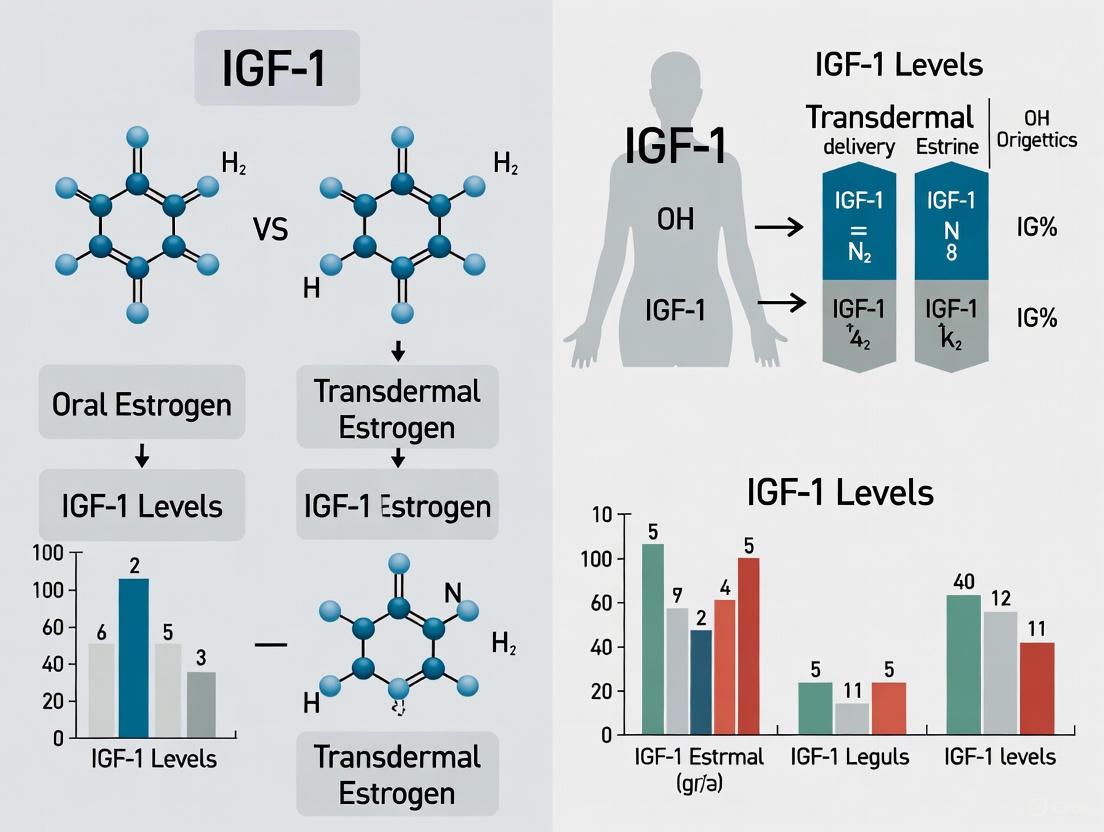
Oral Estrogen Administration: Oral estrogen passes first through the portal circulation to the liver, where it exerts potent inhibitory effects on hepatic GH receptor signaling and IGF-1 generation [7] [8]. This first-pass effect results in significantly reduced circulating IGF-1 levels despite increased GH secretion [7].
Transdermal Estrogen Administration: Transdermal delivery bypasses hepatic first-pass metabolism, resulting in more physiological estrogen exposure with minimal impact on IGF-1 levels and GH secretion [7] [8].
Table 2: Comparative Effects of Oral Versus Transdermal Estrogen on the GH/IGF-1 Axis
| Parameter | Oral Estrogen | Transdermal Estrogen | References |
|---|---|---|---|
| IGF-1 Levels | Significant reduction (mean 42.7% ± 41.4) | No significant change | [7] [8] |
| GH Secretion | Increased spontaneous secretion | No significant alteration | [8] |
| IGFBP-1 Levels | Significant increase (mean 170.2% ± 230.9) | No significant change | [7] |
| IGFBP-3 Levels | No significant change | Decreased (particularly in older women) | [7] [8] |
| Hepatic Impact | Significant first-pass effect | Minimal hepatic exposure | [3] [4] |
| Clinical Implication | May require GH dose adjustment in replacement therapy | More physiological GH/IGF-1 axis profile | [7] |
Experimental Methodologies for GH/IGF-1 Axis Assessment
Standard Assessment Protocols
Rigorous evaluation of the GH/IGF-1 axis requires specialized methodologies capable of capturing its dynamic nature:
GH Secretion Profiling: Comprehensive assessment involves 12-hour overnight blood sampling at 20-minute intervals to characterize pulsatile secretion patterns, including burst frequency, amplitude, and duration [8]. Deconvolution analysis is then applied to quantify secretory events and hormone half-lives [2].
GH Stimulation Testing: Standard provocative tests include GHRH stimulation (1 μg/kg IV bolus) with serial GH measurements over 120 minutes to assess pituitary responsiveness [8]. Additional stimulation paradigms may employ insulin-induced hypoglycemia, clonidine, L-dopa, or arginine as secretagogues.
IGF-1 System Evaluation: Serum IGF-1 measurement via chemiluminescent immunoassay (e.g., IDS iSYS platform) with age- and sex-adjusted reference ranges [9]. Complementary assessment of IGFBP-3 and ALS provides additional axis characterization.
Specialized Research Approaches
Advanced research methodologies enable deeper investigation of axis dynamics and regulatory mechanisms:
Network Modeling: Mathematical modeling of the GH neuroendocrine axis as a nonlinear dynamical system incorporating temporal delays, feedback interactions, and receptor dynamics to simulate pulsatile behavior and predict system responses to perturbations [2].
Tracer Infusion Studies: Stable isotope-labeled amino acid infusion techniques to quantify IGF-1 kinetics, including production rates and metabolic clearance, under different physiological conditions and intervention states.
Diagram 2: Experimental Workflow for Comprehensive GH/IGF-1 Axis Assessment. The flowchart outlines the sequential steps for rigorous evaluation of axis function, from subject preparation through specialized testing protocols to data analysis.
The Scientist's Toolkit: Essential Research Reagents
Table 3: Essential Research Reagents for GH/IGF-1 Axis Investigation
| Reagent/Category | Specific Examples | Research Application | Key Considerations |
|---|---|---|---|
| GH Assays | Chemiluminescent immunoassays, ELISA, RIA | Quantification of GH concentrations in serum/plasma | Must account for pulsatile secretion; assay-specific reference ranges |
| IGF-1 Assays | IDS iSYS, Immunoassays with extraction | Measurement of total IGF-1 levels | Requires acid-ethanol extraction to remove binding proteins; age-stratified reference values |
| IGFBP Assays | IGFBP-1, IGFBP-3 specific immunoassays | Assessment of IGF-binding protein profiles | IGFBP-1 shows diurnal variation; IGFBP-3 is GH-dependent |
| Stimulation Agents | GHRH, GHRP-6, Insulin, Clonidine | Pituitary reserve and responsiveness testing | Different mechanisms of action; safety monitoring required for insulin tolerance test |
| Recombinant Hormones | rhGH, rhIGF-1 | Intervention studies, replacement protocols | Dose-response characterization essential; metabolic effects monitoring |
| Binding Protein Reagents | Recombinant IGFBPs, ALS antibodies | Mechanistic studies of IGF-1 bioavailability | Impact on IGF-1 half-life and receptor activation |
| Signal Transduction Assays | Phospho-specific antibodies for IGF-1R, Akt, MAPK | Assessment of downstream pathway activation | Tissue-specific signaling patterns; time-course considerations |
Regulatory Considerations and Methodological Challenges
Special Populations and Contextual Factors
Accurate interpretation of GH/IGF-1 axis parameters requires consideration of numerous modifying factors:
Age-Related Changes: The GH/IGF-1 axis undergoes significant changes across the lifespan, with a progressive decline in both GH secretion and IGF-1 levels with advancing age [5] [9]. This senescence-related decline complicates the use of population-based reference intervals in elderly populations [9].
Nutritional Modulation: Nutritional status represents a potent regulator of IGF-1 generation, with malnutrition and fasting suppressing IGF-1 production despite elevated GH levels, creating a state of hepatic GH resistance [1] [3]. This nutritional regulation is mediated, in part, through insulin-dependent mechanisms and increased fibroblast growth factor 21 [1].
Hepatic and Renal Function: Both liver and kidney function significantly impact IGF-1 system components, with hepatic dysfunction reducing IGF-1, IGFBP-3, and ALS production, and renal impairment altering IGFBP clearance and compounding the complexity of axis interpretation [3] [9].
Analytical Considerations and Limitations
Several methodological challenges complicate the assessment and interpretation of the GH/IGF-1 axis:
IGF-1 Reliability in Elderly Populations: Serum IGF-1 demonstrates substantial intra-individual variability in elderly individuals (mean coefficient of variation: 14.7%), with high reference change values (44.3% increase, 30.7% decrease) that complicate the interpretation of single measurements [9]. The low index of individuality (0.44) further limits the utility of population-based reference intervals in geriatric populations [9].
Assay-Specific Variability: Different IGF-1 immunoassays may yield substantially different absolute values, necessitating method-specific reference ranges and complicating cross-study comparisons and meta-analyses.
Pulsatility Challenges: The pulsatile nature of GH secretion makes single random GH measurements clinically uninformative, necessitating either dynamic testing or serial sampling protocols to adequately characterize axis activity.
The GH/IGF-1 axis represents a sophisticated neuroendocrine system characterized by complex regulatory feedback loops, multiple levels of control, and significant modulation by sex steroids, nutritional status, and age. The route-dependent effects of estrogen administration highlight the intricate relationship between hepatic processing and endocrine function, with oral estrogen substantially reducing IGF-1 levels while transdermal delivery preserves a more physiological axis profile. These distinctions have profound implications for both basic research and clinical management, particularly in the context of hormone replacement strategies and their impact on growth, metabolism, and age-related physiological decline. A comprehensive understanding of the core physiology and regulatory mechanisms of this axis provides the essential foundation for rational experimental design, accurate data interpretation, and therapeutic innovation targeting this critical endocrine system.
The growth hormone (GH) and insulin-like growth factor-1 (IGF-1) axis represents a crucial regulatory system controlling somatic growth, metabolism, and tissue repair [10] [11]. GH secretion from the pituitary stimulates IGF-1 production primarily in the liver, creating a classic endocrine axis. Meanwhile, estrogen receptors, particularly ERα and ERβ, function as ligand-dependent transcription factors that regulate gene expression in numerous tissues [12] [13]. The interface between these two signaling systems represents a complex cross-talk mechanism that significantly impacts physiological processes and therapeutic outcomes. Understanding these molecular interactions is particularly relevant when comparing the metabolic effects of different estrogen administration routes, as oral and transdermal estrogens differentially impact IGF-1 levels and biological responses [7].
Molecular Mechanisms of GH Signaling and IGF-1 Gene Regulation
GH Receptor Activation and Downstream Signaling
The GH receptor (GHR) is a member of the class I cytokine receptor family and exists as a pre-formed homodimer on the cell surface [14]. Upon GH binding, the receptor undergoes a conformational change that brings the intracellular domains into closer proximity, activating the associated JAK2 tyrosine kinase [15] [14]. This activation initiates multiple signaling cascades:
- JAK-STAT Pathway: Activated JAK2 phosphorylates tyrosine residues on the GHR, creating docking sites for STAT proteins, particularly STAT5b [10] [14]. Once phosphorylated by JAK2, STAT5b forms dimers that translocate to the nucleus and bind to specific response elements in target genes, including IGF-1 [10].
- MAPK Pathway: GH activates the Ras-Raf-MEK-ERK pathway through Shc adapter proteins that bind to the GHR-JAK2 complex [15] [16].
- PI3K-Akt Pathway: GH stimulates tyrosine phosphorylation of insulin receptor substrate (IRS) proteins, leading to activation of phosphatidylinositol-3-kinase and Akt [15] [16].
Table 1: Key Signaling Molecules in GH Receptor Activation
| Signaling Component | Function | Role in IGF-1 Regulation |
|---|---|---|
| GHR | Cell surface receptor for GH | Initiates entire signaling cascade |
| JAK2 | Tyrosine kinase associated with GHR | Phosphorylates STAT5b and other substrates |
| STAT5b | Transcription factor | Directly binds and activates IGF-1 gene |
| Shc | Adapter protein | Links GHR to MAPK pathway |
| IRS1/2 | Insulin receptor substrates | Activate PI3K-Akt pathway |
Transcriptional Regulation of the IGF-1 Gene
The human IGF-1 gene spans approximately 90 kb of genomic DNA and consists of six exons that undergo complex alternative splicing [11]. The gene contains two alternative leader exons (exon 1 and exon 2), resulting in distinct transcript classes (Class 1 and Class 2) with different 5' untranslated regions but identical coding potential for the mature IGF-1 peptide [11]. STAT5b activation by GH represents the primary mechanism driving IGF-1 transcription, with STAT5b binding to multiple conserved regulatory elements within the IGF-1 gene locus [10]. Research has demonstrated that physiological levels of GH rapidly stimulate human IGF-1 gene transcription through these STAT5b-binding enhancer elements [10].
Estrogen Receptor-Mediated Modulation of GH Signaling
Distinct Functions of ERα and ERβ
ERα and ERβ, while structurally similar, exert different and often opposing effects on cellular functions [12] [13]. ERα is primarily associated with cell proliferation in tissues such as breast and uterus, while ERβ has been linked to pro-apoptotic and anti-proliferative effects [17] [12]. The ratio between these receptors appears to be a critical factor determining cellular responses to estrogen signaling [17].
Direct Molecular Interactions at Multiple Levels
Estrogen receptors interface with GH signaling through several distinct mechanisms:
- Receptor Expression Cross-Regulation: Suppression of IGF-IR expression in breast cancer cells leads to reduced ERα levels but increased ERβ expression, altering the ERα:ERβ ratio and consequently changing cellular responses to estradiol [17].
- Membrane-Initiated Signaling: Both ERα and ERβ can localize to the cell membrane and participate in rapid, non-genomic signaling that converges with GH-activated pathways [17] [18]. The IGF-IR appears essential for non-genomic, membrane-associated ERα activity [17].
- Transcriptional Integration: ERs can modulate the expression of components of the GH signaling pathway, with estradiol enhancing IGF-IR expression in certain tissues [17].
- Signal Transduction Cross-Talk: In breast cancer cells with suppressed IGF-IR, treatment with estradiol increases phosphorylation of p38 MAPK, which in turn phosphorylates the p53 tumor suppressor and accelerates apoptosis [17].
Diagram 1: ER and GH-IGF1 signaling pathway integration
Experimental Evidence: Methodologies and Key Findings
Cellular Models of ER-IGF-IR Interaction
Stable transfection of MCF-7 cells with siRNA targeting IGF-IR has been used to generate IGF-IR-deficient cell lines (IGF-IR.low cells) [17]. These models demonstrate that suppressing IGF-IR expression concomitantly reduces ERα and progesterone receptor expression while elevating ERβ [17]. The experimental workflow typically involves:
- Cell Culture: MCF-7 human breast cancer cells maintained in phenol red-free medium with 10% fetal bovine serum [17].
- Transfection: Using LipofectAMINE PLUS method with siRNA expression vectors targeting IGF-IR [17].
- Selection: Hygromycin-resistant colonies isolated using clone rings and expanded [17].
- Characterization: Western blotting for receptor expression, BrdU incorporation for proliferation, and specific ELISA assays for apoptosis measurement [17].
Clinical Evidence: Route of Estrogen Administration Matters
A randomized controlled trial comparing oral versus transdermal estrogen administration in women with hypopituitarism demonstrated significant route-dependent effects on IGF-1 levels [7]:
Table 2: Effects of Estrogen Administration Route on IGF-1 System Parameters
| Parameter | Oral Estradiol (2mg) | Transdermal 17β-Estradiol (50μg/day) |
|---|---|---|
| IGF-1 Levels | Significant reduction (42.7% ± 41.4, p=0.046) | No significant difference |
| IGFBP1 Levels | Significant increase (170.2% ± 230.9, p=0.028) | No significant difference |
| IGFBP3 Levels | No significant difference | No significant difference |
| HDL Cholesterol | Significant increase (27.8 ± 9.3, p=0.003) | Not reported |
| Clinical Implication | Requires higher GH dosage | No GH dosage adjustment needed |
This clinical finding has significant implications for patients receiving GH replacement therapy, as those using oral estrogen require increased GH dosage to achieve therapeutic effects [7]. The first-pass hepatic metabolism of oral estrogens is thought to mediate these suppressive effects on IGF-1, whereas transdermal administration avoids this effect [7].
The Scientist's Toolkit: Essential Research Reagents
Table 3: Key Research Reagents for Studying ER-GH-IGF1 Interactions
| Reagent/Cell Line | Application | Key Features |
|---|---|---|
| MCF-7 Human Breast Cancer Cells | Model system for ERα-positive breast cancer | Express both ERα and IGF-IR; responsive to estradiol and IGF-1 |
| IGF-IR.low Cells | Studying IGF-IR and ER interactions | Stable transfection with siRNA targeting IGF-IR; altered ERα:ERβ ratio |
| Custom siRNA Expression Vectors | Gene suppression studies | Target specific sequences of human IGF-IR cDNA |
| BrdU Incorporation ELISA | Cell proliferation measurement | Quantifies DNA synthesis during cell proliferation |
| M30-Apoptosense ELISA | Apoptosis detection | Measures caspase-cleaved cytokeratin 18 neo-epitope |
| Recombinant Rat GH | GH signaling studies | Available from National Hormone and Pituitary Program |
| Phospho-Specific Antibodies | Signaling pathway analysis | Detect activated signaling molecules (e.g., p-STAT5, p-p38 MAPK) |
The molecular interface between estrogen receptors and GH signaling represents a sophisticated regulatory network with significant implications for both basic research and clinical applications. The opposing effects of ERα and ERβ on cell proliferation and apoptosis, combined with their differential modulation of IGF-IR signaling, create a complex balance that influences tissue-specific responses [17] [12]. The route of estrogen administration emerges as a critical factor, with oral estrogens reducing IGF-1 levels while transdermal estrogens avoid this first-pass hepatic effect [7]. This distinction has direct clinical relevance for patients requiring GH replacement therapy and highlights the importance of considering administration route in hormonal therapeutics. Further research into the precise molecular mechanisms governing ER subtype-specific effects on the GH-IGF-1 axis may yield novel therapeutic approaches for conditions ranging from hormone-responsive cancers to metabolic disorders.
The route of estrogen administration represents a critical therapeutic variable that fundamentally dictates hormonal bioavailability, metabolic consequences, and clinical outcomes. Oral estrogen preparations undergo extensive first-pass hepatic metabolism before reaching systemic circulation, triggering a cascade of hepatic-mediated effects that alter the growth hormone (GH)/insulin-like growth factor-1 (IGF-1) axis and other metabolic parameters. In contrast, transdermal estrogen delivery provides direct systemic absorption bypassing the gastrointestinal tract and liver, resulting in a more physiological hormonal profile. This distinction is not merely pharmacokinetic but has profound implications for IGF-1 signaling, bone metabolism, cardiovascular risk markers, and potentially clinical efficacy across various therapeutic areas. Understanding these route-dependent differences is essential for researchers investigating endocrine pathways, drug development professionals designing targeted therapies, and clinicians optimizing treatment regimens for hormone-sensitive conditions.
Comparative Pharmacokinetics and Metabolic Consequences
Fundamental Pharmacokinetic Differences
The hepatic first-pass effect creates dramatic differences in how the body processes oral versus transdermal estrogens. When administered orally, estrogens are absorbed from the gastrointestinal tract and transported directly to the liver via the portal circulation, where they undergo significant metabolism before entering the systemic bloodstream. This process results in non-physiological metabolite ratios, particularly the estrone-to-estradiol ratio, which approaches 5:1 with oral administration compared to approximately 1:1 with transdermal delivery—the ratio typical of premenopausal women [19] [20]. Transdermal systems, including patches, gels, and sprays, deliver 17β-estradiol directly through the skin into systemic circulation, avoiding this initial hepatic passage and preserving more natural estrogen proportions.
Table 1: Comparative Pharmacokinetic Profiles of Oral vs. Transdermal Estrogen
| Parameter | Oral Estrogen | Transdermal Estrogen |
|---|---|---|
| First-Pass Hepatic Metabolism | Extensive | Minimal to none |
| Estradiol/Estrone Ratio | Approximately 0.2-0.5 (non-physiological) [20] | Approximately 1.0 (physiological) [20] |
| Hepatic Protein Stimulation | Significant increase | Minimal stimulation |
| Dose Requirements | Higher due to first-pass metabolism | Lower due to direct delivery |
| Accumulation Potential | Signs of retention after multiple doses [20] | No accumulation with continuous use [20] |
Impact on the GH/IGF-1 Axis
The route-dependent effects on the GH/IGF-1 axis represent one of the most significant metabolic differences between oral and transdermal estrogen. Multiple randomized controlled trials demonstrate that oral estrogen administration significantly suppresses IGF-1 levels while concurrently increasing GH concentrations, effectively dissociating the normal GH/IGF-1 axis [21] [22] [23]. This phenomenon occurs because the liver serves as the primary source of circulating IGF-1, and the high estrogen concentrations achieved during first-pass metabolism directly suppress hepatic IGF-1 production. In a randomized, placebo-controlled study of healthy postmenopausal women, oral 17β-estradiol significantly decreased IGF-1 levels compared to both placebo (P = 0.04) and transdermal estrogen (P = 0.004), whereas transdermal estrogen had no significant effect on IGF-1 levels compared to placebo (P = 0.56) [22].
This dissociation occurs rapidly, with studies in premenopausal women showing that just 5 days of oral estrogen administration significantly suppressed IGF-I levels and increased fasting GH levels [23]. The suppressive effect of oral estrogen on IGF-1 appears to be consistent across different estrogen formulations, including ethinyl estradiol, conjugated equine estrogens, and micronized 17β-estradiol, confirming that the route of administration rather than the specific estrogen compound drives this metabolic effect [21].
Table 2: Effects on the GH/IGF-1 Axis and Downstream Tissues
| Parameter | Oral Estrogen | Transdermal Estrogen |
|---|---|---|
| IGF-1 Levels | Significant decrease [21] [22] [24] | No significant change or moderate increase [21] [22] |
| GH Levels | Significant increase [21] [23] | No significant change [21] |
| Bone Formation Markers | Suppression [21] | Stimulation [21] |
| Connective Tissue Metabolism | Reduced collagen synthesis [21] | Increased nonbone collagen synthesis [21] |
| IGFBP-3 Levels | No significant change [22] | No significant change [22] |
Figure 1: Metabolic Pathways of Oral vs. Transdermal Estrogen Administration
Experimental Evidence and Clinical Correlations
Bone and Connective Tissue Metabolism
The route-dependent effects on IGF-1 have significant implications for bone and connective tissue metabolism. In a seminal study investigating impact on connective and bone tissue metabolism, transdermal estrogen administration significantly (p < 0.05) increased IGF-1, procollagen III, procollagen I, osteocalcin, and urinary hydroxyproline/creatinine ratio (a marker of bone turnover) [21]. Conversely, oral estrogen administration had a suppressive effect on these parameters, with significant differences observed between the two routes for IGF-1 (p = 0.001), procollagen III (p = 0.018), procollagen I (p = 0.002), osteocalcin (p = 0.015), and urinary hydroxyproline/creatinine ratio (p = 0.004) [21]. These findings demonstrate that transdermally delivered estrogen stimulates IGF-1 production, increases osteoblastic function, and enhances both bone and nonbone collagen synthesis, while oral administration produces opposite effects.
The relationship between IGF-1 changes and bone metabolism markers during estrogen therapy was statistically significant (p < 0.05), with IGF-1 changes correlating with changes in procollagen III, procollagen I, osteocalcin, and urinary hydroxyproline/creatinine ratio [21]. This correlation underscores the importance of IGF-1 as a mediator of estrogen's effects on mesenchymal tissues.
Cardiovascular Risk Markers
The hepatic first-pass effect also significantly influences cardiovascular risk markers, particularly C-reactive protein (CRP). In a randomized, crossover, placebo-controlled study of postmenopausal women, transdermal estradiol had no effect on CRP levels, whereas eight weeks of oral conjugated estrogens caused a more than twofold increase in CRP (p < 0.01) in the same women [24]. This increase in CRP was accompanied by a significant reduction in IGF-1, and the magnitude of CRP increase was inversely correlated with the decrease in IGF-1 (r = -0.49, p = 0.008) [24]. Neither oral nor transdermal administration affected plasma concentrations of inflammatory cytokines such as IL-1β, IL-6, and TNF-α that promote CRP synthesis, indicating that the CRP elevation results specifically from the first-pass hepatic effect rather than systemic inflammation [24].
Special Populations: Anorexia Nervosa Case Study
The differential effects of estrogen route have been particularly studied in conditions of low bone density, such as anorexia nervosa (AN), where hormonal alterations contribute to significant bone impairment. A 12-month randomized, double-blind, placebo-controlled study investigated transdermal 17-beta estradiol with and without recombinant human IGF-1 (rhIGF-1) administration in young women with AN [25]. While this study found no additive benefit of rhIGF-1 administration over transdermal estrogen replacement alone, it highlighted the importance of route selection in this population. Previous research had demonstrated that adolescent girls with AN receiving transdermal physiologic 17-β-estradiol for 18 months showed increases in spine and hip bone mineral density at a rate equivalent to age-matched normal-weight controls, an effect not observed with placebo [25]. This effect was attributed to the ability of transdermal estrogen to provide estrogen replacement without suppressing endogenous IGF-1 production, which is already compromised in AN.
Research Methods and Experimental Protocols
Key Experimental Designs
The fundamental differences between oral and transdermal estrogen have been elucidated through carefully designed clinical trials employing specific methodological approaches:
Randomized Crossover Designs: Several pivotal studies utilized randomized, crossover, placebo-controlled designs to compare the effects of different estrogen routes within the same subjects [23] [24]. For example, Vongpatanasin et al. conducted a study where 21 postmenopausal women received transdermal estradiol (100 μg/day), oral conjugated estrogen (0.625 mg/day), or placebo, each for eight weeks in random order [24]. This design effectively controls for interindividual variability and enhances statistical power.
Longitudinal Intervention Studies: Longer-term studies have typically employed parallel-group randomized designs with baseline and endpoint measurements. For instance, Sonnet et al. conducted a randomized study of 196 healthy postmenopausal women allocated to receive either oral 17β-estradiol (1 mg) plus progesterone, transdermal 17β-estradiol (50 μg) plus progesterone, or placebo over a 6-month period [22]. This design allows for assessment of medium-term metabolic adaptations.
Biochemical Assessment Protocols: Standardized biochemical assessments across studies typically include:
- IGF-1 measurement by radioimmunoassay or immunochemiluminometric assay
- GH profiling through frequent sampling over 24 hours or fasting levels
- Bone turnover markers (procollagen I, procollagen III, osteocalcin, NTX, P1NP)
- Inflammatory markers (CRP, IL-6, TNF-α)
- Hormonal profiles (estradiol, estrone, gonadotropins)
Figure 2: Experimental Workflow for Estrogen Route Comparison Studies
The Scientist's Toolkit: Essential Research Reagents and Materials
Table 3: Key Research Reagents and Materials for Estrogen Route Studies
| Reagent/Material | Specification Examples | Research Application |
|---|---|---|
| 17β-estradiol | Micronized for oral administration; Patches (0.025-0.1 mg/day), gels, or sprays for transdermal | Active pharmaceutical ingredient for both routes [19] [22] |
| Conjugated Equine Estrogens (CEE) | 0.625 mg/day standard dose | Comparative oral estrogen preparation [24] |
| Progesterone | 100 mg/day micronized | Endometrial protection in non-hysterectomized subjects [22] |
| IGF-1 Assay | Radioimmunoassay (RIA) or immunochemiluminometric assay (ICMA) | Quantification of IGF-1 levels [21] [26] |
| GH Assay | Immunoradiometric assay (IRMA) or chemiluminescence | Measurement of GH concentrations and pulsatility [21] |
| Bone Turnover Markers | Procollagen I, procollagen III, osteocalcin, P1NP, NTX | Assessment of bone formation and resorption [21] [25] |
| Inflammatory Marker Panels | High-sensitivity CRP, IL-6, TNF-α, IL-1β | Evaluation of inflammatory pathways [24] |
| Hormonal Profiling Assays | Estradiol, estrone, FSH, LH | Assessment of hormonal status and pharmacokinetics [19] [20] |
The evidence consistently demonstrates that the first-pass hepatic metabolism of oral estrogen versus direct systemic delivery of transdermal estrogen produces fundamentally different biological effects, particularly on the GH/IGF-1 axis. Oral administration dissociates the GH/IGF-1 axis through hepatic suppression of IGF-1 production, while transdermal administration preserves physiological relationships between these critical growth factors. These route-dependent effects extend to multiple tissue systems, influencing bone formation markers, connective tissue metabolism, and cardiovascular risk profiles.
For researchers and drug development professionals, these findings highlight the importance of considering administration route as a critical variable in study design and therapeutic development. The selection between oral and transdermal delivery should be guided by specific research objectives and clinical goals—whether aiming to manipulate the GH/IGF-1 axis or maintain its physiological function. Future research should continue to explore the long-term clinical implications of these metabolic differences, particularly in special populations with compromised bone health, and investigate novel delivery systems that can precisely target specific tissues while minimizing unwanted hepatic effects.
The growth hormone/insulin-like growth factor-1 (GH/IGF-1) axis represents a crucial regulatory system for metabolic homeostasis, body composition, and tissue maintenance. Within this system, IGF-1 circulates primarily bound to binding proteins, most notably IGF binding protein-3 (IGFBP-3), which forms a ternary complex with an acid-labile subunit (ALS) to extend IGF-1 half-life from minutes to hours [27] [28]. IGFBP-1, another key binding protein, undergoes rapid regulation by nutritional status and hormones, providing short-term control of IGF-1 bioavailability [29] [27].
Emerging evidence demonstrates that estrogen replacement therapy exerts profoundly different effects on this axis depending solely on its administration route. Oral estrogen preparations undergo first-pass hepatic metabolism, triggering significant alterations in hepatic protein synthesis, while transdermal delivery bypasses this effect, providing more physiological estrogen exposure without dramatic impacts on the IGF system [7] [8] [29]. This review synthesizes current clinical evidence contrasting these administration routes, providing researchers and drug development professionals with quantitative comparisons and methodological frameworks for investigating this phenomenon.
Quantitative Comparison of Route-Dependent Effects
Effects on Circulating IGF-1 and Binding Proteins
Table 1: Comparative Effects of Oral versus Transdermal Estrogen on IGF-1 Axis Biomarkers
| Biomarker | Oral Estrogen Effect | Magnitude of Change | Transdermal Estrogen Effect | Magnitude of Change | Study References |
|---|---|---|---|---|---|
| Total IGF-1 | Significant decrease | ↓ 13-42.7% | No significant change | Stable | [7] [8] [29] |
| IGFBP-1 | Significant increase | ↑ 104-170% | No significant change | Stable | [7] [29] |
| IGFBP-3 | Significant decrease | ↓ Significant (p≤0.005) | No significant change (except in older women) | Mostly stable | [7] [8] [30] |
| ALS | Significant decrease | ↓ Significant (p≤0.005) | No significant change | Stable | [30] |
| GH Secretion | Increased | ↑ Spontaneous nocturnal secretion | No significant change | Stable | [8] |
The data consistently demonstrate that oral estrogen administration reduces circulating IGF-1 levels substantially (13-42.7% across studies), while transdermal estrogen maintains baseline IGF-1 concentrations [7] [8] [29]. This divergence highlights the first-pass hepatic effect of oral estrogen, which suppresses hepatic IGF-1 production despite elevated GH levels observed in some studies [8].
Similarly, oral estrogen profoundly increases IGFBP-1 concentrations (104-170%), creating a potent inhibitory influence on free, bioactive IGF-1 through sequestration [7] [29]. IGFBP-3, the primary carrier of IGF-1 in circulation, decreases significantly with oral but not transdermal administration, further reducing the circulating IGF-1 reservoir [7] [30]. The acid-labile subunit (ALS), essential for ternary complex stability, follows a similar pattern of suppression with oral estrogen only [30].
Clinical Implications of Route-Dependent Effects
Table 2: Clinical Implications of Estrogen Administration Route on IGF-1 Axis
| Parameter | Oral Estrogen Impact | Transdermal Estrogen Impact | Clinical Significance |
|---|---|---|---|
| GH Therapy Requirements | Increased GH dosage needed | Stable GH requirements | Critical for hypopituitary patients [7] |
| Metabolic Effects | Reduced beneficial GH effects on body composition | Preservation of GH anabolic effects | Impacts fat/protein metabolism [7] |
| Lipoprotein(a) Modulation | Significant reduction (↓23%) | No significant effect | Cardiovascular risk implications [29] |
| Hepatic Impact | First-pass metabolism, protein synthesis alterations | Minimal hepatic impact | Basis for differential effects [29] |
The route-dependent effects carry significant clinical implications, particularly for patients receiving GH replacement therapy. Individuals taking oral estrogen require approximately 50% higher GH doses to achieve equivalent IGF-1 levels compared to those using transdermal delivery [7] [31]. Oral estrogen may also attenuate the beneficial metabolic effects of GH replacement on body composition and quality of life [7].
Interestingly, the IGFBP-1 increase associated with oral estrogen correlates inversely with lipoprotein(a) [Lp(a)] reduction, suggesting a potential mechanism for the cardioprotective lipid effects of oral estrogen [29]. This relationship underscores the complex interplay between estrogen administration routes, the IGF system, and cardiovascular risk factors.
Experimental Protocols and Methodologies
Representative Clinical Study Designs
Randomized, Comparative Study in Hypopituitarism (Isotton et al., 2012)
- Participants: 11 women with hypopituitarism receiving stable GH replacement [7]
- Study Design: Randomized allocation to either oral estradiol (2mg/day, n=6) or transdermal 17β-estradiol (50μg/day, n=5) for 3 months [7]
- Measurements: Serum IGF-1, IGFBP-3, IGFBP-1 at baseline and after 3 months; additional metabolic parameters including lipid profiles [7]
- Key Findings: Oral group showed significant reduction in IGF-1 (42.7±41.4%, p=0.046) and increase in IGFBP-1 (170.2±230.9%, p=0.028); transdermal group showed no significant changes [7]
Placebo-Controlled Crossover Trial in Postmenopausal Women (Bellantoni et al., 1996)
- Participants: 16 healthy postmenopausal women, ages 49-75 years [8]
- Study Design: Randomized, placebo-controlled, crossover trial of 6 weeks of oral conjugated estrogen (1.25mg daily) or transdermal estradiol (100μg/day) separated by 8-week washout [8]
- Measurements: Spontaneous nocturnal GH secretion (12-hour sampling), GHRH-stimulated GH release, serum IGF-1, IGFBP-3 [8]
- Key Findings: Oral estrogen increased spontaneous GH secretion but decreased IGF-1; transdermal estrogen did not alter GH secretion or IGF-1 levels [8]
Double-Blind, Placebo-Controlled Study (Paassilta et al., 2000)
- Participants: 73 hysterectomized postmenopausal women randomized to oral (n=35) or transdermal (n=38) estrogen [29]
- Study Design: 6-month double-blind, placebo-controlled trial with measurements at baseline, 3, and 6 months [29]
- Measurements: Plasma IGFBP-1, IGF-I, lipoprotein(a) [29]
- Key Findings: Oral estrogen increased IGFBP-1 by 104% (p<0.001) and decreased IGF-I by 13%; transdermal estrogen showed no significant changes [29]
Laboratory Methodologies for Biomarker Assessment
Serum IGF-1 Measurement
- Sample Collection: Fasting blood samples, serum separation, storage at -80°C until analysis [32]
- Assay Techniques: Enzyme-linked immunosorbent assay (ELISA) or electrochemiluminescence immunoassay (ECLIA) [32] [33]
- Standardization: Age-specific reference ranges required due to age-dependent decline (~30.1 ng/mL per decade in adult males) [33]
- Quality Control: Duplicate measurements, internal controls, acceptable intra- and inter-assay coefficients of variation [32]
IGFBP-3 and IGFBP-1 Assessment
- IGFBP-3 Measurement: ELISA or ECLIA; accounts for ~90% of IGF:ALS ternary complexes [33] [27]
- IGFBP-1 Measurement: ELISA; rapid regulation by insulin and nutritional status [29] [27]
- Western Ligand Blotting: Alternative method for identifying IGFBPs in secretion media from cell cultures or tissue explants [34]
Signaling Pathways and Molecular Mechanisms
Pathway 1: Oral Estrogen Disruption of Hepatic IGF-1 Axis
The diagram illustrates the fundamental mechanistic divergence between estrogen administration routes. Oral estrogen undergoes extensive first-pass hepatic metabolism, triggering alterations in hepatic protein synthesis that reduce IGF-1 production while simultaneously increasing IGFBP-1 and decreasing IGFBP-3 and ALS [29] [30]. This coordinated response disrupts the ternary complex formation, substantially reducing bioactive IGF-1 availability despite compensatory increases in GH secretion [8] [27]. Transdermal estrogen bypasses hepatic first-pass metabolism, preserving normal IGF-1 axis homeostasis [7] [8].
The molecular basis for these effects involves estrogen receptor-mediated modulation of gene expression in hepatocytes. Oral administration creates supraphysiological estrogen concentrations in the portal circulation, directly impacting the synthesis of components of the IGF system [29]. This effect is dose-dependent, with higher estrogen doses producing more pronounced suppression of IGF-1 and IGFBP-3 [30].
The Scientist's Toolkit: Essential Research Reagents
Table 3: Key Research Reagents for Investigating Estrogen Effects on IGF Axis
| Reagent Category | Specific Examples | Research Application | Functional Role |
|---|---|---|---|
| Estrogen Formulations | Oral estradiol (2mg), Transdermal 17β-estradiol (50-100μg/day), Conjugated equine estrogen (1.25mg) | Route comparison studies | Experimental interventions to test administration route effects [7] [8] |
| IGF System Assays | IGF-1 ELISA, IGFBP-3 ECLIA, IGFBP-1 ELISA, Western ligand blotting | Biomarker quantification | Precise measurement of IGF axis components [34] [32] [33] |
| Molecular Biology Tools | Recombinant human IGF-1, IGFBP-3, PAPP-A proteases, IGF-1R inhibitors | Mechanistic studies | Pathway manipulation to establish causal relationships [34] [27] |
| Cell Culture Systems | Primary hepatocytes, OA chondrocytes, Human OA cartilage explants | In vitro modeling | Isolated system analysis of estrogen effects [34] |
These research tools enable comprehensive investigation of estrogen's route-dependent effects, from clinical correlation to mechanistic insight. The combination of specific estrogen formulations with validated IGF system assays allows researchers to replicate and extend the foundational findings of route-dependent effects [7] [8]. Molecular biology tools facilitate exploration of the underlying signaling pathways, while specialized cell culture systems provide controlled environments for dissecting specific biological mechanisms [34] [27].
The administration route of estrogen therapy fundamentally determines its impact on the GH/IGF-1 axis, with oral estrogen substantially reducing IGF-1, IGFBP-3, and ALS while increasing IGFBP-1, and transdermal estrogen largely preserving baseline homeostasis. These differences stem from first-pass hepatic metabolism of oral estrogen and have profound implications for clinical management, particularly in patients requiring GH replacement therapy.
For researchers and drug development professionals, these findings highlight the importance of considering administration route in study design, clinical trial interpretation, and therapeutic development. The consistent experimental evidence across multiple study populations provides a robust foundation for future investigations into tissue-specific effects, long-term outcomes, and the molecular mechanisms underlying these route-dependent phenomena. As therapeutic strategies targeting the IGF axis continue to evolve, understanding these fundamental endocrine principles remains essential for optimizing metabolic outcomes and minimizing unintended consequences of hormone therapy.
Clinical Translation: Route-Dependent IGF-1 Responses in Disease Management and Hormone Therapy
The administration of hormone replacement therapy (HRT) in postmenopausal women represents a critical intervention for managing menopausal symptoms and preventing long-term health consequences associated with estrogen deficiency. Beyond its clinical indications, HRT serves as a powerful model for investigating how administration routes dictate metabolic consequences, particularly within the growth hormone/insulin-like growth factor-1 (GH/IGF-1) axis. The divergent effects of oral versus transdermal estrogen delivery on hepatic and peripheral metabolic pathways underscore the fundamental principle that route-specific pharmacokinetics can drive substantially different physiological outcomes.
This review systematically compares the metabolic impacts of oral and transdermal HRT, with particular emphasis on their differential effects on IGF-1 signaling—a key regulator of metabolism, body composition, and long-term health outcomes. By synthesizing evidence from clinical trials, mechanistic studies, and comparative analyses, we provide researchers and drug development professionals with a comprehensive framework for understanding how administration routes fundamentally alter the biological actions of estrogens in postmenopausal women.
Metabolic Fate of Estrogens: Fundamental Route-Dependent Divergence
The route of estrogen administration dictates a fundamentally different metabolic fate, with profound implications for downstream physiological effects. Oral estrogen administration undergoes extensive first-pass metabolism in the liver, resulting in elevated hepatic estrogen exposure and consequent modulation of hepatic protein synthesis [26] [35]. This pathway leads to the synthesis of binding proteins, coagulation factors, and lipoproteins, while simultaneously suppressing hepatic IGF-1 production [26] [36].
In contrast, transdermal estrogen delivery bypasses first-pass hepatic metabolism, providing a more physiological pattern of hormone delivery that maintains stable serum levels and avoids the high hepatic exposure characteristic of oral administration [35] [37]. This fundamental pharmacokinetic difference underlies the route-specific effects on the GH/IGF-1 axis and associated metabolic parameters.
Table 1: Fundamental Pharmacokinetic Differences Between HRT Administration Routes
| Parameter | Oral Estrogen | Transdermal Estrogen |
|---|---|---|
| First-Pass Metabolism | Extensive hepatic metabolism | Bypasses hepatic first-pass |
| Hepatic Estrogen Exposure | Significantly elevated | Minimal increase |
| IGF-1 Axis Impact | Suppresses hepatic IGF-1 production | Minimal direct IGF-1 suppression |
| Binding Protein Effects | Decreases IGFBP-3 | Decreases IGFBP-3 (age-dependent) |
| Metabolic Consequences | Marked effects on lipid metabolism, coagulation | Minimal metabolic interference |
Route-Specific Modulation of the GH/IGF-1 Axis
Experimental Evidence from Clinical Studies
The differential effects of estrogen administration routes on the GH/IGF-1 axis have been demonstrated in multiple controlled trials. A crossover study investigating both oral conjugated estrogen (1.25 mg/day) and transdermal estradiol (100 μg/day) in postmenopausal women found distinctly different patterns of regulation [36]:
- Oral estrogen significantly increased spontaneous GH secretion (146 ± 25% of baseline, P < 0.01) while simultaneously decreasing circulating IGF-1 levels (76 ± 4% of baseline, P < 0.01)
- Transdermal estrogen did not significantly alter nocturnal GH secretion or morning IGF-1 levels
- IGF-binding protein-3 (IGFBP-3) levels decreased with both oral (in younger women only) and transdermal (both younger and older women) administration
These findings demonstrate that oral and transdermal estrogens exert fundamentally different effects on the GH/IGF-1 axis, with oral administration creating a paradoxical state of increased GH secretion coupled with reduced IGF-1 production—a pattern consistent with hepatic IGF-1 resistance.
Impact of Baseline IGF-1 Status
The effect of HRT on IGF-1 levels appears modulated by baseline IGF-1 status. A study of 66 postmenopausal women receiving either oral (n=44) or transdermal (n=22) HRT for 6 months demonstrated that women with low basal IGF-1 levels experienced significant increases in IGF-1 after therapy (65% increase in oral group, 77% in transdermal group) [26]. Conversely, women with high basal IGF-1 levels showed decreases (-8% and -16%, respectively). This suggests that the metabolic context of the individual significantly influences the response to HRT.
Methodological Approaches to Investigating Route-Specific Effects
Experimental Protocols for Assessing IGF-1 Responses
Protocol 1: Cross-Over Design for GH/IGF-1 Axis Evaluation [36]
- Population: Postmenopausal women (ages 49-75), stratified by age (≤62 vs. >62 years)
- Intervention: 6 weeks of oral conjugated estrogen (1.25 mg daily) or transdermal estradiol (100 μg/day) in random order, separated by 8-week washout period
- Assessment Methods:
- Spontaneous GH secretion: 12-hour overnight blood sampling at 20-minute intervals
- GH responsiveness: IV bolus injection of GHRH (1 μg/kg)
- Serum analyses: IGF-1 and IGFBP-3 levels before and after GHRH stimulation
- Key Outcome Measures: Nocturnal GH secretion, GHRH-stimulated GH release, circulating IGF-1 and IGFBP-3 concentrations
Protocol 2: Randomized Controlled Trial of Transdermal Estrogen with IGF-1 Supplementation [25]
- Population: 75 adolescent and young adult women with anorexia nervosa (AN) ages 14-22 years
- Intervention: Transdermal 17-beta estradiol 0.1 mg/day with either:
- rhIGF-1 (30 mcg/kg/dose subcutaneously twice daily) [AN-IGF-1+]
- Placebo [AN-IGF-1-]
- Duration: 12-month randomized, double-blind, placebo-controlled longitudinal study
- Assessment Methods:
- Bone turnover markers: P1NP (formation), NTX (resorption)
- Bone density: DXA for areal BMD
- Bone microarchitecture: HRpQCT at distal radius and tibia
- Strength estimates: Finite element analysis
- Key Findings: No additive benefit of rhIGF-1 over transdermal estrogen alone for bone outcomes
Diagram 1: Metabolic pathway divergence between oral and transdermal HRT administration. Oral administration creates hepatic IGF-1 suppression despite increased GH secretion, while transdermal delivery preserves normal GH/IGF-1 axis function.
The Scientist's Toolkit: Essential Research Reagents and Methodologies
Table 2: Key Research Reagent Solutions for Investigating HRT Metabolic Effects
| Reagent/Assay | Research Application | Functional Significance |
|---|---|---|
| 17-β-estradiol patches (Vivelle-Dot) | Transdermal delivery system | Provides consistent estradiol delivery while bypassing hepatic first-pass metabolism [25] |
| Recombinant human IGF-1 (rhIGF-1) | Intervention for IGF-1 supplementation | Directly tests IGF-1 contribution to observed metabolic effects [25] |
| IGF-1 & IGFBP-3 ELISA Kits | Serum level quantification | Measures circulating levels of IGF-1 and its primary binding protein [36] |
| GHRH (1 μg/kg) | GH stimulation testing | Assesses pituitary GH responsiveness and reserve capacity [36] |
| High-resolution peripheral quantitative CT (HRpQCT) | Bone microarchitecture analysis | Provides 3D assessment of bone density, geometry, and microarchitecture [25] |
Clinical Implications of Route-Specific Metabolic Effects
Cardiovascular Risk Profiles
The differential metabolic effects of HRT administration routes translate into clinically meaningful risk profile differences:
- Venous Thromboembolism (VTE) Risk: Systematic review evidence indicates transdermal HRT carries significantly lower VTE risk compared to oral administration (OR 0.6-0.8 for transdermal vs. oral) [37]. This likely reflects the avoidance of first-pass hepatic effects on coagulation factors.
- Lipid Metabolism: Oral estrogen consistently reduces LDL cholesterol but increases triglycerides and HDL, while transdermal estrogen has more neutral effects on triglycerides and variable effects on HDL [37].
- Cardiometabolic Risk in Special Populations: Women with type 2 diabetes using transdermal HRT demonstrate no increased cardiovascular risk, whereas oral HRT is associated with doubled pulmonary embolism risk and 21% higher heart disease incidence [38].
Bone Metabolism and Body Composition
The GH/IGF-1 axis plays a crucial role in bone metabolism, with route-specific effects influencing bone health outcomes:
- Bone Mineral Density: Both oral and transdermal administration improve BMD, with no clear superiority between routes despite their differential effects on IGF-1 [37].
- Bone Microarchitecture: In populations with anorexia nervosa, transdermal estrogen replacement improves bone outcomes, though with no additive benefit from rhIGF-1 coadministration [25].
Diagram 2: Comprehensive experimental workflow for investigating route-specific HRT effects. Studies should include careful population characterization, controlled interventions, and multidimensional outcome assessment.
The comparison between oral and transdermal hormone replacement therapy provides a compelling paradigm for route-specific metabolic consequences in postmenopausal women. The fundamental pharmacokinetic differences between these administration routes—particularly regarding first-pass hepatic metabolism—drive substantially divergent effects on the GH/IGF-1 axis and associated metabolic parameters.
For researchers and drug development professionals, these route-specific effects highlight critical considerations for future therapeutic development:
- Individualized Therapy Selection: Metabolic context, including baseline IGF-1 status and individual risk profiles, should guide route selection
- Hepatic vs. Peripheral Targeting: Administration route represents a strategic choice between hepatic metabolic effects versus more targeted endocrine actions
- Research Design Imperatives: Future investigations must account for route-specific effects when evaluating metabolic outcomes of hormone therapies
The mechanistic insights gained from comparing oral versus transdermal HRT not only inform clinical management of menopausal women but also provide a broader framework for understanding how administration routes fundamentally alter drug actions and metabolic consequences across therapeutic domains.
The management of growth hormone (GH) deficiency in hypopituitarism requires careful consideration of concomitant hormone replacement therapies, particularly estrogen in females. Estrogen administration route exerts a profound influence on GH dosing and metabolic efficacy, creating a critical therapeutic consideration for endocrinologists and pharmaceutical developers. This review synthesizes evidence demonstrating that oral estrogen therapy attenuates GH action by reducing insulin-like growth factor-1 (IGF-1) production and impairing substrate utilization, whereas transdermal estrogen largely avoids these hepatic effects. We present quantitative comparisons of IGF-1 suppression, detailed experimental methodologies from key studies, and visualizations of the underlying mechanisms. The evidence supports individualized GH dosing strategies based on estrogen replacement route to optimize metabolic outcomes and body composition in hypopituitary patients.
Hypopituitarism, characterized by deficiency of one or more pituitary hormones, affects approximately 37.5–45.5 cases per 100,000 individuals [39]. Growth hormone deficiency (GHD) specifically contributes to abnormal body composition, reduced bone mass, adverse cardiovascular risk profiles, and impaired quality of life [40]. The complex interplay between GH and other hormonal axes necessitates careful management when replacing multiple hormones, particularly in females requiring estrogen therapy.
The route of estrogen administration emerges as a critical factor influencing GH replacement efficacy. This review examines the mechanistic basis and clinical evidence for route-dependent interactions between estrogen and GH/IGF-1 axis, focusing on implications for dosing strategies and treatment outcomes. Understanding these interactions is essential for optimizing hormonal replacement in hypopituitarism, particularly as recent evidence indicates that morbidity and mortality approach normal in hypopituitary patients receiving modern replacement therapy including GH [40].
Estrogen Pharmacology and Hepatic First-Pass Metabolism
Estrogen compounds are available in various formulations with distinct pharmacological properties. Natural estrogens (e.g., micronized 17β-estradiol), prodrug formulations (e.g., estradiol valerate, conjugated equine estrogen), and synthetic compounds (e.g., ethinyl estradiol) demonstrate markedly different potencies and metabolic effects [41]. The average daily production rate of 17β-estradiol ranges between 50-100 μg during the follicular phase, rising more than 5-fold during mid-cycle, informing replacement dosing strategies [41].
Route-Dependent Hepatic Exposure
The fundamental difference between estrogen administration routes lies in their hepatic exposure patterns:
Oral administration: Estradiol undergoes rapid metabolism and inactivation by the liver, requiring micronized or prodrug formulations to achieve sufficient bioavailability. Oral delivery results in unnaturally high estrogen concentrations in portal blood, inducing major effects on liver function [41]. For replacement therapy, 2 mg of oral 17β-estradiol is equivalent to 100 μg delivered transdermally - a 20-fold difference [41].
Transdermal administration: This route delivers estrogen directly to the systemic circulation, avoiding first-pass hepatic metabolism. Consequently, transdermal estrogen does not produce the high portal blood concentrations observed with oral therapy [41].
Synthetic estrogens: Ethinyl estradiol, a synthetic estrogen resistant to hepatic metabolism, demonstrates particularly high potency - less than 20 μg is sufficient for contraception, reflecting 50-100 times the potency of 17β-estradiol [41].
Table 1: Estrogen Formulations and Equivalent Dosing
| Formulation Type | Examples | Typical Doses | Relative Potency |
|---|---|---|---|
| Oral Natural Estrogens | Micronized 17β-estradiol | 1-2 mg | Baseline |
| Oral Prodrugs | Estradiol valerate | 1-2 mg | Similar to natural |
| Conjugated Estrogens | Conjugated equine estrogen | 0.3-1.25 mg | Variable |
| Synthetic Estrogens | Ethinyl estradiol | 20-50 μg | 50-100x natural |
| Transdermal Estrogens | 17β-estradiol patch | 50-100 μg/day | 20x more efficient than oral |
Mechanisms of Estrogen-GH/IGF-1 Axis Interactions
The growth hormone and insulin-like growth factor-1 (GH/IGF-1) axis exhibits complex interactions with estrogen signaling, predominantly mediated through hepatic effects.
Hepatic GH Receptor Signaling
Estrogen administration route differentially affects GH receptor signaling in the liver. Oral estrogen inhibits hepatic GH receptor signaling, reducing IGF-1 production [41]. This occurs in parallel with estrogen-sensitive hepatic proteins such as sex hormone-binding globulin (SHBG), corticosteroid-binding globulin (CBG), thyroxine-binding globulin (TBG), and GH-binding protein [41]. The reduction in IGF-1 levels diminishes negative feedback on pituitary GH secretion, resulting in increased GH levels [41].
IGF Binding Proteins
Oral estrogen increases concentrations of IGF binding protein-1 (IGFBP-1), reducing the bioavailability and biological activity of already diminished IGF-1 levels [41] [7]. This effect is not observed with transdermal estrogen at replacement doses [41] [7].
Figure 1: Mechanism of Route-Dependent Estrogen Effects on GH/IGF-1 Axis. Oral estrogen administration causes high hepatic exposure, inhibiting GH receptor signaling and increasing IGFBP-1 production, ultimately reducing bioactive IGF-1. Transdermal estrogen avoids these effects through low hepatic exposure.
Quantitative Effects on IGF-1 Levels and Metabolic Parameters
Clinical studies consistently demonstrate route-dependent effects of estrogen on IGF-1 levels and metabolic parameters in hypopituitary patients.
IGF-1 Suppression with Oral Estrogen
In a randomized study of hypopituitary patients during GH treatment, oral estradiol (2 mg) resulted in a significant 42.7% reduction in IGF-1 levels, while transdermal estradiol (50 μg/day) showed no significant change [7]. Oral estrogen also increased IGFBP-1 levels by 170.2%, further reducing IGF-1 bioavailability [7].
The degree of IGF-1 suppression exhibits dose-dependence relative to estrogen potency. Among three different oral formulations in postmenopausal women, 20 μg ethinyl estradiol induced the greatest dissociation between GH and IGF-1, followed by 1.25 mg conjugated equine estrogen, then 2 mg estradiol valerate [41].
Metabolic Consequences
Oral estrogen reduces lipid oxidation and increases carbohydrate oxidation compared to transdermal administration [42]. This shift in substrate utilization has significant implications for body composition:
- Oral estrogen resulted in a 1.2 kg increase in fat mass and a 1.2 kg decrease in lean mass compared to transdermal estrogen [42].
- Transdermal estrogen produced no significant changes in lean body mass (0.4 kg increase) or fat mass (0.1 kg increase) [42].
Table 2: Comparative Effects of Oral vs. Transdermal Estrogen on Metabolic Parameters
| Parameter | Oral Estrogen | Transdermal Estrogen | Study Reference |
|---|---|---|---|
| IGF-1 Levels | 42.7% reduction | No significant change | Isotton et al. [7] |
| IGFBP-1 Levels | 170.2% increase | No significant change | Isotton et al. [7] |
| Lipid Oxidation | Significant reduction | Preserved | O'Sullivan et al. [42] |
| Fat Mass | 1.2 kg increase | 0.1 kg increase | O'Sullivan et al. [42] |
| Lean Body Mass | 1.2 kg decrease | 0.4 kg increase | O'Sullivan et al. [42] |
| GH Secretion | Increased | No significant change | Bellantoni et al. [43] |
Implications for GH Replacement Dosing
The route of estrogen administration significantly impacts GH dose requirements during replacement therapy in hypopituitary patients.
GH Dose Adjustments
Women with hypopituitarism receiving oral estrogen require higher GH doses to achieve equivalent IGF-1 levels compared to those using transdermal estrogen [41] [40]. Observations that women generally need higher GH doses than men during replacement therapy primarily apply to those receiving oral estrogen; when estrogen is administered via transdermal route, GH requirements approximate those for men [41].
In hypopituitary women already exhibiting low IGF-1 levels, oral estrogen therapy (2 mg 17β-estradiol) causes a further 30% reduction, worsening the severity of GH deficiency and necessitating higher GH replacement doses [41].
Clinical Prescribing Patterns
Despite established evidence, clinical practice often fails to incorporate these pharmacological principles. A UK study involving over 300 estrogen-treated hypopituitary women reported that the vast majority received oral therapy, with up to half inappropriately treated with contraceptive steroids rather than replacement doses [41]. Less than one-fifth of hypopituitary women appropriately received transdermal estrogen replacement [41].
Experimental Methodologies in Estrogen-GH Research
Understanding key experimental approaches provides context for interpreting research findings and designing future studies.
Randomized Cross-Over Design
O'Sullivan et al. employed an open-label randomized crossover study comparing 24 weeks each of oral (Premarin 1.25 mg) and transdermal (Estraderm 100TTS) estrogen in 18 postmenopausal women [42]. Metabolic assessments included:
- Energy expenditure and substrate oxidation: Measured by indirect calorimetry in fasted and fed states before treatment and after 2 and 6 months of treatment.
- Body composition: Assessed by dual X-ray absorptiometry before and after 6 months of treatment.
- Hormonal measurements: Luteinizing hormone (LH) and IGF-1 levels monitored throughout the study.
Hypopituitary Patient Studies
Isotton et al. conducted a prospective comparative study in 11 patients with hypopituitarism randomly allocated to receive either 2 mg oral estradiol (n=6) or 50 μg/day transdermal 17β-estradiol (n=5) for 3 months [7]. Outcome measures included:
- Serum concentrations of IGF-1, IGFBP-3, and IGFBP-1
- Lipid profiles (total cholesterol, HDL, LDL, triglycerides)
- Glucose homeostasis parameters (glucose, insulin, C-peptide, HOMA-IR)
- Anthropometric measurements and vital signs
Figure 2: Experimental Workflow for Estrogen Route Studies. Typical methodology for clinical investigations comparing metabolic effects of oral versus transdermal estrogen in either hypopituitary or postmenopausal populations.
The Scientist's Toolkit: Essential Research Reagents and Methodologies
Table 3: Key Research Reagents and Analytical Methods
| Reagent/Method | Application | Function in Estrogen-GH Research |
|---|---|---|
| Recombinant Human GH | GH replacement therapy | Pharmaceutical-grade GH for clinical trials |
| 17β-estradiol | Estrogen replacement | Natural estrogen for oral/transdermal delivery |
| Ethinyl Estradiol | Synthetic estrogen reference | High-potency estrogen for comparative studies |
| IGF-1 Immunoassays | Serum quantification | Measure circulating IGF-1 concentrations |
| IGFBP-1 Immunoassays | Serum quantification | Assess IGF-1 bioavailability |
| Indirect Calorimetry | Metabolic assessment | Measure substrate oxidation rates |
| Dual X-ray Absorptiometry (DEXA) | Body composition | Quantify lean mass, fat mass, and bone density |
| GH Receptor Binding Assays | Mechanistic studies | Evaluate estrogen effects on GH signaling |
| Hepatic Cell Cultures | In vitro studies | Investigate first-pass metabolism mechanisms |
The route of estrogen administration significantly influences GH replacement dosing and efficacy in hypopituitary patients. Oral estrogen therapy reduces IGF-1 production, increases IGFBP-1, impairs lipid oxidation, and promotes adverse body composition changes - effects largely avoided with transdermal estrogen. Consequently, women receiving oral estrogen require higher GH doses to achieve therapeutic IGF-1 levels.
For optimal management of hypopituitarism:
- Transdermal estrogen should be preferred over oral formulations in hypopituitary women requiring GH replacement
- GH dosing must be adjusted based on estrogen route, with higher doses typically needed for oral estrogen users
- Monitoring should include IGF-1 levels and body composition assessments beyond routine biochemical parameters
Future research should explore genetic modifiers of treatment response, long-term cardiovascular outcomes, and optimized dosing algorithms incorporating estrogen route as a key variable. Pharmaceutical development should prioritize transdermal delivery systems for hormonal replacement in hypopituitarism.
Anorexia Nervosa (AN) precipitates a severe bone health crisis, characterized by significant deficits in bone mineral density (BMD) and elevated fracture risk. This review systematically compares the efficacy of oral versus transdermal estrogen replacement therapy (ERT) in mitigating these deficits, with a specific focus on the critical role of the growth hormone/insulin-like growth factor-1 (GH/IGF-1) axis. We evaluate compelling evidence that transdermal estrogen uniquely preserves the anabolic IGF-1 environment and improves BMD, in contrast to oral estrogen which suppresses IGF-1 and demonstrates limited efficacy. Furthermore, we explore the therapeutic potential of adjunctive recombinant human IGF-1 (rhIGF-1), which has been shown to stimulate bone formation markers directly. By synthesizing current clinical data and mechanistic insights, this guide provides a rigorous, evidence-based comparison for researchers and clinicians developing optimized bone-directed therapies for patients with AN.
Anorexia Nervosa exerts a devastating impact on skeletal health, with over 50% of adult women with AN affected by low bone mass, leading to a significantly increased risk of fragility fractures [44]. This risk persists for decades, with one cohort study of 80,000 individuals finding that patients with AN were 2.26 times more likely (women) and 1.86 times more likely (men) to be hospitalized for fractures compared to matched controls [45]. The pathophysiological basis for bone loss in AN is multifactorial, stemming from a synergistic blend of hypogonadism, nutritional deficits, and hormonal alterations [46] [44]. Central to these alterations is a nutritionally acquired resistance to growth hormone (GH), resulting in low levels of insulin-like growth factor-1 (IGF-1), a key bone trophic factor [46]. During adolescence—a critical period for peak bone mass accrual—this hormonal disruption is particularly detrimental, as IGF-1 levels naturally peak to support rapid skeletal growth [46].
The primary clinical goal of bone-directed therapy in AN is to reduce long-term fracture risk by improving bone density and quality. While weight restoration and menses resumption remain the cornerstones of management, pharmacological interventions are often necessary [44]. Estrogen replacement is a logical consideration; however, its efficacy is highly dependent on the route of administration, largely due to differential effects on the GH/IGF-1 axis. This guide provides a detailed, data-driven comparison of oral and transdermal estrogen, assesses the promise of rhIGF-1 as an adjunctive anabolic agent, and outlines the experimental protocols underpinning these findings.
Mechanistic Insights: Estrogen Route and Its Critical Impact on the GH/IGF-1 Axis
The route of estrogen administration fundamentally determines its impact on hepatic metabolism and, consequently, the GH/IGF-1 axis. Oral estrogen undergoes first-pass metabolism in the liver, which stimulates the production of hormone-binding proteins and suppresses the production of IGF-1. In contrast, transdermal delivery provides non-cyclic, physiologic estrogen levels directly into the systemic circulation, bypassing the liver and avoiding this suppression [47] [8].
Table 1: Metabolic and IGF-1 Axis Impacts of Oral vs. Transdermal Estrogen
| Parameter | Oral Estrogen | Transdermal Estrogen |
|---|---|---|
| First-Pass Liver Metabolism | Significant | Negligible |
| Impact on Serum IGF-1 Levels | Decreases [8] | No significant change [8] |
| Impact on GH Secretion | Increases (likely compensatory) [8] | No significant alteration [8] |
| Impact on IGFBP-3 | Variable (no change or decrease) [8] | Decreases [8] |
| Therapeutic Implication in AN | Counterproductive due to IGF-1 suppression | Protects the anabolic IGF-1 environment |
This mechanistic distinction is crucial for AN, where patients already have inherently low IGF-1 levels due to undernutrition. Using an oral estrogen formulation that further suppresses IGF-1 is counterproductive, whereas transdermal estrogen offers a more physiological replacement that preserves this critical anabolic pathway [47].
The following diagram illustrates the divergent signaling pathways activated by oral versus transdermal estrogen delivery, highlighting the key differential impact on hepatic IGF-1 production.
Comparative Clinical Efficacy: Oral vs. Transdermal Estrogen for BMD
Clinical evidence overwhelmingly supports the superiority of transdermal estrogen over oral estrogen for improving bone health in adolescents with AN.
Evidence for Transdermal Estrogen
A landmark randomized, double-blind trial by Misra et al. demonstrated that physiologic estrogen replacement via a transdermal 100 μg 17β-estradiol patch (changed twice weekly) in mature girls, or low-dose oral ethinyl estradiol in immature girls, significantly increased bone mineral density Z-scores at the spine and hip over 18 months compared to placebo [47]. This study established that a physiological approach, which does not suppress IGF-1, is effective for bone accrual in AN.
Evidence Against Oral Estrogen
In contrast, studies using higher-dose oral estrogen, such as that found in oral contraceptive pills, have consistently failed to improve BMD in adolescents with AN [48] [47]. This lack of efficacy is attributed to the suppression of IGF-1, an essential anabolic hormone for bone formation [47].
Table 2: Clinical Outcomes of Estrogen Therapy in Adolescent AN
| Study Component | Oral Estrogen (High-Dose/OCP) | Transdermal Estrogen (Physiologic Dose) |
|---|---|---|
| Effect on Spine BMD | No improvement [48] [47] | Significant increase [47] |
| Effect on Hip BMD | No improvement [48] [47] | Significant increase [47] |
| Effect on IGF-1 Level | Suppression [47] [8] | No suppression / Preservation [47] [8] |
| Overall Conclusion | Not effective for BMD in AN | Effective for increasing BMD in AN |
A 2024 systematic review of 27 publications further concluded that transdermal estrogen replacement therapy is among the most significant pharmacological interventions for improving bone density in females with AN [44] [49].
rhIGF-1 as a Novel Anabolic Therapy: Experimental Data and Protocol
Given the central role of IGF-1 deficiency in AN-related bone loss, direct administration of recombinant human IGF-1 (rhIGF-1) has been investigated as a potential anabolic therapy.
Experimental Protocol and Workflow
A pioneering study administered rhIGF-1 subcutaneously to 10 adolescent girls with AN for 7-9 days to assess its effect on bone turnover markers [46].
- Subjects: 10 consecutive girls with AN (12-18 years) received rhIGF-1; 10 age-matched girls with AN served as controls.
- Dosing: A graded escalation was used, starting at 30 mcg/kg twice daily. Doses were increased to 35-40 mcg/kg twice daily based on pubertal stage to approximate physiologic replacement.
- Safety Monitoring: The first dose was administered in a clinical setting with glucose monitoring every 30 minutes for 3 hours due to the risk of hypoglycemia. Subsequent doses were self-administered at home 20 minutes after meals to mitigate this risk.
- Outcome Measures: Serum levels of IGF-1, PINP (a bone formation marker), and CTX (a bone resorption marker) were measured at baseline, day 4/5, and day 8/9.
The experimental workflow is summarized in the following diagram:
Key Quantitative Findings
The short-term administration of rhIGF-1 produced a clear anabolic effect on bone turnover, as summarized in the table below.
Table 3: Surrogate Marker Response to Short-Term rhIGF-1 Administration [46]
| Biomarker | Group | Percent Change from Baseline | P-Value vs. Baseline | P-Value (Group Comparison) |
|---|---|---|---|---|
| PINP (Formation) | rhIGF-1 | Significant Increase | p=0.004 | p=0.02 |
| PINP (Formation) | Control | No Change | Not Significant | - |
| CTX (Resorption) | rhIGF-1 | No Change | Not Significant | p=0.006 |
| CTX (Resorption) | Control | Significant Increase | p=0.01 | - |
| IGF-1 | rhIGF-1 | Significant Increase | p<0.0001 | - |
The study concluded that rhIGF-1 was well-tolerated without significant side effects like hypoglycemia, supporting its safety for further investigation [46].
The Scientist's Toolkit: Key Research Reagents and Materials
For researchers aiming to investigate this field, the following table details essential reagents and their experimental functions as derived from the cited protocols.
Table 4: Essential Research Reagents and Materials
| Reagent / Material | Experimental Function | Example from Cited Research |
|---|---|---|
| Recombinant Human IGF-1 (rhIGF-1) | The investigative anabolic agent to directly stimulate osteoblast function and bone formation. | Administered subcutaneously at 30-40 mcg/kg twice daily [46]. |
| Transdermal 17β-Estradiol Patches | Provides physiologic, non-suppressive estrogen replacement; the active comparator against oral estrogen. | 100 μg patch applied twice weekly in mature girls [47]. |
| Oral Ethinyl Estradiol | Used in low, escalating doses to mimic early pubertal estrogen rise in immature subjects without suppressing IGF-1. | Escalating doses from 3.75 mg to 11.25 mg daily over 18 months [47]. |
| PINP Immunoassays | Quantifies the N-terminal propeptide of type 1 procollagen, a sensitive surrogate marker of bone formation. | Used as a primary outcome measure to track changes in bone formation [46]. |
| CTX Immunoassays | Quantifies C-terminal telopeptide of type 1 collagen, a marker of bone resorption. | Used to monitor bone resorption and ensure anabolic selectivity [46]. |
| Dual-Energy X-ray Absorptiometry (DXA) | The gold-standard non-invasive method for measuring areal Bone Mineral Density (BMD) at key skeletal sites. | Used to measure spine and hip BMD as a primary endpoint in long-term trials [47]. |
Integrated Therapeutic Approach and Future Directions
The evidence supports an integrated treatment model for bone health in AN. Weight restoration remains the foundational intervention. In patients requiring pharmacological support, transdermal estrogen should be considered the preferred hormonal therapy due to its positive BMD outcomes and favorable impact on the GH/IGF-1 axis. For patients with persistent, severe bone deficits despite weight and hormonal stabilization, adjunctive rhIGF-1 presents a promising anabolic strategy to directly stimulate bone formation, as evidenced by rapid increases in PINP.
Future research should focus on several key areas:
- Long-term rhIGF-1 Trials: Investigating the effects of prolonged rhIGF-1 administration on actual BMD and fracture risk, not just surrogate markers.
- Combination Therapy: Evaluating the potential synergistic effects of transdermal estrogen and rhIGF-1 in large-scale, randomized controlled trials.
- Non-Pharmacological Interventions: Exploring modalities like low-magnitude mechanical signals (LMMS), which have shown promise in normalizing bone turnover in some studies [48].
- Personalized Medicine: Developing algorithms that tailor treatment based on pubertal status, bone age, baseline BMD, and specific hormonal profiles [44] [49].
In conclusion, a nuanced understanding of the differential effects of estrogen administration routes on IGF-1 is paramount. Transdermal estrogen emerges as the superior choice for hormonal therapy, while rhIGF-1 represents a novel and directly anabolic avenue. Together, they form a compelling two-pronged therapeutic strategy to address the multifactorial bone pathology in Anorexia Nervosa.
The management of acromegaly, a rare endocrine disorder characterized by chronic growth hormone (GH) and insulin-like growth factor-1 (IGF-1) excess, primarily relies on surgical resection, somatostatin receptor ligands (SRLs), GH receptor antagonists, and dopamine agonists [50] [51]. Despite these advanced therapeutic options, a significant challenge remains as biochemical control is not achieved in all patients, with first-generation SRLs normalizing IGF-1 in only approximately 55% of cases [51]. This therapeutic gap has prompted the re-evaluation of historical treatments, specifically estrogens and selective estrogen receptor modulators (SERMs), as potential adjuvant therapies [52]. These compounds, once sidelined due to the side effects of high-dose estrogen formulations and the development of more targeted drugs, are now experiencing a resurgence of interest due to their low cost and unique mechanism of action [51] [53].
The therapeutic rationale for estrogens and SERMs in acromegaly is rooted in their ability to antagonize GH activity, a phenomenon consistently observed in the setting of GH replacement therapy where women taking oral estrogen require higher GH doses to achieve normal IGF-1 levels [51] [52]. This review synthesizes current evidence on the efficacy and application of estrogens and SERMs, focusing on their role within modern, personalized acromegaly management protocols. It further contextualizes their use within ongoing research comparing the differential effects of oral versus transdermal estrogen on the GH/IGF-1 axis.
Molecular Mechanisms: How Estrogens and SERMs Modulate the GH/IGF-1 Axis
Estrogens exert their physiological effects primarily through the estrogen receptor (ER), a nuclear receptor that functions as a transcriptional regulator [51]. The impact of estrogen on the GH/IGF-1 axis is complex and exhibits a tissue-specific duality. In the liver, estrogen antagonizes GH signaling, leading to a reduction in IGF-1 synthesis. This occurs through several mechanisms, including the downregulation of JAK2 phosphorylation and the subsequent dampening of STAT5 activation, a critical pathway for IGF-1 gene transcription [51]. Simultaneously, estrogen has a stimulatory effect at the pituitary level, enhancing GH secretion [51]. The net clinical result of these opposing actions is a reduction in circulating IGF-1, which is the primary therapeutic goal in acromegaly.
The following diagram illustrates the key molecular pathways through which estrogens and SERMs modulate the GH/IGF-1 axis in acromegaly:
Diagram 1: Molecular mechanism of estrogens and SERMs in acromegaly. Estrogens/SERMs antagonize hepatic GH signaling, reducing JAK2/STAT5 activation and IGF-1 gene transcription, while potentially stimulating GH secretion at the pituitary level. The net effect is a reduction in serum IGF-1.
SERMs, such as raloxifene and tamoxifen, mimic the effects of estrogen in certain tissues while acting as anti-estrogens in others [51]. This selective action allows them to reproduce the beneficial IGF-1-lowering effect of estrogen in the liver while potentially mitigating unwanted estrogenic effects in other tissues, such as the breast and endometrium, making them a more attractive therapeutic option, particularly for male patients and those with concerns about long-term estrogen therapy [51] [52].
Clinical Evidence and Efficacy Data
Quantitative Synthesis of Therapeutic Outcomes
Clinical evidence for estrogens and SERMs in acromegaly is primarily derived from observational studies and small trials. A 2014 meta-analysis of published observational studies found that overall, estrogen and SERM treatment led to a pooled mean reduction in IGF-1 of -29.09 nmol/L (95% CI: -37.23 to -20.95) [53]. The analysis further revealed important subgroup differences, indicating that the route of estrogen administration and the type of agent significantly influence the magnitude of IGF-1 reduction.
Table 1: Summary of Clinical Efficacy of Estrogens and SERMs in Acromegaly
| Therapeutic Agent | Study Design | Patient Population | IGF-1 Reduction | Key Findings |
|---|---|---|---|---|
| Oral Estrogen | Meta-analysis (Stone et al.) [53] | Acromegalic women | -38.12 nmol/L (95% CI: -46.78 to -29.45) | Most potent IGF-1 reduction. Significant first-pass hepatic effect. |
| SERMs (e.g., Raloxifene) | Meta-analysis (Stone et al.) [53] | Acromegalic women | -22.91 nmol/L (95% CI: -32.73 to -13.09) | Moderate efficacy with a potentially safer side-effect profile. |
| SERMs (e.g., Tamoxifen) | Observational Studies [52] | Acromegalic men | -11.41 nmol/L (95% CI: -30.14 to 7.31) | Trend of IGF-1 reduction, but not statistically significant. Requires further study. |
| Transdermal Estrogen | RCT in Postmen. Women [8] | Healthy postmenopausal women | No significant change in IGF-1 | Avoids first-pass liver metabolism, minimizing impact on hepatic IGF-1 synthesis. |
The data demonstrates that oral estrogen is the most potent IGF-1-lowering agent among these options, particularly in women [53]. The significant first-pass hepatic metabolism of oral estrogens is believed to underpin this strong suppressive effect on IGF-1 synthesis [26] [8]. In contrast, transdermal estrogen, which avoids first-pass metabolism, has been shown to have little to no significant effect on circulating IGF-1 levels in healthy postmenopausal women, highlighting the critical importance of the administration route [8]. SERMs offer a middle ground, providing a statistically significant, though more modest, IGF-1 reduction in women, with insufficient evidence currently to confirm their efficacy in men [53].
The Route of Administration: Oral vs. Transdermal Estrogen
The differential effects of oral and transdermal estrogen on the GH/IGF-1 axis are a key concept in understanding their potential application in acromegaly. This is rooted in the "first-pass" hepatic effect.
- Oral Estrogen: When administered orally, estrogen is absorbed through the gastrointestinal tract and travels directly to the liver via the portal vein. This high hepatic concentration potently inhibits the synthesis and secretion of IGF-1 from the liver, leading to a pronounced decrease in serum IGF-1 levels [26] [8]. This is the desired effect in acromegaly. Studies have also shown that oral estrogen increases spontaneous GH secretion, likely via feedback mechanisms, but the net effect on the axis is a lower IGF-1 [8].
- Transdermal Estrogen: This route delivers estrogen directly into the systemic circulation, bypassing the initial liver passage. Consequently, the liver is exposed to lower, more physiological concentrations of estrogen, which results in a markedly blunted effect on hepatic IGF-1 production [26] [8]. Research in postmenopausal women confirms that transdermal estrogen does not significantly alter nocturnal GH secretion or morning IGF-1 levels, making it a less suitable option for acromegaly management where the primary goal is IGF-1 reduction [8].
Experimental Protocols and Research Methodologies
Key Research Workflow
To evaluate the efficacy of novel adjuvant therapies in acromegaly, researchers typically employ a structured clinical trial workflow. The following diagram outlines a generalized protocol for a randomized controlled trial (RCT) investigating estrogen/SERM therapy, synthesized from methodologies used in acromegaly and related endocrine research [50] [25].
Diagram 2: Generalized workflow for a clinical trial evaluating estrogen/SERM therapy in acromegaly. The protocol involves baseline biochemical and clinical assessment, randomization to intervention or control, a follow-up period, and final endpoint analysis.
The Scientist's Toolkit: Essential Research Reagents and Materials
Research into the molecular mechanisms and therapeutic effects of estrogens and SERMs relies on a specific set of reagents and tools. The following table details key components of the experimental toolkit.
Table 2: Essential Research Reagents and Materials for Investigating Estrogens/SERMs in Acromegaly
| Tool/Reagent | Function/Application | Example from Literature |
|---|---|---|
| Ultrasensitive GH Assay | Precise measurement of low GH concentrations for OGTT and random GH testing. Critical for defining remission (e.g., nadir <0.4 ng/mL) [50]. | Assays calibrated to international standard IS 98/574 [54]. |
| IGF-1 Immunoassay | Quantification of age-adjusted and sex-adjusted IGF-1 levels, the primary biomarker for disease activity and treatment response. | Commercially available kits (e.g., IDS iSYS) [9]. |
| Recombinant Human IGF-1 (rhIGF-1) | Used in controlled studies to understand axis dynamics or as an experimental therapy in conditions like anorexia nervosa [25]. | Investigational drug for combination therapy studies. |
| SERM Compounds | Active pharmaceutical ingredients for in vitro mechanistic studies and in vivo clinical trials (e.g., Raloxifene, Tamoxifen). | Clinical-grade formulations for human trials [52] [53]. |
| Transdermal Estradiol Patches | Provides consistent delivery of 17-β-estradiol, bypassing first-pass liver metabolism for route-of-administration studies. | Vivelle-Dot (0.1 mg/day) [25]. |
| Cell Culture Models | In vitro systems using somatotroph adenoma cells to study ER expression and direct drug effects on GH secretion [51]. | Primary human pituitary adenoma cell cultures. |
Practical Application and Clinical Integration
Patient Selection and Therapeutic Algorithm
The consensus from the literature indicates that estrogens and SERMs are not first-line treatments for acromegaly but serve as ancillary tools in specific clinical scenarios [51] [52]. Their use may be considered in the following contexts:
- Women with Refractory Disease: Female patients with acromegaly who have not achieved biochemical control with surgery, SRLs, or pegvisomant (due to inefficacy, intolerance, or cost) are the most suitable candidates [53]. The robust IGF-1-lowering effect of oral estrogen, as confirmed by meta-analysis, makes it a viable option in this subgroup [53].
- Resource-Limited Settings: Given their low cost compared to second-generation SRLs or pegvisomant, estrogens and SERMs represent a cost-effective therapeutic strategy in healthcare systems where access to advanced drugs is restricted [52] [53].
- Mild Disease in Women: SERMs could be considered for women with mild biochemical activity and significant contraindications or aversions to injectable therapies [51].
The 2023 consensus recommendations highlight that therapy must be personalized, managed by a multidisciplinary team, and that the choice of medical treatment should be based on patient characteristics, including adenoma features and comorbidities [50] [51].
Limitations and Future Research Directions
Despite the promising data, several limitations exist. The evidence base is primarily composed of observational studies and small, often short-term, clinical trials [53]. Long-term data on safety, tumor behavior, and hard clinical outcomes are scarce. The side-effect profile of high-dose estrogen, including thromboembolic risk, remains a concern, particularly for male patients, in whom the efficacy of SERMs is also less established [52] [53]. Furthermore, the reliability of IGF-1 as a monitoring biomarker in the elderly, a growing segment of the acromegaly population, is challenged by high intra-individual variability, complicating treatment titration [9].
Future research should focus on well-designed prospective trials that stratify outcomes by gender and treatment type. Investigating the molecular determinants of response, such as estrogen receptor status in somatotroph adenomas, could enable better patient selection [51]. Finally, exploring the role of newer, more selective SERMs with improved safety profiles could help expand the utility of this treatment class.
Estrogens and SERMs represent a valuable, though niche, therapeutic option in the modern management of acromegaly. The robust evidence for oral estrogen's efficacy in lowering IGF-1 in women, combined with the more favorable safety profile of SERMs, warrants their consideration in the personalized treatment algorithm for acromegaly, particularly for women with refractory disease. The route of estrogen administration is a critical determinant of its effect on the GH/IGF-1 axis, with oral delivery being essential for the desired hepatic suppression of IGF-1. As the acromegaly treatment landscape evolves, these re-emerging adjuvant therapies offer a low-cost and effective strategy for achieving biochemical control in select patient populations, underscoring the importance of continuing to explore all available avenues for managing this complex endocrine disorder.
Overcoming Clinical Challenges: Dose Adjustment, Patient Selection, and Therapeutic Monitoring
The impact of estrogen replacement therapy (ERT) on the insulin-like growth factor-1 (IGF-1) axis demonstrates a fundamental divergence based solely on administration route. Extensive clinical research has established that oral estrogen administration consistently suppresses circulating IGF-1 levels, while transdermal estrogen typically exhibits neutral or modestly stimulatory effects [55] [7] [56]. This phenomenon is attributed to the first-pass hepatic effect: orally administered estrogen is absorbed through the portal circulation and directly impacts liver metabolism before reaching systemic circulation [56]. This hepatic exposure inhibits IGF-1 synthesis and mRNA expression, resulting in significantly lowered circulating IGF-1 concentrations [56]. Understanding which patient populations are most vulnerable to this suppression is crucial for optimizing therapeutic outcomes and mitigating potential negative metabolic consequences.
Susceptible Patient Populations: Clinical Evidence
Clinical investigations across diverse patient groups have consistently identified specific populations that experience significant IGF-1 suppression following oral estrogen administration.
Postmenopausal Women
Postmenopausal women undergoing hormone replacement represent the most extensively studied population. A randomized cross-over study by Helle et al. demonstrated that oral estrogen (17β-estradiol 2 mg daily) caused a significant 16% decrease in plasma IGF-I levels, while transdermal treatment (estradiol 50 μg/24 h) showed no significant effect [55]. This suppression occurred despite comparable plasma estradiol levels between the two routes, highlighting the critical importance of administration method.
Women with Hypopituitarism on GH Therapy
Patients with hypopituitarism requiring growth hormone (GH) replacement represent a particularly vulnerable subgroup. A randomized study by Isotton et al. found that women with hypopituitarism receiving oral estradiol (2 mg daily) experienced a dramatic 42.7% mean reduction in IGF-1 levels, whereas the transdermal group (50 μg/day) showed no significant change [7]. This substantial suppression has direct therapeutic implications, as patients receiving oral estrogen require increased GH dosages to achieve therapeutic IGF-1 levels, potentially diminishing the beneficial effects of GH replacement on body composition and metabolism [7].
Table 1: Magnitude of IGF-1 Suppression in Different Patient Populations
| Patient Population | Oral Estrogen Dose | Study Duration | IGF-1 Reduction | Transdermal Effect | Citation |
|---|---|---|---|---|---|
| Postmenopausal Women | 17β-estradiol 2 mg daily | 6 months | 16% | No significant effect | [55] |
| Women with Hypopituitarism | Estradiol 2 mg daily | 3 months | 42.7% | No significant effect | [7] |
Quantitative Data Comparison: Oral vs. Transdermal Estrogen
The differential effects of estrogen administration routes extend beyond IGF-1 to include important metabolic parameters. The following table summarizes key comparative findings from clinical studies:
Table 2: Comprehensive Metabolic Effects of Oral vs. Transdermal Estrogen
| Parameter | Oral Estrogen Effect | Transdermal Estrogen Effect | Clinical Implications |
|---|---|---|---|
| IGF-1 Levels | Significant decrease (16-43%) [55] [7] | No significant change or mild increase [55] [56] | Altered growth hormone axis, potential impact on tissue maintenance |
| IGFBP-1 Levels | Significant increase (46-170%) [55] [7] | No significant change [55] [7] | Further reduces IGF-1 bioavailability |
| Lipid Oxidation | Significantly reduced [42] | Preserved [42] | Potential for increased fat mass |
| Body Composition | Increased fat mass, decreased lean body mass [42] | Minimal change [42] | Negative impact on metabolic health |
| GH Secretion | Marked increase (250%) [56] | Minimal change [56] | Compensatory response to IGF-1 suppression |
| GH-Binding Protein | Significant increase [56] | No significant change [56] | Altered GH bioavailability |
Underlying Mechanisms and Signaling Pathways
The mechanistic basis for route-dependent IGF-1 suppression involves hepatic metabolism and feedback loops within the GH/IGF-1 axis.
Diagram 1: Metabolic Pathways of Oral vs. Transdermal Estrogen (55 characters)
The diagram illustrates how oral estrogen undergoes first-pass hepatic metabolism, leading to direct inhibition of IGF-1 synthesis and increased production of IGF binding protein-1 (IGFBP-1). This creates a double-hit effect: reduced total IGF-1 and increased binding proteins that further decrease IGF-1 bioavailability [55] [56]. The suppressed IGF-1 levels trigger compensatory increases in GH secretion through reduced negative feedback, characteristic of oral estrogen administration [56].
Research Methodology and Experimental Protocols
Standardized Experimental Approach
To systematically evaluate route-dependent IGF-1 suppression, researchers have employed rigorous clinical study designs:
Randomized Cross-over Design [55]:
- Population: 14 postmenopausal women
- Intervention 1: Oral 17β-estradiol (2 mg daily days 1-22, 1 mg daily days 23-28) with norethisterone (1 mg days 13-22)
- Intervention 2: Transdermal estradiol (50 μg/24 h days 1-28) with norethisterone (250 μg/24 h days 15-28)
- Duration: 6 months per treatment with cross-over to alternative treatment
- Assessments: Fasting blood samples at baseline, 3, 6, 9, and 12 months
- Analyses: IGF-I, IGF-II, IGFBP-1, IGFBP-3 by radioimmunoassays; Western ligand blots for IGFBPs
Hypopituitary Patient Protocol [7]:
- Population: 11 women with hypopituitarism
- Randomization: Oral estradiol (2 mg, n=6) vs. transdermal 17β-estradiol (50 μg/day, n=5)
- Duration: 3 months
- Outcomes: IGF-I, IGFBP-3, IGFBP-1, lipid profiles, glucose metabolism
- Significance: Specifically assessed impact on GH replacement efficacy
The Scientist's Toolkit: Essential Research Reagents
Table 3: Key Research Reagents for IGF-1/Estrogen Studies
| Reagent/Assay | Specific Application | Research Function | Example Implementation |
|---|---|---|---|
| Radioimmunoassays (RIA) | Quantification of IGF-I, IGFBPs, hormones | Gold-standard for peptide hormone measurement | Measured IGF-I, IGF-II, IGFBP-1, IGFBP-3, estradiol, norethisterone [55] |
| Western Ligand Blotting | IGFBP pattern evaluation | Detects binding protein profiles and modifications | Evaluated IGFBPs in patient subgroups [55] |
| Indirect Calorimetry | Metabolic substrate oxidation | Measures lipid vs. carbohydrate utilization | Quantified reduced lipid oxidation with oral estrogen [42] |
| Dual X-ray Absorptiometry (DXA) | Body composition analysis | Precisely measures fat and lean mass changes | Detected increased fat mass with oral vs. transdermal estrogen [42] |
| Sensitive GH Assays | Pulsatile GH secretion assessment | Measures GH dynamics and pulsatility | Documented increased GH secretion with oral estrogen [56] |
Clinical Implications and Risk Stratification
Identifying High-Risk Patients
Based on the accumulated evidence, several patient profiles emerge as particularly susceptible to oral estrogen-induced IGF-1 suppression:
Women with Hypopituitarism on GH Therapy: This population experiences the most profound suppression (mean 42.7%) and represents the highest priority for transdermal estrogen consideration [7]
Postmenopausal Women at Risk for Metabolic Syndrome: Oral estrogen-induced reductions in lipid oxidation and increases in fat mass may exacerbate metabolic dysfunction [42]
Patients with Osteoporosis Concerns: Given IGF-1's anabolic role in bone metabolism, suppression may potentially attenuate bone density benefits of HRT
Individuals Requiring Precise GH/IGF-1 Axis Function: Conditions where normal IGF-1 signaling is crucial for tissue maintenance and metabolic health
Clinical Decision Framework
The route of estrogen administration represents more than mere convenience—it fundamentally alters endocrine physiology. Transdermal estrogen offers a physiologically superior profile for patients vulnerable to IGF-1 suppression consequences, bypassing first-pass hepatic metabolism and preserving normal GH/IGF-1 axis function [55] [7] [56]. This understanding enables clinicians to identify at-risk patients and select estrogen therapy routes that optimize therapeutic outcomes while minimizing unintended metabolic consequences.
Susceptibility to oral estrogen-induced IGF-1 suppression is most pronounced in specific patient populations, particularly women with hypopituitarism requiring GH therapy and those at risk for metabolic complications. The mechanistic basis—first-pass hepatic metabolism—is well-established, and the clinical consequences extend beyond IGF-1 suppression to include altered body composition and substrate utilization. Recognition of these risk profiles enables targeted therapeutic decisions, reserving transdermal estrogen for vulnerable populations while acknowledging that oral estrogen may remain appropriate for others. This individualized approach ensures optimal endocrine outcomes based on comprehensive understanding of route-dependent metabolic effects.
For researchers and clinicians managing growth hormone (GH) replacement therapy, the concomitant use of estrogen represents a critical pharmacological interaction that significantly influences treatment efficacy and dosing requirements. A substantial body of evidence demonstrates that the route of estrogen administration—oral versus transdermal—fundamentally alters the GH-insulin-like growth factor-1 (IGF-1) axis through distinct mechanisms. This interaction poses practical challenges in clinical settings where women with hypopituitarism require both GH and estrogen replacement therapy. Understanding these mechanistic differences is essential for drug development professionals aiming to optimize hormone replacement protocols and for clinicians seeking to achieve therapeutic IGF-1 targets without exceeding safe dosing parameters. The first-pass hepatic metabolism of oral estrogens appears to be the primary determinant of this route-dependent effect, creating a state of relative GH resistance that necessitates significant dose adjustments—a consideration that does not apply to transdermal delivery systems.
Mechanistic Insights: How Estrogen Administration Route Modulates the GH-IGF-1 Axis
Divergent Molecular Pathways
The route of estrogen administration activates divergent molecular pathways that profoundly influence GH signaling and IGF-1 generation. Oral estrogens, after absorption, travel directly to the liver via the portal circulation, where they exert potent hepatic effects that alter GH sensitivity. Research indicates that oral estrogen upregulates suppressor of cytokine signaling 2 (SOCS2) expression, which inhibits GH-induced JAK2 phosphorylation, thereby creating a state of hepatic GH resistance [57]. Additionally, oral estrogen administration increases circulating GH binding protein levels, potentially reducing the amount of free GH available for receptor binding [57].
In contrast, transdermal estrogen delivery provides non-portal systemic circulation that avoids this first-pass hepatic effect. The transdermal route maintains relatively normal GH signaling kinetics and preserves hepatic IGF-1 generation capacity. This fundamental difference in bioavailability and hepatic exposure underlies the clinically significant variations in IGF-1 response observed between the two administration routes during concomitant GH therapy.
Visualizing the Route-Dependent Mechanisms
The diagram below illustrates the key mechanistic differences between oral and transdermal estrogen administration and their impacts on the GH-IGF-1 axis:
Quantitative Evidence: Comparative Effects on IGF-1 and Metabolic Parameters
Impact on IGF-1 Levels and GH Dosing
Robust clinical evidence demonstrates that oral estrogen significantly reduces circulating IGF-1 levels in patients receiving GH therapy, while transdermal estrogen has minimal effects.
Table 1: Effects of Estrogen Route on IGF-1 Levels and GH Dosing Requirements
| Parameter | Oral Estrogen | Transdermal Estrogen | Study Details |
|---|---|---|---|
| IGF-1 Change | 42.7% ± 41.4% reduction [7] | No significant difference [7] | 3-month RCT in hypopituitary women |
| IGFBP-1 Change | 170.2% ± 230.9% increase [7] | No significant change [7] | 3-month RCT in hypopituitary women |
| GH Dose Requirement | Substantially higher doses needed [58] [57] | Minimal dose adjustment needed [58] | Clinical guidance based on IGF-1 monitoring |
| Clinical Implication | May reduce beneficial effects of GH on metabolism and body composition [7] | Preserves GH metabolic benefits [7] | Expert consensus |
A randomized study of women with hypopituitarism during GH treatment found that the oral estrogen group showed a significant reduction in IGF-1 levels (mean: 42.7%±41.4, P=0.046), while no difference was observed in the transdermal estrogen group [7]. This profound suppression of IGF-1 with oral estrogen occurs despite increased GH secretion, indicating a state of hepatic GH resistance.
Metabolic Consequences Beyond the GH-IGF-1 Axis
The route of estrogen administration also exerts distinct effects on substrate metabolism and body composition, with potential implications for overall treatment outcomes.
Table 2: Metabolic and Body Composition Differences Between Estrogen Routes
| Parameter | Oral Estrogen | Transdermal Estrogen | Study Details |
|---|---|---|---|
| Lipid Oxidation | Significantly reduced (36±5 mg/min) [42] | Higher (54±5 mg/min) [42] | 24-week crossover study in postmenopausal women |
| Carbohydrate Oxidation | Higher (147±13 mg/min) [42] | Lower (109±12 mg/min) [42] | 24-week crossover study in postmenopausal women |
| Body Fat Mass | Increased by 1.2±0.5 kg [42] | No significant change (0.1±0.4 kg) [42] | 24-week crossover study in postmenopausal women |
| Lean Body Mass | Decreased by 1.2±0.4 kg [42] | No significant change (0.4±0.2 kg) [42] | 24-week crossover study in postmenopausal women |
| HDL Cholesterol | Significant increase [7] [59] | Moderate increase [59] | Multiple clinical trials |
| Triglycerides | Significant increase (MD=19.82 mg/dL) [59] | Minimal change [59] | Meta-analysis of RCTs |
Oral estrogen resulted in a significant increase in fat mass (1.2±0.5 kg, P<0.05) and decrease in lean mass (1.2±0.4 kg, P<0.01) compared with transdermal estrogen, which preserved both lean and fat mass [42]. These route-dependent changes in body composition may have important implications for postmenopausal health and the efficacy of GH replacement therapy.
Experimental Approaches: Methodologies for Investigating Route-Dependent Effects
Key Study Designs and Protocols
Research investigating the interaction between estrogen administration routes and GH therapy has employed several methodological approaches:
Randomized Crossover Trials
The most robust evidence comes from randomized crossover trials where participants receive both oral and transdermal estrogen in random order, separated by washout periods. One seminal study employed a design where 16 healthy postmenopausal women received 6 weeks of oral conjugated estrogen (1.25 mg daily) or transdermal estradiol (100 μg/day) in random order, separated by an 8-week, treatment-free interval [8] [43]. Measurements included spontaneous GH secretion (assessed by 12-h overnight blood sampling at 20-min intervals), GH responsiveness to intravenous GHRH injection, and levels of serum IGF-I and IGFBP-3 before and after GHRH stimulation.
Prospective Comparative Studies
In patients with hypopituitarism, prospective comparative studies have been instrumental in establishing clinical guidelines. One such study randomized 11 patients with hypopituitarism to receive either 2 mg oral estradiol or 50 μg/day of transdermal 17β-estradiol for 3 months during stable GH treatment [7]. This design allowed direct comparison of the effects on IGF-1 parameters, lipid profiles, and metabolic markers between the two administration routes in a clinically relevant population.
Metabolic Studies with Indirect Calorimetry
To investigate the mechanisms underlying body composition changes, researchers have employed indirect calorimetry to measure energy expenditure, lipid oxidation, and carbohydrate oxidation in both fasted and fed states. One open-label randomized crossover study compared these parameters after 24 weeks each of oral and transdermal estrogen therapy in 18 postmenopausal women [42]. This methodology provided direct evidence of the route-dependent effects on substrate utilization that precede changes in body composition.
Experimental Workflow
The typical experimental workflow for investigating estrogen route effects on GH responsiveness involves multiple assessment timepoints and specialized measurements:
The Scientist's Toolkit: Essential Research Reagents and Methodologies
Table 3: Essential Research Tools for Investigating Estrogen-GH Interactions
| Research Tool | Specific Application | Research Utility |
|---|---|---|
| Recombinant Human GH | GH replacement therapy in clinical trials | Establishing baseline GH therapy before testing estrogen effects [58] |
| Oral Estradiol (1.25-2 mg/day) | Oral estrogen intervention | Testing first-pass hepatic effects on GH-IGF-1 axis [7] [8] |
| Transdermal Estradiol (50-100 μg/day) | Transdermal estrogen intervention | Comparing non-portal estrogen delivery effects [7] [8] |
| IGF-1 Immunoassays | Quantifying circulating IGF-1 levels | Primary endpoint for assessing GH bioactivity [7] [60] |
| IGFBP-3 and IGFBP-1 Assays | Measuring IGF binding proteins | Evaluating GH-IGF axis regulation [7] |
| Indirect Calorimetry | Measuring substrate oxidation | Assessing metabolic effects of estrogen routes [42] |
| Dual X-ray Absorptiometry (DEXA) | Body composition analysis | Quantifying lean mass and fat mass changes [42] |
| Overnight GH Sampling | Assessing spontaneous GH secretion | Evaluating pulsatile GH release patterns [8] [43] |
| GHRH Stimulation Tests | Testing pituitary GH reserve | Assessing GH responsiveness to secretagogues [8] [43] |
Clinical Translation: Implications for GH Dosing and Treatment Monitoring
Evidence-Based Dosing Recommendations
Current clinical guidelines explicitly acknowledge the significant impact of estrogen administration route on GH dosing requirements. The Medscape Medical Reference for Growth Hormone Deficiency in Adults specifies distinct dosing regimens based on estrogen therapy: "Age <30 years or women on oral estrogen therapy: 0.4-0.5 mg/day" compared to "Age 30-60 years: 0.2-0.3 mg/day" for those not on oral estrogen [58]. This approximately two-fold higher starting dose for women on oral estrogen reflects the profound hepatic GH resistance induced by the first-pass metabolism of oral preparations.
Furthermore, guidelines explicitly state: "Women who are taking oral estrogen replacement therapy usually need higher doses of GH, but those on transdermal estrogen preparations may not need a dose change" [58]. This distinction highlights the clinical importance of considering the estrogen administration route when initiating or adjusting GH therapy.
Monitoring and Titration Strategies
Serum IGF-1 levels remain the primary biochemical marker for guiding GH dose adjustments in patients receiving concomitant estrogen therapy. The target range is typically the upper half of the age-adjusted normal range, with more frequent monitoring recommended during initial therapy or after changes in estrogen regimen [58]. For women switching from oral to transdermal estrogen (or vice versa), close monitoring of IGF-1 levels is essential, as significant dose adjustments may be necessary to maintain therapeutic efficacy while avoiding overdosage.
Clinical follow-up should include assessment of body composition changes, lipid profiles, and metabolic parameters, as these may be differentially affected by the estrogen route independent of IGF-1 levels [7] [42]. The potential for oral estrogen to attenuate the beneficial effects of GH on body composition underscores the importance of considering the estrogen administration route in the overall treatment strategy.
The route-dependent effects of estrogen on GH dosing requirements represent a critical consideration for both clinical management and drug development. The mechanistic evidence demonstrates that oral estrogen induces hepatic GH resistance through upregulation of SOCS2 and inhibition of JAK2 phosphorylation, necessitating higher GH doses to achieve therapeutic IGF-1 targets. In contrast, transdermal estrogen bypasses first-pass hepatic metabolism and preserves normal GH sensitivity.
From a research perspective, these findings highlight the importance of considering estrogen administration route in the design of clinical trials investigating GH therapeutics. Future studies should explore whether the metabolic disadvantages of oral estrogen—including reduced lipid oxidation and unfavorable body composition changes—can be overcome by GH dose adjustments, or whether route switching represents a preferable strategy for optimizing metabolic outcomes. For drug development professionals, these observations suggest potential opportunities for novel estrogen formulations that provide the metabolic benefits of oral therapy without compromising GH sensitivity.
The insulin-like growth factor (IGF) system, comprising IGF-1 and its binding proteins (IGFBPs), along with traditional lipid parameters, has emerged as a critical modulator of metabolic and cardiovascular health. This review explores the utility of these biomarkers in guiding and optimizing therapeutic interventions, with a specific focus on the comparative effects of oral versus transdermal estrogen administration. We synthesize evidence from recent clinical studies to illustrate how biomarker profiling can inform treatment selection, predict therapeutic response, and ultimately pave the way for more personalized medical approaches. The analysis reveals that the route of estrogen administration produces distinct biomarker signatures, with oral estrogen significantly increasing IGFBP-1 and decreasing IGF-1 levels, while transdermal estrogen preserves baseline IGF-1/IGFBP-1 homeostasis. These differential effects underscore the importance of biomarker-guided strategies for optimizing hormonal, metabolic, and cardiovascular therapeutics.
The insulin-like growth factor system represents a complex network of ligands, receptors, and binding proteins that plays a fundamental role in growth, metabolism, and overall homeostasis. Insulin-like growth factor 1 (IGF-1), a 70-amino acid peptide hormone produced primarily in the liver in response to growth hormone stimulation, serves as a key regulator of cellular proliferation, differentiation, and survival across numerous tissues [61]. The bioactivity of IGF-1 is predominantly controlled by a family of high-affinity binding proteins, IGFBP-1 through IGFBP-6, which regulate its transport, stability, and receptor accessibility [62]. Among these, IGFBP-1, a ~30 kDa protein synthesized in the liver, serves as a dynamic regulator of IGF-1 bioavailability and has emerged as a particularly sensitive biomarker of hepatic insulin action and metabolic health [63].
Circulating IGFBP-1 levels exhibit significant diurnal variation influenced by nutritional status, with insulin serving as its primary suppressor. This inverse relationship between insulin and IGFBP-1 positions this binding protein as a valuable indicator of hepatic insulin sensitivity [63]. Lower fasting IGFBP-1 concentrations are consistently associated with unfavorable metabolic parameters, including obesity, insulin resistance, and components of metabolic syndrome [64]. Conversely, elevated IGFBP-1 levels have been linked to improved metabolic outcomes in some contexts but predict worse cardiovascular disease outcomes in others, reflecting the complex physiology of this system [65] [63].
The integration of IGF axis biomarkers with traditional lipid profiling offers a multifaceted approach to treatment optimization across various therapeutic domains. This review examines the current evidence supporting biomarker-guided therapy, with particular emphasis on the differential effects of medication administration routes on these parameters, as exemplified by oral versus transdermal estrogen replacement.
Biomarker Profiles in Oral vs. Transdermal Estrogen Therapy
Differential Impact on the IGF-1/IGFBP-1 Axis
The route of estrogen administration produces markedly distinct effects on the IGF system, with significant implications for biomarker-guided treatment optimization. Multiple randomized controlled trials have demonstrated that oral estrogen administration significantly alters the IGF-1/IGFBP-1 axis, while transdermal delivery largely preserves baseline homeostasis.
Table 1: Effects of Estrogen Administration Route on IGF System Biomarkers
| Biomarker | Oral Estrogen | Transdermal Estrogen | Study Details |
|---|---|---|---|
| IGF-1 | Decrease of 13-43% [7] [29] [26] | No significant change [7] [29] [26] | 2-6 month RCTs in postmenopausal women |
| IGFBP-1 | Increase of 104-170% [7] [29] | No significant change [7] [29] | 3-6 month RCTs in postmenopausal women |
| IGF-I/IGFBP-1 Ratio | Significant decrease | Minimal change | Calculated from above changes |
| Proposed Mechanism | First-pass hepatic effect; enhanced IGFBP-1 synthesis; suppressed hepatic IGF-1 production [63] | Avoidance of first-pass metabolism; steady-state delivery [29] |
A randomized study of women with hypopituitarism receiving growth hormone treatment found that oral estradiol (2 mg/day) resulted in a marked 42.7% reduction in IGF-1 levels and a 170.2% increase in IGFBP-1, whereas transdermal estradiol (50 μg/day) produced no significant changes in either biomarker [7]. Similarly, a double-blind, placebo-controlled trial in hysterectomized postmenopausal women demonstrated that oral estradiol valerate (2 mg/day) increased IGFBP-1 levels by 104% and decreased IGF-1 by 13% over six months, while transdermal estradiol gel (1 mg/day) maintained stable levels of both parameters [29].
The underlying mechanism for these route-dependent differences appears to be the first-pass hepatic effect of oral estrogen. When administered orally, estrogen concentrates in the portal circulation, directly modulating hepatic protein synthesis, including enhanced IGFBP-1 production and suppressed IGF-1 expression [63]. Transdermal administration, which bypasses this first-pass metabolism, delivers estrogen steadily into systemic circulation without disproportionately affecting hepatic function [29].
Lipid Profile Modifications by Administration Route
The differential hepatic effects of estrogen administration routes extend to lipid metabolism, with important implications for cardiovascular risk management.
Table 2: Lipid Profile Changes in Response to Estrogen Therapy
| Lipid Parameter | Oral Estrogen | Transdermal Estrogen | Clinical Implications |
|---|---|---|---|
| Lp(a) | Decrease of 23-30% [29] | No significant change [29] | Potentially reduced cardiovascular risk |
| HDL Cholesterol | Significant increase [7] | Minimal change | Potentially beneficial effect |
| LDL Cholesterol | Significant decrease | Minimal change | Potentially beneficial effect |
| Triglycerides | Variable increase | Minimal change | Potential adverse effect |
Oral estrogen administration produces a more pronounced impact on lipid parameters, significantly reducing lipoprotein(a) [Lp(a)] levels by 23% in one study, while transdermal estrogen maintained stable Lp(a) concentrations [29]. This reduction in Lp(a), an independent genetic risk factor for cardiovascular disease, may partially explain the cardioprotective effects associated with oral estrogen therapy. Additionally, oral estrogen typically increases high-density lipoprotein (HDL) cholesterol and reduces low-density lipoprotein (LDL) cholesterol, while transdermal estrogen has more modest effects on these parameters [7].
The correlation between increased IGFBP-1 and decreased Lp(a) observed during oral estrogen therapy (r = -0.40) suggests a potential mechanistic relationship between the IGF axis and lipid metabolism that warrants further investigation [29].
Predictive Capacity of IGF Biomarkers in Therapeutic Contexts
Predicting Intervention Responsiveness
Baseline IGF-1 and IGFBP-1 levels show promising potential for predicting individual responses to therapeutic interventions, enabling more personalized treatment approaches.
In prediabetes, higher baseline IGF-1 levels have been associated with significantly better metabolic outcomes following lifestyle interventions. A 2024 study of 345 high-risk prediabetic individuals demonstrated that stratification by baseline IGF-1 percentiles revealed substantial differences in intervention success [66]. Participants with higher baseline IGF-1 levels experienced significantly greater reductions in visceral adipose tissue (VAT) and intrahepatic lipids (IHL) following one-year lifestyle interventions compared to those with lower IGF-1 levels [66]. Conversely, lower baseline IGFBP-1 levels predicted greater improvements in intrahepatic lipids and 2-hour glucose levels after intervention [66].
These findings suggest that the IGF system components may serve as valuable biomarkers for identifying individuals most likely to benefit from lifestyle interventions versus those who might require more intensive approaches or early pharmacological therapy. The predictive capacity of these biomarkers extends to hormone replacement therapy, where baseline IGF-1 levels influence treatment response. Postmenopausal women with lower baseline IGF-1 levels experienced dramatic increases (65-77%) in IGF-1 following HRT, while those with higher baseline levels showed minimal change or slight decreases [26].
Prognostic Value in Cardiovascular Disease
IGFBP-1 has emerged as a significant predictor of cardiovascular outcomes, particularly in high-risk populations. In patients with peripheral artery disease (PAD), baseline IGFBP-1 levels independently predicted major adverse cardiovascular events (MACE) over a two-year follow-up period [65]. Those experiencing MACE had significantly higher baseline IGFBP-1 levels (20.66 pg/mL) compared to event-free patients (13.94 pg/mL) [65].
After adjustment for clinical and demographic factors, including history of coronary and cerebrovascular disease, IGFBP-1 remained a significant independent predictor of 2-year MACE occurrence (adjusted hazard ratio 1.57, 95% CI 1.21-1.97) [65]. The prognostic utility of IGFBP-1 was consistent across sexes, with significant associations observed in both females (adjusted HR 1.52) and males (adjusted HR 1.04) [65].
Incorporating IGFBP-1 into clinical risk prediction models significantly enhanced prognostic performance, increasing the area under the receiver operating characteristic curve from 0.73 to 0.79, with a net reclassification improvement of 0.21 [65]. This demonstrates the additive value of IGFBP-1 measurement beyond traditional risk factors for cardiovascular risk stratification.
Experimental Protocols and Methodological Considerations
Standardized Biomarker Assessment Protocols
Accurate measurement of IGF system components requires careful attention to methodological details, as these biomarkers exhibit dynamic fluctuations in response to various physiological conditions.
Sample Collection and Processing:
- Timing: Fasting blood samples are essential due to the significant suppression of IGFBP-1 by insulin following food intake [63]. Samples should be collected in the morning after an overnight fast.
- Processing: Blood should be collected in EDTA-containing tubes, centrifuged at 3,000×g for 10 minutes at room temperature, with plasma separated from cellular components and stored at -80°C until analysis [67].
- Stability Considerations: IGFBPs are generally stable, but repeated freeze-thaw cycles should be minimized to prevent degradation.
Analytical Techniques:
- Immunoassays: Enzyme-linked immunosorbent assays (ELISA) represent the most common methodology for quantifying IGF-1 and IGFBP-1 levels in both clinical and research settings [64] [67]. Commercial kits are widely available with varying degrees of specificity and sensitivity.
- Multiplex Platforms: Bead-based multiplex immunoassays enable simultaneous measurement of multiple IGFBPs (e.g., IGFBP-1, IGFBP-3, IGFBP-7) using minimal sample volume [64]. Median fluorescence intensities are typically analyzed on systems like the Bioplex-200 with appropriate curve-fitting software.
- Special Considerations: Phosphorylation status significantly influences IGFBP-1 affinity for IGF-1, with phosphorylated forms exhibiting greater inhibitory capacity [63]. Standard immunoassays may not distinguish phosphorylation states, potentially affecting biological interpretation.
Contextual Interpretation:
- Age and Sex Stratification: IGFBP-1 levels are generally higher in females compared to males and increase with advancing age [63]. Reference ranges should account for these demographic factors.
- Hormonal Status: Oral contraceptive use, menopausal status, and hormone replacement therapy significantly influence IGFBP-1 concentrations [63].
- Inflammatory States: Proinflammatory cytokines (IL-1β, IL-6, TNF-α) stimulate IGFBP-1 production, potentially elevating levels during acute illness or chronic inflammatory conditions [63].
Research Reagent Solutions
Table 3: Essential Research Reagents for IGF System Investigation
| Reagent/Category | Specific Examples | Research Application | Technical Considerations |
|---|---|---|---|
| ELISA Kits | IGF-1 and IGFBP-1 assay kits (Cusabio) [67] | Quantitative measurement in plasma/serum | Validate for specific sample matrix; check cross-reactivity |
| Multiplex Immunoassay Systems | Bead-based multiplexing (R&D Systems) [64] | Simultaneous measurement of multiple IGFBPs | Use Bioplex-200 system with Bio-Plex Manager Software v6 [64] |
| Specialized Assays | Hs-CRP ELISA (Hycult Biotech) [64]; Ox-LDL ELISA (Immundiagnostik) [64] | Assessment of cardiovascular risk markers | Optimal dilutions determined empirically (1:1000 for Hs-CRP; 1:10 for Ox-LDL) |
| Cell-Based Systems | HepG2 human hepatoma cells [63] | In vitro investigation of IGFBP-1 regulation | Respond to insulin suppression and cytokine stimulation |
Visualization of Signaling Pathways and Metabolic Relationships
IGF-1 Signaling Pathway and Regulatory Mechanisms
Diagram 1: IGF-1 Signaling Pathway and Regulation. This diagram illustrates the production, circulation, and activity of IGF-1, highlighting key regulatory nodes. Growth hormone (GH) stimulates hepatic IGF-1 production through GH receptor activation. Circulating IGF-1 exists in ternary complexes with IGFBP-3 and acid-labile subunit (ALS), binary complexes with IGFBP-1, or as free, bioactive IGF-1. Oral estrogen stimulates IGFBP-1 production while suppressing IGF-1, whereas transdermal estrogen has minimal effect. Insulin suppresses IGFBP-1 production, creating nutritional regulation of IGF-1 bioavailability. Only free IGF-1 can activate IGF-1 receptors to mediate cellular effects.
Hepatic First-Pass Mechanism of Oral Estrogen
Diagram 2: Hepatic First-Pass Mechanism of Route-Dependent Effects. This diagram contrasts the metabolic consequences of oral versus transdermal estrogen administration. Oral administration results in high portal vein concentrations that directly stimulate hepatic protein synthesis, significantly increasing IGFBP-1 production while suppressing IGF-1. This first-pass effect also produces more substantial lipid modifications, including Lp(a) reduction and HDL increases. Transdermal administration bypasses first-pass metabolism, delivering steady-state hormone concentrations that produce minimal hepatic effects, thereby preserving IGF-1/IGFBP-1 homeostasis and producing less pronounced lipid changes.
The integration of IGF-1, IGFBP-1, and lipid profiling into therapeutic decision-making represents a promising approach for treatment optimization across multiple medical domains. The distinct biomarker signatures associated with different medication administration routes, particularly evident in oral versus transdermal estrogen therapy, underscore the importance of considering these parameters in treatment selection.
Oral estrogen administration produces a characteristic biomarker profile marked by significantly increased IGFBP-1, decreased IGF-1, and favorable Lp(a) reductions—changes mediated primarily through first-pass hepatic effects. Transdermal estrogen, in contrast, largely preserves the baseline IGF-1/IGFBP-1 axis while exerting more modest effects on lipid parameters. These differential effects highlight the potential for biomarker-guided selection of administration routes based on individual patient profiles and therapeutic goals.
Beyond hormonal therapeutics, IGF system components show considerable promise for predicting intervention responsiveness in metabolic disorders and stratifying cardiovascular risk. The emerging evidence supporting IGFBP-1 as an independent predictor of major adverse cardiovascular events in peripheral artery disease patients illustrates the potential clinical utility of these biomarkers for identifying high-risk individuals who might benefit from more intensive therapeutic interventions.
Future research should focus on validating standardized biomarker assessment protocols, establishing clinically relevant reference ranges, and conducting prospective trials to determine whether biomarker-guided treatment algorithms actually improve patient outcomes. As we advance toward more personalized medical approaches, the IGF system appears poised to provide valuable insights for optimizing therapeutic interventions across multiple disease states.
Hormone therapy for transfeminine individuals aims to induce physical feminization while minimizing long-term health risks. The route of estrogen administration—primarily oral versus transdermal—represents a critical therapeutic choice with significant implications for metabolic health. This review provides a comprehensive comparison of these administration routes, focusing on their differential effects on insulin-like growth factor 1 (IGF-1) signaling and metabolic parameters, to guide researchers and drug development professionals in optimizing treatment strategies.
Pharmacological Foundations: Oral vs. Transdermal Estrogen
Fundamental Pharmacokinetic Differences
The metabolic fate of estrogen differs substantially between oral and transdermal administration due to first-pass hepatic metabolism.
Oral administration: Estradiol undergoes extensive first-pass metabolism in the liver, converting primarily to estrone and creating a non-physiological estrone-to-estradiol ratio of approximately 5:1 to 20:1 [68] [20]. This route generates supraphysiologic estrogen concentrations in the liver, profoundly affecting hepatic protein synthesis and metabolic pathways [69].
Transdermal administration: Bypasses first-pass metabolism, delivering estradiol directly into systemic circulation. This maintains physiological estrone-to-estradiol ratios near 1:1 and avoids disproportionate hepatic exposure [68] [20].
Table 1: Pharmacokinetic Comparison of Estrogen Administration Routes
| Parameter | Oral Estradiol | Transdermal Estradiol |
|---|---|---|
| First-Pass Metabolism | Extensive | Negligible |
| Estrone:Estradiol Ratio | 5:1 to 20:1 [68] | ~1:1 [68] [20] |
| Hepatic Exposure | Supraphysiologic | Physiological |
| Dose Equivalency | 1-2 mg/day | 50 μg/day [68] |
IGF-1 Axis and Metabolic Significance
IGF-1 plays crucial roles in growth, metabolism, and tissue development. The liver is the primary source of circulating IGF-1, and its production is highly dependent on nutritional status and hormonal milieu [70]. The IGF-1 receptor (IGF-1R) shares structural homology with the insulin receptor and activates similar downstream signaling pathways, particularly the Akt pathway, which regulates glucose uptake, lipid metabolism, and cell growth [71]. Estrogen administration routes differentially impact this metabolic axis through their varying effects on hepatic function.
Experimental Evidence: Methodologies and Key Findings
IGF-1 Response in Growth Hormone Deficiency Models
A pivotal study investigated the interaction between estrogen route and IGF-1 levels during growth hormone (GH) replacement in GH-deficient adults [72].
Experimental Protocol:
- Design: Open prospective study with 29 GH-deficient adults (18 women, 11 men)
- Intervention: GH replacement therapy with regular monitoring
- Subgroups: Female participants received either oral (n=9) or transdermal (n=9) estrogen replacement
- Measurements: Serum IGF-1 levels assessed every 4 weeks during titration, then every 3 months
- Duration: 2 years of follow-up
- Analysis: Comparison of IGF-1 levels and GH dosage requirements between administration routes
Key Findings:
- Women using transdermal estrogen achieved higher IGF-1 levels during GH therapy compared to those using oral estrogen
- The mean GH maintenance dose was significantly lower in the transdermal group (25% reduction, p<0.01)
- Men required lower GH doses than women to maintain similar IGF-1 levels
- Transdermal estrogen was associated with more efficient GH utilization and higher IGF-1 bioavailability [72]
Table 2: IGF-1 Response to GH Therapy by Estrogen Route
| Parameter | Oral Estrogen | Transdermal Estrogen |
|---|---|---|
| IGF-1 Levels | Lower | Higher |
| GH Maintenance Dose | Higher | 25% lower [72] |
| IGF-1/GH Efficiency | Reduced | Enhanced |
Metabolic and Cardiovascular Risk Profiles
Systematic Review Methodology (Doma et al.) [59]:
- Data Sources: PubMed, Scopus, Web of Science, ClinicalTrials.gov
- Inclusion Criteria: RCTs comparing oral vs. transdermal estrogen in postmenopausal women
- Participants: 885 total (453 oral, 432 transdermal)
- Outcomes: Lipid parameters, blood pressure, heart rate
- Analysis: Pooled mean differences with random effects models
Key Metabolic Findings [59]:
- Oral estrogen produced greater increases in high-density lipoprotein (HDL) (MD=3.48 mg/dL, 95% CI: 1.54-5.43, P<0.01)
- Oral estrogen significantly increased triglyceride levels (MD=19.82 mg/dL, 95% CI: 6.85-32.78, P<0.01)
- No significant differences in systolic/diastolic blood pressure, heart rate, total cholesterol, or low-density lipoprotein
Feminization Efficacy Studies
Observational Studies in Transfeminine Populations:
- Design: Multiple cohort studies comparing physical feminization outcomes
- Participants: Transgender women using cyproterone acetate with either oral or transdermal estradiol
- Measurements: Breast circumference, breast-chest difference, breast volume
- Duration: 6 months to 3 years follow-up
- Findings: No significant differences in breast development or body composition changes between routes when controlling for BMI and age [68]
Metabolic Pathway Analysis: Estrogen Route and IGF-1 Signaling
The following diagram illustrates the differential impact of estrogen administration routes on metabolic signaling pathways:
Pathway Interpretation: Oral estrogen administration triggers extensive first-pass hepatic metabolism, leading to altered IGF-1 production and reduced bioavailability. This impairs growth hormone sensitivity and disrupts metabolic signaling. In contrast, transdermal delivery preserves physiological IGF-1 signaling, maintaining metabolic homeostasis while supporting feminizing effects.
Research Reagent Solutions for Estrogen Pathway Studies
Table 3: Essential Research Tools for Investigating Estrogen Administration Effects
| Reagent/Category | Specific Examples | Research Applications |
|---|---|---|
| Estrogen Formulations | Micronized 17β-estradiol, Conjugated equine estrogens, Estradiol valerate | Comparative pharmacokinetic studies [20] |
| IGF-Axis Assays | Serum IGF-1 ELISA, IGFBP-3 Western blot, IGF-1R phosphorylation assays | Quantifying IGF-1 bioavailability and signaling [72] [71] |
| Lipid Profiling | Enzymatic triglyceride assays, HDL/LDL cholesterol quantification | Metabolic impact assessment [59] |
| Molecular Tools | IGF-1R inhibitors, Akt pathway reporters, Estrogen receptor antagonists | Mechanistic pathway analysis [71] |
| Clinical Parameters | Breast volumetry, DEXA body composition, Blood pressure monitoring | Feminization efficacy and safety endpoints [68] |
The evidence demonstrates that transdermal estrogen offers distinct metabolic advantages over oral administration while maintaining equivalent feminization efficacy. The preservation of physiological IGF-1 signaling with transdermal delivery represents a key mechanism for its superior metabolic profile. For drug development professionals, these findings highlight the importance of administration route selection in optimizing the therapeutic index of gender-affirming hormones. Future research should focus on long-term prospective studies in transfeminine populations and the development of novel delivery systems that further minimize metabolic perturbations while maintaining efficacy.
Head-to-Head Comparison: Weighing Biochemical Efficacy, Clinical Outcomes, and Safety Profiles
The route of estrogen administration is a critical determinant of its physiological effects, particularly on the growth hormone (GH)/insulin-like growth factor-1 (IGF-1) axis. This systematic review synthesizes evidence from randomized controlled trials (RCTs) and comparative studies to evaluate the differential impact of oral versus transdermal estrogen delivery on IGF-1 suppression. The IGF-1 signaling pathway plays a fundamental role in cellular growth, metabolism, and tissue maintenance [73] [74]. Understanding how estrogen formulations modulate this axis has significant implications for therapeutic drug development, treatment personalization, and management of metabolic and bone health outcomes across diverse patient populations.
Estrogen influences the GH/IGF-1 axis through complex endocrine interactions. The hepatic first-pass effect associated with oral administration triggers distinct metabolic consequences compared to the direct systemic delivery of transdermal formulations [75]. This meta-perspective quantitatively assesses the potency of IGF-1 suppression between these administration routes, providing researchers and pharmaceutical developers with evidence-based insights for optimizing estrogen-based therapeutics.
Methodology
Search Strategy and Selection Criteria
A comprehensive literature search was conducted using electronic databases including PubMed, MEDLINE, Cochrane Library, and Embase for relevant publications from inception through 2024. The search employed key terms including "estrogen," "administration route," "oral," "transdermal," "IGF-1," "insulin-like growth factor," "randomized controlled trial," and "RCT" in various combinations.
Studies were included if they: (1) were RCTs or prospective comparative studies; (2) directly compared oral and transdermal estrogen administration; (3) reported pre- and post-intervention IGF-1 levels or change scores; (4) included adult human participants; and (5) were published in English. Exclusion criteria included: (1) non-comparative study designs; (2) lack of specific IGF-1 outcome data; (3) combined interventions with other hormonal treatments; and (4) conference abstracts without full methodological details.
Data Extraction and Quality Assessment
Data were systematically extracted using a standardized form capturing study characteristics, participant demographics, estrogen formulations and dosages, study duration, and IGF-1 outcomes. Methodological quality was assessed using the Cochrane Risk of Bias Tool for RCTs [76]. For the meta-analysis, IGF-1 values were extracted as mean ± standard deviation where available, and percentage changes were calculated for studies reporting alternative metrics.
The IGF-1 Signaling Pathway: Molecular Mechanisms
Insulin-like growth factor-1 is a critical polypeptide hormone that regulates anabolic processes in virtually all tissues. The canonical IGF-1 signaling pathway begins when IGF-1 binds to its specific transmembrane tyrosine-kinase receptor (IGF-1R), triggering autophosphorylation of tyrosine residues on the intracellular β-subunits [74]. This activation initiates two primary signaling cascades:
The PI3K/AKT Pathway: Phosphorylation of insulin receptor substrate (IRS) proteins recruits and activates phosphoinositide 3-kinase (PI3K), which converts PIP2 to PIP3. This leads to membrane recruitment and activation of AKT, a central regulator of cell survival, growth, and metabolism [73] [74]. Activated AKT phosphorylates numerous downstream targets including mTOR, GSK-3β, and FoxO transcription factors.
The Ras/MAPK Pathway: SHC-Grb2-SOS complex activation stimulates Ras, initiating a phosphorylation cascade through Raf, MEK, and ERK that ultimately regulates gene expression, cell proliferation, and differentiation [74].
The following diagram illustrates these core signaling pathways:
Estrogen modulates this signaling axis through both direct and indirect mechanisms. Oral estrogen administration triggers a hepatic first-pass effect that reduces hepatic IGF-1 production and increases IGF-binding protein-1 (IGFBP-1) levels, thereby decreasing bioavailable IGF-1 [7] [75]. In contrast, transdermal estrogen bypasses this first-pass metabolism, resulting in minimal impact on circulating IGF-1 concentrations.
Comparative Efficacy: Quantitative Analysis of IGF-1 Modulation
Primary Outcomes from Randomized Controlled Trials
A pivotal randomized study by Isotton et al. directly compared the effects of oral versus transdermal estrogen on IGF-1 parameters in women with hypopituitarism during GH treatment [7]. The findings demonstrated striking differences between administration routes:
Table 1: IGF-1 Pathway Modulation by Estrogen Administration Route
| Parameter | Oral Estradiol (2mg) | Transdermal Estradiol (50μg/day) | P-value |
|---|---|---|---|
| IGF-1 Reduction | 42.7% ± 41.4 | No significant change | 0.046 |
| IGFBP-1 Increase | 170.2% ± 230.9 | No significant change | 0.028 |
| IGFBP-3 Levels | No significant change | No significant change | NS |
| GH Dosage Requirement | Increased | Unchanged | - |
This study demonstrated that oral estrogen significantly reduced IGF-1 levels (mean reduction: 42.7% ± 41.4, P=0.046) and substantially increased IGFBP-1 levels (mean increase: 170.2% ± 230.9, P=0.028), while transdermal estrogen showed no significant effects on these parameters [7]. These findings indicate that oral estrogen may compromise the metabolic efficacy of GH therapy, necessitating dosage adjustments.
Meta-Perspective Across Patient Populations
The differential effects of estrogen administration routes on the GH/IGF-1 axis extend across various clinical populations:
Table 2: Route-Dependent Effects on GH/IGF-1 Axis Across Populations
| Population | Oral Estrogen Impact | Transdermal Estrogen Impact | Clinical Implications |
|---|---|---|---|
| Postmenopausal Women | Increased GH, decreased IGF-1 [75] | Increased or unchanged IGF-1 [75] | Altered bone metabolism patterns |
| Premenopausal Women (COC) | Increased GH, decreased IGF-1 [75] | Unchanged IGF-1 [75] | Potential impact on bone accrual in adolescents [77] |
| Hypopituitary Patients | Significant IGF-1 reduction [7] | Minimal IGF-1 change [7] | Increased GH dosage requirements with oral route |
| Functional Hypothalamic Amenorrhea | Decreased BMD in some studies [78] | Increased BMD in some studies [78] | Variable bone health outcomes |
A comprehensive review of exogenous estrogen administration across the lifespan confirmed that oral estrogen consistently increases GH concentrations while decreasing circulating IGF-1 in both premenopausal and postmenopausal women [75]. This pattern reflects the hepatic first-pass effect of oral estrogen, which alters hepatic production of IGF-1 and its binding proteins. In contrast, transdermal estrogen delivery either maintains or slightly increases IGF-1 levels, demonstrating a fundamentally different impact on the GH/IGF-1 axis.
Molecular Mechanisms: Administration Route and Hepatic First-Pass Effect
The fundamental difference in IGF-1 response between oral and transdermal estrogen stems from the distinct pharmacokinetic pathways they engage:
The hepatic first-pass effect of oral estrogen administration significantly influences hepatic gene expression, reducing IGF-1 production while increasing synthesis of IGF-binding proteins, particularly IGFBP-1 [7] [75]. This dual effect substantially decreases bioavailable IGF-1, potentially compromising IGF-1-mediated anabolic processes in peripheral tissues.
In contrast, transdermal delivery provides direct systemic absorption that minimizes hepatic exposure, thereby preserving normal hepatic IGF-1 production and binding protein profiles. This fundamental pharmacokinetic difference explains the divergent effects on the GH/IGF-1 axis and accounts for the differential impact on downstream physiological processes.
Implications for Bone Health and Metabolism
The route-dependent modulation of IGF-1 has clinically significant implications for bone metabolism and overall skeletal health. IGF-1 is a critical trophic factor for bone formation, stimulating osteoblast differentiation and activity [73] [75]. The reduction in bioavailable IGF-1 associated with oral estrogen may therefore compromise bone mineral density (BMD) acquisition and maintenance.
In functional hypothalamic amenorrhea, where bone loss is a significant concern, transdermal estrogen has demonstrated potential advantages. Some RCTs have reported increased lumbar spine BMD with transdermal estrogen, while oral contraceptive pills have shown variable effects, with some studies demonstrating decreased BMD [78]. A systematic review of RCTs concluded that transdermal estrogen may be considered as second-line comanagement to optimize bone health in amenorrheic athletes [78].
The skeletal implications are particularly relevant for adolescents, as the GH/IGF-1 axis plays a crucial role in peak bone mass acquisition. Studies indicate that combined oral contraceptives can compromise bone mineral accrual in adolescents, especially within the first three years post-menarche [77]. This effect is attributed to the IGF-1 suppressing effect of oral ethinyl estradiol, which may hinder the normal trajectory of bone mass accumulation during this critical developmental period.
The Scientist's Toolkit: Key Research Reagents and Methodologies
Table 3: Essential Research Reagents and Methodologies for IGF-1 Pathway Analysis
| Reagent/Methodology | Function/Application | Research Context |
|---|---|---|
| Dual-Energy X-ray Absorptiometry (DXA) | Quantifies bone mineral density (BMD) | Primary outcome for bone health studies [78] |
| ELISA/RIA Kits | Measures serum IGF-1, IGFBP-1, IGFBP-3 | Standardized quantification of pathway components [7] |
| PI3K/AKT Pathway Inhibitors | Mechanistic studies of IGF-1 signaling | Elucidating specific pathway contributions [73] |
| High Resolution peripheral QCT (HRpQCT) | Assesses bone microarchitecture | Detailed skeletal phenotyping beyond BMD [77] |
| Transdermal Estradiol Patches | Controlled transdermal delivery | Experimental intervention with precise dosing [7] [78] |
| Oral Estradiol Formulations | Standard oral administration | Comparative intervention with hepatic first-pass [7] [75] |
These research tools enable comprehensive investigation of the IGF-1 signaling pathway and its modulation by different estrogen formulations. Standardized methodologies for assessing serum IGF-1 and related binding proteins are essential for generating comparable data across studies [7]. Advanced imaging techniques like DXA and HRpQCT provide structural and functional outcomes that link molecular changes to tissue-level effects [78] [77].
This systematic review demonstrates a consistent pattern of route-dependent effects of estrogen on the GH/IGF-1 axis. Oral administration produces significant IGF-1 suppression—approximately 40% reduction in controlled studies—while transdermal delivery maintains stable IGF-1 levels. This differential impact stems from the hepatic first-pass effect unique to oral estrogen, which alters hepatic production of IGF-1 and its binding proteins.
The clinical implications of these findings are substantial. For conditions where IGF-1-mediated anabolic processes are crucial—such as bone health maintenance, adolescent development, or GH replacement therapy—transdermal estrogen offers a distinct metabolic advantage. Conversely, in clinical scenarios where IGF-1 suppression may be desirable, oral formulations provide this additional pharmacodynamic effect.
These findings underscore the importance of considering administration route as a critical variable in estrogen-based therapeutic development and clinical practice. Future research should focus on long-term outcomes associated with these differential metabolic effects and explore personalized approaches to estrogen therapy based on individual metabolic profiles and treatment goals.
The route of administration for estrogen therapy is a critical determinant of its systemic effects, influencing metabolic pathways and thrombotic risk beyond hepatic first-pass metabolism. While the liver serves as a primary site for estrogen action, oral and transdermal formulations exert distinct and often divergent impacts on lipid metabolism, glucose homeostasis, and coagulation cascades. Understanding these differences is essential for personalized therapeutic strategies, particularly for patients with specific metabolic risk profiles. This review synthesizes experimental data and clinical findings to objectively compare how administration routes shape estrogen's extrahepatic physiological effects, providing researchers and drug development professionals with a structured analysis of supporting experimental data.
Table 1: Quantitative Comparison of Metabolic and Thrombotic Parameters
| Parameter | Oral Estrogen | Transdermal Estrogen | Key Supporting Findings |
|---|---|---|---|
| Lipid Metabolism | |||
| HDL-C Change | ↑ MD=3.48 mg/dL [59] | Neutral [59] | Systematic review of 885 participants [59] |
| Triglycerides | ↑ MD=19.82 mg/dL [59] | Neutral [59] | Significant rise with oral therapy (P<0.01) [59] |
| LDL-C | ↓ 9-18 mg/dL [79] | Neutral effect [79] | Oral reduces LDL more effectively [79] |
| Glucose Homeostasis | |||
| Fasting Glucose | ↓ ~20 mg/dL [79] | Improves insulin sensitivity [79] | Meta-analysis findings [79] |
| HbA1c | ↓ up to 0.6% [79] | Beneficial effects [79] | Particularly in women with T2DM [79] |
| Insulin Resistance | Improves [79] | Improves [79] | Initiation early in menopause is key [79] |
| Thrombotic Risk | |||
| Venous Thromboembolism | Increased risk [80] [81] | Lower risk [80] [81] | Oral associated with higher VTE risk [81] |
| Pulmonary Embolism | 2x higher risk [80] | No increased risk [80] | Study of women with T2DM [80] |
| Ischemic Heart Disease | 21% higher risk [80] | 25% lower risk [80] | Compared to non-HRT users with T2DM [80] |
| GH/IGF-1 Axis | |||
| IGF-I Levels | Decreased [8] | No significant change [8] | Clinical Research Center study [8] |
| Spontaneous GH Secretion | Increased [8] | No alteration [8] | 12-h overnight sampling assessment [8] |
Detailed Experimental Protocols and Methodologies
Protocol for GH/IGF-I Axis Clinical Investigation
A landmark Clinical Research Center study employed a rigorous crossover design to compare the effects of oral and transdermal estrogen on the growth hormone/insulin-like growth factor I axis [8].
- Study Population: 16 healthy postmenopausal women (ages 49-75 years), stratified into younger (≤62 years, n=8) and older (>62 years, n=8) cohorts [8].
- Intervention Design: Randomized, placebo-controlled, cross-over trial featuring two 6-week treatment periods separated by an 8-week, treatment-free washout interval [8].
- Formulations and Dosing:
- Oral conjugated estrogen (1.25 mg daily)
- Transdermal estradiol (100 μg/day)
- Assessment Methods:
- GH Secretion: 12-hour overnight blood sampling at 20-minute intervals to measure spontaneous nocturnal GH secretion [8].
- GH Responsiveness: Intravenous bolus injection of GHRH (GH-releasing hormone) with subsequent GH measurement [8].
- IGF-I and IGFBP-3: Serum levels measured before and after GHRH stimulation at enrollment and after each treatment period [8].
- Key Findings: Oral estrogen significantly increased spontaneous GH secretion while decreasing circulating IGF-I levels. Transdermal estrogen did not alter nocturnal GH secretion or morning IGF-I levels, demonstrating fundamentally different physiological impacts based on administration route [8].
Protocol for Cardiovascular Risk and Lipid Profile Assessment
Multiple systematic reviews and meta-analyses have established standardized methodologies for evaluating cardiovascular and metabolic parameters.
- Data Extraction Framework: The meta-analysis by Doma et al. provides a template for systematic comparison [59].
- Study Selection: Inclusion of randomized clinical trials (RCTs) comparing oral and transdermal estrogen therapy in postmenopausal women [59].
- Primary Outcomes: Changes from baseline in systolic and diastolic blood pressure, heart rate, total cholesterol, low-density lipoprotein (LDL), high-density lipoprotein (HDL), and triglyceride levels [59].
- Statistical Analysis: Pooled mean differences (MDs) with 95% confidence intervals calculated using random effects model [59].
- Population Metrics: Analysis of 885 participants across 8 RCTs, with 453 (51.2%) receiving oral estrogen therapy [59].
Signaling Pathways and Mechanistic Insights
Estrogen-Mediated Metabolic Regulation Pathways
Diagram Title: Metabolic Signaling Pathways
Thrombotic Risk Pathway Differentiation
Diagram Title: Thrombotic Risk Mechanisms
The Scientist's Toolkit: Essential Research Reagents and Materials
Table 2: Key Research Reagent Solutions for Estrogen Pathway Investigation
| Reagent/Material | Function/Application | Experimental Context |
|---|---|---|
| Conjugated Equine Estrogen (CEE) | Synthetic oral estrogen formulation | Standard comparator in WHI and other major trials; 1.25 mg daily dose [8] |
| 17β-estradiol Transdermal Patches | Transdermal estrogen delivery | Provides consistent systemic delivery; 100 μg/day dose used in controlled studies [8] |
| GHRH (GH-Releasing Hormone) | Stimulation of GH secretion | Assess pituitary responsiveness via IV bolus injection [8] |
| IGF-I and IGFBP-3 Assays | Quantification of circulating levels | ELISA or RIA methods for measuring serum concentrations [8] |
| Nocturnal Blood Sampling System | Assessment of spontaneous GH secretion | 12-hour overnight sampling at 20-minute intervals [8] |
| Lipid Profile Assays | Quantification of cholesterol fractions | Standardized measurements of HDL, LDL, and triglycerides [59] |
| HbA1c and Glucose Assays | Assessment of glycemic control | Monitoring metabolic parameters in study populations [79] |
The route of estrogen administration fundamentally determines its physiological effects beyond hepatic metabolism. Oral estrogen, while favorably modulating LDL cholesterol and HDL cholesterol, significantly increases triglyceride levels, decreases IGF-I concentrations, and elevates thrombotic risk. Transdermal estrogen provides a more neutral metabolic profile, with stable IGF-I levels and significantly lower thrombotic potential. These differential effects are mediated primarily through the first-pass hepatic metabolism of oral formulations versus direct systemic delivery of transdermal preparations. For researchers and drug development professionals, these findings highlight the importance of administration route selection in trial design and therapeutic development, particularly for populations with specific metabolic risk factors. Future research should continue to elucidate the molecular mechanisms underlying these route-dependent effects to optimize targeted estrogen therapies.
For researchers and clinicians developing and prescribing gender-affirming hormone therapy (GAHT) for transgender women and nonbinary individuals, a critical question remains: does the administration route of estrogen confer distinct, measurable effects on key feminizing outcomes? Breast development and body composition are two primary physical markers of therapy efficacy and are significant factors in patient satisfaction and psychological well-being [82]. The broader physiological context for this comparison is a well-documented divergence in how these routes influence the growth hormone/insulin-like growth factor-I (GH/IGF-I) axis [42]. This guide objectively compares the experimental data and clinical outcomes associated with oral versus transdermal estrogen administration, providing a synthesis of current evidence for drug development and clinical protocol optimization.
Metabolic Mechanisms: The Oral vs. Transdermal Divide
The fundamental difference between administration routes lies in their metabolic processing, which directly impacts IGF-I levels—a key hormone for tissue development and metabolism.
- First-Pass Hepatic Metabolism: Oral estrogen undergoes significant first-pass metabolism in the liver. This exposure to high estrogen concentrations potently suppresses the hepatic synthesis of IGF-I [7] [42].
- Direct Systemic Absorption: In contrast, transdermal estrogen bypasses the portal system, entering the circulation directly. This results in a more physiological hormone profile and avoids the significant suppression of IGF-I seen with the oral route [42].
Table 1: Metabolic and Pharmacokinetic Profile of Estrogen Administration Routes
| Parameter | Oral Estrogen | Transdermal Estrogen |
|---|---|---|
| Primary Metabolism | Extensive first-pass hepatic | Direct systemic absorption |
| Impact on Serum IGF-I | Significant decrease [7] [42] | No significant change or minimal decrease [7] [26] |
| Impact on SHBG | Increases | Minimal to no change |
| Impact on Lipid Oxidation | Decreases [42] | Preserved [42] |
| Associated VTE Risk | Higher [76] | Lower [76] |
The following diagram illustrates the divergent metabolic pathways and their key physiological consequences.
Comparative Analysis of Key Feminizing Outcomes
The metabolic differences between administration routes translate into measurable, though sometimes subtle, differences in physical feminization outcomes.
Breast Development
Achieving satisfactory breast development is a primary goal for many transgender women undergoing GAHT, yet results from hormone therapy alone are often variable and unpredictable [82].
- Efficacy of Estrogen Monotherapy: Evidence suggests that the route of estrogen administration does not independently determine final breast size. A 2023 review concluded that breast growth from hormone therapy alone is highly variable, with most individuals achieving minimal cup size (AAA or A) after 1-3 years, regardless of the estrogen type used [83] [82]. Maximal growth is typically reached within 2-3 years of continuous therapy [82].
- The Role of Progestins: The addition of progesterone to estrogen therapy is a key area of ongoing research. A recent randomized controlled trial (2021-2024) demonstrated that adding progesterone to a regimen of at least one year of estrogen therapy led to a significant increase in breast volume—up to 30%—compared to estrogen alone. Participants also reported higher satisfaction with breast size and shape [84]. The mechanism is thought to involve progestin-driven ductal branching and alveolar development, mimicking later stages of pubertal breast development.
Table 2: Breast Development Outcomes Across Hormonal Regimens
| Regimen | Reported Efficacy & Outcomes | Time to Maximal Effect | Key Supporting Evidence |
|---|---|---|---|
| Oral/Transdermal Estrogen | Highly variable; majority achieve ≤ A-cup; no clear route superiority [82]. | 2-3 years [82] | Longitudinal cohort studies [82] |
| Estrogen + Progesterone | Up to 30% increase in breast volume; improved patient satisfaction [84]. | Study reported at 1 year of add-on therapy [84] | Randomized Controlled Trial [84] |
Body Composition
Feminizing body composition changes involve a decrease in lean body mass (LBM) and an increase in fat mass, particularly with a gynoid (hips and thighs) distribution. The route of estrogen administration appears to have a more measurable impact here, mediated by IGF-I.
- Lean Body Mass (LBM): The suppression of IGF-I by oral estrogen has a catabolic effect on muscle tissue. A crossover study in postmenopausal women found that oral estrogen resulted in a significant decrease in lean body mass (1.2 kg), whereas transdermal estrogen did not cause a significant change [42]. This suggests that transdermal estrogen may better preserve LBM, though this requires further study in transgender populations.
- Fat Mass and Distribution: The same study found oral estrogen increased fat mass by 1.2 kg compared to transdermal, which was neutral [42]. This may be linked to oral estrogen's suppression of lipid oxidation [42]. A 2025 prospective study in treatment-naïve transgender women found that low-dose estradiol (both oral and sublingual) effectively initiated a feminine body shape, decreasing the android-to-gynoid fat ratio within 6 months. However, the sublingual route (similar to transdermal in avoiding first-pass metabolism) resulted in a less pronounced increase in total and visceral fat compared to oral estradiol combined with cyproterone acetate [85].
Table 3: Body Composition Changes by Estrogen Administration Route
| Body Composition Parameter | Oral Estrogen | Transdermal Estrogen |
|---|---|---|
| Lean Body Mass (LBM) | Significant decrease (≈ -1.2 kg) [42] | No significant change [42] |
| Fat Mass | Significant increase (≈ +1.2 kg) [42] | No significant change [42] |
| Visceral Fat Area | More pronounced increase [85] | Less pronounced increase [85] |
| Android-to-Gynoid Ratio | Decreases (achieving more feminine shape) [85] | Decreases (achieving more feminine shape) [85] |
| Lipid Oxidation | Reduced [42] | Preserved [42] |
Experimental Protocols & Measurement Techniques
Robust assessment of therapy efficacy relies on standardized experimental protocols and precise measurement tools.
Key Experimental Designs
- Randomized Controlled Trials (RCTs): The gold standard for evaluating drug efficacy. For example, the study on progesterone involved randomizing 90 participants already on estrogen to receive either progesterone or no add-on therapy for one year, with outcomes measured via 3D scanning and surveys [84].
- Longitudinal Cohort Studies: These observational studies follow a group of individuals over time. The study by de Blok et al., which tracked breast development in 224 transgender women over 12 months of estrogen therapy, is a prime example [82].
- Open-Label Randomized Crossover Studies: This design, used in studies comparing administration routes, involves participants receiving one treatment (e.g., oral) for a period, then "crossing over" to the other (e.g., transdermal) after a washout period. Each participant serves as their own control, increasing statistical power [42].
Body Composition Measurement Techniques
- Dual-Energy X-ray Absorptiometry (DXA): Considered a reference standard for in-vivo body composition analysis. It provides highly accurate and precise measurements of lean mass, fat mass, and bone mineral density, and can assess regional fat distribution (android/gynoid) [85].
- Bioelectrical Impedance Analysis (BIA): A more accessible and portable method that estimates body composition by measuring the resistance of a small electrical current passed through the body. A 2025 study validated that BIA showed good agreement with DXA in transgender women, making it a viable alternative for clinical monitoring [85].
- 3D Surface Scanning: Used to quantify breast volume and morphology changes. This method provides a high-resolution, digital representation of the torso, allowing for precise calculation of volume changes over time, as utilized in the progesterone RCT [84].
- Indirect Calorimetry: The standard method for measuring energy expenditure and substrate utilization (lipid vs. carbohydrate oxidation) at the whole-body level, key to understanding the metabolic differences between estrogen routes [42].
The workflow for a comprehensive efficacy study integrates these designs and tools, as shown below.
The Scientist's Toolkit: Essential Research Reagents & Materials
Table 4: Key Reagents and Materials for GAHT Research
| Item | Function/Application in Research | Examples / Notes |
|---|---|---|
| 17β-Estradiol | The primary active estrogen in most GAHT regimens; available in oral, transdermal, and sublingual formulations. | Used as the independent variable in route-comparison studies. Purity and formulation are critical. |
| Anti-Androgens | Suppress endogenous testosterone to enhance feminization. | Cyproterone Acetate, Spironolactone, GnRH agonists (e.g., Leuprolide) [82]. |
| Progesterone/Progestins | Investigated for potential to enhance breast development and body feminization. | Micronized progesterone; medroxyprogesterone acetate [82] [84]. |
| IGF-I Immunoassay | Quantifies serum IGF-I levels, a key biomarker affected by estrogen route. | ELISA or CLIA-based kits; requires careful standardization due to IGF-binding proteins [7] [42]. |
| DXA (Dual-Energy X-ray Absorptiometry) System | Gold-standard method for precise measurement of lean mass, fat mass, and bone density. | Hologic, GE Lunar systems; allows for regional analysis [85]. |
| 3D Surface Scanner | Objectively quantifies breast volume and morphology changes over time. | Used in modern RCTs to replace subjective assessments [84]. |
| Bioelectrical Impedance Analyzer | Accessible tool for estimating body composition in clinical or research settings. | Validated against DXA for use in transgender populations [85]. |
The choice between oral and transdermal estrogen in feminizing therapy involves a trade-off grounded in measurable physiological differences. Oral estrogen induces a less favorable metabolic profile, characterized by IGF-I suppression, which is associated with decreased lean mass and increased fat mass. Transdermal estrogen maintains a more physiological IGF-I level and is associated with a superior safety profile concerning venous thromboembolism risk [76]. Current evidence does not strongly suggest that one route is superior for breast development as monotherapy; the addition of progesterone appears to be a more significant factor for enhancing breast volume [84]. For researchers and drug developers, these findings highlight that "efficacy" must be defined multidimensionally. Optimizing future GAHT protocols will likely involve not only selecting an estrogen route based on individual patient metabolic risk and body composition goals but also investigating the timing and sequencing of add-on therapies like progesterone to maximize desired physical outcomes safely and effectively.
The route of estrogen administration is a critical determinant of its systemic effects, extending far beyond mere bioavailability. A substantial body of evidence reveals that oral and transdermal estrogen therapies exert profoundly different impacts on the growth hormone/insulin-like growth factor-1 (GH/IGF-1) axis, lipid metabolism, and inflammatory markers. These biologically significant differences have immediate implications for both research design and clinical decision-making. In research settings, failure to account for these route-dependent effects can confound study outcomes, particularly in investigations involving bone metabolism, body composition, and cardiovascular risk assessment. In clinical practice, understanding these distinctions enables more precise, personalized treatment aligned with patient-specific metabolic profiles and therapeutic goals.
This analysis synthesizes evidence from randomized controlled trials, systematic reviews, and mechanistic studies to develop a comprehensive decision framework for estrogen route selection. The complex interplay between estrogen administration, hepatic metabolism, and the GH/IGF-1 axis represents a paradigm example of how pharmaceutical formulation can fundamentally alter biological responses. By integrating quantitative data on IGF-1 levels, lipid changes, inflammatory markers, and body composition effects, this framework aims to guide researchers in study design and clinicians in therapeutic individualization for postmenopausal women and other populations requiring estrogen therapy.
Metabolic Pathways: Differential Effects on the GH/IGF-1 Axis
Mechanistic Insights and Hepatic First-Pass Effects
The route of estrogen administration fundamentally determines its metabolic handling and subsequent effects on the GH/IGF-1 axis. Oral estrogen undergoes significant first-pass metabolism in the liver, where it directly modulates hepatic gene expression and protein synthesis. This results in substantial reductions in circulating IGF-1 levels—a key mediator of growth hormone effects—while simultaneously increasing GH secretion through reduced negative feedback [7] [43]. In contrast, transdermal estrogen delivery bypasses hepatic first-pass metabolism, maintaining more physiological concentrations of both IGF-1 and GH by avoiding direct hepatic effects [43] [86].
The clinical consequences of this mechanistic difference are profound. IGF-1 reduction during oral estrogen therapy may attenuate the anabolic effects of GH, potentially impacting protein metabolism, body composition, and quality of life [7]. This has particular relevance for patients receiving GH replacement therapy, as oral estrogen may necessitate higher GH dosages to achieve therapeutic IGF-1 levels [7]. The hepatic effects of oral estrogen also extend to increased production of binding proteins and inflammatory markers, creating a metabolic profile distinct from transdermal delivery.
Diagram Title: Metabolic Pathways of Oral vs Transdermal Estrogen
Quantitative Effects on IGF-1 and Binding Proteins
Table 1: Effects of Estrogen Route on IGF-1 Axis Parameters
| Parameter | Oral Estrogen | Transdermal Estrogen | Study Population | Citation |
|---|---|---|---|---|
| IGF-1 Levels | ↓ 42.7% ± 41.4 (P=0.046) | No significant change | Hypopituitary women on GH | [7] |
| IGFBP-1 | ↑ 170.2% ± 230.9 (P=0.028) | No significant change | Hypopituitary women on GH | [7] |
| IGFBP-3 | No significant change | ↓ in older women only | Postmenopausal women | [43] |
| Spontaneous GH Secretion | Significantly increased | No significant change | Postmenopausal women | [43] |
| GHRH-Stimulated GH | No significant change | No significant change | Postmenopausal women | [43] |
The data consistently demonstrate that oral estrogen significantly reduces circulating IGF-1 levels while increasing IGFBP-1, creating a pattern of hepatic impact that is largely avoided with transdermal administration [7]. This suppression of IGF-1 during oral therapy occurs despite increased GH secretion, indicating the development of hepatic GH resistance [43]. The implications for research are substantial: studies investigating body composition, bone metabolism, or physical function in populations using estrogen therapy must account for and document the administration route, as it fundamentally alters the hormonal milieu.
Cardiovascular and Metabolic Profiles: Beyond the IGF-1 Axis
Lipid Metabolism and Inflammatory Markers
The route-dependent hepatic effects of estrogen extend to lipid metabolism and inflammation, creating distinct cardiovascular risk profiles. Oral estrogen therapy produces a favorable increase in HDL cholesterol (mean increase of 27.8 ± 9.3, P=0.003) but simultaneously elevates triglycerides (mean difference: 19.82 mg/dL; 95% CI: 6.85-32.78, P<0.01) [7] [59]. This pattern contrasts with transdermal estrogen, which has minimal impact on triglyceride levels while still offering some cardioprotective lipid effects [59]. Perhaps more importantly, oral estrogen uniquely increases C-reactive protein (CRP) levels—a marker of inflammation—by more than twofold, while transdermal administration has no such effect [86].
These differential effects create a complex risk-benefit profile that must be individualized based on patient characteristics. For women with hypertriglyceridemia or established cardiovascular disease, the triglyceride-elevating and pro-inflammatory effects of oral estrogen may be concerning, favoring transdermal administration. Conversely, for those with isolated low HDL cholesterol and normal triglycerides, the oral route might provide superior lipid benefits. This nuanced understanding enables more precise matching of therapy to individual metabolic phenotypes.
Table 2: Cardiovascular and Metabolic Parameters by Estrogen Route
| Parameter | Oral Estrogen | Transdermal Estrogen | Clinical Significance | Citation |
|---|---|---|---|---|
| HDL Cholesterol | ↑ 3.48 mg/dL (CI: 1.54-5.43, P<0.01) | Minimal change | Potentially cardioprotective | [59] |
| Triglycerides | ↑ 19.82 mg/dL (CI: 6.85-32.78, P<0.01) | No significant change | Concern in hypertriglyceridemia | [59] |
| C-reactive Protein | >2-fold increase | No significant change | Pro-inflammatory effect | [86] |
| LDL Cholesterol | No significant difference | No significant difference | Neutral effect | [59] |
| Fasting Glucose | No significant change | No significant change | Neutral effect | [7] |
| Insulin Sensitivity | No major impact | Slight improvement in lipid oxidation | Modest metabolic advantage | [42] |
Body Composition and Substrate Oxidation
The metabolic consequences of route-dependent estrogen effects extend to body composition and fuel utilization. Compared to transdermal delivery, oral estrogen reduces lipid oxidation (36±5 versus 54±5 mg/min, P<0.01) and increases carbohydrate oxidation after a standardized meal (147±13 versus 109±12 mg/min, P<0.05) [42]. This shift in substrate partitioning has clinically measurable effects on body composition, with oral estrogen resulting in a significant increase in fat mass (1.2±0.5 kg, P<0.05) and decrease in lean body mass (1.2±0.4 kg, P<0.01) compared to transdermal estrogen [42].
These findings suggest that the suppression of IGF-1 with oral estrogen may have catabolic effects on muscle tissue while promoting fat accumulation through reduced lipid utilization. For researchers studying body composition in menopausal women or other estrogen-treated populations, these route-specific effects represent potentially significant confounding variables that must be controlled in study design. For clinicians, these differences support individualizing estrogen therapy based on body composition goals and metabolic priorities, particularly for women with sarcopenic obesity or metabolic syndrome.
Experimental Methodologies: Standardizing Route Comparison Studies
Protocol for Assessing GH/IGF-1 Axis Effects
The following methodology represents a standardized approach for investigating route-dependent effects of estrogen on the GH/IGF-1 axis, derived from published clinical research [43]:
Study Design: Randomized, crossover, placebo-controlled trial with three treatment phases:
- Oral estrogen phase: Conjugated equine estrogen (0.625-1.25 mg/day) or estradiol (2 mg/day) for 6-12 weeks
- Transdermal estrogen phase: 17β-estradiol (50-100 μg/day) for 6-12 weeks
- Washout period: 4-8 weeks of treatment-free interval between phases
Participant Selection: Postmenopausal women (ages 49-75) or women with hypopituitarism, excluding those with liver disease, thrombotic disorders, or uncontrolled thyroid conditions.
Key Measurements:
- Spontaneous GH secretion: Assessed via 12-hour overnight blood sampling at 20-minute intervals
- IGF-1 and IGFBP-3 levels: Measured before and after each treatment phase
- GHRH-stimulated GH secretion: Administer 1 μg/kg GHRH IV bolus with serial measurements
- Additional parameters: Lipid profile, inflammatory markers (CRP), body composition (DEXA)
This protocol enables direct within-subject comparison of estrogen routes while controlling for interindividual variability. The crossover design provides robust statistical power with smaller sample sizes, while the inclusion of both spontaneous and stimulated GH assessment captures different aspects of axis regulation.
Diagram Title: Experimental Protocol for Estrogen Route Comparison
The Scientist's Toolkit: Essential Research Reagents and Methods
Table 3: Key Reagents and Methodologies for Estrogen Route Studies
| Reagent/Method | Specific Application | Research Purpose | Example Products/Assays |
|---|---|---|---|
| Conjugated Equine Estrogens | Oral estrogen arm | First-pass metabolism model | Premarin (0.625-1.25 mg/day) |
| 17β-Estradiol Patches | Transdermal estrogen arm | Non-hepatic metabolism model | Estraderm (50-100 μg/day) |
| IGF-1 Immunoassays | Quantify IGF-1 levels | Assess GH axis activity | DSL-10-5600 Active ELISA Kit |
| IGFBP-1 & IGFBP-3 Assays | Measure binding proteins | Evaluate IGF-1 bioavailability | DSL IGFBP-1/3 ELISA Kits |
| High-Sensitivity CRP | Inflammation marker | Cardiovascular risk assessment | hs-CRP immunonephelometry |
| Indirect Calorimetry | Substrate oxidation measurement | Assess lipid vs carbohydrate utilization | Metabolic cart systems |
| Dual X-ray Absorptiometry | Body composition analysis | Quantify fat and lean mass changes | DEXA scanners |
| Overnight GH Sampling | Pulsatile GH secretion | Evaluate hypothalamic-pituitary function | 12-hour serial sampling |
This toolkit enables comprehensive assessment of the multidimensional effects of estrogen administration routes. The combination of hormonal assays, metabolic measurements, and body composition analysis provides complementary data streams that capture both mechanistic and clinical outcomes. Standardization of these methodologies across studies facilitates meta-analyses and evidence synthesis, strengthening the overall knowledge base supporting route selection decisions.
Decision Framework: Integrating Evidence for Research and Clinical Applications
Phenotype-Guided Route Selection Algorithm
The following evidence-based framework integrates key research findings to guide estrogen route selection for specific metabolic phenotypes and research scenarios:
Scenario 1: GH/IGF-1 Axis as Primary Research Focus
- Preferred route for mechanistic studies: Transdermal estrogen, as it minimizes confounding effects on the GH/IGF-1 axis
- Rationale: Avoids artificial suppression of IGF-1 and elevation of GH, allowing study of physiological relationships
- Exception: When specifically investigating hepatic first-pass effects or IGF-1 regulation
Scenario 2: Cardiovascular Outcome Studies
- Preferred route for elevated triglycerides: Transdermal estrogen (avoids further triglyceride elevation)
- Preferred route for isolated low HDL: Oral estrogen (produces greater HDL increase)
- Preferred route for inflammatory markers: Transdermal estrogen (avoids CRP elevation)
Scenario 3: Body Composition or Sarcopenia Research
- Preferred route for lean mass preservation: Transdermal estrogen (avoids IGF-1 suppression)
- Rationale: Oral estrogen associated with 1.2 kg loss of lean mass compared to transdermal
Scenario 4: Women with Hypopituitarism on GH Therapy
- Strong preference: Transdermal estrogen
- Rationale: Oral estrogen may necessitate higher GH doses and attenuate metabolic benefits
This framework emphasizes that route selection should be guided by specific research objectives and individual metabolic contexts rather than uniform application. The decision process should explicitly consider whether the hepatic first-pass effects of oral estrogen represent a confounding variable (favoring transdermal route) or a mechanism of interest (favoring oral route).
Research Gaps and Future Directions
Despite robust evidence for route-dependent metabolic effects, significant knowledge gaps remain. Future research should prioritize:
- Long-term comparative outcomes beyond 6-12 months
- Dose-response relationships for both administration routes
- Interaction effects with progestogens in combined hormone therapy
- Molecular mechanisms underlying the hepatic versus peripheral effects
- Personalized approaches based on genetic polymorphisms in estrogen metabolism
Additionally, more research is needed in special populations including women with obesity, metabolic syndrome, and hepatic steatosis, where route-dependent effects may be magnified or altered. The development of novel estrogen formulations with tissue-selective effects may further complicate route comparisons but also offer opportunities for more precise therapeutic targeting.
This integrative analysis demonstrates that the route of estrogen administration is far from pharmaceutically equivalent—it fundamentally alters metabolic responses, body composition effects, and potential long-term health outcomes. The divergent effects on the GH/IGF-1 axis represent a particularly compelling example of how administration route can modulate hormonal systems and their downstream biological consequences.
For researchers, this evidence mandates careful consideration of estrogen administration routes in study design, with explicit reporting and appropriate stratification in data analysis. For clinicians, these findings support a personalized approach to estrogen therapy that considers individual metabolic profiles, body composition goals, and cardiovascular risk factors. The decision framework presented here provides a structured approach to route selection that moves beyond one-size-fits-all recommendations toward precision medicine in estrogen therapy.
As the field advances, continued attention to route-dependent effects will be essential for both optimizing research methodologies and enhancing clinical outcomes in estrogen-treated populations.
Conclusion
The route of estrogen administration is a critical determinant of its impact on the GH/IGF-1 axis, with oral estrogen consistently demonstrating a potent suppressive effect on circulating IGF-1 due to first-pass hepatic metabolism, an effect notably absent with transdermal delivery. This fundamental pharmacokinetic difference has profound clinical implications, influencing GH dosing requirements in hypopituitarism, modulating metabolic parameters, and informing risk-benefit assessments in various therapeutic contexts. For future research, the focus should shift towards long-term outcome studies correlating these biochemical differences with hard clinical endpoints such as bone density, cardiovascular health, and cancer risk. In drug development, these insights pave the way for designing targeted estrogen therapies that can selectively harness tissue-specific effects, minimizing unwanted hepatic impacts while achieving desired clinical outcomes.
References
- 1. The growth hormone–insulin-like growth factor-I axis in the ... [https://pmc.ncbi.nlm.nih.gov/articles/PMC5987361/]
- 2. Systems-Level Analysis of Physiological Regulatory ... [https://www.sciencedirect.com/science/article/abs/pii/S1043947106800376]
- 3. Growth hormone/insulin-like growth factor I axis in health ... [https://www.frontiersin.org/journals/endocrinology/articles/10.3389/fendo.2024.1456195/full]
- 4. Growth Hormone/Insulin Growth Factor Axis in Sex Steroid ... [https://www.frontiersin.org/journals/cell-and-developmental-biology/articles/10.3389/fcell.2021.630503/full]
- 5. The somatotropic axis in human aging - PubMed Central - NIH [https://pmc.ncbi.nlm.nih.gov/articles/PMC4919980/]
- 6. Role of IGF-1 in the Growth Hormone/IGF Axis [https://www.alpco.com/resources/role-igf-1-growth-hormoneigf-axis?srsltid=AfmBOoocyX_Zy0Eo9XzzVUFFvTDUb-ibdsRE8RY_d0nJw-DnE3W_3zBd]
- 7. Effects of oral and transdermal estrogen on IGF1, IGFBP3, ... [https://pubmed.ncbi.nlm.nih.gov/22108915/]
- 8. Effects of oral versus transdermal estrogen on the growth ... [https://pure.johnshopkins.edu/en/publications/effects-of-oral-versus-transdermal-estrogen-on-the-growth-hormone-5]
- 9. Serum IGF-1 May be less reliable when assessing the GH- ... [https://link.springer.com/article/10.1007/s11102-025-01575-4]
- 10. Defining human insulin-like growth factor I gene regulation [https://pmc.ncbi.nlm.nih.gov/articles/PMC5005972/]
- 11. Frontiers | The Regulation of IGF-1 Gene Transcription and ... [https://www.frontiersin.org/journals/endocrinology/articles/10.3389/fendo.2013.00039/full]
- 12. Estrogen Receptors Alpha (ERα) and Beta (ERβ) [https://pmc.ncbi.nlm.nih.gov/articles/PMC4192010/]
- 13. Role of estrogen receptors in health and disease [https://www.frontiersin.org/journals/endocrinology/articles/10.3389/fendo.2022.839005/full]
- 14. The Growth Hormone Receptor: Mechanism of ... [https://www.frontiersin.org/journals/endocrinology/articles/10.3389/fendo.2018.00035/full]
- 15. Growth hormone signaling pathways - PMC [https://pmc.ncbi.nlm.nih.gov/articles/PMC7644140/]
- 16. Growth hormone signaling pathways - PubMed - NIH [https://pubmed.ncbi.nlm.nih.gov/26421979/]
- 17. Interactions between insulin-like growth factor-I, estrogen ... [https://pmc.ncbi.nlm.nih.gov/articles/PMC3242433/]
- 18. Estrogen Receptor Alpha - an overview [https://www.sciencedirect.com/topics/biochemistry-genetics-and-molecular-biology/estrogen-receptor-alpha]
- 19. comparison with conventional oral estrogens used for ... [https://pubmed.ncbi.nlm.nih.gov/2992279/]
- 20. Comparison with conventional oral estrogens used for ... [https://www.sciencedirect.com/science/article/pii/0002937885905691]
- 21. Impact of short-term estrogen administration on growth ... [https://pubmed.ncbi.nlm.nih.gov/1642149/]
- 22. Effects of the route of oestrogen administration on IGF-1 ... [https://pubmed.ncbi.nlm.nih.gov/17492948/]
- 23. Short-term oral oestrogen therapy dissociates the growth ... [https://www.sciencedirect.com/science/article/abs/pii/S1096637408001123]
- 24. Differential effects of oral versus transdermal estrogen ... [https://pubmed.ncbi.nlm.nih.gov/12706932/]
- 25. Effect of Transdermal Estradiol and Insulin-like Growth ... [https://pmc.ncbi.nlm.nih.gov/articles/PMC8427708/]
- 26. Effects of HRT on serum levels of IGF-I in postmenopausal ... [https://www.sciencedirect.com/science/article/abs/pii/S0378512201002304]
- 27. Pathologic Signaling and Disease Implications of Insulin ... [https://www.mdpi.com/1422-0067/26/21/10248]
- 28. Role of IGFBP-3 in Human Diseases Relying on Different Cell ... [https://pmc.ncbi.nlm.nih.gov/articles/PMC11981043/]
- 29. Insulin-like growth factor binding protein-1 (IGFBP-1) and ... [https://www.sciencedirect.com/science/article/abs/pii/S0021915099003184]
- 30. and dose-dependent effects on insulin-like growth factor ... [https://pubmed.ncbi.nlm.nih.gov/10843175/]
- 31. Effects of oral and transdermal estrogen on IGF1, IGFBP3 ... [https://www.semanticscholar.org/paper/Effects-of-oral-and-transdermal-estrogen-on-IGF1%2C-a-Isotton-Wender/2d7005e39284357de994f0a43587988c45fc7838]
- 32. Association of IGF-1 and IGFBP-3 with metabolic ... [https://www.frontiersin.org/journals/endocrinology/articles/10.3389/fendo.2025.1579107/full]
- 33. Normative data for insulin-like growth factor-1 and ... [https://www.nature.com/articles/s41598-025-25235-6]
- 34. Pharmacological disruption of insulin-like growth factor 1 ... [https://pmc.ncbi.nlm.nih.gov/articles/PMC546277/]
- 35. Comparative Evidence Between Transdermal and Oral ... - NCBI [https://www.ncbi.nlm.nih.gov/books/NBK614934/]
- 36. Effects of oral versus transdermal estrogen on the growth ... [https://scispace.com/papers/effects-of-oral-versus-transdermal-estrogen-on-the-growth-k95bkgvt8c]
- 37. Effects of transdermal versus oral hormone replacement ... [https://link.springer.com/article/10.1007/s00404-022-06647-5]
- 38. Oral HRT linked to higher heart disease risk in women ... [https://www.medicalnewstoday.com/articles/oral-hrt-linked-to-higher-heart-disease-risk-in-women-with-diabetes]
- 39. An Update on Advances in Hypopituitarism: Etiology, ... [https://www.mdpi.com/2077-0383/13/20/6161]
- 40. Personalized approach to growth hormone replacement in ... [https://pmc.ncbi.nlm.nih.gov/articles/PMC10522239/]
- 41. Managing Estrogen Therapy in the Pituitary Patient - PMC [https://pmc.ncbi.nlm.nih.gov/articles/PMC10153416/]
- 42. The route of estrogen replacement therapy confers ... [https://www.jci.org/articles/view/2773]
- 43. Effects of oral versus transdermal estrogen on the growth ... [https://pubmed.ncbi.nlm.nih.gov/8768841/]
- 44. Medication for bone loss in female patients with anorexia ... [https://link.springer.com/article/10.1007/s40519-025-01758-9]
- 45. Anorexia nervosa and risk of fractures: a matched cohort ... [https://www.sciencedirect.com/science/article/pii/S0002934325007041]
- 46. Effects of rhIGF-1 administration on surrogate markers ... [https://pmc.ncbi.nlm.nih.gov/articles/PMC3590307/]
- 47. Effective Therapy Identified for Improving Bone Density in ... [https://www.massgeneral.org/children/anorexia-nervosa/effective-therapy-identified-for-improving-bone-density-adolescent-girls]
- 48. Addressing Bone Health in Adolescents With Anorexia ... [https://pmc.ncbi.nlm.nih.gov/articles/PMC11613611/]
- 49. Medication for bone loss in female patients with anorexia ... [https://pmc.ncbi.nlm.nih.gov/articles/PMC12321915/]
- 50. Consensus on acromegaly therapeutic outcomes: an update [https://www.nature.com/articles/s41574-025-01148-2]
- 51. Role of Estrogen and Estrogen Receptor in GH-Secreting ... [https://www.mdpi.com/1422-0067/24/12/9920]
- 52. Estrogens and selective estrogen receptor modulators in ... [https://pubmed.ncbi.nlm.nih.gov/27704479/]
- 53. A meta-analysis of published observational studies [https://research.bond.edu.au/en/publications/estrogen-and-selective-estrogen-receptor-modulators-serms-for-the/]
- 54. Growth Hormone Assay-Adjusted Standardization Reveals ... [https://www.sciencedirect.com/science/article/abs/pii/S1530891X25012546]
- 55. Effects of oral and transdermal oestrogen replacement ... [https://pubmed.ncbi.nlm.nih.gov/9039339/]
- 56. Contrasting Effects of Oral and Transdermal Routes ... [https://pubmed.ncbi.nlm.nih.gov/1991807/]
- 57. Treatment of the adult growth hormone deficiency ... [https://www.sciencedirect.com/science/article/abs/pii/S1096637420300253]
- 58. Growth Hormone Deficiency in Adults Treatment & ... [https://emedicine.medscape.com/article/120767-treatment]
- 59. Efficacy of oral versus transdermal estrogen therapy on ... [https://pubmed.ncbi.nlm.nih.gov/41186572/]
- 60. Growth Hormone (GH) and Growth Hormone Antagonists [https://www.aetna.com/cpb/medical/data/100_199/0170.html]
- 61. The Insulin-like Growth Factor Family as a Potential ... [https://www.mdpi.com/1422-0067/26/6/2561]
- 62. Insulin-Like Growth Factor Binding Proteins in Autoimmune ... [https://www.frontiersin.org/journals/endocrinology/articles/10.3389/fendo.2018.00499/full]
- 63. Insulin-like Growth Factor-Binding Protein-1 (IGFBP-1) as a ... [https://pmc.ncbi.nlm.nih.gov/articles/PMC11592324/]
- 64. Profiling of Insulin-Like Growth Factor Binding Proteins ... [https://pmc.ncbi.nlm.nih.gov/articles/PMC8355984/]
- 65. The Prognostic Potential of Insulin-like Growth Factor-Binding ... [https://pmc.ncbi.nlm.nih.gov/articles/PMC12296022/]
- 66. IGF-1 and IGFBP-1 as Possible Predictors of Response to ... [https://pmc.ncbi.nlm.nih.gov/articles/PMC11203659/]
- 67. The Value of IGF-1 and IGFBP-1 in Patients With Heart ... [https://www.frontiersin.org/journals/cardiovascular-medicine/articles/10.3389/fcvm.2021.772105/full]
- 68. A Comparison of Oral and Transdermal Estradiol in Transfeminine ... [https://transfemscience.org/articles/oral-vs-transdermal-e2/]
- 69. Are all estrogens created equal? A review of oral vs. transdermal ... [https://pubmed.ncbi.nlm.nih.gov/22011208/]
- 70. IGF-1 and IGF-2 as Molecules Linked to Causes and ... [https://pmc.ncbi.nlm.nih.gov/articles/PMC11012406/]
- 71. Insulin-like growth factor-1 deficiency and metabolic ... [https://translational-medicine.biomedcentral.com/articles/10.1186/s12967-015-0762-z]
- 72. Insulin-like growth factor I levels during growth hormone ... [https://www.sciencedirect.com/science/article/abs/pii/S1096637404001285]
- 73. Mechanisms of IGF-1-Mediated Regulation of Skeletal ... [https://pmc.ncbi.nlm.nih.gov/articles/PMC7564605/]
- 74. The signaling landscape of insulin-like growth factor 1 - PMC [https://pmc.ncbi.nlm.nih.gov/articles/PMC11748690/]
- 75. A summary of the influence of exogenous estrogen ... [https://www.sciencedirect.com/science/article/abs/pii/S1096637416300521]
- 76. Effects of transdermal versus oral hormone replacement ... [https://pmc.ncbi.nlm.nih.gov/articles/PMC10147786/]
- 77. Hormonal Contraception and Bone Health in Adolescents [https://www.frontiersin.org/journals/endocrinology/articles/10.3389/fendo.2020.00603/full]
- 78. Effect of oral and transdermal oestrogen therapy on bone ... [https://pmc.ncbi.nlm.nih.gov/articles/PMC8264872/]
- 79. Cardiovascular Risk Associated with Menopause and ... [https://pmc.ncbi.nlm.nih.gov/articles/PMC12511246/]
- 80. Oral HRT linked to higher heart disease risk in women with ... [https://www.aol.com/articles/oral-hrt-linked-higher-heart-180000378.html]
- 81. Menopausal Hormone Therapy—Risks, Benefits and ... [https://www.mdpi.com/1422-0067/26/22/11098]
- 82. Chest Feminization in Male-to-Female Transgender Patients [https://pmc.ncbi.nlm.nih.gov/articles/PMC8664122/]
- 83. The Impact of Gender-Affirming Hormone Therapy on ... [https://pmc.ncbi.nlm.nih.gov/articles/PMC10795902/]
- 84. Addition of Progesterone Leads to Increased Breast Growth ... [https://amsterdamumc.org/en/spotlight/addition-of-progesterone-leads-to-increased-breast-growth-for-transgender-women.htm]
- 85. Early body composition changes in trans women on low ... [https://pubmed.ncbi.nlm.nih.gov/39985177/]
- 86. Differential effects of oral versus transdermal estrogen ... [https://www.sciencedirect.com/science/article/pii/S0735109703001566]