Melatonin vs. Cortisol: A Comparative Analysis of Circadian Phase Markers for Biomedical Research and Chronotherapy
This article provides a comprehensive analysis for researchers and drug development professionals on the use of melatonin and cortisol as key circadian phase markers.
Melatonin vs. Cortisol: A Comparative Analysis of Circadian Phase Markers for Biomedical Research and Chronotherapy
Abstract
This article provides a comprehensive analysis for researchers and drug development professionals on the use of melatonin and cortisol as key circadian phase markers. It covers the foundational neuroendocrinology of these rhythms, compares advanced methodologies for their precise quantification, and addresses critical confounders in measurement. A dedicated comparative analysis evaluates the relative strengths, precision, and clinical applicability of Dim Light Melatonin Onset (DLMO) and the Cortisol Awakening Response (CAR), concluding with the translational potential of these biomarkers in chronotherapy and circadian medicine for optimizing treatment timing and efficacy.
The Neuroendocrine Clock: Foundational Biology of Melatonin and Cortisol Rhythms
The suprachiasmatic nucleus (SCN) serves as the master circadian pacemaker in mammals, coordinating near-24-hour rhythms in physiology and behavior. This review synthesizes current understanding of the SCN's neural architecture, molecular timekeeping mechanisms, and its coordination of peripheral oscillators. We examine the transcriptional-translational feedback loops (TTFLs) formed by core clock genes that generate cellular rhythmicity, and the neural pathways through which the SCN communicates timing information to regulate sleep-wake cycles, metabolism, and hormone secretion. Particular emphasis is placed on the experimental evidence establishing melatonin and cortisol as primary circadian phase markers in human research, with comparative analysis of their methodological applications. Understanding SCN function provides critical insights for developing chronotherapeutic interventions for circadian rhythm sleep disorders, mood disorders, and metabolic conditions.
The suprachiasmatic nucleus (SCN) is a bilateral structure located in the anterior hypothalamus, directly above the optic chiasm, consisting of approximately 10,000 neurons per hemisphere [1] [2]. As the principal circadian pacemaker, the SCN coordinates most circadian rhythms in the body, maintaining internal synchronization across tissues and systems [1]. The SCN exhibits autonomous rhythmicity in metabolic and electrical activity that persists ex vivo, demonstrating its intrinsic timekeeping capacity [3]. This master clock receives direct photic input from the environment and coordinates subordinate cellular clocks throughout the body, ensuring temporal organization of physiological processes [2] [4].
Disruption to SCN function correlates with various pathological conditions, including sleep disorders, mood disorders, metabolic syndrome, and drug addiction [1] [5] [6]. The SCN's robustness as a circadian oscillator enables it to maintain coherent rhythmicity even under genetic and pharmacological manipulations that substantially alter its intrinsic period [3]. This review comprehensively examines the neural pathways, molecular mechanisms, and experimental approaches essential for understanding SCN function and its role as the central circadian pacemaker.
Neural Architecture and Connectivity of the SCN
Structural and Functional Organization
The SCN displays a complex heterogeneous architecture with two primary subregions: the ventrolateral "core" and the dorsomedial "shell" [1] [2]. These compartments demonstrate distinct neurochemical properties and functional specializations. The core region, characterized by vasoactive intestinal peptide (VIP) and gastrin-releasing peptide (GRP) expression, serves as the primary recipient of afferent inputs [1]. The shell region, dominated by arginine vasopressin (AVP)-expressing neurons, shows more pronounced rhythmicity and projects extensively to other hypothalamic areas [1] [3].
This compartmentalization enables specialized information processing within the SCN network. The core receives and processes environmental light information, while the shell maintains robust endogenous rhythmicity and coordinates output signals [7]. The neural network within the SCN is heterogeneous despite most neurons utilizing GABA as their primary neurotransmitter, with neuropeptides including VIP, AVP, GRP, prokineticin-2, and neuromedin-S demarcating spatially restricted subpopulations [3].
Afferent and Efferent Neural Pathways
The SCN receives multiple monosynaptic inputs that convey environmental and internal state information, with four principal afferent pathways identified:
Table 1: Major Afferent Pathways to the Suprachiasmatic Nucleus
| Pathway | Origin | Primary Neurotransmitters | Functional Role |
|---|---|---|---|
| Retinohypothalamic Tract (RHT) | Retinal Ganglion Cells | Glutamate, PACAP | Photic entrainment; mediates light regulation of circadian rhythmicity |
| Geniculohypothalamic Tract (GHT) | Intergeniculate Leaflet (IGL) | NPY, GABA, Enkephalin | Secondary, indirect photic input; non-photic modulation (activity, behavior) |
| Median Raphe Nuclei | Midbrain Raphe Nuclei | Serotonin | Modulates pacemaker responses to light; potentiates glutamate input by day, inhibits at night |
| Brainstem Tegmentum | Pedunculopontine, Parabigeminal, Laterodorsal Tegmentum | Acetylcholine | Non-photic regulation; modulates SCN activity |
The efferent projections from the SCN primarily target hypothalamic and thalamic nuclei, including the medial preoptic nucleus, subparaventricular zone, ventromedial nucleus, dorsomedial nucleus, and paraventricular nucleus of the thalamus [1]. These connections mediate the SCN's influence over diverse physiological functions including sleep-wake regulation, feeding rhythms, and hormone secretion. Recent research has identified a direct projection from the SCN to the horizontal limbs of the diagonal band (HDB) in the basal forebrain that promotes NREM sleep during the dark phase in mice [8]. The SCN also regulates the pineal gland via a polysynaptic pathway to control melatonin production, representing a crucial humoral output [1].
Figure 1: Neural Pathways of the Suprachiasmatic Nucleus. The SCN receives direct light input via the retinohypothalamic tract (RHT), processes this information through its core-shell organization, and regulates physiological rhythms through neural and humoral outputs.
Molecular Mechanisms of the Circadian Clock
Core Clock Genes and the TTFL Mechanism
At the molecular level, circadian rhythms are generated by cell-autonomous transcriptional-translational feedback loops (TTFLs) involving core clock genes and their protein products. This molecular clockwork is present not only in SCN neurons but also in most cell types throughout the body [4] [3]. The core mechanism consists of interconnected positive and negative regulatory elements:
- Positive Elements: CLOCK (Circadian Locomotor Output Cycles Kaput) and BMAL1 (Brain-muscle Arnt-like 1) form heterodimers that act as transcriptional activators, binding to E-box elements (CANNTG) in promoter regions of target genes [6] [4].
- Negative Elements: Period (PER1, PER2, PER3) and Cryptochrome (CRY1, CRY2) proteins accumulate, form complexes, and translocate to the nucleus to inhibit CLOCK-BMAL1-mediated transcription, completing the feedback loop [6] [3].
An adjoining oscillatory loop involves nuclear receptors REV-ERBα/β and RORα/γ, which competitively bind ROR response elements (ROREs) to rhythmically regulate BMAL1 expression, with REV-ERBs repressing and RORs activating transcription [6] [4]. This secondary loop stabilizes the core circadian rhythm and provides additional regulatory control.
Figure 2: Molecular Feedback Loops of the Circadian Clock. Core clock genes form interconnected transcriptional-translational feedback loops that generate approximately 24-hour rhythms. The CLOCK-BMAL1 heterodimer activates Per and Cry transcription, while PER-CRY complexes provide negative feedback. REV-ERB and ROR proteins form a stabilizing loop that regulates Bmal1 expression.
Post-Translational Regulation and Emerging Mechanisms
Post-translational modifications critically regulate clock protein stability, localization, and function, ensuring circadian precision. Key regulatory mechanisms include:
- Phosphorylation: Casein kinase 1δ/ε (CK1δ/ε) and glycogen synthase kinase 3β (GSK3β) phosphorylate PER proteins, marking them for ubiquitination and proteasomal degradation [5] [6].
- Ubiquitination: F-Box and Leucine-Rich Repeat Protein 3 (FBXL3) mediates CRY protein ubiquitination and degradation [6].
- SUMOylation: Recent evidence indicates SUMO modification of BMAL1 enhances transcriptional activity, while excessive SUMOylation promotes degradation through crosstalk with ubiquitination pathways [6].
These dynamic modifications provide critical fine-tuning of the circadian period and allow adaptability to environmental cues. The discovery of SUMOylation as a regulatory mechanism represents an emerging layer of circadian control that influences oscillation amplitude and robustness [6].
Experimental Approaches for Investigating SCN Function
Genetic and Molecular Manipulations
Advanced genetic techniques have been instrumental in elucidating SCN function and circadian mechanisms. Key approaches include:
Table 2: Genetic Manipulation Techniques in Circadian Research
| Technique | Application | Key Findings |
|---|---|---|
| Conditional Knockout | Cell-type specific deletion of clock genes | Pan-neuronal Bmal1 knockout does not cause arrhythmia if ~70% SCN cells retain Bmal1 [3] |
| Clock Gene Mutations | Disruption of specific TTFL components | Bmal1 knockout ablates rhythms; Clock mutation shortens period; Per2 mutations alter phase stability [6] [3] |
| Kinase Manipulations | Altering clock protein stability | Ck1δ excision lengthens period; demonstrates tissue-specific period regulation [3] |
| Real-time Reporters | Monitoring TTFL dynamics in living systems | Per1-Luc and Per2-Venus reporters reveal circadian dynamics in SCN slices [3] |
Circuit Mapping and Functional Analysis
Neural pathway tracing and functional manipulation techniques have revealed SCN connectivity and circuit-level organization:
- Anterograde and Retrograde Tracing: Cholera toxin subunit B and viral tracers identify direct projections between SCN and target regions like the horizontal limbs of the diagonal band (HDB) [8].
- Optogenetics: Precise light-controlled activation of specific pathways, such as SCN→HDB projections, demonstrating increased NREM sleep during the dark phase [8].
- Chemogenetics (DREADDs): Chemogenetic activation of SCN→HDB pathway using hM3Dq receptors and CNO administration confirmed NREM sleep promotion, particularly during the active/dark phase [8].
These approaches have revealed that the SCN does not function as a homogeneous population but rather as a network of specialized neuronal subpopulations that interact to generate coherent circadian outputs [3]. The emergent properties of this network include robustness, precise phase determination, and the ability to maintain rhythmicity even when individual cellular oscillators are compromised.
Circadian Biomarkers: Methodological Comparison in Research and Clinical Applications
Melatonin and Cortisol as Circadian Phase Markers
In human research, where direct SCN measurement is infeasible, hormonal biomarkers serve as reliable proxies for circadian phase assessment. Melatonin and cortisol represent the best-characterized endocrine markers of circadian timing [9].
Table 3: Comparative Analysis of Circadian Biomarkers
| Parameter | Melatonin | Cortisol |
|---|---|---|
| Rhythm Pattern | Nadir by day, peaks at night | Peaks early morning, nadir around midnight |
| Primary Phase Marker | Dim Light Melatonin Onset (DLMO) | Cortisol Awakening Response (CAR) |
| Phase Determination Precision | Standard deviation: 14-21 min [9] | Standard deviation: ~40 min [9] |
| Sampling Matrix | Saliva, blood, urine, sweat [9] [10] | Saliva, blood, urine, sweat [9] [10] |
| Optimal Sampling Window | 4-6 hours (before bedtime) [9] | 1 hour post-awakening (for CAR) [9] |
| Key Influencing Factors | Light exposure, beta-blockers, NSAIDs [9] | Stress, awakening time, HPA axis activity [9] |
| Analytical Methods | Immunoassays, LC-MS/MS [9] | Immunoassays, LC-MS/MS [9] |
| Advantages | Gold standard phase marker; direct SCN output | Easier sampling; established stress marker |
| Limitations | Low concentrations; light-sensitive | Lower phase precision; multiple influences |
Methodological Protocols and Emerging Technologies
Standardized protocols are essential for reliable circadian phase assessment. Key methodological considerations include:
Dim Light Melatonin Onset (DLMO) Protocol:
- Sampling environment: Maintain dim light (<10-30 lux) before and during collection
- Sampling frequency: Every 30-60 minutes for 4-6 hours before habitual bedtime
- Sample type: Saliva (most common) or plasma
- DLMO calculation: Time when melatonin concentration exceeds threshold of 3-4 pg/mL (saliva) or 10 pg/mL (plasma), or two standard deviations above baseline mean [9]
Cortisol Awakening Response (CAR) Protocol:
- Sampling timing: Immediately upon awakening, then 15, 30, and 45 minutes post-awakening
- Sample type: Saliva
- Protocol adherence: Precise recording of awakening time; minimal stress during sampling
- Analysis: Area under the curve or peak concentration [9]
Emerging technologies are revolutionizing circadian biomarker measurement. Recent research demonstrates successful continuous monitoring of cortisol and melatonin using passive perspiration-based wearable sensors, showing strong correlation with salivary levels (Pearson r = 0.92 for cortisol, r = 0.90 for melatonin) [10]. This enables real-time, dynamic assessment of circadian phase relationships in naturalistic environments. The simultaneous measurement of both hormones reveals differential rhythmicity, with melatonin typically peaking around 2 AM and cortisol around 8 AM in healthy adults, though these phases shift with age and environmental factors [10].
Figure 3: Experimental Workflow for Circadian Phase Assessment. The flowchart outlines key decision points in measuring circadian phase using hormonal biomarkers, from biomarker selection through analytical methods to final phase determination.
The Scientist's Toolkit: Essential Research Reagents and Materials
Table 4: Key Research Reagents for Circadian Rhythm Investigation
| Reagent/Material | Application | Function/Utility |
|---|---|---|
| AAV-hEF1a-hChR2(H134R)-eGFP | Optogenetic activation | Enables light-controlled stimulation of specific neural pathways [8] |
| AAV-hSyn-DIO-HM3D(Gq)-eGFP | Chemogenetic activation | Permits receptor-based cellular activation using CNO [8] |
| Per1-Luciferase Reporters | Real-time rhythm monitoring | Allows bioluminescence tracking of Per1 expression in SCN slices [3] |
| Cholera Toxin Subunit B | Neural pathway tracing | Retrograde tracer identifying direct projections to injection site [8] |
| LC-MS/MS Systems | Hormone quantification | Gold-standard analytical method for melatonin/cortisol measurement [9] |
| Salivary Collection Kits | Biomarker sampling | Non-invasive hormone collection for DLMO/CAR assessment [9] |
| Passive Perspiration Sensors | Continuous biomarker monitoring | Wearable technology for dynamic hormone tracking [10] |
| CK1δ/ε Inhibitors | Clock period manipulation | Pharmacological alteration of PER stability and circadian period [6] |
The suprachiasmatic nucleus represents a remarkable biological system that integrates cellular oscillators into a robust network capable of coordinating circadian timing throughout the organism. Its molecular timekeeping mechanism, based on transcriptional-translational feedback loops, provides the foundation for cellular rhythmicity, while its neural architecture enables precise environmental entrainment and physiological coordination.
The comparative analysis of melatonin and cortisol as circadian phase markers reveals distinct advantages and limitations for research applications. Melatonin's DLMO provides superior phase precision, while cortisol's CAR offers practical sampling advantages and stress axis insights. Emerging technologies, particularly wearable biosensors that simultaneously track multiple biomarkers in passive perspiration, promise to revolutionize circadian assessment by enabling continuous, real-time monitoring in naturalistic environments [10].
Future research directions should focus on several key areas: (1) understanding how clock proteins behave in complexes and how their modifications regulate circadian timing; (2) identifying specific SCN neuronal populations that act as pacemakers and their signaling mechanisms; (3) elucidating the role of astrocytes and other glial cells within the SCN network; and (4) developing targeted chronotherapeutic interventions that leverage circadian principles for treating sleep, metabolic, and neuropsychiatric disorders. As our understanding of SCN function deepens, so too will our ability to manipulate circadian timing for therapeutic benefit across a range of conditions characterized by circadian disruption.
Melatonin, an ancient indoleamine often termed the "hormone of darkness," serves as a critical neuroendocrine transducer of photoperiodic information in mammals. While its role in circadian regulation and sleep-wake cycles is well-established, emerging research reveals multifaceted physiological functions extending far beyond chronobiotic regulation. This review comprehensively examines melatonin biosynthesis, secretion dynamics, and its function as a primary circadian phase marker in comparison to cortisol. We explore its potent antioxidant, anti-inflammatory, and mitochondrial regulatory properties, along with its emerging therapeutic potential in neurodegenerative, cardiovascular, and metabolic disorders. Methodological advancements in biomarker detection and analytical techniques are critically evaluated to provide researchers and drug development professionals with practical frameworks for investigating melatonin's diverse mechanisms of action.
Pineal Physiology and Melatonin Biosynthesis
Anatomical and Functional Organization
The pineal gland is a small (100-150 mg), highly vascularized neuroendocrine organ located in the midline of the brain, outside the blood-brain barrier and attached to the roof of the third ventricle [11]. In humans, the gland typically shows age-related calcification, providing a useful imaging marker. The principal innervation is sympathetic, arising from the superior cervical ganglia, with arterial vascularization supplied by both anterior and posterior circulation [11]. The predominant cell type is pinealocytes (∼95%), which are responsible for melatonin synthesis and secretion, interspersed with scattered glial cells (astrocytic and phagocytic subtypes) [11].
The pineal gland's primary function is to receive information about environmental light-dark cycles and convey this information through the production and secretion of melatonin during the dark phase [11]. In mammals, photic information is detected by melanopsin-containing retinal ganglion cells that project to the suprachiasmatic nucleus (SCN), the master circadian pacemaker. The SCN relays this information through a complex polysynaptic pathway to the pineal gland [11].
Melatonin Synthetic Pathway
Melatonin (N-acetyl-5-methoxytryptamine) synthesis follows a well-characterized biochemical pathway within pinealocytes:
Figure 1: Melatonin Biosynthesis Pathway. The conversion of tryptophan to melatonin involves enzymatic steps regulated by norepinephrine (NE) release in response to darkness signals from the suprachiasmatic nucleus (SCN). TPH: Tryptophan hydroxylase; AADC: Aromatic L-amino acid decarboxylase; AA-NAT: Arylalkylamine N-acetyltransferase; ASMT: Acetylserotonin O-methyltransferase.
Synthesis begins with the essential amino acid tryptophan, which is hydroxylated to 5-hydroxytryptophan by tryptophan hydroxylase (TPH) and then decarboxylated to form serotonin [11] [12]. The critical regulatory step involves serotonin N-acetylation by the enzyme arylalkylamine N-acetyltransferase (AA-NAT), forming N-acetylserotonin (NAS) [11]. This rate-limiting enzyme exhibits a 10- to 100-fold increase in activity during the dark phase, primarily regulated by norepinephrine release from sympathetic nerve terminals acting through β1- and α1b-adrenergic receptors [11]. Finally, NAS is converted to melatonin by acetylserotonin O-methyltransferase (ASMT) [11]. The synthetic process is characterized by robust circadian regulation, with light exposure at night rapidly suppressing melatonin production through proteasomal degradation of AA-NAT [11].
Melatonin and Cortisol as Circadian Phase Markers
Comparative Rhythmicity and Physiological Significance
In circadian research, melatonin and cortisol serve as crucial peripheral biomarkers of internal timing, with their secretion patterns providing complementary information about circadian phase and HPA axis function.
Table 1: Comparative Analysis of Circadian Biomarker Characteristics
| Parameter | Melatonin | Cortisol |
|---|---|---|
| Primary Rhythm | Nocturnal secretion, peaks 2-4 AM | Diurnal rhythm, peaks 30-45 min after awakening |
| Phase Marker | Dim Light Melatonin Onset (DLMO) | Cortisol Awakening Response (CAR) |
| Amplitude Range | 10-60 pg/mL (serum) | 5-23 μg/dL (morning peak) |
| Sampling Matrix | Serum, saliva, urine, sweat | Serum, saliva, urine, sweat |
| Phase Precision | ±14-21 minutes | ±40 minutes |
| Primary Regulator | Light-dark cycle | HPA axis + circadian input |
| Major Confounders | β-blockers, NSAIDs, light exposure | Stress, depression, medications |
Melatonin secretion exhibits a robust circadian pattern characterized by low daytime levels (<10 pg/mL) and elevated nocturnal concentrations (10-60 pg/mL), with precise onset timing 2-3 hours before habitual sleep [13] [14]. The Dim Light Melatonin Onset (DLMO) represents the most reliable marker of endogenous circadian phase, typically determined as the time when melatonin concentrations cross a fixed threshold (commonly 10 pg/mL in serum or 3-4 pg/mL in saliva) or exceed two standard deviations above baseline values [13] [14]. In contrast, cortisol follows a roughly inverse rhythm, with lowest levels around midnight and a characteristic sharp increase immediately after morning awakening (Cortisol Awakening Response), serving as an index of hypothalamic-pituitary-adrenal axis activity [14] [10].
Methodologically, DLMO assessment typically requires 4-6 hours of sampling from 5 hours before to 1 hour after habitual bedtime, while CAR evaluation necessitates precise sampling at awakening and at 15-, 30-, and 45-minute post-awakening [14]. Recent technological advances enable continuous monitoring of both hormones through passive perspiration using wearable biosensors, demonstrating strong correlation with salivary measures (r=0.90 for melatonin, r=0.92 for cortisol) and facilitating dynamic circadian assessment [10].
Analytical Methodologies and Technical Considerations
Accurate quantification of circadian biomarkers requires careful consideration of analytical methodologies and potential confounders:
Table 2: Methodological Comparison for Hormone Detection
| Analytical Platform | Sensitivity | Specificity | Matrix Compatibility | Limitations |
|---|---|---|---|---|
| LC-MS/MS | High (pg/mL) | Excellent | Serum, saliva, sweat | Cost, technical expertise |
| Immunoassay | Moderate | Moderate (cross-reactivity) | Serum, saliva | Limited specificity for low concentrations |
| Saliva Collection | Non-invasive | Subject to contamination | Home collection possible | Low concentrations, requires compliance |
| Serum Collection | High | High | Clinical settings | Invasive, discontinuous |
Liquid chromatography-tandem mass spectrometry (LC-MS/MS) has emerged as the gold standard for hormone quantification due to superior specificity, sensitivity, and reproducibility, particularly for low-abundance analytes like salivary melatonin [13] [14]. Immunoassays, while more accessible, suffer from cross-reactivity issues, especially problematic for melatonin measurement where metabolites may interfere [14]. Salivary sampling offers non-invasive ambulatory collection but presents analytical challenges due to low hormone concentrations, while serum provides higher analyte levels but requires venipuncture [14].
Critical methodological confounders include ambient light exposure (>30 lux can suppress melatonin), body posture (standing increases nighttime levels), and various medications (β-blockers suppress melatonin; antidepressants may alter rhythms) [11] [14]. Standardized protocols controlling these variables are essential for reliable circadian phase assessment.
Physiological Roles Beyond Circadian Regulation
Antioxidant and Mitochondrial Functions
Melatonin's evolutionary history reveals its primordial function as a potent free radical scavenger, with this capacity conserved across species from bacteria to humans [15] [12]. Unlike conventional antioxidants, melatonin participates in a "cascade reaction," generating metabolites including cyclic 3-hydroxymelatonin, N1-acetyl-N2-formyl-5-methoxykynuramine (AFMK), and N-acetyl-5-methoxykynuramine (AMK) that themselves possess significant antioxidant activity [12]. This multi-level defense system enables melatonin to neutralize multiple reactive oxygen and nitrogen species through non-receptor-mediated mechanisms.
Recent research demonstrates that melatonin is synthesized within the mitochondrial matrix and activates a mitochondrial MT1 signal-transduction pathway that inhibits stress-mediated cytochrome c release and caspase activation, representing a novel automitocrine signaling mechanism for cellular protection [11]. This mitochondrial localization is evolutionarily conserved, originating from the endosymbiotic incorporation of melatonin-producing α-proteobacteria that evolved into modern mitochondria [12]. The concentration of melatonin within mitochondria significantly exceeds plasma levels, providing critical protection for these organelles that are major sites of cellular oxidative metabolism [15].
Neuroprotection and Cognitive Function
Emerging evidence indicates significant neuroprotective properties mediated through multiple mechanisms. Melatonin and its metabolites modulate memory-related signaling pathways, including phosphorylation of extracellular signal-regulated kinase (ERK), calcium/calmodulin-dependent kinases (CaMKs), and cAMP-response element binding protein (CREB) - proteins essential for long-term memory formation [16]. Experimental models demonstrate that the metabolite AMK particularly enhances object memory and improves age-related memory decline [16].
Melatonin exhibits additional neuroprotective activities through inhibition of the NLRP3 inflammasome (a protein complex initiating inflammatory cell death), reduction of toxic protein aggregates including β-amyloid and tau, and support of metabolic waste clearance during sleep [16]. The decline in endogenous melatonin production with aging may consequently represent a potentially modifiable risk factor for neurodegenerative conditions, with therapeutic supplementation showing promise in preclinical models of Alzheimer's disease [16].
Metabolic, Cardiovascular, and Systemic Effects
Beyond the nervous system, melatonin exerts pleiotropic effects throughout the body:
Cardiometabolic Regulation: Recent clinical data indicates that high-dose melatonin (40-200 mg daily) significantly improves cardiovascular and metabolic parameters, including reductions in arterial hypertension, ischemic heart disease, and diabetes mellitus incidence in elderly populations [17]. Proposed mechanisms include improved mitochondrial function, reduced oxidative stress, and anti-inflammatory actions.
Immune Modulation: Melatonin enhances immune responses through direct actions on immune cells and indirect antioxidant effects, potentially explaining its investigated role in inflammatory conditions and as an adjuvant in cancer therapy [15] [17].
Bone and Reproductive Function: Evidence supports roles in bone formation regulation and pubertal development, though these functions require further clinical characterization [11] [15].
Therapeutic Implications and Safety Considerations
Dose-Dependent Effects and Clinical Applications
Melatonin exhibits dose-dependent differential effects, with chronobiotic actions (circadian rhythm regulation) typically achieved at lower doses (0.5-10 mg daily), while cytoprotective, antioxidant, and anti-inflammatory effects generally require higher doses (≥40 mg daily) [17]. Allometric scaling from animal studies suggests optimal cytoprotective doses of 75-112.5 mg for a 75 kg adult, with Phase 1 trials demonstrating no significant toxicity at doses up to 100 mg in healthy volunteers [17].
Current research explores melatonin's therapeutic potential in diverse conditions including sleep disorders, neurodegenerative diseases, cardiovascular conditions, and as an adjunct in cancer therapy [15] [17] [16]. Particularly promising is its differential receptor regulation, with MT1 receptors primarily modulating REM sleep and MT2 receptors regulating NREM sleep, suggesting potential for targeted receptor-specific therapies [18].
Safety Profile and Emerging Concerns
While traditionally considered safe with mild, self-limited adverse effects (headache, dizziness), recent observational studies raise important safety considerations. A five-year review of over 130,000 adults with insomnia found that long-term melatonin use (≥1 year) was associated with significantly increased risks of heart failure diagnosis (4.6% vs. 2.7%), heart failure hospitalization (19.0% vs. 6.6%), and all-cause mortality (7.8% vs. 4.3%) compared to matched non-users [19]. Importantly, this association does not establish causation and may reflect confounding by indication or other unmeasured variables. Researchers noted the need for prospective studies to clarify melatonin's cardiovascular safety profile [19].
Regulatory considerations are noteworthy, as melatonin is available over-the-counter in many countries (including the U.S.) without rigorous quality control, resulting in potential variability in formulation purity and concentration [19]. This contrasts with prescription-only status in other jurisdictions (e.g., United Kingdom), highlighting important regulatory disparities.
The Scientist's Toolkit: Essential Research Methodologies
Core Experimental Protocols
DLMO Assessment Protocol:
- Participant Preparation: Maintain dim light (<30 lux) for 3 hours prior to and during sampling; verify medication status excluding confounders (β-blockers, NSAIDs, antidepressants)
- Sampling Schedule: Collect saliva or blood every 30-60 minutes for 4-6 hours, typically from 18:00 to 01:00
- Sample Processing: Centrifuge saliva samples immediately, store at -80°C until analysis
- Analytical Method: LC-MS/MS preferred for optimal sensitivity and specificity
- Phase Calculation: Apply fixed threshold (3-4 pg/mL saliva, 10 pg/mL serum) or variable threshold (2 SD above baseline) methods
- Data Validation: Visual inspection of melatonin profile with alternative threshold comparison
Wearable Biosensor Implementation:
- Sensor Calibration: Individual calibration against paired saliva samples
- Continuous Monitoring: 24-48 hour wear time with passive perspiration collection
- Data Integration: Synchronization with sleep logs and light exposure records
- Rhythmicity Analysis: Application of Cosinor analysis or CircaCompare algorithms for phase determination [10]
Essential Research Reagents and Materials
Table 3: Key Research Reagent Solutions
| Reagent/Material | Application | Functional Purpose |
|---|---|---|
| Melatonin Antibodies | Immunoassays (RIA, ELISA) | Molecular recognition for quantification |
| Deuterated Melatonin Standards | LC-MS/MS analysis | Internal standardization for precise quantification |
| Salivary Collection Devices | Ambulatory sampling | Non-invasive sample matrix collection |
| Passive Perspiration Sensors | Continuous monitoring | Wearable circadian biomarker tracking |
| MT1/MT2 Selective Agonists/Antagonists | Receptor studies | Pharmacological dissection of receptor-specific functions |
| Melatonin Synthesis Inhibitors | Pathway analysis | Experimental manipulation of endogenous production |
Melatonin represents a phylogenetically ancient molecule that has evolved from its primordial antioxidant functions to become a sophisticated regulator of circadian timing while retaining its fundamental cytoprotective properties. As a circadian phase marker, DLMO provides superior precision for circadian phase assessment compared to cortisol-based measures, with advancing methodologies enabling increasingly dynamic monitoring through wearable biosensing platforms. The hormone's diverse physiological roles—encompassing neuroprotection, metabolic regulation, immune modulation, and mitochondrial function—underscore its therapeutic potential beyond sleep disorders. However, emerging safety data regarding long-term cardiovascular effects highlights the necessity for rigorous prospective studies, particularly as high-dose applications expand. For researchers and drug development professionals, meticulous methodological standardization remains paramount when investigating this versatile hormone, whose multifunctional nature continues to reveal novel therapeutic pathways and biological insights.
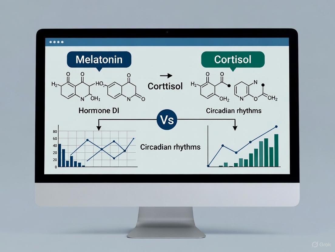
Within the broader field of circadian rhythm research, the quest for robust, measurable phase markers is paramount for both basic science and clinical application. While melatonin is widely regarded as the gold-standard marker for assessing the phase of the endogenous circadian clock, its measurement, particularly the Dim Light Melatonin Onset (DLMO), requires controlled conditions that can be logistically challenging [13] [14]. Consequently, cortisol, with its pronounced diurnal rhythm, presents itself as a potential alternative or complementary biomarker. The hypothalamic-pituitary-adrenal (HPA) axis tightly regulates cortisol secretion, producing a characteristic pattern upon which a superimposed pulsatile rhythm allows for rapid dynamic responses to environmental changes [20]. This review objectively compares the performance of cortisol against melatonin as a circadian phase marker, evaluating supporting experimental data on its regulation, reliability, and methodological constraints for research and drug development professionals.
Biomarker Comparison: Cortisol vs. Melatonin
The direct comparison of cortisol and melatonin reveals critical differences in their utility as circadian phase markers. The following table summarizes their key characteristics side-by-side.
Table 1: Direct Comparison of Circadian Phase Markers: Cortisol vs. Melatonin
| Feature | Cortisol | Melatonin |
|---|---|---|
| Primary Role | Stress response, metabolism, catabolic signal [20] [21] | Sleep-onset signaling, "darkness" signal [14] |
| Circadian Pattern | Peak ~30-45 min after awakening (CAR), nadir at night [21] [14] | Onset (DLMO) 2-3h before habitual sleep, peak at night [14] |
| Key Phase Marker | Cortisol Awakening Response (CAR); Cortisol acrophase [13] [20] | Dim Light Melatonin Onset (DLMO) [13] [14] |
| Phase Precision | Lower precision (SD ~40 min for SCN phase) [14] | High precision (SD 14-21 min for SCN phase) [14] |
| Major Confounders | Stress, awakening process, circadian misalignment, HPA axis dysregulation [20] [22] [23] | Ambient light, NSAIDs, beta-blockers, antidepressants, sleep deprivation [14] |
| Ideal Sampling Matrix | Saliva (for CAR), Blood [13] [14] | Saliva (for DLMO), Blood [13] [14] |
| Gold-Standard Assay | LC-MS/MS (superior specificity/sensitivity) [13] [14] | LC-MS/MS (superior specificity/sensitivity) [13] [14] |
A crucial development in this field is the recent evidence challenging the traditional interpretation of the Cortisol Awakening Response (CAR). A 2025 study measured tissue cortisol both before and after waking and found that awakening itself does not activate an increase in cortisol release [22]. The observed increase in levels after waking is more likely the tail end of a circadian-driven rise that began in the early hours of the morning, peaking shortly after habitual wake time [22]. This finding necessitates a careful re-evaluation of CAR's physiological basis and its interpretation in clinical studies.
Experimental Protocols for Circadian Assessment
The Constant Routine Protocol
To isolate the endogenous circadian component of cortisol rhythm from external influences, researchers employ the Constant Routine (CR) protocol. This method is the gold-standard for assessing intrinsic rhythms [24].
- Objective: To remove or uniformly distribute external and behavioral influences (e.g., sleep-wake cycles, feeding, posture, light exposure) that can mask the endogenous rhythm [20] [24].
- Key Procedure: Participants remain awake in a semi-recumbent posture for at least 24 hours in a controlled, dimly lit environment. They consume identical hourly isocaloric snacks and have blood samples collected frequently via an indwelling intravenous catheter [20] [24].
- Application: Studies using this protocol have demonstrated that the circadian rhythm of cortisol persists under these conditions, confirming its endogenous nature. Recent work using CR has been pivotal in redefining the CAR, showing that the cortisol increase around waking is part of a pre-awakening circadian rise rather than a response to waking itself [22].
Forced Desynchrony Protocol
This protocol is used to explicitly separate the effects of the endogenous circadian pacemaker from the imposed sleep-wake cycle.
- Objective: To induce circadian misalignment and study its effects on cortisol rhythms and other physiological outputs [20].
- Key Procedure: Participants are placed on sleep-wake cycles that are outside the range of entrainment for the human circadian pacemaker (e.g., a 28-hour "day" in an environment free of time cues). This causes the endogenous rhythm and the behavioral cycle to move in and out of alignment [20].
- Outcome Data: One study using forced desynchrony found that prolonged circadian misalignment can decrease overall cortisol exposure (24-hour area under the curve) and increase variability in the timing of individual cortisol peaks [20].
Measurement Methodologies and Data Interpretation
Accurate quantification is critical for reliable data. Immunoassays (e.g., ELISA) are widely used but can suffer from cross-reactivity with other steroids. Liquid chromatography–tandem mass spectrometry (LC-MS/MS) has emerged as a superior alternative, offering enhanced specificity, sensitivity, and reproducibility for both salivary and serum hormone measurements [13] [14].
When interpreting cortisol data, particularly the CAR, the recent 2025 findings dictate a paradigm shift. Researchers should exercise extreme caution when interpreting post-wake cortisol values without information about the pre-waking state [22]. The major cause of changes in cortisol around awakening is now understood to be predominantly related to the endogenous circadian rhythm, not the act of waking [22].
Table 2: Impact of Common Conditions on Cortisol Rhythms and Measurement
| Condition/Intervention | Impact on Cortisol Rhythm | Key Experimental Findings |
|---|---|---|
| Acute Circadian Misalignment (e.g., 3-day night shift) | Minimal change in mesor or amplitude; small but significant delay in acrophase (~26.5 minutes) [20]. | Studied under constant routine protocol after simulated shifts; circadian rhythmicity remains apparent [20]. |
| Chronic Circadian Misalignment (e.g., forced desynchrony) | Can decrease overall 24-hour cortisol exposure (AUC); greater variability in peak timing [20]. | Differs from acute misalignment, suggesting duration of misalignment is a key variable [20]. |
| Sleep Restriction | Increases late afternoon/early evening cortisol; does not significantly alter 24-hour cortisol output [20]. | Effect is distinct from that of circadian misalignment [20]. |
| Burnout / Chronic Stress | Associated with cortisol dysregulation and circadian misalignment; can lead to hyper- or hypocortisolemia [21] [25]. | Observed in shift-working healthcare professionals; linked to suppressed melatonin and poor sleep [25]. |
| Post-Stroke | Common HPA axis dysregulation; hypercortisolemia linked to poor functional outcomes and mortality; loss of diurnal variation in severe cases [21]. | High cortisol levels post-stroke are correlated with neurological deficits and inferior memory [21]. |
The Scientist's Toolkit: Key Research Reagent Solutions
- LC-MS/MS Kits and Reagents: Essential for achieving high-specificity measurement of low-abundance cortisol in saliva and serum. These kits provide the necessary columns, internal standards (e.g., deuterated cortisol), and mobile phases to minimize cross-reactivity and provide high sensitivity, which is crucial for accurate salivary CAR assessment [13] [14].
- Salivary Cortisol Immunoassay Kits: Despite limitations in specificity, these kits remain popular for high-throughput analysis due to their lower cost and technical demands. They are ideal for large-scale field studies where the extreme precision of LC-MS/MS is not required, but researchers must be aware of potential cross-reactivity [13] [14].
- Automated Saliva Sampling Systems: Crucial for standardized, at-home collection of saliva for CAR measurement. These systems often include neutral cotton-based swabs and stabilizing buffers to preserve sample integrity. The use of such systems was key in the recent 2025 study that measured cortisol before and after waking [22].
- Radioimmunoassay (RIA) Kits: Used in earlier and some current constant routine studies for measuring plasma melatonin and cortisol. While providing good sensitivity, they are being progressively supplanted by LC-MS/MS due to the latter's superior specificity and lack of radioactivity [24].
- Actigraphy Devices: Worn on the wrist to objectively monitor sleep-wake cycles and physical activity in the days leading up to and during a study. This data is vital for ensuring participant compliance with pre-study sleep schedules and for correlating cortisol measures with rest-activity cycles [24].
Signaling Pathways and Experimental Workflows
HPA Axis Regulation of Cortisol
The following diagram illustrates the core signaling pathway of the HPA axis, which governs cortisol secretion and its subsequent feedback and synchronizing functions.
Experimental Workflow for Circadian Rhythm Assessment
This workflow outlines the key methodological steps for a rigorous investigation of cortisol's circadian rhythm, incorporating protocols like the Constant Routine.
For researchers and drug development professionals, the choice between cortisol and melatonin as a circadian phase marker involves a clear trade-off between convenience and precision. Melatonin's DLMO remains the superior marker for accurately pinpointing the phase of the central circadian pacemaker due to its high precision and direct responsiveness to the light-dark cycle [14]. However, cortisol measurement, particularly the CAR, offers a non-invasive and practical alternative for large-scale studies where the logistical burden of DLMO assessment is prohibitive. The critical caveat is that the physiological basis of the CAR has been reinterpreted, and its value may lie more in reflecting the integrity of the HPA axis and the preceding circadian rise than as a pure response to awakening [22]. Future research integrating both markers under standardized protocols and utilizing high-fidelity assays like LC-MS/MS will provide the most comprehensive view of circadian health and its disruption in disease.
In the realm of chronobiology, melatonin and cortisol represent two fundamental endocrine markers that exhibit a striking antagonistic relationship crucial for maintaining optimal physiological function. These two hormones operate in a precisely coordinated, inverse rhythmic pattern that orchestrates the body's sleep-wake cycle, energy metabolism, and stress adaptation [14] [26]. This diurnal dance is governed by the suprachiasmatic nucleus (SCN) in the hypothalamus, which serves as the body's master clock, synchronizing peripheral clocks throughout tissues and organs [14] [27] [26].
The phase opposition between these hormones is not merely coincidental but represents an evolutionarily conserved temporal architecture that optimizes biological processes according to anticipated environmental demands. Understanding this relationship extends far beyond academic interest, as disruptions in this delicate balance have been implicated in numerous pathological conditions including neurodegenerative diseases, metabolic syndrome, psychiatric illnesses, sleep disorders, and even certain cancers [14] [27]. For researchers and pharmaceutical developers working in chronotherapeutics, unraveling the complexities of this hormonal interplay opens avenues for more precisely timed interventions that align with intrinsic biological rhythms.
Physiological Mechanisms of Antagonistic Regulation
Central and Peripheral Timing Systems
The antagonistic relationship between melatonin and cortisol emerges from a sophisticated hierarchical regulatory system beginning with light detection and culminating in hormonal secretion. The SCN receives photic input directly from specialized melanopsin-containing retinal ganglion cells via the retinohypothalamic tract, enabling entrainment to the external light-dark cycle [27] [26]. This light information is then translated into hormonal signals that synchronize peripheral clocks throughout the body.
The SCN regulates cortisol secretion through the hypothalamic-pituitary-adrenal (HPA) axis, which results in cortisol release from the adrenal cortex [28] [27]. Conversely, the SCN inhibits melatonin production during daylight hours via a multisynaptic pathway that extends from the paraventricular nucleus through the superior cervical ganglia to the pineal gland [26]. As daylight diminishes, this inhibitory signal is lifted, allowing melatonin synthesis and secretion to proceed [26]. This creates the fundamental antagonism: light suppresses melatonin while stimulating cortisol, and darkness enables melatonin while cortisol declines.
Molecular Crosstalk and Systemic Integration
Beyond their centralized regulation, melatonin and cortisol engage in bidirectional modulation at the molecular level. Cortisol functions as a key zeitgeber (time-giver) for peripheral clocks by influencing the expression of clock genes in tissues throughout the body [26]. The rhythmic fluctuation of cortisol—with its characteristic morning peak and evening trough—helps synchronize metabolic processes with the active phase [26]. Melatonin, often called the "molecular expression of darkness," provides the counter-signal that entrains the body to the inactive phase, promoting sleep-related processes and exerting antioxidant effects through both receptor-mediated and receptor-independent pathways [26].
This hormonal opposition creates a coordinated temporal framework that partitions physiological processes appropriately across the 24-hour cycle. The system ensures that energy mobilization (cortisol-driven) coincides with anticipated activity periods, while restorative functions (melatonin-facilitated) dominate the night [27] [26]. Recent research indicates that this antagonism extends to immune regulation, oxidative stress management, and cellular repair mechanisms, suggesting why disruption of this rhythm has such far-reaching health consequences [14] [26].
Figure 1: Physiological Regulation of Melatonin and Cortisol Secretion. The suprachiasmatic nucleus (SCN) coordinates antagonistic hormonal outputs in response to light input. Light stimulates cortisol production via the HPA axis while suppressing melatonin synthesis through sympathetic inhibition of the pineal gland. The resulting inverse relationship synchronizes peripheral clocks throughout the body.
Quantitative Assessment of Circadian Phase Markers
Methodological Approaches for Hormone Quantification
Accurately measuring the phase opposition between melatonin and cortisol requires careful consideration of methodological approaches. Researchers have several analytical platforms and biological matrices at their disposal, each with distinct advantages and limitations for circadian studies [14].
Liquid chromatography-tandem mass spectrometry (LC-MS/MS) has emerged as the gold standard for hormone quantification due to its superior specificity, sensitivity, and ability to measure multiple analytes simultaneously [14] [13]. This method significantly reduces cross-reactivity issues that plague traditional immunoassays (ELISA), particularly problematic for low-abundance analytes like melatonin [14]. For circadian applications, the capacity to measure both hormones from a single sample without additional cost or time makes LC-MS/MS particularly advantageous [14].
The choice of biological matrix represents another critical consideration. Salivary measurement has gained popularity for its non-invasive nature, allowing frequent sampling in ambulatory settings that capture dynamic hormonal fluctuations [14] [29]. Blood sampling provides higher analyte concentrations and improved reliability but presents logistical challenges for frequent sampling [14]. Recently, passive perspiration has emerged as a novel matrix enabling continuous, non-invasive monitoring through wearable biosensors, showing strong correlation with salivary levels (Pearson r = 0.92 for cortisol and r = 0.90 for melatonin) [10].
Table 1: Analytical Method Comparison for Melatonin and Cortisol Detection
| Method | Sensitivity | Specificity | Throughput | Cost | Best Applications |
|---|---|---|---|---|---|
| LC-MS/MS | High (pg/mL) | Excellent | Moderate | High | Gold standard for research; simultaneous multi-analyte quantification |
| ELISA/Immunoassay | Moderate | Variable cross-reactivity | High | Moderate | High-throughput screening; clinical diagnostics |
| Wearable Sensors | Developing | Correlates with reference methods | Continuous | Varies | Ambulatory monitoring; real-time circadian tracking |
Defining Circadian Phase Markers
The antagonistic relationship between melatonin and cortisol is most precisely captured through specific circadian phase markers that have been standardized for research and clinical applications.
The Dim Light Melatonin Onset (DLMO) represents the most reliable marker of internal circadian timing, typically defined as the time when melatonin concentrations cross a threshold of 3-4 pg/mL in saliva or 10 pg/mL in serum under dim light conditions [14]. DLMO usually occurs 2-3 hours before habitual bedtime and can be determined through several analytical approaches including fixed threshold, variable threshold (2 standard deviations above baseline), or the "hockey-stick" algorithm for objective automated assessment [14].
The Cortisol Awakening Response (CAR) serves as the complementary marker, characterized by a sharp 50-150% increase in cortisol levels within 30-45 minutes after waking [14] [27]. This response prepares the body for anticipated demands of the day and is influenced by both circadian timing and sleep quality [14]. A blunted CAR (increase <50%) indicates impaired HPA axis function and is observed in various pathological conditions including burnout and shift work disorder [27] [30].
Table 2: Characteristic Patterns of Circadian Phase Markers in Healthy Adults
| Parameter | Melatonin Rhythm | Cortisol Rhythm | Phase Relationship |
|---|---|---|---|
| Evening (6PM-10PM) | DLMO: Rise begins 2-3h before bedtime | Steady decline | Melatonin rise begins as cortisol reaches nadir |
| Night (10PM-4AM) | Peak: 80-100 pg/mL (saliva) | Lowest: 0.54-2.28 ng/mL (saliva) | Maximal separation: high melatonin, low cortisol |
| Morning (4AM-10AM) | Sharp decline to <3 pg/mL | CAR: 50-150% increase within 45min of waking | Cortisol peak coincides with melatonin offset |
| Day (10AM-6PM) | Low: <3 pg/mL | Gradual decline | Both at lower levels with cortisol dominant |
Experimental Models and Research Applications
Standardized Protocols for Circadian Assessment
Robust investigation of the melatonin-cortisol antagonism requires strict adherence to standardized protocols that minimize confounding variables. For DLMO assessment, researchers should implement a 4-6 hour sampling window from 5 hours before to 1 hour after habitual bedtime, with samples collected every 30-60 minutes under dim light conditions (<10 lux) [14]. Participants should avoid substances that interfere with melatonin secretion including NSAIDs, antidepressants, beta-blockers, and estrogen-containing medications for an appropriate washout period before testing [14] [31].
CAR assessment requires precise timing relative to awakening, with samples collected immediately upon waking and at 15, 30, and 45 minutes post-awakening [14] [27]. Participants must be trained to record exact awakening times and provide samples without delay, as the dynamic response unfolds rapidly. For comprehensive circadian profiling, extended sampling every 2-4 hours throughout the 24-hour cycle captures the full antagonistic pattern, though this imposes significant participant burden [32].
Recent technological innovations have introduced wearable biosensors that continuously monitor both hormones in passive perspiration, enabling unprecedented temporal resolution in free-living conditions [10]. These devices demonstrate strong agreement with traditional salivary measures (Bland-Altman bias near zero with narrow limits of agreement: -6.09 to 5.94 ng/mL for cortisol and -7.54 to 10.77 pg/mL for melatonin) and facilitate long-term circadian tracking without disrupting natural behaviors [10].
Figure 2: Experimental Workflow for Concurrent Melatonin and Cortisol Assessment. Standardized protocols for circadian hormone measurement require strict control at multiple points to ensure data quality and reproducibility, particularly regarding light exposure, timing precision, and confounding substances.
Research Models of Circadian Disruption
Several experimental and observational models effectively illustrate the consequences of disrupted melatonin-cortisol antagonism. Night-shift work represents a natural experiment in circadian misalignment, characterized by altered cortisol secretion patterns including blunted morning peaks, delayed acrophase (peak timing), and elevated nighttime levels [27]. Shift workers consistently demonstrate approximately 40% reduction in melatonin amplitude and 2-3 hour shifts in cortisol peak timing compared to day workers [27] [30].
Occupational burnout provides another compelling model of hormonal dysregulation, with healthcare professionals showing suppressed melatonin secretion and flattened cortisol rhythms proportional to burnout severity [30]. Across 14 studies systematically reviewed, burnout was associated with both melatonin suppression and CAR blunting, creating a pathological state of both chronic activation and circadian misalignment [30].
Sleep disorders such as obstructive sleep apnea (OSA) further demonstrate the interdependence of these hormonal systems. Patients with OSA show altered melatonin profiles (mean 80.80 ± 52.48 pg/mL at baseline) that partially normalize following CPAP treatment (63.78 ± 39.85 pg/mL after 8 weeks), though corresponding cortisol changes are more variable [29]. These clinical observations reinforce the functional significance of maintained antagonism for overall health.
The Scientist's Toolkit: Essential Research Reagents and Materials
Table 3: Essential Research Materials for Circadian Hormone Investigation
| Category | Specific Items | Research Application | Technical Considerations |
|---|---|---|---|
| Sample Collection | Salivette collection devices; Sterile polyethylene tubes; Portable cold packs | Ambulatory sample collection for CAR and DLMO assessment | Maintain cold chain; record exact collection times; ensure participant compliance |
| Storage | -70°C freezer; Cryogenic vials; Centrifuge with temperature control | Sample preservation before analysis | Avoid freeze-thaw cycles; centrifuge saliva before storage to remove mucins |
| Analytical | LC-MS/MS system; HPLC columns; Mass spectrometry reagents | Gold standard quantification of melatonin and cortisol | Method validation required; consider deuterated internal standards for precision |
| Immunoassay | Human Melatonin ELISA Kit; Human Cortisol ELISA Kit; Microplate reader | High-throughput screening alternative to LC-MS/MS | Check cross-reactivity; validate against gold standard; optimize sensitivity |
| Circadian Software | CircaCompare; Cosinor analysis; "Hockey-stick" algorithm | Rhythm analysis and phase marker determination | Select appropriate algorithm based on sampling density and hormone profile |
| Wearable Tech | Passive perspiration sensors; Actigraphy devices | Continuous ambulatory monitoring | Validate against serum/saliva standards; ensure proper skin contact |
Implications for Chronotherapy and Drug Development
The precise antagonism between melatonin and cortisol offers compelling opportunities for chronotherapeutic interventions that align treatment timing with intrinsic biological rhythms. The circadian regulation of drug targets presents untapped potential for optimizing efficacy and minimizing side effects [14]. For instance, evidence suggests that approximately 80% of protein-coding genes exhibit circadian expression patterns, including those involved in drug metabolism, transport, and mechanism of action [14].
Pharmaceutical development can leverage this hormonal antagonism in several strategic approaches. First, timed administration of medications can coincide with peaks in relevant hormone receptors or metabolic pathways. Second, circadian biomarker stratification in clinical trials may identify patient subgroups most likely to respond to chronotherapeutic approaches. Third, drug delivery systems can be engineered to release active compounds at optimal biological times based on an individual's melatonin-cortisol phase relationship.
For researchers developing treatments for sleep disorders, metabolic conditions, or psychiatric illnesses, monitoring this hormonal axis provides crucial insights into therapeutic mechanisms and timing. The development of wearable biosensors that continuously track both hormones [10] promises to personalize chronotherapy based on individual circadian phase rather than arbitrary clock time, potentially revolutionizing treatment for the approximately 20% of the workforce engaged in shift work [27] and the broader population suffering from circadian rhythm sleep-wake disorders.
In mammals, the circadian clock is an endogenous timekeeping system that generates 24-hour rhythms in physiology and behavior, synchronizing them with the external environment. At its core lies a meticulously orchestrated transcription-translation feedback loop (TTFL) composed of core clock genes and their protein products: CLOCK, BMAL1, PER, and CRY. This molecular oscillator not only governs daily cycles but also influences broader physiological processes, from sleep-wake patterns to drug metabolism [14]. The precision of this clock is increasingly recognized as a critical factor in human health, with its disruption implicated in various disorders, including sleep disturbances, metabolic syndrome, and neurodegenerative diseases [6] [14].
The study of circadian rhythms in humans relies on accurately measuring the output of this internal clock. Hormonal biomarkers like melatonin and cortisol serve as vital, measurable proxies for the underlying circadian phase, as direct measurement of the master pacemaker—the suprachiasmatic nucleus (SCN)—is not feasible [14]. Melatonin, the "hormone of darkness," and cortisol, which peaks around waking, provide a window into the clock's timing. Understanding the fundamental TTFL that drives these rhythms is therefore essential for both basic research and the growing field of circadian medicine [14].
The Core Circadian Feedback Loop: A Molecular Oscillator
The stability of our daily rhythms is founded upon an interlocked series of molecular events. The core TTFL can be dissected into a primary negative feedback loop and a stabilizing auxiliary loop.
The Primary Negative Feedback Loop
The core circadian loop is initiated by the CLOCK-BMAL1 heterodimer. BMAL1 (also known as ARNTL1) and CLOCK are transcriptional activators that form a complex and bind to E-box enhancer elements in the promoter regions of target genes, including the Period (Per1, Per2, Per3) and Cryptochrome (Cry1, Cry2) genes [6] [33]. This binding drives the transcription of Per and Cry genes.
Following translation, PER and CRY proteins accumulate in the cytoplasm. After undergoing specific post-translational modifications, they form multimeric complexes that translocate back into the nucleus. Once there, they act as the negative limb of the feedback loop by directly inhibiting the transcriptional activity of the CLOCK-BMAL1 complex. This action represses their own transcription, completing the core cycle [6] [34].
The Stabilizing Auxiliary Loop
An interlocking auxiliary loop provides robustness to the core oscillator. The CLOCK-BMAL1 heterodimer also activates the transcription of the nuclear receptor genes Rev-erbα and Rora [6] [35]. Their protein products, REV-ERBα and RORα, compete for binding to ROR response elements (RREs) in the Bmal1 promoter. RORα acts as a transcriptional activator, while REV-ERBα functions as a repressor, creating a second, interlocking feedback loop that rhythmically regulates Bmal1 transcription [6] [35]. This dual-loop architecture is crucial for system stability.
Table 1: Core Components of the Circadian Transcription-Translation Feedback Loop (TTFL)
| Component | Gene Symbol | Role in TTFL | Functional Classification |
|---|---|---|---|
| BMAL1 | ARNTL1 | Forms heterodimer with CLOCK; binds E-box to activate transcription of Per and Cry genes. | Transcriptional Activator |
| CLOCK | CLOCK | Forms heterodimer with BMAL1; histone acetyltransferase activity. | Transcriptional Activator |
| Period | PER1/2/3 | Forms repressor complex with CRY; inhibits CLOCK-BMAL1 activity. | Transcriptional Repressor |
| Cryptochrome | CRY1/2 | Forms repressor complex with PER; inhibits CLOCK-BMAL1 activity. | Transcriptional Repressor |
| REV-ERBα/β | NR1D1/2 | Represses Bmal1 transcription by binding ROREs. | Transcriptional Repressor (Auxiliary Loop) |
| RORα/γ | RORA/RORC | Activates Bmal1 transcription by binding ROREs. | Transcriptional Activator (Auxiliary Loop) |
The entire process from transcription to nuclear repression introduces an approximately 24-hour time delay, which is critical for generating sustained oscillations. This delay is fine-tuned by post-translational modifications (PTMs), such as phosphorylation by kinases like casein kinase 1δ/ε (CK1δ/ε), which mark PER proteins for degradation via the ubiquitin-proteasome system [6]. Furthermore, recent studies highlight the role of SUMOylation in modulating CLOCK-BMAL1 transcriptional activity and stability, adding another layer of regulatory control [6].
Experimental Insights into TTFL Dynamics and Stability
The intricate dynamics of the TTFL have been elucidated through sophisticated cellular and animal models, combined with mathematical modeling. Key experiments have moved beyond simply identifying components to quantifying their contributions to the system's robustness.
Disrupting the Auxiliary Loop: The ΔRRE Model
To investigate the specific role of the RRE-mediated auxiliary loop, researchers used CRISPR-Cas9 to generate mutant cells and mice (ΔRRE mutants) by deleting two highly conserved ROREs upstream of the Bmal1 gene [35]. This disruption abrogated the rhythmic transcription of Bmal1, resulting in its constitutive expression. Despite this loss of Bmal1 mRNA rhythm, both mutant cells and mice exhibited apparently normal circadian rhythms in behavior and tissue-level bioluminescence reporters [35].
However, a combination of experimental data and mathematical modeling revealed the subtle yet critical function of the auxiliary loop. The Kim-Forger mathematical model of the circadian clock was adapted to simulate the ΔRRE mutant. While the simulated mutant maintained oscillations, its circadian period and amplitude were more susceptible to perturbations when the CRY1 protein rhythm was disturbed [35]. This demonstrates that the RRE-mediated rhythmic transcription of Bmal1 is not strictly necessary for oscillation generation but is crucial for conferring perturbation-resistant stability to the entire timekeeping system.
Post-Translational Regulation and System-Level Integration
The TTFL is reinforced by robust post-translational regulation. For example, in the ΔRRE mutant, a BMAL1 phosphorylation rhythm persists even in the absence of its transcriptional rhythm [35]. This indicates that the protein phosphorylation cycle, potentially driven by the rhythmic activity of other clock components, can operate semi-independently to stabilize the clock. Furthermore, recent research has uncovered a layer of post-transcriptional regulation where the clock controls the sequestration of mRNAs into biomolecular condensates like stress granules, adding another dimension to the rhythmic control of gene expression [33].
Table 2: Key Experimental Models for Investigating the Core TTFL
| Experimental Model | Genetic Manipulation | Key Phenotypic Observations | Functional Insight |
|---|---|---|---|
| ΔRRE Mutant Cells/Mice [35] | Deletion of RRE elements in the Bmal1 promoter. | Loss of Bmal1 mRNA rhythm; preserved circadian oscillations in reporter genes and behavior. | The RRE-mediated loop is dispensable for rhythm generation but essential for robustness against perturbations. |
| Bmal1 Knockout Mice [6] [35] | Complete knockout of the Bmal1 gene. | Complete loss of circadian rhythmicity in constant darkness; severe sleep fragmentation. | BMAL1 is non-redundant and essential for the core oscillator function. |
| Human ARNTL Haploinsufficiency [34] | Heterozygous loss-of-function variants in the ARNTL (BMAL1) gene. | Neurodevelopmental disorders, early-onset epilepsy, hypotonia; circadian sleep disorders not a primary feature. | BMAL1 has developmental roles potentially separable from its core circadian function. |
| Per2 Mutant Models [6] | Mutations in the Per2 gene. | Altered circadian phase and sleep instability; shortened circadian period in some cases. | PER proteins are critical for maintaining the correct period and phase of the circadian cycle. |
Connecting Molecular Mechanisms to Circadian Phase Markers
The rhythmic output of the SCN's TTFL is communicated to the rest of the body via neural, hormonal, and behavioral signals. The hormones melatonin and cortisol are key peripheral markers of this central timing.
Melatonin: The Hormone of Darkness
The SCN regulates the pineal gland to secrete melatonin, a process tightly coupled to the core clock. Melatonin synthesis is suppressed by light and peaks during the biological night [14] [36]. The timing of its evening rise, measured as the Dim Light Melatonin Onset (DLMO), is considered the gold standard for assessing human circadian phase [14]. The molecular clock drives this rhythm, with CLOCK-BMAL1 heterodimers ultimately regulating the enzymes in the melatonin synthesis pathway through multisynaptic outputs [36].
Cortisol: The Glucocorticoid Rhythm
Cortisol secretion, under the control of the hypothalamic-pituitary-adrenal (HPA) axis, also exhibits a strong circadian rhythm, with a peak around wake-up time and a nadir around midnight [14] [37]. The Cortisol Awakening Response (CAR) is a sharp increase within 30-45 minutes of waking used as a marker of HPA axis rhythm [14]. While its rhythm is also driven by the SCN, cortisol is more susceptible to masking by external factors like stress and sleep-wake transitions than melatonin [14].
Table 3: Comparison of Key Circadian Phase Markers in Humans
| Parameter | Melatonin | Cortisol |
|---|---|---|
| Primary Role | Signals onset of biological night; promotes sleep. | Prepares body for activity; response to stress. |
| Phase Marker | Dim Light Melatonin Onset (DLMO). | Cortisol Awakening Response (CAR). |
| Peak Timing | Middle of the night (e.g., 02:00-04:00). | Early morning, shortly after awakening. |
| Relationship to Core TTFL | Synthesis driven by SCN via multisynaptic pathway. | Secretion driven by SCN via HPA axis and direct neural pathways. |
| Susceptibility to Masking | Low (requires strict dim-light conditions). | High (influenced by stress, sleep, posture, meals). |
| Phase Precision (Standard Deviation) | High (~14-21 minutes) [14]. | Lower (~40 minutes) [14]. |
| Key Analytical Challenge | Low concentrations in saliva; requires sensitive assays like LC-MS/MS. | Strong pulsatile secretion; requires high-frequency sampling. |
Analytical Methodologies for Circadian Research
Accurately measuring circadian phase requires rigorous protocols and analytical methods. The choice of biomarker, sampling matrix, and analysis technique significantly impacts data reliability.
Measuring Melatonin and Cortisol
- Sampling Matrices: Melatonin and cortisol can be measured in blood (plasma/serum), saliva, and urine. Saliva is favored for ambulatory studies due to its non-invasive nature, though it has lower hormone concentrations. Serum provides higher analyte levels but is more invasive [14].
- Analytical Techniques: Immunoassays are traditional but can suffer from cross-reactivity. Liquid chromatography-tandem mass spectrometry (LC-MS/MS) has emerged as a superior method due to its high specificity, sensitivity, and reproducibility, particularly for low-abundance salivary analytes [14].
- Determining DLMO: The most common method is the fixed threshold (e.g., melatonin concentration exceeding 3-4 pg/mL in saliva). Alternative methods include a variable threshold (e.g., two standard deviations above baseline) or the "hockey-stick" algorithm, which objectively identifies the breakpoint from baseline to rise [14] [38]. The choice of method depends on the individual's melatonin profile amplitude and the assay's sensitivity.
Critical Experimental Protocols
Controlled conditions are paramount. Studies must be conducted under dim light conditions (< 10 lux) to prevent melatonin suppression [36]. For high-precision phase assessment, researchers often use a Constant Routine (CR) protocol, which eliminates masking effects by keeping participants awake in a semi-recumbent posture with constant dim light and evenly distributed, isocaloric snacks [36]. The timing, intensity, and pattern of light exposure are critical variables that must be standardized to draw valid conclusions about circadian phase and phase-shifting responses [36].
The Scientist's Toolkit: Essential Research Reagents and Materials
Table 4: Key Reagent Solutions for Circadian TTFL and Biomarker Research
| Reagent / Material | Function / Application | Key Considerations |
|---|---|---|
| CRISPR-Cas9 System | Generation of knockout/knock-in cell and animal models (e.g., ΔRRE mutants) [35]. | Enables precise genetic manipulation of core clock genes and regulatory elements. |
| Bioluminescence Reporters (e.g., PER2::LUC) | Real-time monitoring of circadian rhythms in live cells and tissue explants [35]. | Provides high-temporal resolution of clock gene expression without destructive sampling. |
| LC-MS/MS (Liquid Chromatography-Tandem Mass Spectrometry) | Gold-standard quantification of melatonin and cortisol in biological matrices [14]. | Offers high specificity and sensitivity, crucial for low-concentration salivary measurements. |
| Dim Light Environment (< 10 lux) | Essential for unmasked assessment of melatonin secretion and DLMO calculation [14] [36]. | Requires controlled laboratory settings with calibrated lighting. |
| Constant Routine (CR) Protocol | Gold-standard human study protocol to minimize masking effects on circadian outputs [36]. | Logistically demanding; controls for sleep, posture, activity, food intake, and light. |
| Specific Immunoassays | Alternative method for hormone quantification (e.g., ELISA for melatonin/cortisol). | Potential for cross-reactivity; requires validation against LC-MS/MS for sensitivity/specificity. |
| Mathematical Models (e.g., Kim-Forger Model) | Theoretical framework to simulate and predict circadian dynamics following perturbations [35]. | Allows in silico testing of hypotheses about network robustness and gene function. |
The transcription-translation feedback loop of CLOCK, BMAL1, PER, and CRY genes forms the bedrock of circadian biology. This interlocked system, stabilized by auxiliary loops and post-translational modifications, generates a robust ~24-hour oscillation that coordinates our physiology. The experimental dissection of this loop, using models from mutant mice to human biomarker studies, has revealed its dual nature: capable of generating rhythms even with simplified architecture, yet dependent on its full complexity for stability.
Understanding these molecular mechanisms is more than an academic exercise; it is the foundation of circadian medicine. The TTFL regulates the expression of countless clock-controlled genes, influencing drug metabolism pathways and disease pathophysiology [6] [14]. The precise measurement of circadian phase via melatonin and cortisol is therefore critical for diagnosing circadian rhythm disorders and for developing chronotherapeutic strategies—timing medications to align with the body's internal clock to maximize efficacy and minimize side effects [14]. As research continues to unravel how clock disruption contributes to diseases from insomnia to neurodevelopmental disorders [6] [34], targeting the molecular gears of the circadian clock offers a promising path for future therapeutics.
From Lab to Clinic: Methodologies for Quantifying Circadian Phase
The suprachiasmatic nucleus (SCN) in the hypothalamus acts as the master pacemaker, coordinating circadian rhythms (~24-hour cycles) throughout the body [14]. These rhythms regulate critical physiological processes, including sleep-wake cycles, hormone secretion, metabolism, and behavior. Since direct measurement of SCN activity in humans is not feasible, reliable peripheral biomarkers are essential for assessing circadian phase in both research and clinical settings [14]. Among these, Dim Light Melatonin Onset (DLMO) and the Cortisol Awakening Response (CAR) have emerged as the two most prominent endocrine markers. DLMO, marking the evening onset of melatonin secretion, is widely regarded as the gold standard for assessing the central circadian phase [39] [14] [40]. In contrast, CAR, which measures the rapid increase in cortisol levels following morning awakening, provides a distinct perspective, reflecting both circadian influences and the activation of the hypothalamic-pituitary-adrenal (HPA) axis in response to stress [41] [42] [43]. This guide provides a comparative analysis of these two key phase markers, detailing their underlying physiology, measurement methodologies, and applicability in research and drug development.
Physiological Underpinnings and Molecular Basis
The secretion of melatonin and cortisol is governed by distinct yet interconnected neuroendocrine pathways. The following diagram illustrates the primary signaling pathways that regulate these two crucial circadian hormones.
Diagram 1: Signaling pathways regulating melatonin and cortisol secretion. The suprachiasmatic nucleus (SCN) integrates light input to synchronize both pathways. The melatonin pathway (red) is activated by the nocturnal SCN signal via a multisynaptic sympathetic pathway. The cortisol pathway (blue) involves SCN input to the paraventricular nucleus (PVN), activating the HPA axis. Both systems are under circadian control from core clock genes [44] [14] [43].
Melatonin: The Darkness Hormone
Melatonin is primarily synthesized and secreted by the pineal gland during the night. Its production is tightly controlled by the SCN via a multisynaptic pathway in the sympathetic nervous system [14]. The critical event is a disinhibition of the pineal gland, where the SCN removes its GABA-ergic suppression, allowing for the release of melatonin into the circulation [39]. The primary function of this hormonal signal is to convey timing information about the "biological night" to tissues throughout the body, thus promoting sleep and coordinating various circadian functions [14]. Melatonin rhythms are notably stable and are influenced very little by environmental factors such as sleep-wake state, exercise, or mood, making it a robust marker [44].
Cortisol: The Stress & Awakening Hormone
Cortisol, a glucocorticoid hormone produced by the adrenal cortex, exhibits a diurnal rhythm opposite to that of melatonin, with levels peaking in the morning and reaching a nadir around midnight [14] [43]. The CAR is a specific phenomenon characterized by a sharp increase (50-160%) in cortisol secretion that occurs within the first 30-45 minutes after waking [42] [40] [43]. This response was historically thought to be a direct reaction to the stress of waking up. However, a recent 2025 study using in-home microdialysis found that the rate of cortisol increase after waking was not significantly different from the rate of increase before waking, challenging the notion that waking itself is the primary driver [41]. This suggests CAR is more tightly regulated by the underlying circadian rhythm, though it remains highly sensitive to HPA axis activation from both anticipated challenges and psychological stress [41] [43].
Methodological Comparison: Protocols and Analytical Techniques
Accurate assessment of DLMO and CAR requires strict adherence to standardized protocols, as both are susceptible to confounding factors. The experimental workflow for measuring each marker is distinct, as outlined below.
Diagram 2: Comparative experimental workflows for DLMO and CAR assessment. The DLMO protocol (red) requires strict dim light conditions and evening sampling. The CAR protocol (blue) depends on precise post-awakening sampling and normal morning light exposure. LC-MS/MS = Liquid Chromatography with Tandem Mass Spectrometry [39] [42] [14].
Standardized Protocols for Phase Assessment
DLMO Protocol: The core requirement for DLMO assessment is sampling under dim light conditions (<30 lux) to prevent light-induced suppression of melatonin [39] [14]. Participants provide saliva samples every 30-60 minutes over a 4-6 hour window in the evening, typically starting 5 hours before and ending 1 hour after habitual bedtime [14]. The "dim light" requirement is absolute, and participants often use a pen-sized dim flashlight if movement is necessary [44]. Key confounders to control for include beta-blockers, non-steroidal anti-inflammatory drugs (NSAIDs), antidepressants, and melatonin supplements, all of which can alter the natural melatonin profile [14].
CAR Protocol: In contrast, CAR measurement requires participants to provide saliva samples immediately upon waking (within 5 minutes), then again at 30 and 60 minutes after waking [42] [40] [43]. The precise timing of sample collection is critical, as minutes can alter the results. Unlike DLMO, normal morning light exposure is essential, as light is a primary stimulus for the cortisol awakening response [43]. Adherence to the collection schedule must be verified, ideally with electronic monitoring. Factors such as sleep duration, wake time variability, and chronic stress levels significantly influence CAR and must be documented [41] [40].
Analytical Methods and Data Interpretation
DLMO Calculation: There is no universal standard for calculating DLMO. The two most common methods are:
- Fixed Threshold: The time when interpolated melatonin concentration crosses a pre-defined value (e.g., 3 pg/mL or 4 pg/mL in saliva) [39] [14].
- Variable Threshold: The time when melatonin levels rise two standard deviations above the mean of three or more baseline (pre-rise) values [14]. A comparison found the variable method estimated an earlier DLMO by 22-24 minutes in 76% of cases, though some studies favor the fixed threshold for its stability against fluctuating baselines [14]. Alternative methods like the "hockey-stick" algorithm offer more objective, automated assessment [14].
CAR Calculation: The CAR is typically quantified using:
- Area Under the Curve (AUC): Captures the total cortisol output during the awakening period.
- Mean Increase: The difference between the peak level (usually at 30 or 60 minutes) and the waking level. A normal CAR shows a 50-160% increase within the first 30 minutes after waking [43]. A blunted CAR (a low or absent rise) is associated with chronic stress and burnout, while an excessively high CAR may indicate high job strain or heightened stress reactivity [40] [43].
Table 1: Direct Comparison of DLMO and CAR as Circadian Phase Markers
| Feature | Dim Light Melatonin Onset (DLMO) | Cortisol Awakening Response (CAR) |
|---|---|---|
| Primary Physiological Role | Marker of biological night onset; promotes sleep [14] | Prepares body for anticipated energy demands; stress response [41] [43] |
| Gold-Standard Matrix | Saliva (blood also used) [14] | Saliva (urine also used) [42] [14] |
| Optimal Sampling Window | 4-6 hours in the evening (e.g., 18:00-00:00) [14] | 1 hour in the morning (samples at 0, 30, 60 min post-waking) [42] [43] |
| Critical Environmental Control | Dim light (<30 lux) to prevent suppression [39] [14] | Normal light exposure; strict adherence to sample timing [43] |
| Key Confounding Factors | Beta-blockers, NSAIDs, antidepressants, melatonin supplements [14] | Sleep duration/quality, wake time variability, chronic stress [41] [40] |
| Phase Marker Precision (Standard Deviation) | ~14-21 minutes [14] | ~40 minutes [14] |
| Stability | High; less influenced by sleep or exercise [44] [45] | Moderate; highly sensitive to state factors (stress, sleep) [41] |
Research Applications and Commercial Testing
Utility in Clinical and Research Settings
DLMO and CAR serve distinct but complementary purposes in research and clinical diagnostics.
DLMO Applications: DLMO is the preferred marker for diagnosing circadian rhythm sleep-wake disorders, such as Delayed and Advanced Sleep-Wake Phase Disorder [14] [40]. It is also critical in studies investigating the impact of shift work, jet lag, and light therapy, where precise determination of the central circadian phase is required [39]. Furthermore, research is exploring its role in neurodegenerative diseases like Alzheimer's, where nocturnal melatonin secretion is often suppressed [14].
CAR Applications: CAR is widely used as a biomarker of HPA axis dynamics and stress reactivity in psychiatric and metabolic research [41] [43]. A blunted CAR is frequently reported in individuals with chronic fatigue syndrome, post-traumatic stress disorder (PTSD), and major depressive disorder [42] [40] [43]. Conversely, an elevated CAR may be associated with current job strain or anxiety [40]. It is also studied in relation to general health outcomes and cardiometabolic parameters [41] [39].
Commercial Testing and Emerging Technologies
Several commercial laboratories offer DLMO and CAR testing, primarily using saliva samples.
Commercial CAR Tests: Companies like ZRT Laboratory and US BioTek offer saliva test kits that measure cortisol at multiple points across the day, including the key CAR time points (waking, +30 min, +60 min) [46] [42]. Precision Analytical (DUTCH test) offers a comprehensive CAR profile that also includes cortisone, the inactive metabolite of cortisol, which can provide additional insight into HPA axis activity [43].
Commercial DLMO Tests: While not as widely offered as CAR tests, home-based DLMO kits are available and have been validated in research settings. These kits include detailed instructions for dim light compliance and saliva collection in the evening [39].
Emerging Technologies: Research is rapidly advancing toward non-invasive, continuous monitoring. A 2025 study demonstrated a wearable biosensor that measures cortisol and melatonin in passive perspiration, showing strong agreement with salivary levels (Pearson r = 0.92 for cortisol and r = 0.90 for melatonin) [10]. This technology promises to revolutionize circadian research by enabling real-world, high-resolution hormonal profiling.
Essential Research Toolkit
Table 2: Key Reagents and Materials for Circadian Phase Marker Analysis
| Item | Function | Example Use Case |
|---|---|---|
| Saliva Collection Tubes | Non-invasive sample collection for hormone analysis. | Collecting serial samples for CAR (0, 30, 60 min) or DLMO (every 30 min in evening) [46] [42]. |
| Portient Actigraphs | Objective monitoring of sleep-wake cycles and physical activity. | Verifying sleep schedules and timing of sample collection in home-based studies [44] [39]. |
| LC-MS/MS Instrumentation | Gold-standard analytical method for hormone quantification; offers high sensitivity and specificity [14]. | Precisely measuring low concentrations of salivary melatonin and cortisol, avoiding cross-reactivity of immunoassays [14]. |
| Dim Light Meter | Verifies ambient light levels are below the suppression threshold (<30 lux) [39]. | Ensuring compliance with dim light protocols during evening DLMO sampling at participants' homes. |
| Electronic Monitoring Adherence | Tracks compliance with saliva sample collection times. | Monitoring and verifying that CAR samples were taken at the exact required times after waking [40]. |
DLMO and CAR are fundamental, yet distinct, pillars of human circadian assessment. DLMO stands as the most precise and reliable marker of the central circadian phase, directly reflecting the timing signal emitted by the SCN with minimal influence from behavioral or state-dependent factors [44] [14]. Its precision, with a standard deviation of 14-21 minutes, makes it indispensable for basic chronobiology and diagnosing circadian rhythm disorders [14]. In contrast, CAR is a composite measure, reflecting the interplay of the endogenous circadian rhythm, the sleep-wake transition, and, prominently, the individual's stress system and HPA axis reactivity [41] [43]. Its greater variability (~40 minutes standard deviation) and sensitivity to daily experiences make it less ideal for pinpointing pure circadian phase but highly valuable for investigating stress physiology and its links to physical and mental health [14].
The choice between these markers is not a matter of superiority but of scientific or clinical question. DLMO is the unambiguous choice for mapping the phase of the master clock. CAR is more appropriate for studies focused on stress, allostatic load, and the metabolic preparedness for the day ahead. Emerging evidence from 2025 even suggests that the cortisol rise at waking may be more a continuation of a pre-awakening circadian rhythm than a direct response to awakening itself, further refining our understanding of CAR [41].
Future directions in the field point toward the integration of these markers in longitudinal studies and the adoption of new technologies, such as wearable sweat sensors [10], which will allow for unobtrusive, continuous monitoring of both hormones in naturalistic environments. This will provide unprecedented insight into the dynamic relationship between the circadian system, stress, and real-world behaviors, ultimately advancing the field of circadian medicine and the development of chronotherapeutic interventions.
The selection of an appropriate biological matrix is a critical determinant of success in biomedical research and clinical diagnostics, particularly in the precise field of circadian rhythm research. The accurate measurement of endocrine markers such as melatonin and cortisol depends fundamentally on the biological fluid chosen for analysis [13] [14]. Each matrix—serum, saliva, and urine—offers a unique combination of advantages and limitations, influencing data reliability, participant compliance, and methodological feasibility [47] [48] [49].
This guide provides an objective comparison of these three major biological matrices, with special emphasis on their application in circadian phase assessment. We synthesize current methodological insights and experimental data to help researchers and drug development professionals make evidence-based decisions for their specific investigative contexts.
Comparative Analysis of Biological Matrices
The table below summarizes the core characteristics, advantages, and challenges of serum, saliva, and urine as sampling matrices in biomedical research.
Table 1: Comprehensive Comparison of Serum, Saliva, and Urine Sampling Matrices
| Characteristic | Serum/Plasma | Saliva | Urine |
|---|---|---|---|
| Invasiveness | Invasive (venipuncture) [50] | Minimally invasive/non-invasive [47] [50] | Non-invasive [48] |
| Collection Simplicity | Requires trained phlebotomist [48] | Simple; potential for self-collection [47] | Simple; can be self-collected [48] |
| Patient Compliance | Lower, especially for repeated sampling [47] | High, suitable for children and elderly [47] [48] | High [48] |
| Risk of Infection | Higher (sharp objects) [48] | Very low | Very low |
| Analyte Stability | Generally good, but requires specific processing [47] | More vulnerable to enzymatic degradation [47] | Generally good; depends on analyte [48] |
| Representativeness | Systemic circulation; "gold standard" [47] | Reflects free, biologically active fraction of analytes [50] | Represents metabolic waste and clearance [48] |
| Ideal for Circadian Studies | Yes, but limited by frequency | Excellent for high-frequency, ambulatory sampling [13] [14] | Excellent for long-term rhythm assessment (e.g., 24h profiles) [48] |
Performance in Circadian Rhythm Research
The assessment of circadian phase relies heavily on the robust measurement of rhythmic biomarkers, primarily melatonin and cortisol. The performance of different matrices varies significantly for these applications.
Table 2: Matrix Performance for Key Circadian Biomarkers
| Biomarker & Application | Serum/Plasma | Saliva | Urine |
|---|---|---|---|
| Melatonin (DLMO Assessment) | Sensitivity: HighConsiderations: Gold standard for Dim Light Melatonin Onset (DLMO); measures total hormone [13] [14] | Sensitivity: High for free hormoneConsiderations: Correlates with free serum levels; less sensitive for low producers; ideal for frequent sampling [13] [14] | Sensitivity: Metabolites, not parent compoundConsiderations: Used for long-term rhythm patterns (e.g., 7-day rhythms) [48] |
| Cortisol (CAR Assessment) | Sensitivity: HighConsiderations: Measures total hormone; invasive for the tight sampling of the Cortisol Awakening Response (CAR) [14] | Sensitivity: High for free hormoneConsiderations: Method of choice for CAR due to non-invasive, high-frequency sampling [14] | Sensitivity: MetabolitesConsiderations: Suitable for assessing daily or longer-term integrated output [48] |
| Metabolomics (COVID-19 Study Example) | Sensitivity: 0.97, Specificity: 0.97 [49] | Sensitivity: 0.78, Specificity: 0.83 [49] | Not available in cited study |
| Key Diagnostic Strengths | Systemic overview; high sensitivity and specificity for many analytes [47] [49] | Non-invasive dynamic monitoring; reflects bioavailable fraction [47] [50] | Cumulative metabolic picture; ideal for compliance-challenging populations [48] |
Experimental Protocols for Circadian Phase Assessment
Protocol for Determining Dim Light Melatonin Onset (DLMO) from Saliva
Dim Light Melatonin Onset is the gold standard marker for assessing the timing of the central circadian clock [14]. Saliva is the preferred matrix for its practicality in frequent sampling.
- Sample Collection: Collect saliva samples every 30-60 minutes over a 4-6 hour window, typically starting 5 hours before and ending 1 hour after the individual's habitual bedtime [14].
- Environmental Control: Sampling must occur under dim light conditions (< 10-30 lux) to prevent light-induced suppression of melatonin secretion [13] [14].
- Sample Handling: Participants should not eat, drink (except water), or smoke during the sampling period. Saliva is collected using specialized synthetic swabs (e.g., Salivette), then centrifuged and stored at -20°C or -80°C until analysis [14].
- Analytical Technique: Liquid Chromatography-Tandem Mass Spectrometry (LC-MS/MS) is recommended for its high specificity and sensitivity, overcoming cross-reactivity issues inherent in immunoassays [13] [14].
- Data Analysis - DLMO Calculation: The most common method is the fixed threshold approach, where DLMO is defined as the time when the melatonin concentration continuously exceeds a threshold of 3-4 pg/mL. Alternative methods include the variable threshold (2 standard deviations above the mean of baseline samples) and the "hockey-stick" algorithm for objective, automated estimation [14].
Protocol for Assessing Cortisol Awakening Response (CAR) from Saliva
The CAR is a distinct rise in cortisol levels peaking 30-45 minutes after awakening, reflecting HPA axis activity and influenced by the circadian system [14].
- Sample Collection: Participants collect saliva samples immediately upon waking (Sample 1), then at 30, and 45 minutes post-awakening. Exact timing must be strictly adhered to and verified [14].
- Sample Handling: Similar to melatonin protocols, samples are collected using appropriate devices, stored refrigerated after collection, and then frozen long-term.
- Analytical Technique: LC-MS/MS is again preferred for its accuracy, though validated immunoassays are also used [13] [14].
- Data Analysis: The area under the curve (AUC) with respect to ground (AUCg) or increase (AUCi) is commonly calculated. The absolute increase (peak minus waking value) can also be used [14].
Circadian Biomarker Assessment Workflow
Methodological Signaling Pathways and Physiological Basis
Understanding the origin and regulation of circadian biomarkers is crucial for selecting the appropriate matrix. The following diagram illustrates the physiological pathway from the central clock to the measurable biomarkers in different matrices.
Physiological Pathway of Circadian Biomarkers
The Scientist's Toolkit: Essential Research Reagent Solutions
The table below details key materials and reagents essential for conducting high-quality research on circadian biomarkers across different matrices.
Table 3: Essential Research Reagents and Materials for Circadian Biomarker Analysis
| Reagent/Material | Function/Application | Key Considerations |
|---|---|---|
| LC-MS/MS System | Gold-standard for hormone quantification in serum, saliva, and urine [13] [14]. | Provides high specificity and sensitivity; required to distinguish low salivary melatonin from interfering substances. |
| Salivette Collection Devices | Standardized saliva collection using synthetic swabs [14]. | Minimizes interference; allows for easy centrifugation to obtain clear saliva sample. |
| Stable Isotope-Labeled Internal Standards | Used in LC-MS/MS for melatonin and cortisol quantification [14]. | Corrects for matrix effects and loss during sample preparation; essential for analytical accuracy. |
| Antioxidants/Protease Inhibitors | Added to saliva and urine samples post-collection [47]. | Preserves analyte integrity by preventing enzymatic degradation during storage. |
| Specialized Urine Preservatives | Stabilizes urine metabolites during 24-hour collections [48]. | Prevents bacterial growth and metabolite degradation; crucial for accurate long-term rhythm assessment. |
| Immunoassay Kits (ELISA) | Alternative method for cortisol/melatonin analysis [13] [14]. | Higher throughput but potential for cross-reactivity; requires rigorous validation against LC-MS/MS. |
Serum, saliva, and urine each occupy a distinct and valuable niche in circadian rhythm research. Serum remains the reference matrix for total hormone concentration and maximum diagnostic sensitivity. Saliva is unparalleled for the non-invasive, high-frequency sampling required for precise phase markers like DLMO and CAR, reflecting the biologically active free fraction of hormones. Urine provides a unique window into integrated, longer-term hormone secretion and metabolic output [47] [14] [48].
The choice of matrix is not a question of which is universally superior, but which is most appropriate for the specific research question, participant population, and analytical resources. Future research will benefit from integrated multi-matrix approaches that leverage the complementary strengths of each biofluid to construct a more holistic atlas of human circadian physiology [49].
In the field of circadian biology and biomarker research, the accurate measurement of key hormones like melatonin and cortisol is paramount. These hormones serve as crucial phase markers for the internal circadian clock, with Dim Light Melatonin Onset (DLMO) and the Cortisol Awakening Response (CAR) being central to understanding circadian rhythmicity and its disruption [9]. The choice of analytical platform—Enzyme-Linked Immunosorbent Assay (ELISA) or Liquid Chromatography-Tandem Mass Spectrometry (LC-MS/MS)—significantly influences the sensitivity, specificity, and ultimate reliability of this critical data. This guide provides an objective, data-driven comparison of these two predominant methodologies to inform researchers, scientists, and drug development professionals.
Core Technology and Principle Comparison
The fundamental difference between these platforms lies in their detection philosophy: ELISA is an indirect, antibody-based method, whereas LC-MS/MS provides direct, physical measurement of the analyte.
- ELISA (Enzyme-Linked Immunosorbent Assay): This technique relies on the specific binding between an antibody and the target antigen (e.g., cortisol or melatonin). The detection is achieved via an enzyme-linked conjugate that produces a colorimetric, fluorescent, or chemiluminescent signal. The intensity of this signal is inversely proportional to the amount of analyte in a competitive ELISA, a common format for small molecules [51] [52].
- LC-MS/MS (Liquid Chromatography-Tandem Mass Spectrometry): This method combines the physical separation capabilities of liquid chromatography with the high-specificity detection of tandem mass spectrometry. Analytes are first separated by LC based on their chemical properties. They are then ionized, and the mass spectrometer selectively filters and detects ions based on their specific mass-to-charge ratio (m/z), ultimately isolating and quantifying characteristic fragment ions [53] [54] [55].
Table 1: Fundamental Characteristics of ELISA and LC-MS/MS
| Feature | ELISA | LC-MS/MS |
|---|---|---|
| Principle | Antibody-antigen interaction [53] | Separation by chromatography and fragmentation by mass spectrometry [53] |
| Assay Complexity | Simple, often single-step assay [53] | Multistep, complex technique [53] |
| Assay Duration | ~1.5 to 2 hours [51] [52] | Typically longer due to chromatography and complex prep |
| Sample Throughput | High (39-78 samples per kit in duplicate) [51] [52] | Lower, but allows for multiplexing [9] [55] |
| Cost | Relatively inexpensive [53] | More expensive (instrumentation, maintenance) [53] |
Performance Comparison in Circadian Biomarker Analysis
When applied to circadian biomarkers, the technical differences between the platforms translate into distinct performance outcomes, particularly concerning specificity, sensitivity, and quantitative accuracy.
Specificity and Cross-Reactivity
- ELISA: A primary limitation is the potential for antibody cross-reactivity with structurally similar compounds. For instance, an ELISA kit for cortisol may cross-react with other endogenous steroids like cortisone, which has a closely related structure [52]. This can lead to overestimation of the target analyte's concentration, a critical issue in complex biological matrices.
- LC-MS/MS: This method excels in specificity. It can differentiate between cortisol and cortisone, as well as between melatonin and its metabolites (e.g., N-acetyl-serotonin, 6-hydroxymelatonin, 6-sulfatoxymelatonin) based on their distinct chromatographic retention times and unique mass fragmentation patterns [9] [55]. This allows for the precise measurement of specific molecular isoforms and post-translational modifications that are often obscured by ELISA [53].
Sensitivity and Dynamic Range
Sensitivity is critical for measuring the low, basal levels of hormones like melatonin in saliva, especially during the day.
- ELISA: Commercial ELISA kits demonstrate good sensitivity. For example, a standard Salivary Cortisol ELISA has a reported sensitivity of <0.007 µg/dL and a range of 0.012-3.000 µg/dL [52]. Another kit reports a sensitivity of 36 pg/mL [51].
- LC-MS/MS: Generally offers superior sensitivity and a wider dynamic range. It is particularly adept at trace-level detection, which is essential for accurately determining the onset of melatonin secretion (DLMO) at very low concentrations in saliva [53] [9]. Methods have been developed to simultaneously quantify hormones and metabolites in challenging matrices like hair at sensitivities as low as 0.05 pg/mg [55].
Table 2: Quantitative Performance and Operational Factors
| Performance Metric | ELISA | LC-MS/MS |
|---|---|---|
| Sensitivity | Good for moderate concentrations [53] | Excellent for trace-level detection [53] |
| Specificity | Can be affected by cross-reactivity [53] | Highly specific [53] |
| Quantitative Accuracy | Can be biased by matrix effects and cross-reactivity [56] [54] | High accuracy, especially with isotope-labeled internal standards [54] |
| Multiplexing Capability | Typically single-analyte | High (can measure multiple analytes simultaneously) [9] [55] |
| Expertise Required | Standard laboratory training | Specialized expertise [53] |
Experimental Data and Methodological Workflows
Direct Comparative Studies
Studies directly comparing the two methods for specific analytes highlight the practical implications of their performance differences.
- Tacrolimus Monitoring: A study comparing ELISA and LC-MS/MS for measuring the drug tacrolimus in transplant recipients found that the two methods were not automatically interchangeable. The ELISA showed less accuracy at lower concentrations, and the choice of assay led to dosage prediction differences of up to 30% in some patients [56].
- Desmosine Quantification: A 2025 study comparing a new ELISA with an isotope-dilution LC-MS/MS method for the biomarker desmosine found a high correlation coefficient (0.9941). However, the LC-MS/MS measurements showed a greater deviation from theoretical values before methodological recalibration. After recalibration, both methods demonstrated high accuracy and a correlation of 0.9889, showing that a well-validated ELISA can perform comparably to LC-MS/MS for specific applications [54].
Workflow and Protocol Details
ELISA Protocol for Salivary Cortisol (Summarized from [51] [52]):
- Coating: A monoclonal antibody against cortisol is pre-coated onto a microtiter plate.
- Incubation: Standards or diluted saliva samples are added to the wells. A cortisol-horseradish peroxidase (HRP) conjugate and a cortisol monoclonal antibody are added, initiating a competitive binding reaction. Incubation proceeds for 1 hour with shaking.
- Wash: Unbound components are washed away.
- Detection: A Tetramethylbenzidine (TMB) substrate is added. The bound cortisol-HRP conjugate catalyzes the conversion of TMB to a blue product.
- Stop and Read: The reaction is stopped with an acid, turning the solution yellow. The optical density is measured at 450 nm. The signal is inversely proportional to the cortisol concentration in the sample.
LC-MS/MS Protocol for Hair Melatonin and Cortisol (Summarized from [55]):
- Sample Preparation: A 20 mg hair segment is cut and washed to remove surface contaminants.
- Extraction: Analytes are extracted by incubating the hair sample in 1 mL of methanol at ~27°C.
- Chromatography: The extract is injected into an LC system. Analytes are separated on a chromatographic column using a mobile phase of 95% methanol and 5% 5 mM ammonium acetate.
- Ionization and Detection: Eluted analytes are ionized using an Electrospray Ionization (ESI) source in positive ion mode. The tandem mass spectrometer filters precursor ions and selects characteristic product ions for each compound (e.g., m/z 232.10 and 397.25 for melatonin). Quantification is achieved by comparing the peak areas to a calibration curve.
The following diagram illustrates the core logical relationship and workflow differences between the two analytical platforms:
Essential Research Reagent Solutions
The following table details key materials and reagents required for implementing ELISA and LC-MS/MS methodologies in circadian research.
Table 3: Key Research Reagents for Circadian Biomarker Analysis
| Reagent / Material | Function / Description | Example Application in Circadian Research |
|---|---|---|
| Salivary Cortisol ELISA Kit | A ready-to-use kit containing pre-coated plates, standards, antibodies, and substrates for quantifying cortisol in saliva. | Measuring the Cortisol Awakening Response (CAR) and diurnal rhythm from non-invasively collected saliva samples [52]. |
| Melatonin ELISA Kit | An immunoassay kit configured for the detection of melatonin, often in saliva or serum. | Determining the onset of melatonin secretion (DLMO) for circadian phase assessment [9]. |
| Isotope-Labeled Internal Standards | Chemical analogs of the target analyte (e.g., cortisol-d4, isodesmosine-13C3,15N1) with stable isotope labels. | Used in LC-MS/MS to correct for sample loss during preparation and ion suppression/enhancement in the mass spectrometer, ensuring high quantitative accuracy [54]. |
| LC-MS/MS Grade Solvents | High-purity solvents (e.g., methanol, acetonitrile, ammonium acetate) for mobile phase preparation and sample extraction. | Essential for achieving low background noise, stable chromatographic baselines, and preventing instrument contamination in LC-MS/MS [55]. |
| Solid Phase Extraction (SPE) Cartridges | Columns used to purify and concentrate analytes from complex biological samples before LC-MS/MS analysis. | Cleaning up saliva, serum, or hair extracts to remove interfering salts, proteins, and lipids that can suppress ionization [55]. |
Both ELISA and LC-MS/MS are powerful analytical platforms with distinct roles in circadian biomarker research.
- ELISA offers a cost-effective, relatively simple, and high-throughput solution. It is well-suited for studies where budget and operational simplicity are primary concerns, and where the target analyte is present in sufficient concentrations with minimal risk of cross-reactivity from known metabolites.
- LC-MS/MS is the gold standard for applications demanding the highest levels of specificity, sensitivity, and quantitative accuracy. Its ability to multiplex—simultaneously measuring melatonin, cortisol, and their metabolites—provides a more comprehensive view of the endocrine landscape [9] [55]. This is invaluable in advanced drug development, complex biomarker discovery, and rigorous clinical diagnostics where precise phase estimation of DLMO is critical [53] [9].
The choice between these platforms should be guided by the specific research question, required data quality, available resources, and the need to either profile a single biomarker with high efficiency or multiple biomarkers with absolute precision.
The Dim Light Melatonin Onset (DLMO) is widely regarded as the gold standard marker for assessing the phase of the human circadian system [13] [14]. Its accurate determination is crucial for both research and clinical applications, particularly in the context of circadian rhythm sleep-wake disorders, shift work studies, and the growing field of circadian medicine. Unlike cortisol, which demonstrates greater variability due to stress reactivity, melatonin provides a more stable and precise phase marker with a standard deviation of 14-21 minutes for SCN phase determination compared to approximately 40 minutes for cortisol-based methods [14].
This guide provides a comprehensive comparison of standardized protocols for DLMO assessment, focusing specifically on sampling methodologies and analytical approaches for determining melatonin onset. We objectively evaluate current practices, technological platforms, and emerging alternatives to equip researchers and clinicians with the necessary tools for precise circadian phase assessment.
DLMO Sampling Protocols: Biological Matrices and Collection Standards
The accurate determination of DLMO requires careful attention to sampling protocols, including the choice of biological matrix, sampling frequency, and duration. Standardized collection procedures are essential for minimizing confounding variables and ensuring reliable, reproducible results.
Sampling Matrices and Methodological Considerations
Table 1: Comparison of Biological Matrices for DLMO Assessment
| Matrix | Sampling Window | Sampling Frequency | Key Advantages | Major Limitations |
|---|---|---|---|---|
| Saliva | 4-6 hours (typically 5 hours before to 1 hour after habitual bedtime) [14] | Every 30-60 minutes [14] | Non-invasive; suitable for ambulatory settings; correlates well with plasma levels [13] [10] | Low hormone concentrations challenge analytical sensitivity; requires strict dim light conditions (<10-30 lux) [14] |
| Blood Plasma/Serum | 4-6 hours (similar to saliva) [14] | Every 30-60 minutes [14] | Higher analyte concentrations; better reliability; considered reference standard [14] | Invasive; logistically demanding for frequent sampling; not ideal for ambulatory monitoring [13] |
| Passive Perspiration (Sweat) | Continuous monitoring possible [10] | Continuous (wearable sensors) [10] | Non-invasive; enables real-time, continuous monitoring; strong correlation with saliva (r=0.90 for melatonin) [10] | Emerging technology; requires further validation; potential influence of sweat rate on analyte concentrations [10] |
Critical Pre-Analytical Factors
Standardized DLMO assessment requires strict control over several confounding variables:
- Dim Light Conditions: Sampling must occur under dim light conditions (<10-30 lux) to prevent melatonin suppression [14]. Light, particularly blue wavelengths (~460-480 nm), suppresses melatonin via SCN-mediated inhibition of arylalkylamine N-acetyltransferase (AANAT) [57].
- Posture and Activity: Body posture should be controlled as it can influence melatonin secretion patterns.
- Substance Avoidance: Participants should avoid alcohol, caffeine, and certain medications (NSAIDs, beta-blockers, antidepressants) that may alter melatonin secretion [14].
- Meal Timing: Food intake should be standardized as it can influence circadian phase.
The following workflow diagram illustrates the standard experimental protocol for DLMO assessment:
DLMO Calculation: Threshold Methodologies and Analytical Approaches
The determination of DLMO from melatonin concentration data employs various threshold methodologies, each with distinct advantages and limitations. The choice of analytical method significantly impacts the precision and reliability of phase markers.
Comparative Analysis of Threshold Methods
Table 2: Threshold Methods for DLMO Calculation
| Method | Definition | Typical Threshold Values | Advantages | Limitations |
|---|---|---|---|---|
| Fixed Threshold | Time when interpolated melatonin concentrations reach a predetermined absolute value [14] | Serum: 10 pg/mL [14]Saliva: 3-4 pg/mL [14]Low producers: 2 pg/mL (plasma) [14] | Simple to implement; widely used; standardized across studies [14] | Problematic for low melatonin producers; inter-individual variation in amplitude not accounted for [14] |
| Variable/Dynamic Threshold | Time when melatonin levels exceed two standard deviations above the mean of three or more baseline values [14] | Based on individual baseline characteristics [14] | Accounts for individual differences in baseline secretion; avoids issues with low producers [14] | Unreliable with insufficient baseline samples; potentially inaccurate with unstable baselines [14] |
| Hockey-Stick Algorithm | Objective, automated estimation of the point of change from baseline to rise in melatonin levels [14] | Algorithmically determined inflection point [14] | Automated and objective; shows better agreement with expert visual assessment than threshold methods [14] | Requires specific software implementation; less familiarity among researchers [14] |
Analytical Platforms for Melatonin Quantification
The choice of analytical platform significantly impacts the sensitivity, specificity, and reliability of melatonin measurements:
Table 3: Analytical Methods for Melatonin Quantification
| Method | Sensitivity | Specificity | Throughput | Best Applications |
|---|---|---|---|---|
| LC-MS/MS | High (suitable for low salivary concentrations) [13] [14] | Excellent (minimal cross-reactivity) [13] [14] | Moderate | Gold standard for research; low melatonin producers; method validation [13] [14] |
| Immunoassays (ELISA, RIA) | Variable (may challenge low salivary levels) [13] [14] | Moderate (potential for cross-reactivity) [13] [14] | High | Large epidemiological studies; clinical screening when LC-MS/MS unavailable [13] |
| Wearable Biosensors | Emerging (correlates with saliva: r=0.90 for melatonin) [10] | To be fully established [10] | Continuous | Ambulatory monitoring; circadian rhythm dynamics; personalized chronotherapy [10] |
The following diagram illustrates the decision-making process for selecting appropriate threshold methodologies based on sample characteristics and research objectives:
The Scientist's Toolkit: Essential Research Reagents and Materials
Table 4: Essential Research Reagents and Materials for DLMO Assessment
| Item | Function | Technical Specifications | Application Notes |
|---|---|---|---|
| Salivary Collection Devices (Salivette, passive drool) | Non-invasive saliva collection | Polyester or cotton rolls; preservative-free | Ensure compatibility with downstream analytical method (LC-MS/MS vs. immunoassay) [14] |
| Dim Light Source | Maintain melatonin secretion during sampling | <10-30 lux; minimal blue light emission [14] | Verify with lux meter; use red light when illumination necessary [14] |
| Portable Cooler/Freezer | Sample preservation during collection | Maintain -20°C for short-term storage | Critical for preserving melatonin integrity before analysis [14] |
| LC-MS/MS System | Gold standard melatonin quantification | High sensitivity (pg/mL range); specificity against metabolites [13] [14] | Required for low melatonin producers; method validation studies [14] |
| Melatonin Immunoassay Kits | Alternative melatonin quantification | Verify cross-reactivity with metabolites; sufficient sensitivity for saliva [14] | Suitable for large-scale studies; confirm correlation with LC-MS/MS when possible [14] |
| Wearable Sweat Sensors | Continuous melatonin monitoring | Passive perspiration sampling; correlation with salivary levels (r=0.90) [10] | Emerging technology for dynamic circadian assessment [10] |
The standardized assessment of DLMO through appropriate sampling windows and threshold methodologies provides an essential tool for circadian rhythm research. While salivary sampling with a 4-6 hour window and fixed threshold analysis remains the most widely adopted approach, emerging technologies including wearable sweat sensors and automated analytical algorithms show promise for advancing the field. The choice between methodologies should be guided by research objectives, participant characteristics, and available analytical resources. As circadian medicine continues to evolve, standardized DLMO assessment will play an increasingly important role in both clinical diagnostics and therapeutic monitoring.
The Cortisol Awakening Response (CAR) is a crucial component of the human circadian system, defined as the sharp increase in cortisol levels that occurs within the first 30 to 45 minutes after waking [14]. This dynamic phenomenon serves as a distinct marker of hypothalamic-pituitary-adrenal (HPA) axis activity, reflecting the intricate interplay between the central circadian clock and the endocrine stress system [13] [14]. Unlike melatonin, which rises in the evening to signal the onset of the biological night, cortisol peaks in the early morning hours, preparing the body for anticipated daytime demands [14]. In circadian research, CAR provides valuable insights into circadian phase and rhythm integrity, complementing the information provided by Dim Light Melatonin Onset (DLMO) [14]. While melatonin remains the gold standard for circadian phase assessment with higher precision, CAR offers a practical and psychologically informative alternative that is particularly relevant for studies investigating stress physiology, health outcomes, and the impacts of circadian disruption [14].
Accurate measurement of CAR presents unique methodological challenges that distinguish it from other circadian biomarkers. The rapid fluctuations in cortisol concentration during the post-awakening period require precise sampling protocols to capture the true response magnitude [58]. Furthermore, the CAR is highly susceptible to influences from sampling delays, waking time, and participant compliance, making rigorous methodological control essential for valid assessment [58] [59]. This guide systematically compares the performance of various ambulatory salivary cortisol sampling approaches, providing researchers with evidence-based protocols for obtaining reliable CAR measurements in field-based settings.
Methodological Framework for CAR Assessment
Core Sampling Protocol and Timing
The fundamental protocol for capturing CAR requires saliva samples at multiple time points relative to awakening. The standard sampling schedule includes:
Immediately upon waking (T1): This sample serves as the baseline measurement before the cortisol surge begins. Participants should be instructed to collect this sample before any morning activities, including sitting up in bed, eating, drinking, or brushing teeth [58] [60].
30 minutes post-awakening (T2): This sample typically captures the peak cortisol level, as the CAR usually reaches its maximum approximately 30 minutes after waking [58] [14].
45 minutes post-awakening (T3): This optional time point can help capture the peak in individuals with delayed or prolonged CAR and provides a more complete response curve [58].
Adherence to exact sampling times is critical, as even minor deviations can significantly alter CAR parameters. Research indicates that delays exceeding 15 minutes in collecting the awakening sample can lead to blunted CAR estimates and steeper diurnal slopes [58].
Compliance Verification Methods
Participant compliance represents the most significant challenge in ambulatory CAR research. Several verification methods have been developed to address this issue:
Electronic monitoring: Devices such as MEMS Caps (MicroElectroMechanical Systems) provide objective timestamps of container opening, offering verification of sample collection times [58].
Accelerometry: Tri-axial accelerometers embedded in vests or worn on the body can detect postural changes from lying to upright positions, providing objective verification of wake time against which self-reported sampling times can be validated [58].
Supervised collection: In studies with children or clinical populations, parent or teacher verification of sampling times can improve protocol adherence [58].
Studies using accelerometry to verify compliance have found that children and adolescents show high compliance with awakening salivary sampling protocols (ICC = 0.98), though sampling delays in children were associated with a steeper diurnal slope (β = -0.23, p = 0.037) and greater awakening cortisol levels (β = 0.24, p = 0.024) [58].
Comparative Reliability of Diurnal Cortisol Metrics
The reliability of cortisol metrics varies considerably, with CAR parameters generally showing lower test-retest reliability compared to other diurnal cortisol measures. A comprehensive meta-analysis and investigation of diurnal cortisol features revealed large variability in day-to-day test-retest reliability [59].
Table 1: Reliability of Common Diurnal Cortisol Metrics
| Cortisol Metric | Definition | Meta-Analytic ICC | Reliability Classification |
|---|---|---|---|
| Cortisol Awakening Response (CAR) | Increase from waking to 30-45 minutes post-awakening | 0.28-0.46 | Poor |
| Awakening (T1) Cortisol | Level immediately upon waking | 0.22-0.52 | Poor to Fair |
| Bedtime (T6) Cortisol | Level immediately before sleep | 0.21-0.78 | Poor to Fair |
| Area Under the Curve with Respect to Ground (AUCg) | Total cortisol output across day | 0.56-0.74 | Fair to Good |
| Diurnal Slope | Rate of decline from peak to bedtime | 0.27-0.71 | Poor to Fair |
Data derived from reliability meta-analysis of 11 studies (total n = 3,307) [59].
The relatively poor reliability of CAR (ICC = 0.28-0.46) indicates substantial day-to-day variability, suggesting that single-day measurements may be insufficient for individual differences research [59]. In contrast, Area Under the Curve with respect to ground (AUCg) demonstrates fair-to-good reliability (ICC = 0.56-0.74) and may represent a more stable measure for detecting associations between cortisol output and health outcomes [59].
Diagram 1: CAR Sampling and Analysis Workflow. This workflow illustrates the critical steps in capturing an accurate Cortisol Awakening Response, from timed sample collection through compliance verification to analytical method selection.
Analytical Methodologies for Salivary Cortisol
Comparison of Detection Platforms
The selection of analytical methodology significantly impacts the sensitivity, specificity, and reliability of cortisol measurements. Immunoassays and liquid chromatography-tandem mass spectrometry (LC-MS/MS) represent the two primary analytical platforms used in cortisol research.
Table 2: Comparison of Cortisol Analytical Methods
| Parameter | Immunoassays | Liquid Chromatography-Tandem Mass Spectrometry (LC-MS/MS) |
|---|---|---|
| Sensitivity | Moderate | High |
| Specificity | Subject to cross-reactivity with similar steroids | High specificity for cortisol |
| Throughput | High | Moderate |
| Cost | Lower | Higher |
| Sample Volume | Higher requirements | Lower requirements |
| Ideal for | Large-scale studies with sufficient sample volume | Studies requiring high precision and sensitivity |
LC-MS/MS has emerged as a superior alternative for salivary cortisol measurement, offering enhanced specificity, sensitivity, and reproducibility compared to traditional immunoassays [14]. This method is particularly valuable for measuring low-abundance analytes in saliva and avoids cross-reactivity issues that can compromise immunoassay accuracy [14]. However, immunoassays remain widely used due to their lower cost and higher throughput, making them suitable for large-scale studies where resources are constrained [59] [60].
Correlation with Serum Cortisol
Salivary cortisol reflects the biologically active free fraction of cortisol in circulation and demonstrates good correlation with serum levels under certain conditions. Research comparing salivary to serum cortisol during ACTH stimulation tests found:
For high-dose ACTH tests (HDT), correlation coefficients between serum and salivary cortisol were 0.80 at baseline, 0.48 at 30 minutes, and 0.75 at 60 minutes [60].
For low-dose ACTH tests (LDT), correlations were weaker: 0.59 at baseline and 0.33 at peak cortisol response [60].
Sensitivity and specificity calculations for diagnosing adrenal insufficiency showed that at a salivary cortisol cut-off of <15 nmol/L, sensitivity was 73.9% with specificity of 69.6% [60].
These findings suggest that salivary cortisol performs more reliably as a substitute for serum cortisol in high-dose stimulation tests compared to low-dose tests, where its diagnostic accuracy may be insufficient for clinical applications [60].
Alternative Matrices for Cortisol Assessment
While saliva remains the primary matrix for CAR assessment due to its non-invasive nature and correlation with free cortisol, other biological matrices offer complementary information:
Dried Urine: Recent research demonstrates that dried urine sampling provides a viable alternative for measuring cortisol and cortisol metabolites. Four-spot urine collections show excellent consistency with 24-hour urine collections (ICCs = 0.89-0.95) and reflect similar diurnal patterns to salivary cortisol [61].
Sweat: Emerging wearable biosensor technology enables continuous monitoring of cortisol and melatonin in passive perspiration. Strong correlations have been observed between sweat and salivary concentrations (Pearson r = 0.92 for cortisol, r = 0.90 for melatonin), opening new possibilities for dynamic circadian rhythm assessment [10].
Each matrix offers distinct advantages: saliva provides instantaneous assessment at collection time, while urine reflects cumulative production over hours. The choice of matrix should align with research questions, with saliva remaining optimal for capturing rapid CAR dynamics [62].
The Scientist's Toolkit: Essential Research Reagents and Materials
Table 3: Essential Materials for Salivary Cortisol Research
| Item | Function | Specifications |
|---|---|---|
| Salivette Sampling Device | Saliva collection | Cotton swab placed under tongue for ≥30 seconds [58] |
| Salivette Tubes | Sample storage and transport | Refrigerate until returned to laboratory [58] |
| Tri-axle Accelerometer | Compliance verification | Detects postural changes from supine to upright; defines wake time [58] |
| Electronic Monitor (MEMS Cap) | Compliance verification | Date- and time-stamps container opening [58] |
| Saliva Collection Log | Documentation | Participant records date/time of each sample; verified by parent/teacher initialing [58] |
| Ultra-Performance Liquid Chromatography-Tandem Mass Spectrometry (UPLC-MS/MS) | Cortisol analysis | High specificity/sensitivity; minimal cross-reactivity [14] [60] |
| Immunoassay Kits | Cortisol analysis | Cost-effective alternative for large studies [59] |
Diagram 2: Circadian Phase Markers: Cortisol and Melatonin Pathways. This diagram illustrates the parallel neuroendocrine pathways governing cortisol and melatonin rhythms, highlighting CAR and DLMO as key phase markers with different precision characteristics.
Based on current evidence, the following recommendations emerge for optimizing ambulatory salivary cortisol sampling for CAR assessment:
Prioritize compliance verification through objective methods like accelerometry or electronic monitoring, as self-reported sampling times are frequently inaccurate [58].
Implement multiple sampling days to account for the relatively poor day-to-day reliability of CAR metrics, with AUCg serving as a more stable alternative when single-day measurements are unavoidable [59].
Standardize sampling protocols with strict timing intervals (0, 30, and 45 minutes post-awakening) and clear participant instructions regarding prohibited activities before sample collection [58] [60].
Consider analytical methodology carefully, with LC-MS/MS preferred for studies requiring high precision and sensitivity, while acknowledging the continued utility of immunoassays for large-scale investigations [14] [60].
Explore emerging technologies including sweat-based biosensors for continuous monitoring and dried urine sampling for complementary assessment of cortisol metabolism [10] [61].
While CAR presents measurement challenges due to its dynamic nature and sensitivity to methodological confounders, rigorous protocol implementation can yield valuable insights into HPA axis function and circadian regulation. When precision of circadian phase assessment is the primary research objective, DLMO remains the gold standard; however, CAR provides unique information about the stress system activation that complements melatonin assessment in comprehensive circadian rhythm profiling [14].
Confounding Factors and Best Practices for Robust Circadian Assessment
In the field of circadian biology, accurate phase assessment of hormonal markers is fundamental for both research and clinical diagnostics. Among these markers, melatonin stands as a gold standard for determining the phase of the endogenous circadian clock, largely because its secretion rhythm is generated by the central pacemaker in the suprachiasmatic nucleus (SCN) and is highly resistant to masking by non-photic stimuli. However, this robustness does not extend to photic influences; melatonin secretion is exquisitely sensitive to light, which exerts a powerful suppressive effect on its production. This creates a critical methodological imperative: controlling ambient light conditions is not merely a best practice but an absolute necessity for obtaining valid melatonin measurements. The hormone cortisol, while also a valuable circadian marker with a rhythm roughly opposite to melatonin, demonstrates different light sensitivity characteristics and presents its own measurement challenges. This guide systematically compares the impact of light on these two key circadian phase markers, providing researchers and drug development professionals with experimental data, standardized protocols, and technical recommendations to ensure measurement accuracy and cross-study comparability in circadian research.
Melatonin vs. Cortisol: Differential Sensitivity to Light as a Phase Marker
The selection of a circadian phase marker involves careful consideration of physiological characteristics, sensitivity to environmental confounders, and methodological practicality. Melatonin and cortisol, while both representing outputs of the central circadian clock, exhibit fundamentally different relationships with light, which directly impacts their reliability and the conditions required for their measurement.
Melatonin is a hormone produced by the pineal gland, with secretion characteristically rising in the evening, peaking during the night, and falling to low levels during the day. This "hormone of darkness" provides the body's internal biological signal of darkness. Its most significant advantage as a circadian phase marker is that its rhythm is influenced very little by environmental factors such as sleep-wake state, exercise, and mood under controlled conditions. However, it is profoundly suppressed by light exposure, especially short-wavelength light. The most reliable marker derived from melatonin is the Dim Light Melatonin Onset (DLMO), which requires strict control of ambient light levels (typically < 20 lux) for several hours before and during sampling to prevent suppression and thus ensure an accurate phase determination [44] [14].
Cortisol, a glucocorticoid hormone produced by the adrenal cortex, exhibits a diurnal rhythm with a sharp peak shortly after morning awakening (the Cortisol Awakening Response, or CAR) and a nadir around midnight. While its rhythm is also generated by the SCN, it is more susceptible to masking by a wider range of non-photic factors, including psychological and physical stress, food intake, and the sleep-wake cycle itself. Although cortisol's rhythm can be influenced by bright light, its acute suppression by evening light is less pronounced than that of melatonin. This can make it seem like a more practical marker; however, this very characteristic limits its precision. Comparative studies have shown that melatonin allows for SCN phase determination with a standard deviation of 14 to 21 minutes, whereas cortisol-based methods are less precise, with a standard deviation of about 40 minutes [14].
Table 1: Comparative Analysis of Circadian Phase Markers
| Feature | Melatonin | Cortisol |
|---|---|---|
| Primary Rhythm | Levels rise in the evening, peak at night | Levels peak in the morning, nadir at night |
| Key Phase Marker | Dim Light Melatonin Onset (DLMO) | Cortisol Awakening Response (CAR) |
| Sensitivity to Light | High acute suppression by evening/night light [63] | Less acute suppression; rhythm is more stable to light exposure |
| Other Major Confounders | Beta-blockers, NSAIDs [14] | Stress, food intake, awakening routine, medication [14] |
| Phase Precision | High (SD: 14-21 min) [14] | Moderate (SD: ~40 min) [14] |
| Recommended Sampling Matrix | Saliva, Plasma | Saliva, Plasma |
| Ideal Sampling Condition | Strict dim light (< 20 lux) | Can be measured under normal room light |
The following diagram illustrates the fundamental physiological relationship between these two hormones and the pivotal role of the master clock in their regulation.
Diagram 1: Circadian Hormone Regulation Pathway. This diagram illustrates the differential regulatory pathways for melatonin and cortisol secretion from the central SCN master clock. Light input via intrinsically photosensitive Retinal Ganglion Cells (ipRGCs) influences the SCN, which in turn signals the pineal gland to suppress melatonin production. The SCN also regulates cortisol release via the Hypothalamic-Pituitary-Adrenal (HPA) axis. The crucial distinction is that melatonin secretion is directly inhibited by light, whereas cortisol's rhythm is more stable under light exposure.
Experimental Evidence: Quantifying Light-Induced Melatonin Suppression
The impact of light on melatonin is not a matter of subtle adjustment but of profound physiological disruption. A foundational study demonstrated this effect conclusively: exposure to room light (<200 lux) before bedtime suppressed melatonin levels in 99% of individuals, resulting in a later melatonin onset and shortening the total duration of melatonin production by approximately 90 minutes. Furthermore, exposure to room light during usual sleep hours suppressed melatonin by more than 50% in the majority (85%) of trials [63]. This level of suppression, elicited by ordinary indoor lighting, underscores why dim light conditions are non-negotiable for valid assessment.
The relationship follows a dose-response pattern, influenced by intensity, duration, and spectral composition of light. Research has established that the half-maximal response for melatonin suppression is observed at about 100 lux [63], which is substantially dimmer than typical office lighting (∼350–500 lux). This high sensitivity means that even modest light exposure during sampling can invalidate results. The problem is particularly acute for adolescents, a population often studied for circadian phase delays. A 2025 study found that afternoon-to-early evening bright light exposure had carry-over effects, reducing melatonin production later in the evening, highlighting that prior light history can also modulate subsequent circadian photosensitivity [64].
The effects of different light intensities are further illustrated in shift work studies. One investigation exposed security guards to different light intensities during night shifts. While plasma cortisol levels increased significantly under very bright (9000 lux) light, plasma melatonin levels were already significantly reduced under moderate (4500 lux) light, demonstrating melatonin's greater vulnerability to light suppression in a real-world setting [65].
Table 2: Experimental Evidence of Light Effects on Melatonin and Cortisol
| Study & Design | Light Intervention | Effect on Melatonin | Effect on Cortisol |
|---|---|---|---|
| Laboratory Study (n=116) [63] | Room light (<200 lux) in the 8h before bedtime vs. dim light (<3 lux) | Suppression in 99% of subjects; duration shortened by ~90 min; >50% suppression during sleep in 85% of trials. | Not reported in this study. |
| Shift Worker Study (n=20) [65] | 4500 lux vs. natural light during night shifts | Statistically significant decrease in plasma melatonin levels. | No significant difference from natural light. |
| Shift Worker Study (n=20) [65] | 9000 lux vs. natural light during night shifts | No significant further decrease compared to 4500 lux effect. | Statistically significant increase in plasma cortisol levels. |
| Adolescent Crossover Study (n=22) [64] | Afternoon-to-early evening bright light (2500 lx) vs. dim (6.5 lx) | Decreased evening melatonin levels later in the evening. | Not the focus of this study. |
Methodological Protocols: Establishing Valid DLMO Assessment
The gold-standard protocol for circadian phase assessment is the determination of DLMO in dim light conditions. The core principle is to measure the time at which endogenous melatonin concentration begins to rise in the absence of the suppressing effect of light. Deviations from strict dim light protocols introduce significant error and compromise data integrity.
Core DLMO Protocol
The standard protocol involves collecting serial samples (saliva or plasma) in the hours leading up to and slightly past an individual's habitual bedtime. The sampling window typically spans from 5 hours before to 1 hour after habitual bedtime [14]. Throughout this period, participants must remain in dim light conditions (< 20 lux), verified at the angle of gaze. Participants should avoid activities that could confound results, such as eating, drinking caffeine, brushing teeth, or vigorous physical activity for 20-30 minutes before each sample. Posture should ideally be controlled, as it can influence hormone levels [66] [14].
At-Home vs. In-Lab Validation
Given the practical and financial constraints of laboratory studies, validated at-home protocols are highly valuable. One key study compared at-home DLMO assessment (with participants using dim lights, dark goggles, and collecting hourly saliva samples) against a controlled in-laboratory protocol. The results demonstrated a strong correlation between the two settings, with DLMO times differing on average by 37 (±19) minutes using a fixed threshold of 3 pg/mL. This indicates that with proper training and equipment, at-home assessment can be a viable and accurate method for determining circadian phase in field studies [66].
The following workflow chart outlines the critical steps for both laboratory and home-based DLMO assessment.
Diagram 2: DLMO Assessment Workflow. This diagram outlines the sequential steps for conducting a valid Dim Light Melatonin Onset (DLMO) assessment, highlighting steps common to both laboratory and home-based protocols. The critical, non-negotiable step is the verification of dim light conditions (< 20 lux) before and during sample collection.
DLMO Calculation Methods
Determining the precise point of DLMO from the melatonin curve can be achieved through several methods, each with strengths and limitations. The choice of method can influence the calculated phase by tens of minutes.
- Fixed Threshold: DLMO is defined as the time when the interpolated melatonin concentration crosses a pre-defined value (e.g., 3 pg/mL for saliva or 10 pg/mL for plasma). This method is straightforward but may be problematic for individuals who are "low secretors" with overall low melatonin amplitudes [66] [14].
- Relative Threshold: DLMO is defined as the time when melatonin levels exceed two standard deviations above the mean of three or more baseline (pre-rise) values. This method adapts to individual baseline variability but can be unstable if too few baseline samples are available [66] [14].
- "Hockey-Stick" Algorithm: A more objective, automated method that estimates the point of change from baseline to the rising phase of melatonin. Studies have shown this method has better agreement with expert visual assessment than fixed or relative threshold methods [14].
The Scientist's Toolkit: Essential Reagents and Materials
Successful circadian biomarker research relies on a suite of specialized reagents and equipment designed to ensure accurate hormone measurement and strict environmental control.
Table 3: Essential Research Reagents and Materials for Circadian Studies
| Item | Function/Description | Key Considerations |
|---|---|---|
| Saliva Collection Kit (e.g., Salivette) | Non-invasive collection of saliva samples for melatonin/cortisol assay. | Must be free of substances that interfere with immunoassays or LC-MS/MS. |
| LC-MS/MS System | Analytical gold standard for hormone quantification. | Provides high specificity and sensitivity for low salivary melatonin levels (low pg/mL range) [14]. |
| Melatonin/Cortisol Immunoassay (RIA/ELISA) | Alternative, accessible method for hormone quantification. | Potential for cross-reactivity with metabolites; requires rigorous validation [14] [65]. |
| Actigraph | Objective monitoring of motor activity to verify sleep-wake schedules and estimate rest/activity cycles. | Critical for ensuring participant compliance with fixed schedules prior to sampling [63]. |
| Calibrated Lux Meter | Precise verification of ambient light levels at the angle of gaze. | Essential for enforcing the < 20 lux dim light condition; calibration is critical. |
| Dark Goggles / Low-Illuminance Lamp | Tools for participants to maintain dim light at home if they need to move around. | Goggles (e.g., red-tinted) can block melatonin-suppressive wavelengths; low-wattage lamps help create a suitable environment [66]. |
| Portable Freezer (-20°C) | For temporary storage of samples in the field or lab before transfer to long-term storage. | Preserves hormone integrity; cold chain must not be broken. |
The accurate measurement of circadian phase is a cornerstone of chronobiological research and its clinical applications. The evidence is unequivocal: ambient light is a profound confounding variable for melatonin assessment, with even ordinary room light capable of suppressing secretion and altering phase markers like DLMO by an hour or more. Cortisol, while less susceptible to acute light suppression, is masked by a wider array of other factors and offers lower precision for phase estimation. Therefore, the rigorous control of light is not a minor technical detail but a fundamental prerequisite for data validity when melatonin is the biomarker of choice.
To achieve reliable results, researchers should adopt the following best practices:
- Standardize Dim Light Protocols: Enforce and verify ambient light levels of < 20 lux in the angle of gaze for at least 3 hours prior to and during melatonin sampling.
- Validate At-Home Methods: When laboratory studies are not feasible, employ and validate at-home DLMO protocols with comprehensive participant kits and training.
- Select Assays Judiciously: Prioritize LC-MS/MS for hormone quantification due to its superior specificity and sensitivity, particularly for the low concentrations found in saliva.
- Report Methods Transparently: Clearly document all aspects of the protocol in publications, including light intensity verification methods, sampling matrix, assay type, and the specific DLMO calculation threshold used.
Adhering to these stringent guidelines ensures the generation of high-quality, reproducible data on circadian phase, which is critical for advancing our understanding of circadian rhythms in health and disease and for developing effective chronotherapeutic interventions.
The accurate assessment of circadian phase is fundamental to advancing our understanding of health and disease. The hormones melatonin and cortisol serve as the primary biochemical markers of the human internal circadian clock [14]. However, a significant challenge in both research and clinical practice is that many commonly prescribed medications can interfere with the production and rhythm of these crucial biomarkers [14]. This guide provides a comparative analysis of the effects of three major drug classes—beta-blockers, NSAIDs, and antidepressants—on melatonin and cortisol circadian phase markers. The objective data and experimental methodologies presented herein are designed to inform the work of researchers, scientists, and drug development professionals in the design and interpretation of chronobiological studies.
Mechanisms of Circadian Interference by Drug Class
The following table summarizes the primary mechanisms through which each drug class interferes with circadian biomarkers, based on current pharmacological understanding.
Table 1: Mechanisms of Circadian Biomarker Interference by Drug Class
| Drug Class | Primary Mechanism of Action | Effect on Melatonin | Effect on Cortisol |
|---|---|---|---|
| Beta-Blockers (e.g., Propranolol, Atenolol) [67] | Beta-adrenergic receptor antagonism [67] | Suppresses secretion [14] | Can mask stress-induced hemodynamic responses; risk of hyperglycemia which may secondarily affect HPA axis [68] [67] |
| NSAIDs (e.g., Ibuprofen, Diclofenac) [69] | Cyclooxygenase (COX) enzyme inhibition [69] | Suppresses secretion [14] | May influence levels via prostaglandin inhibition, which plays a role in HPA axis regulation [69] |
| Antidepressants (SSRIs, TCAs) [70] [71] | Increased monoaminergic neurotransmission (e.g., serotonin); downstream effects on neuroplasticity and HPA axis [70] [71] | Can artificially elevate levels [14] | Modulates HPA axis hyperactivity common in depression; alters cortisol dynamics [71] |
Signaling Pathway Interference
The diagram below illustrates the key physiological pathways regulating melatonin and cortisol secretion, and the points where the featured drug classes are known to interfere.
Comparative Quantitative Data on Biomarker Interference
The quantitative effects of these medications, as reported in experimental studies, are critical for assessing their potential confounding influence.
Table 2: Documented Experimental Effects of Medications on Circadian Biomarkers
| Drug Class / Example | Experimental Effect / Outcome | Reported Quantitative Change | Study Context / Model |
|---|---|---|---|
| Beta-Blockers | Suppression of nocturnal melatonin secretion [14] | Significant reduction in peak plasma/saliva levels [14] | Human experimental studies [14] |
| NSAIDs | Suppression of melatonin secretion [14] | Significant reduction in circulating levels [14] | Human experimental studies [14] |
| Antidepressants (SSRIs) | Artificial elevation of melatonin levels [14] | Increase in measured concentration [14] | Human experimental studies [14] |
| Antidepressants | Modulation of HPA axis hyperactivity [71] | Reduced CRH levels & normalized cortisol rhythm [71] | Animal models of stress/depression [71] |
Essential Experimental Protocols for Biomarker Assessment
To ensure the reliability of circadian data in the presence of potential pharmacological confounders, standardized protocols are essential. Below are detailed methodologies for two key circadian phase assessments.
Protocol 1: Determining Dim Light Melatonin Onset (DLMO)
Objective: To establish the time of evening onset of melatonin secretion under dim light conditions, a gold standard marker of circadian phase [14].
Materials:
- Dim Light Environment: A room with light levels consistently <10 lux [14].
- Sample Collection Kits: Salivary collection tubes (e.g., Salivettes) or materials for plasma/serum collection.
- Analytical Platform: LC-MS/MS is recommended for its superior specificity and sensitivity for salivary melatonin, though immunoassays are also used [14].
- Timer or Sequencer: To manage frequent sampling intervals.
Detailed Workflow:
- Participant Preparation: Instruct participants to avoid bright light for at least 2 hours prior to the session. Restrict food, caffeine, and vigorous exercise during the sampling period. Document all medication use.
- Sampling Window: Collect samples for 4-6 hours, typically from 5 hours before to 1 hour after the participant's habitual bedtime [14].
- Sampling Frequency: Collect saliva or blood every 30-60 minutes [14].
- Sample Handling: Centrifuge saliva samples promptly and store at -20°C or -80°C until analysis.
- DLMO Calculation: The most common method is the fixed threshold, where DLMO is the interpolated time when melatonin concentration crosses a predefined value (e.g., 3-4 pg/mL in saliva or 10 pg/mL in plasma). Alternative methods include the dynamic threshold (2 standard deviations above the mean of baseline samples) or the "hockey-stick" algorithm for objective curve-fitting [14].
Protocol 2: Assessing the Cortisol Awakening Response (CAR)
Objective: To measure the sharp rise in cortisol levels that occurs in the first 30-45 minutes after waking, which serves as an index of HPA axis health and is influenced by circadian timing [14].
Materials:
- At-Home Sampling Kits: Pre-packaged kits with clear instructions and labeled salivettes for patient self-collection.
- Compliance Log: A diary for participants to record exact wake-up time and each sample collection time.
- Analytical Platform: LC-MS/MS or immunoassay for cortisol quantification [14].
- Freezer: For stable sample storage post-collection.
Detailed Workflow:
- Participant Training: Train participants thoroughly on the at-home protocol, emphasizing strict adherence to timing.
- Sampling Schedule: Collect the first sample immediately upon waking (0 min), followed by subsequent samples at +30, and +45 minutes post-awakening [14].
- Compliance Monitoring: Use the participant log to verify sampling times. Electronic monitoring caps can provide superior accuracy.
- Sample Handling: Participants should refrigerate samples before returning them to the lab for centrifugation and long-term freezing.
- CAR Calculation: The area under the curve (AUC) with respect to ground is a common metric to quantify the total CAR. The raw increase from waking (0 min) to the peak (usually at 30 or 45 min) is also analyzed.
The Scientist's Toolkit: Key Research Reagents and Materials
Table 3: Essential Materials for Circadian Biomarker Research
| Item | Function / Application |
|---|---|
| LC-MS/MS System | Gold-standard analytical platform for high-sensitivity, specific quantification of melatonin and cortisol in saliva and serum [14]. |
| Salivary Collection Tubes (e.g., Salivettes) | Non-invasive, patient-friendly collection of saliva samples for hormone analysis, ideal for frequent at-home sampling [14]. |
| Dim Light Environment | Controlled laboratory setting with light <10 lux to prevent suppression of melatonin during DLMO assessment [14]. |
| Electronic Compliance Monitors | Devices (e.g., caps for sample tubes) that record opening times to verify strict adherence to sampling protocols [14]. |
| Validated Immunoassay Kits | Alternative to LC-MS/MS for hormone quantification; requires careful validation due to potential cross-reactivity [14]. |
Pharmacological interference is a critical, often overlooked, confounder in circadian rhythm research. The data and protocols presented in this guide underscore that beta-blockers and NSAIDs can suppress melatonin levels, potentially leading to a misestimation of circadian phase, while antidepressants can elevate them, creating similar challenges for interpretation. Concurrent effects on the HPA axis and cortisol dynamics further complicate the picture. For researchers and drug developers, a rigorous protocol must include a thorough audit of participant medication as a standard control. Future work in this field should aim to quantify the dose-response relationships of these interferences and further refine standardized analytical protocols to isolate the true circadian signal from pharmacological noise.
In the precise field of circadian rhythm research, particularly in studies comparing melatonin and cortisol as phase markers, controlling for behavioral and physiological confounders is paramount. Key factors such as body posture, sleep deprivation, and physical stress can significantly influence the measurement and interpretation of these central circadian biomarkers [13] [14]. The term "confounding variable" refers to a variable that is associated with both the independent variable (e.g., a drug intervention) and the outcome variable (e.g., biomarker concentration), potentially creating a spurious association [72]. A special kind of confounding, "confounding by indication," occurs when the confounding variable itself is the reason for the exposure to the independent variable [72]. Failure to adequately measure and adjust for these confounders in study design can lead to incorrect conclusions about causal relationships [72] [73]. This guide objectively compares the effects of these confounders on experimental data, providing methodologies for their control.
Defining and Quantifying Confounder Effects
The table below summarizes the documented effects of posture, sleep deprivation, and physical activity on key circadian and physiological outcomes, based on experimental data.
Table 1: Documented Effects of Key Confounders on Experimental Outcomes
| Confounder | Experimental Outcome Measure | Documented Effect | Magnitude of Effect / Key Statistic |
|---|---|---|---|
| Sleep Deprivation | Postural Stability (COP Average Velocity) in double stance, eyes closed [74] | Increase (worsening) | Statistically significant interaction effect (Session x Daytime): ( p < 0.01 ), ( \eta^2 > 0.36 ), power > 0.90 [74] |
| Sleep Deprivation | Postural Stability (Spatial Distribution of COP) in double stance [74] | Increase (worsening) | Statistically significant interaction effect (Session x Daytime): ( p < 0.01 ), ( \eta^2 > 0.36 ), power > 0.90 [74] |
| Sleep Deprivation | Postural Stability in one-leg standing (Average Velocities) [74] | No clear decline | Significant interaction effect: increase in control session, no change in sleep deprivation session ( ( p < 0.01 ), ( \eta^2 > 0.37 ), power > 0.90) [74] |
| Physical Activity | Sleep Quality (PSQI Global Score) [75] | Improvement (lower score) | Significant negative correlation: ( r = -0.278, p < 0.05 ) [75] |
| Physical Activity | Body Posture (REEDCO Posture Score) [75] | Improvement (lower score) | Significant negative correlation: ( r = -0.423, p < 0.05 ) [75] |
| Caffeine Consumption | Sleep Quality (PSQI Global Score) [75] | Deterioration (higher score) | Significant positive correlation: ( r = 0.267, p < 0.05 ) [75] |
Detailed Experimental Protocols
To ensure reproducibility and rigorous control of confounders, the following detailed methodologies are provided for key experiments cited in this guide.
Protocol: Sleep Deprivation Effects on Postural Stability
This within-group study design evaluated the effect of one night of total sleep deprivation on postural stability among physically active young adults [74].
- Participants: 22 physical education students (16 men, 6 women), average age ~20.9 years, with a high level of routine physical activity (≥400 min/week of moderate-to-vigorous activity). Inclusion required regular sleep habits and no serious sleep disorders, nervous system disorders, or recent injuries [74].
- Study Design: A one-center within-group study with two separate sessions spaced one week apart:
- Control Session: Normal night of sleep.
- Experimental Session: Total sleep deprivation. In each session, postural stability was measured in the evening (6:00-8:00 PM) and again the next morning (6:00-8:00 AM) [74].
- Postural Stability Measurement:
- Indicator: Center of Pressure (COP) displacements measured via posturography.
- Conditions: Double-leg and one-leg standing, both with eyes open and eyes closed.
- Key Metrics: Average velocities and spatial distribution of COP displacements [74].
- Statistical Analysis: Two-way ANOVA with repeated measures for "session" (control/experimental) and "daytime" (evening/morning) was applied [74].
Table 2: Key Reagents and Equipment for Postural Stability Research
| Item Name | Function/Application | Specific Example / Model |
|---|---|---|
| Posturographic Force Platform | Measures Center of Pressure (COP) displacements to quantify postural sway and stability. | Not specified in search results, but standard in biomechanics labs [74]. |
| REEDCO Posture Evaluation (RPE) | Observational assessment of body posture from lateral and posterior views across 10 features, scored from 0 (poor) to 10 (good) [75]. | Auburn, NY, USA 1974 standard method [75]. |
| APECS-AI Posture System | Photogrammetric posture analysis using a mobile application; analyzes images taken from "front," "back," "right," "left," and "bending" positions [75]. | Apecs-AI Posture Evaluation and Correction System (Apecs Posture Analysis Pro Plus Version 8.2.6) [75]. |
| Pittsburgh Sleep Quality Index (PSQI) | A self-report questionnaire assessing sleep quality and disturbances over a one-month interval, yielding a global score where >5 indicates poor sleep [75]. | Buysse et al., 1989; Turkish adaptation by Ağargün et al., 1996 [75]. |
Protocol: Measuring Core Circadian Biomarkers
Accurate assessment of melatonin and cortisol is fundamental to circadian research, and the methodology is highly sensitive to confounders like light, posture, and stress [13] [14].
- Dim Light Melatonin Onset (DLMO):
- Sampling: Collect saliva or blood samples under dim light conditions (<10 lux) over a 4-6 hour window, typically from 5 hours before to 1 hour after habitual bedtime.
- Analysis: DLMO is most commonly defined as the time when the interpolated melatonin concentration crosses a fixed threshold (e.g., 3-4 pg/mL in saliva, 10 pg/mL in serum). Alternative methods use a dynamic threshold or a "hockey-stick" algorithm [14].
- Cortisol Awakening Response (CAR):
- Sampling: Collect saliva immediately upon waking, followed by samples at 15, 30, and 45 minutes post-awakening. Strict participant adherence to sampling time and recording of wake-up time is critical.
- Analysis: The area under the curve (AUC) or the peak concentration in the first 45-60 minutes after waking is calculated [13] [14].
- Analytical Techniques:
- Immunoassays (ELISA): Widely used but can suffer from cross-reactivity, reducing specificity.
- Liquid Chromatography-Tandem Mass Spectrometry (LC-MS/MS): Considered the gold standard for its high specificity, sensitivity, and reproducibility, especially for low-concentration salivary biomarkers [14].
Visualizing Causal Structures and Workflows
The following diagrams, generated using Graphviz, illustrate the logical relationships and causal pathways involving key confounders in circadian research.
Causal Diagram of Confounding
Experimental Protocol for Sleep Deprivation
Biomarker Measurement Workflow
The Scientist's Toolkit: Research Reagent Solutions
Table 3: Essential Materials for Circadian and Postural Control Research
| Category / Item | Function in Research |
|---|---|
| Salivary Collection Kits (e.g., Salivette) | Non-invasive collection of saliva for cortisol and melatonin analysis, suitable for ambulatory and repeated sampling [14]. |
| Dim Light Melatonin Onset (DLMO) Assay Kits | Immunoassay-based kits for quantifying melatonin levels in saliva or plasma. LC-MS/MS is the gold standard alternative for higher specificity [14]. |
| Cortisol Awakening Response (CAR) Assay Kits | Kits for measuring cortisol concentrations in saliva; used to calculate the CAR, an index of HPA axis activity [13] [14]. |
| Portable Dim Red Light | Provides illumination below the melatonin-suppressing threshold (typically <10 lux) during evening sample collections for DLMO assessment [14]. |
| Posturographic Force Platform | High-precision instrument for measuring Center of Pressure (COP) displacements, the gold standard for quantifying postural stability [74]. |
| Actigraphy Watches | Wearable devices that monitor motor activity and light exposure, used to objectively estimate sleep-wake patterns and validate sleep deprivation protocols [74]. |
In the comparative analysis of melatonin and cortisol as circadian phase markers, inter-individual variation presents a fundamental methodological challenge that significantly impacts data interpretation and clinical applications [14]. Researchers consistently encounter substantial differences in hormonal production and rhythmicity across populations, necessitating specialized strategies for accurate circadian phase assessment. Low melatonin producers, comprising an estimated 5-10% of the general population, exhibit significantly attenuated melatonin secretion that complicates standard measurement protocols and threshold-based determinations of circadian timing [14]. Concurrently, altered hypothalamic-pituitary-adrenal (HPA) axis reactivity manifests as either hyperactive or hypoactive cortisol responses, frequently observed in individuals with history of early life stress, aging populations, and those with specific behavioral phenotypes such as anxiety and depression-like symptoms [76] [77].
This methodological guide systematically compares experimental approaches for addressing these sources of biological variation, providing researchers with evidence-based protocols for obtaining reliable circadian phase data across diverse populations. By implementing these tailored strategies, investigators can enhance measurement precision in both basic circadian research and applied drug development contexts where accurate phase assessment is critical for chronotherapy applications [14].
Analytical Methodologies: Overcoming Technical and Biological Variation
Accurate quantification of circadian hormones requires methodologies capable of addressing both analytical sensitivity challenges and biological variation. Immunoassays (ELISA) historically served as the primary detection method but face significant limitations when measuring low-abundance analytes in saliva samples or studying low melatonin producers due to cross-reactivity with similar molecules and insufficient sensitivity in the lower concentration ranges [14] [9]. By comparison, liquid chromatography-tandem mass spectrometry (LC-MS/MS) has emerged as the gold standard for circadian biomarker assessment, offering superior specificity, enhanced sensitivity (detection limits reaching 0.5-1 pg/mL for melatonin), and the capability for simultaneous analysis of multiple hormonal targets without additional time or cost [14] [9].
Table 1: Comparison of Analytical Platforms for Circadian Hormone Assessment
| Parameter | Immunoassays (ELISA) | LC-MS/MS |
|---|---|---|
| Sensitivity | Limited for low-abundance analytes (melatonin) | High sensitivity (0.5-1 pg/mL for melatonin) |
| Specificity | Subject to cross-reactivity | High specificity with minimal interference |
| Multiplexing | Single analyte typically | Simultaneous analysis of cortisol & melatonin |
| Matrix Flexibility | Variable performance across matrices | Consistent performance across matrices |
| Best Application | High-concentration samples, budget-limited studies | Low melatonin producers, salivary measurements, precision studies |
The selection of biological matrix significantly influences measurement success, particularly for low-concentration samples. Saliva sampling offers non-invasive collection suitable for frequent repeated measurements in ambulatory settings, though its lower hormone concentrations present challenges for low melatonin producers [14] [9]. Serum/plasma provides higher analyte levels but involves invasive collection that may influence HPA axis activity through procedural stress [14]. Emerging evidence supports passive perspiration as a viable alternative matrix, with strong correlation to salivary levels (Pearson r = 0.90 for melatonin, 0.92 for cortisol) enabling continuous monitoring through wearable technologies [10].
Diagram 1: Strategic workflow for addressing inter-individual variation in circadian hormone assessment, outlining specialized approaches for low melatonin producers and altered HPA axis reactivity.
Melatonin Phase Assessment: Protocol Adaptations for Low Producers
Dim Light Melatonin Onset (DLMO) represents the most reliable marker of internal circadian timing, yet its accurate determination in low melatonin producers requires specific methodological adaptations [14]. Standard DLMO assessment typically employs a 4-6 hour sampling window from 5 hours before to 1 hour after habitual bedtime, with sampling frequency of 30-60 minutes under dim light conditions (<10 lux) [14]. For populations where predicting melatonin onset is challenging (blind individuals, irregular sleep-wake cycles, alcoholism), extended sampling periods may be necessary to capture the circadian phase accurately [14].
Threshold Determination Methods
Multiple analytical approaches exist for DLMO calculation, each with distinct advantages for low producer populations:
Fixed Threshold Method: The most commonly applied approach defines DLMO as the time when interpolated melatonin concentrations reach 10 pg/mL in serum or 3-4 pg/mL in saliva. For confirmed low producers, implementing a lower threshold of 2 pg/mL in plasma may be necessary to avoid missing the phase marker [14].
Variable Threshold Method: This approach defines DLMO as the time when melatonin levels exceed two standard deviations above the mean of three or more baseline (pre-rise) values. While this method avoids issues with low producers, it becomes unreliable with insufficient baseline samples (<3) or inconsistent baselines [14].
Hockey-Stick Algorithm: Developed by Danilenko et al. [14], this objective automated method estimates the point of change from baseline to rise in melatonin levels and shows better agreement with expert visual assessment than threshold methods, particularly for atypical profiles [14].
Table 2: Comparison of DLMO Determination Methods for Low Melatonin Producers
| Method | Principle | Advantages for Low Producers | Limitations |
|---|---|---|---|
| Fixed Threshold | Absolute concentration threshold | Simple implementation; Lower threshold (2 pg/mL) applicable | May not capture physiological onset in extreme low producers |
| Variable Threshold | Statistical deviation from baseline | Adapts to individual baseline levels | Unreliable with insufficient baseline samples |
| Hockey-Stick Algorithm | Curve inflection point detection | Objective; Not amplitude-dependent | Requires specialized software implementation |
| Visual Inspection with Threshold Adjustment | Expert pattern recognition | Accommodates profile peculiarities | Subjective component; Requires experience |
Comparative studies indicate that the variable threshold method typically produces DLMO estimates 22-24 minutes earlier than fixed 3 pg/mL thresholds, potentially providing closer approximation to physiological onset in 76% of cases [14]. However, fixed thresholds may provide more reliable results when variable thresholds fall below assay functional sensitivity. Research best practice recommends calculating DLMO using multiple methods with visual inspection confirmation to ensure robust phase estimation in low melatonin producers [14].
HPA Axis Assessment: Accounting for Altered Reactivity Profiles
Cortisol measurement provides complementary circadian phase information through both its diurnal rhythm and the cortisol awakening response (CAR), but altered HPA axis reactivity necessitates careful methodological considerations [14] [76]. The CAR represents a distinct regulatory mechanism from the diurnal rhythm, characterized by a rapid increase (40-60%) in cortisol levels within 20-45 minutes after awakening [9]. Altered HPA reactivity manifests as either hyperactive responses (associated with melancholic depression, chronic stress) or hypoactive responses (observed in PTSD, early life adversity, fatigue states) [76].
Comprehensive HPA Assessment Protocol
Robust HPA axis evaluation requires multi-timepoint sampling across different contexts:
Diurnal Rhythm Assessment: Collect samples at waking, 30 minutes post-waking, 4:00 PM, 9:00 PM, and immediately before bedtime to capture both the CAR and diurnal decline [9].
Controlled Conditions: Standardize sampling conditions including body posture (seated), time since food intake (>30 minutes), and light exposure to minimize confounding influences on HPA activity [14].
Contextual Documentation: Record relevant factors including sleep quality, perceived stress, medication use, and oral contraceptive use, all of which significantly influence cortisol metrics [14] [9].
Early life stress exposure produces markedly different HPA axis alterations depending on stressor type and developmental timing. Prenatal stress, maternal separation, and fragmented maternal care typically result in HPA hyper-reactivity in adulthood, related to increased CRH signaling and impaired glucocorticoid receptor-mediated negative feedback [76]. Conversely, early social deprivation often produces HPA hypo-reactivity, potentially reflecting the altered HPA function observed in post-traumatic stress disorder [76]. According to the match/mismatch theory, early life stress prepares an organism for matching adversities during adulthood, while a mismatching environment increases susceptibility to psychopathology [76].
Diagram 2: Comprehensive HPA axis assessment protocol for identifying altered reactivity profiles, illustrating both hyper-reactive and hypo-reactive patterns with their underlying mechanisms.
Advanced Integrative Approaches and Emerging Technologies
The concurrent measurement of both melatonin and cortisol provides a more comprehensive assessment of circadian system integrity than either marker alone, particularly given their differential sensitivity to various confounding factors [14]. While melatonin demonstrates higher precision for SCN phase determination (standard deviation 14-21 minutes vs. ~40 minutes for cortisol) [14], their combined assessment reveals circadian disruption patterns that might be missed with single-marker approaches.
Emerging Biosensing Technologies
Traditional salivary sampling presents limitations for continuous circadian monitoring. Emerging wearable technologies now enable dynamic, real-time hormone tracking through passive perspiration biosensing [10]. These platforms demonstrate strong agreement with salivary measurements (mean bias near zero with narrow limits of agreement: -6.09 to 5.94 ng/mL for cortisol, -7.54 to 10.77 pg/mL for melatonin) while enabling unobtrusive continuous monitoring [10]. This technological advancement facilitates detection of ultradian rhythms and dynamic hormonal responses to environmental stimuli previously inaccessible through discrete sampling.
Advanced analytical tools like CircaCompare enable differential rhythmicity analysis, revealing age-dependent shifts in circadian hormone organization [10]. Research demonstrates that while young adults typically show distinct melatonin (2:00 AM) and cortisol (8:00 AM) peak phases, older adults exhibit reduced separation between these hormonal rhythms, potentially reflecting age-related circadian disintegration [10].
The Scientist's Toolkit: Essential Research Reagent Solutions
Table 3: Essential Research Materials for Circadian Hormone Assessment
| Reagent/Equipment | Specific Application | Functional Purpose | Considerations for Individual Variation |
|---|---|---|---|
| LC-MS/MS System | Simultaneous melatonin & cortisol quantification | High-sensitivity detection for low-concentration samples | Essential for low melatonin producer studies |
| Salivary Collection Devices | Ambulatory cortisol & melatonin sampling | Non-invasive sample collection for circadian profiles | Use demonstrably low-binding tubes for low melatonin samples |
| Dim Light Compliance Verification | DLMO assessment | Ensures <10 lux exposure during pre-sampling period | Critical as light exposure suppresses melatonin in all individuals |
| Direct Observed Awakening CAR Protocol | Cortisol awakening response | Controls for timing accuracy in CAR assessment | Particularly important for altered HPA reactivity populations |
| Passive Perspiration Wearable Biosensors | Continuous hormonal rhythm assessment | Enables real-time monitoring without sampling burden | Emerging technology for dynamic assessment of individual patterns |
| CircaCompare Software | Differential rhythmicity analysis | Quantifies phase, amplitude, and rhythm robustness | Identifies age-related and individual rhythm differences |
Addressing inter-individual variation in circadian biomarker research requires methodologically nuanced approaches that acknowledge the substantial biological diversity in human populations. For low melatonin producers, implementation of sensitive analytical platforms (LC-MS/MS), adaptive threshold determination methods, and multi-method verification protocols significantly enhances DLMO detection reliability. For populations with altered HPA axis reactivity, comprehensive multi-timepoint sampling under controlled conditions, coupled with detailed contextual documentation, enables accurate characterization of circadian phase despite aberrant reactivity profiles.
The concurrent assessment of both melatonin and cortisol rhythms provides complementary circadian information that robustly captures system-level circadian organization, particularly when implemented through emerging continuous monitoring technologies. By adopting these tailored methodological approaches, researchers can advance both basic circadian science and applied chronotherapeutic development while accounting for the substantial inter-individual variation that characterizes human circadian physiology.
Ensuring Protocol Adherence and Data Integrity in Ambulatory and Clinical Settings
The accurate assessment of circadian rhythms is paramount in both clinical and research settings, particularly in the fields of sleep medicine, drug development, and chronotherapy. Melatonin and cortisol have emerged as the two primary endocrine markers for evaluating circadian phase, yet each presents distinct advantages and methodological challenges. This guide provides an objective comparison of these biomarkers, focusing on their application in ambulatory and clinical environments where maintaining protocol adherence and data integrity is crucial. By examining current detection technologies, analytical platforms, and experimental protocols, we aim to equip researchers and clinicians with the knowledge needed to select appropriate methodologies for robust circadian phase assessment while ensuring data reliability throughout the research lifecycle.
Comparative Analysis of Circadian Biomarkers
Biomarker Characteristics and Methodological Considerations
Table 1: Comparative Analysis of Melatonin and Cortisol as Circadian Phase Markers
| Parameter | Melatonin | Cortisol |
|---|---|---|
| Primary Circadian Pattern | Rises in evening, peaks during night, decreases in early morning [57] | Peaks early morning (∼7-8 AM), declines throughout day [57] |
| Gold Standard Marker | Dim Light Melatonin Onset (DLMO) [13] | Cortisol Awakening Response (CAR) [13] |
| Phase Precision | High (SD: 14-21 min for SCN phase determination) [14] | Moderate (SD: ∼40 min for SCN phase determination) [14] |
| Stability | More sensitive to environmental factors like light exposure [57] | Highly stable and reproducible over time [57] |
| Key Influencing Factors | Light exposure, age, β-blockers, NSAIDs [14] | Stress, sleep quality, physical activity [57] |
| Sampling Requirements | 4-6h sampling window for DLMO; dim light conditions critical [14] | Multiple samples to capture CAR; less sensitive to light conditions [57] |
| Ambulatory Collection Feasibility | Moderate (requires strict light control) [13] | High (less environmental sensitivity) [57] |
Analytical Performance of Detection Platforms
Table 2: Comparison of Analytical Methods for Biomarker Quantification
| Method | Sensitivity | Specificity | Throughput | Cost | Best Applications |
|---|---|---|---|---|---|
| LC-MS/MS | High (sub-pg/mL for melatonin) [14] | Excellent (minimal cross-reactivity) [13] | Moderate | High | Gold-standard reference; low-concentration samples [13] |
| Immunoassays (ELISA) | Moderate | Variable (cross-reactivity issues) [13] | High | Moderate | High-throughput screening; well-validated targets [13] |
| Wearable Biosensors | Emerging (r=0.90-0.92 vs. saliva) [10] | To be fully established | Continuous | Variable (hardware dependent) | Real-time monitoring; longitudinal studies [10] |
Experimental Protocols for Circadian Phase Assessment
Protocol 1: Dim Light Melatonin Onset (DLMO) Assessment
The DLMO protocol represents the gold standard for circadian phase assessment, requiring strict environmental controls and precise timing [14].
Sample Collection:
- Timing: Begin sampling 5 hours before habitual bedtime and continue until 1 hour after bedtime [14]
- Frequency: Collect samples every 30-60 minutes [14]
- Conditions: Maintain dim light conditions (<10-15 lux) throughout sampling period [13]
- Matrix: Saliva (preferred for ambulatory settings) or plasma [14]
DLMO Calculation Methods:
- Fixed Threshold: Time when melatonin concentration exceeds absolute threshold (3-4 pg/mL saliva; 10 pg/mL serum) [14]
- Variable Threshold: Two standard deviations above mean of 3+ baseline values [14]
- Hockey-Stick Algorithm: Objective curve-fitting approach showing better agreement with expert assessment [14]
Critical Protocol Adherence Considerations:
- Document and control ambient light exposure using lux meters [13]
- Standardize participant activities preceding sampling (meals, posture, exercise) [13]
- Record exact sampling times and potential confounding medications [14]
Protocol 2: Cortisol Awakening Response (CAR) Assessment
CAR provides a practical alternative to DLMO, particularly in studies where light control is challenging [57].
Sample Collection:
- Timing: Immediately upon awakening, then at 30, 45, and 60 minutes post-awakening [57]
- Frequency: Multiple samples within first waking hour essential [57]
- Conditions: Record exact awakening time, stress levels, and sleep quality [57]
- Matrix: Saliva (most common), serum, or emerging sweat-based sensors [10]
CAR Calculation:
- Area Under Curve (AUC): Most common analytical approach [57]
- Peak Amplitude: Maximum concentration within first hour post-awakening [57]
Critical Protocol Adherence Considerations:
- Verify awakening time electronically when possible [57]
- Record potential confounders (stress, medication, sleep quality) [57]
- Standardize sample handling procedures across collection sites [13]
Data Integrity Framework for Circadian Research
Implementing robust data integrity practices is essential for generating reliable circadian data. The FDA's ALCOA+ framework provides critical guidance for maintaining data quality throughout the research lifecycle [78].
ALCOA+ Principles Applied to Circadian Research:
- Attributable: Clearly document who collected each sample, using electronic time stamps when possible [78]
- Legible: Ensure all records (including handwritten) are permanently readable [78]
- Contemporaneous: Record sampling times and conditions immediately upon collection [78]
- Original: Maintain primary data sources with audit trails [78]
- Accurate: Implement validation checks for anomalous values (e.g., implausible hormone concentrations) [79]
Procedural Controls for Circadian Data:
- Develop data risk management plans specifying what, when, and how to assess data quality [78]
- Establish vendor selection and oversight procedures for laboratory analyses [78]
- Implement statistical methods for detecting data anomalies or protocol deviations [79]
Emerging Technologies and Methodological Advances
Wearable Biosensors for Continuous Monitoring
Recent advances in biosensor technology enable non-invasive, continuous monitoring of circadian biomarkers, potentially overcoming limitations of discrete sampling.
Sweat-Based Sensing Performance:
- Strong correlation with salivary measures (Pearson r = 0.92 for cortisol; r = 0.90 for melatonin) [10]
- Enables real-time tracking of circadian phase shifts and ultradian rhythms [10]
- Demonstrates age-dependent circadian changes with reduced phase separation in older adults [10]
Methodological Advantages:
- Eliminates discrete sampling burden, improving participant compliance [10]
- Captures dynamic hormone patterns beyond single timepoints [10]
- Facilitates personalized chronotherapy through continuous rhythm assessment [10]
Computational Tools for Rhythm Analysis
CircaCompare Algorithm:
- Enables differential rhythmicity analysis for comparing circadian parameters between groups [10]
- Identifies phase, amplitude, and mesor differences across populations [10]
- Particularly valuable for detecting age-related circadian changes [10]
The Scientist's Toolkit: Essential Research Reagents and Materials
Table 3: Key Research Reagents and Materials for Circadian Biomarker Studies
| Item | Function | Application Notes |
|---|---|---|
| Salivary Collection Devices | Non-invasive sample collection for melatonin/cortisol | Ensure compatibility with downstream analysis (LC-MS/MS vs. immunoassay) [14] |
| LC-MS/MS System | Gold-standard analytical quantification | Provides highest specificity for low-concentration melatonin [13] |
| Dim Light Setup | Controlled lighting for DLMO assessment | Maintain <10-15 lux; verify with calibrated lux meters [14] |
| Wearable Biosensors | Continuous hormone monitoring | Passive perspiration sampling; emerging technology [10] |
| Electronic Compliance Monitors | Verify sampling times and conditions | Critical for ambulatory studies to ensure protocol adherence [57] |
| Standard Reference Materials | Assay calibration and validation | Essential for both immunoassays and LC-MS/MS [13] |
Methodological Workflows
The following diagrams illustrate key experimental workflows and biological relationships in circadian biomarker research.
The selection between melatonin and cortisol as circadian phase markers involves careful consideration of research objectives, practical constraints, and methodological rigor. While DLMO provides superior phase precision for fundamental circadian research, CAR offers practical advantages in clinical and ambulatory settings where strict light control is challenging. Emerging technologies, particularly wearable biosensors, show promise for overcoming current limitations in temporal resolution and participant burden. Regardless of the chosen biomarker, maintaining strict protocol adherence and implementing comprehensive data integrity practices throughout the research lifecycle is essential for generating reliable, reproducible findings that advance our understanding of circadian biology and its clinical applications.
Head-to-Head: A Critical Comparison of Melatonin and Cortisol as Phase Markers
Within the field of circadian biology, the accurate assessment of internal body time is critical for both research and clinical practice. The suprachiasmatic nucleus (SCN), the master circadian clock, is inaccessible for direct measurement in humans, necessitating the use of reliable peripheral biomarkers. Two such endocrine markers—the Dim Light Melatonin Onset (DLMO) and the Cortisol Awakening Response (CAR)—have emerged as key proxies for estimating circadian phase. This guide provides a comparative analysis of the precision and reliability of these two markers, with a specific focus on their standard error of phase estimation. Framed within the broader thesis of melatonin versus cortisol circadian phase markers, this article synthesizes current methodological insights and experimental data to inform researchers, scientists, and drug development professionals in their selection of appropriate biomarkers for circadian phenotyping.
Fundamental Physiology of Circadian Phase Markers
Dim Light Melatonin Onset (DLMO)
DLMO is widely regarded the gold standard marker for assessing the phase of the central circadian pacemaker. Melatonin is a hormone produced by the pineal gland, with secretion tightly restricted to the biological night. Its production is inhibited by light, necessitating measurement under dim light conditions (<1-10 lux) to prevent suppression. DLMO specifically marks the onset of the evening rise in melatonin secretion, signaling the start of the biological night [9] [14]. The neural pathway governing this rhythm is complex and hierarchical, originating in the SCN.
Diagram 1: Neural pathway regulating melatonin secretion. The multisynaptic pathway from the SCN to the pineal gland ensures melatonin production is confined to the biological night.
Cortisol Awakening Response (CAR)
Cortisol, a glucocorticoid hormone produced by the adrenal cortex, exhibits a robust diurnal rhythm that is roughly opposite to that of melatonin, peaking in the early morning and reaching its nadir around midnight [9] [14]. The Cortisol Awakening Response is a distinct phenomenon superimposed on this diurnal variation—a sharp increase in cortisol levels of approximately 38-75% within 20-45 minutes after waking [9]. Unlike the melatonin rhythm, which is primarily under SCN control, cortisol secretion is regulated by multiple interacting systems. The hypothalamic-pituitary-adrenal (HPA) axis is the primary regulator, but the SCN also exerts influence through direct neural connections to the adrenal gland and by modulating ACTH sensitivity [80].
Diagram 2: Multisystem regulation of cortisol secretion. The SCN influences cortisol rhythm through both the HPA axis and direct neural pathways to the adrenal cortex, with awakening acting as a key behavioral trigger.
Quantitative Comparison of Phase Estimation Precision
Direct comparative studies reveal significant differences in the precision of phase estimation between DLMO and CAR. The table below summarizes key quantitative metrics derived from experimental data.
Table 1: Precision Metrics for DLMO and CAR Phase Estimation
| Metric | DLMO | CAR | Experimental Context |
|---|---|---|---|
| Standard Deviation of Phase Estimation | 14-21 minutes [9] | ~40 minutes [9] | Comparative analysis of SCN phase determination |
| Correlation with SCN Phase | High (gold standard) [9] | Moderate [9] | Proxy measurement of central pacemaker |
| Primary Determining Factor | Circadian phase [9] | Circadian phase, awakening time, stress [80] | Multifactorial influence |
| Prediction Accuracy from Ambulatory Data | ±1 hour in 75% of patients [81] | Not consistently established | Statistical model in DSWPD patients |
The nearly two-fold greater standard error for CAR-based phase estimation underscores its limitations as a precise marker of central circadian timing. This reduced precision stems from the multi-system regulation of cortisol secretion, which incorporates inputs beyond the core circadian pacemaker.
Methodological Protocols for Phase Assessment
Standard DLMO Assessment Protocol
Reliable DLMO measurement requires strict adherence to dim light conditions and appropriate sampling strategies. The following workflow outlines a standardized protocol for laboratory-based assessment, though home-based methods are also increasingly used [39].
Diagram 3: Standard DLMO assessment workflow. Protocol requires strict dim light conditions and frequent sampling around expected onset time.
Key Considerations:
- Sampling Window: Typically 4-6 hours, from 5 hours before to 1 hour after habitual bedtime [9]
- Sample Frequency: Every 30-60 minutes in the evening and early night [39]
- DLMO Calculation:
Standard CAR Assessment Protocol
CAR measurement focuses on capturing the rapid increase in cortisol levels following morning awakening, requiring precise timing of samples.
Key Considerations:
- Sampling Protocol: Samples immediately upon awakening (0 min) and at 15, 30, and 45 minutes post-awakening [9]
- Critical Factors: Exact timing of samples relative to awakening; participant compliance with self-sampling protocols
- Calculation: Area under the curve (AUC) with respect to ground or increase with respect to awakening (AUC with respect to increase) [80]
Analytical Techniques and Confounding Factors
Analytical Method Comparison
The choice of analytical technique significantly impacts the reliability of hormone measurements for both DLMO and CAR assessment.
Table 2: Comparison of Analytical Techniques for Hormone Measurement
| Method | Sensitivity | Specificity | Throughput | Cost | Suitability for DLMO/CAR |
|---|---|---|---|---|---|
| LC-MS/MS | High (pg/mL) | High (minimal cross-reactivity) | Moderate | High | Excellent for both [9] |
| ELISA/Immunoassay | Moderate | Moderate (cross-reactivity concerns) | High | Moderate | Acceptable with validation [9] |
| RIA | Moderate | Moderate | Moderate | Moderate | Historically used, declining [65] |
Liquid chromatography-tandem mass spectrometry (LC-MS/MS) has emerged as the superior technique for both melatonin and cortisol quantification, offering enhanced specificity and sensitivity compared to immunoassays, which may suffer from cross-reactivity with similar molecules [9]. This is particularly crucial for melatonin measurement in saliva, where concentrations are low.
Key Confounding Factors
DLMO Confounders:
- Light Exposure: Even brief exposure to room light can suppress melatonin production [36]
- Medications: Beta-blockers and NSAIDs can suppress melatonin; certain antidepressants can elevate it [9]
- Posture: Postural changes can affect hormone levels; recumbent posture is recommended during sampling [9]
CAR Confounders:
- Awakening Time: CAR is more strongly related to awakening time than to circadian phase [80]
- Psychological Stress: Perceived stress and anticipation can amplify the cortisol response [9]
- Smoking and Medication: Particularly glucocorticoids and oral contraceptives [9]
- Sampling Compliance: Precise timing relative to awakening is critical; electronic monitoring is recommended
Emerging Technologies and Future Directions
Novel Sensing Platforms
Emerging technologies are addressing current limitations in circadian phase assessment:
- Wearable Sweat Sensors: Passive perspiration-based biosensors show strong correlation with salivary measurements (Pearson r = 0.92 for cortisol, r = 0.90 for melatonin), enabling continuous, non-invasive monitoring [10]
- Computational Phase Prediction: Mathematical models using ambulatory light exposure and sleep-wake data can predict DLMO with accuracy of ±1 hour in 75% of patients with delayed sleep-wake phase disorder [81]
- State Observer Algorithms: Linear state observers applied to minute-to-minute actigraphy data can provide continuous phase estimates with average absolute error within 1.5 hours [82]
Clinical Applications
Circadian phase markers are finding expanded clinical applications:
- Mood Disorder Prediction: In bipolar disorder patients, daily circadian phase shifts derived from wearables are significant predictors of mood episodes, with phase delays linked to depression and advances to mania [83]
- Obesity Research: Home-based DLMO assessment is feasible in individuals with obesity, with detection rates of 98.2% using individualized thresholds [39]
- Chronotherapy: Precision timing of medications based on individual circadian phase to optimize efficacy and reduce side effects [9]
The Scientist's Toolkit
Table 3: Essential Research Reagent Solutions for Circadian Phase Assessment
| Item | Function | Application Notes |
|---|---|---|
| Salivette Collection Devices | Passive saliva collection for hormone analysis | Inert polyester/polyethylene rolls; suitable for both melatonin and cortisol [80] |
| LC-MS/MS System | Gold-standard quantification of melatonin and cortisol | High specificity and sensitivity; simultaneous analysis of both hormones possible [9] |
| Actiwatch with Light Sensor | Ambulatory monitoring of activity and light exposure | Validated for pre-study screening and light data input for phase prediction models [81] |
| Dim Red Light Source | Safe illumination during DLMO sampling | Wavelength >620 nm minimizes melatonin suppression [9] |
| Electronic Medication Event Monitoring System (MEMS) | Compliance monitoring for home saliva sampling | Documents exact sampling times for CAR assessment [9] |
| Portable Photometer | Verification of dim light conditions | Confirms ambient light <1-10 lux during DLMO assessment [9] |
DLMO demonstrates superior precision for circadian phase estimation with a standard error approximately half that of CAR (14-21 minutes versus ~40 minutes). This precision advantage, coupled with its more direct relationship to SCN activity, establishes DLMO as the gold standard for circadian phase assessment in research and clinical applications. CAR provides complementary information about HPA axis dynamics and stress reactivity but is influenced by multiple non-circadian factors that increase measurement variability. Emerging technologies including wearable biosensors and computational modeling promise to make precise circadian phase assessment more accessible for both research and clinical applications, potentially bridging the gap between these established biomarkers.
Circadian rhythms are endogenous, roughly 24-hour cycles that orchestrate a wide range of physiological processes in humans, including sleep-wake cycles, hormone secretion, metabolism, and behavior [13] [14]. The suprachiasmatic nucleus (SCN) in the hypothalamus serves as the master pacemaker, synchronizing peripheral clocks throughout the body [84] [85]. When these rhythms become misaligned, there is an increased risk for various disorders, including neurodegenerative diseases, psychiatric conditions, sleep disorders, and even certain cancers [13] [14] [86].
Melatonin and cortisol represent crucial biochemical markers of circadian phase, providing valuable insights into the integrity of the circadian system [13] [14]. Melatonin, secreted by the pineal gland in response to darkness, signals the onset of the biological night, while cortisol, a glucocorticoid produced by the adrenal cortex, shows a characteristic diurnal rhythm with a morning peak [14]. The accurate measurement of these hormones has become essential for both research and clinical applications across neurological and psychiatric domains [13] [14]. This review systematically compares the clinical utility of these two circadian biomarkers across sleep, psychiatric, and neurodegenerative conditions, providing researchers and drug development professionals with objective performance comparisons and methodological guidance.
Comparative Analysis of Circadian Biomarkers
Table 1: Fundamental Characteristics of Melatonin and Cortisol as Circadian Biomarkers
| Characteristic | Melatonin | Cortisol |
|---|---|---|
| Primary Rhythm | Rises in evening, peaks at night | Peaks shortly after awakening, declines throughout day |
| Gold Standard Marker | Dim Light Melatonin Onset (DLMO) | Cortisol Awakening Response (CAR) |
| Phase Relationship | Signals biological night onset | Roughly opposite to melatonin rhythm |
| Primary Regulation | Light-dark cycle via SCN | Hypothalamic-pituitary-adrenal (HPA) axis |
| Circadian Precision | High (SD: 14-21 min for SCN phase) [14] | Moderate (SD: ~40 min for SCN phase) [14] |
| Key Clinical Applications | Sleep disorders, ASD, neurodegenerative diseases | Burnout, depression, neurodegenerative diseases |
Table 2: Methodological Considerations for Biomarker Measurement
| Parameter | Melatonin | Cortisol |
|---|---|---|
| Optimal Matrix | Saliva (DLMO: 3-4 pg/mL threshold), Plasma | Saliva (CAR), Serum |
| Sampling Requirement | 4-6h window (e.g., 5h before to 1h after bedtime) [14] | Multiple samples around awakening (0, 30, 45 min post-awakening) |
| Analytical Gold Standard | LC-MS/MS (for sensitivity/specificity) [13] [14] | LC-MS/MS (for sensitivity/specificity) [13] [14] |
| Common Alternatives | ELISA | ELISA, Immunoassays |
| Major Confounders | Ambient light, beta-blockers, NSAIDs [14] | Stress, depression, medication use |
| Emerging Technology | Wearable sweat sensors [10] | Wearable sweat sensors [10] |
Applications in Sleep Disorders
Sleep disorders represent a primary application for circadian biomarker assessment, with Obstructive Sleep Apnea (OSA) being extensively studied. A prospective before-and-after study investigating CPAP therapy in OSA patients demonstrated baseline salivary melatonin concentrations of 80.80 ± 52.48 pg/mL, which decreased to 63.78 ± 39.85 pg/mL after 8 weeks of treatment, though these changes were not statistically significant [29]. Cortisol concentrations showed a slight increase from 7.58 ± 5.45 ng/mL to 8.06 ± 8.08 ng/mL post-treatment [29]. The study noted melatonin was typically higher in the afternoon, while cortisol was higher in the morning, aligning with expected circadian patterns [29].
The Dim Light Melatonin Onset (DLMO) is considered the most reliable marker of internal circadian timing for sleep-related applications [14]. DLMO assessment typically requires a 4-6 hour sampling window, from 5 hours before to 1 hour after habitual bedtime, with a fixed threshold of 3-4 pg/mL in saliva commonly applied [14]. For populations with irregular sleep-wake cycles, such as those with neurodegenerative diseases or blindness, extended sampling periods may be necessary to ensure accurate phase assessment [14].
Shift work, identified by the International Agency for Research on Cancer as a probable carcinogen due to its circadian disruptive effects, has been strongly associated with burnout and sleep disturbances in healthcare professionals [30]. Night-shift nurses consistently display greater circadian disruption and higher burnout scores than day-shift colleagues, demonstrating the clinical significance of circadian misalignment in occupational health [30].
Applications in Psychiatric Disorders
Circadian rhythm disruption has been implicated in various psychiatric conditions through complex pathways involving impaired neurotransmitter release, dysregulated melatonin and cortisol rhythms, metabolic dysfunctions, neuroinflammation, and neural apoptosis [86]. The relationship between circadian dysfunction and psychiatric diseases is bidirectional, with circadian disruptions both contributing to and resulting from psychiatric symptoms [86].
In Autism Spectrum Disorder (ASD), sleep disturbances are extensively reported, with research indicating persistent issues into adulthood [87]. Scientometric analysis has identified ten thematic clusters in the literature, including "HPA-axis dysregulation" (average publication year 2009) and "Use of melatonin for sleep disturbances in ASD" (average publication year 2013) [87]. Future research needs include larger studies allowing population stratification to uncouple confounding factors and longer longitudinal studies assessing melatonin and cortisol across development in ASD [87].
For major depressive disorder and anxiety disorders, cortisol dysregulation represents a significant biomarker. Abnormal secretion of cortisol is associated with depression pathology, while impaired activity of GABAergic neurons and abnormal cortisol secretion are noted in anxiety disorders [86]. Burnout among healthcare professionals—conceptualized as a biopsychosocial syndrome—shows strong associations with suppressed melatonin secretion and cortisol dysregulation, highlighting the role of circadian misalignment in stress-related conditions [30].
Table 3: Biomarker Alterations in Psychiatric Conditions
| Condition | Melatonin Findings | Cortisol Findings | Clinical Implications |
|---|---|---|---|
| Autism Spectrum Disorder | Extensive research on supplementation; safety considerations for long-term use [87] | HPA-axis dysregulation; altered CAR [87] | Need for developmental longitudinal studies; careful melatonin dosing [87] |
| Burnout (Healthcare Workers) | Suppressed secretion, especially in night shifts [30] | Dysregulated rhythm; flattened diurnal pattern [30] | Circadian-oriented interventions; occupational health assessments |
| Depression & Anxiety | Indirect involvement through sleep disruption [86] | Abnormal secretion patterns; hypercortisolemia in depression [86] | Potential biomarker for treatment response; stress management |
Applications in Neurodegenerative Diseases
Disruption of circadian rhythms is a recognized hallmark of age-related neurodegenerative diseases such as Alzheimer's disease (AD), Parkinson's disease (PD), and Huntington's disease (HD) [84]. Emerging evidence suggests these disruptions are not merely symptoms but potential causal factors that can manifest prior to clinical onset, indicating a bidirectional relationship where neurodegenerative processes and circadian dysfunction mutually exacerbate each other [84].
In neurodegenerative conditions, circadian alterations manifest at behavioral, physiological, and molecular levels [88]. Patients with AD, PD, and HD frequently exhibit fragmentation of sleep-wake cycles, dampened amplitude of rest-activity rhythms, and phase shifting of circadian parameters [84] [88]. Weakened rest-activity rhythmicity is robustly associated with the subsequent development of AD and PD years later, suggesting circadian disturbances may act as independent risk factors [88].
Physiological circadian alterations in neurodegenerative diseases include disruptions in cortisol rhythm, reversal of melatonin secretion patterns, dampening of diurnal urine excretion patterns, and impaired body temperature regulation [88]. Molecular studies reveal significant alterations in the timing and amplitude of core clock genes including BMAL1, PER2, and REV-ERBα in PD patients, while polymorphisms in clock-related genes such as ARNTL are proposed as independent risk factors for motor and non-motor symptoms in PD [88].
Table 4: Circadian Biomarker Alterations in Neurodegenerative Diseases
| Disease | Melatonin Alterations | Cortisol Alterations | Molecular Clock Findings |
|---|---|---|---|
| Alzheimer's Disease | Phase desynchrony, amplitude disruption [88]; Suppressed nighttime secretion [14] | Altered circadian amplitude [88] | Loss of diurnal clock gene profile in pineal gland; Decreased SCN neuropeptides (AVP, VIP) [88] |
| Parkinson's Disease | Phase desynchrony, amplitude disruption [88] | Altered circadian amplitude [88] | Loss of circadian BMAL1 expression; Clock gene polymorphisms associated with symptoms [88] |
| Huntington's Disease | Phase desynchrony, amplitude disruption [88] | Altered circadian amplitude [88] | Dysynchronous clock gene expression across brain structures [88] |
Experimental Protocols and Methodologies
DLMO Assessment Protocol
The Dim Light Melatonin Onset (DLMO) represents the gold standard for circadian phase assessment. The recommended protocol involves:
- Sampling Schedule: 4-6 hour sampling window, typically from 5 hours before to 1 hour after habitual bedtime [14]
- Sampling Frequency: Every 30-60 minutes under dim light conditions (<10-30 lux) [14]
- Light Control: Strict dim light conditions maintained throughout sampling period
- Analytical Method: LC-MS/MS preferred for sensitivity; ELISA as alternative [13] [14]
- DLMO Calculation: Fixed threshold method (3-4 pg/mL in saliva) most common; variable threshold (2 SD above baseline) as alternative [14]
Cortisol Awakening Response Protocol
The Cortisol Awakening Response (CAR) assessment requires:
- Sampling Schedule: Immediately upon awakening (0 min), then at 30, and 45 minutes post-awakening
- Sample Collection: Salivary samples collected using salivettes; strict adherence to sampling times
- Participant Compliance: Electronic monitoring recommended to verify sampling times
- Analytical Method: LC-MS/MS preferred; ELISA widely used [13] [14]
- Data Analysis: Area under the curve (AUC) calculations and peak response analysis
Emerging Wearable Sensing Technology
Recent technological advances enable continuous monitoring of circadian biomarkers through passive perspiration. A 2025 study demonstrated:
- Sensing Platform: Wearable biosensor for cortisol and melatonin in passive perspiration [10]
- Validation: Strong agreement between sweat and saliva measurements (Pearson r = 0.92 for cortisol, r = 0.90 for melatonin) [10]
- Data Analysis: CircaCompare algorithm for establishing differential rhythmicity [10]
- Findings: Clear phase separation in healthy adults (melatonin peak at 2 AM, cortisol at 8 AM) with age-dependent alterations [10]
Signaling Pathways and Experimental Workflows
Circadian Regulation Pathway: This diagram illustrates the core regulatory pathways governing melatonin and cortisol rhythms, showing light input through the retinohypothalamic tract to the SCN, which coordinates both melatonin production via the pineal gland and cortisol secretion through the HPA axis, with both hormones subsequently influencing peripheral clocks throughout the body.
Biomarker Assessment Workflow: This workflow outlines the methodological sequence for circadian biomarker assessment, beginning with protocol selection based on research questions, proceeding through standardized sample collection and processing procedures, and concluding with analytical measurement and data interpretation phases.
The Scientist's Toolkit: Research Reagent Solutions
Table 5: Essential Research Materials for Circadian Biomarker Studies
| Reagent/Material | Function/Application | Specifications | Example Use Cases |
|---|---|---|---|
| Salivary Collection Devices | Non-invasive sample collection | Sarstedt Salivettes, passive drool technique | CAR assessment, DLMO measurement [29] |
| LC-MS/MS Systems | Gold standard analytical quantification | High sensitivity (pg/mL), specificity | Precise melatonin/cortisol quantification [13] [14] |
| ELISA Kits | Immunoassay-based quantification | Human Melatonin ELISA, Human Cortisol ELISA | High-throughput screening [29] |
| Dim Light Apparatus | Controlled light conditions for DLMO | <10-30 lux red light | Melatonin sampling environment [14] |
| Actigraphy Devices | Continuous activity/rest monitoring | Wrist-worn accelerometers | Complementary circadian rhythm assessment [30] |
| Electronic Monitoring | Compliance verification for home sampling | Medication Event Monitoring Systems (MEMS) | CAR sampling time verification |
Melatonin and cortisol serve as complementary biomarkers of circadian function with distinct clinical utilities across sleep, psychiatric, and neurodegenerative disorders. Melatonin, particularly through DLMO assessment, provides superior precision for circadian phase determination and finds primary application in sleep disorders and neurodevelopmental conditions like ASD. Cortisol, measured through CAR and diurnal profiles, offers valuable insights into HPA axis function with particular relevance for stress-related disorders and neurodegenerative diseases.
Methodologically, LC-MS/MS emerges as the analytical gold standard for both biomarkers, though ELISA maintains utility for high-throughput applications. Emerging technologies, particularly wearable sweat sensors, promise to revolutionize circadian monitoring through continuous, non-invasive assessment. For researchers and drug development professionals, selection between these biomarkers should be guided by specific research questions, target populations, and methodological considerations outlined in this review.
Future directions include standardization of assessment protocols, integration of multi-omics approaches, development of circadian-targeted interventions, and implementation of wearable technologies for real-world circadian monitoring. As circadian medicine continues to evolve, melatonin and cortisol will remain foundational biomarkers for understanding and treating circadian disruption across diverse clinical conditions.
In the field of chronobiology, understanding how physiological systems respond to perturbations is critical for both basic science and therapeutic development. The circadian system, which governs near-24-hour rhythms in physiology and behavior, can be significantly disrupted by various stressors, including nocturnal light exposure and physical stressors such as critical illness. Melatonin and cortisol have emerged as two primary endocrine markers for tracking circadian phase, yet they exhibit distinctly different temporal dynamics in response to challenges. Melatonin, secreted by the pineal gland primarily during darkness, serves as a reliable marker of the timing of the central circadian pacemaker in the suprachiasmatic nucleus (SCN) [14]. Conversely, cortisol, a glucocorticoid produced by the adrenal cortex, demonstrates a characteristic diurnal rhythm with a sharp peak shortly after awakening—known as the Cortisol Awakening Response (CAR)—and serves as an indicator of hypothalamic-pituitary-adrenal (HPA) axis activity [13] [14]. This guide provides a comparative analysis of these biomarkers' responses to perturbations, supporting researchers in selecting appropriate endpoints for circadian studies in laboratory and real-world settings.
Comparative Dynamics of Circadian Biomarkers
Characteristic Profiles and Measurement Standards
Table 1: Fundamental Properties of Primary Circadian Biomarkers
| Property | Melatonin | Cortisol |
|---|---|---|
| Primary Rhythm | Nocturnal secretion, peaks at night | Diurnal secretion, peaks ~30 min after waking |
| Key Phase Marker | Dim Light Melatonin Onset (DLMO) | Cortisol Awakening Response (CAR) |
| Phase Determination Precision | Standard deviation of 14-21 min [14] | Standard deviation of ~40 min [14] |
| Gold Standard Matrix | Plasma/saliva (DLMO) [13] [14] | Saliva (CAR) [13] [14] |
| Common Sampling Window | 4-6 hours (e.g., 5h before to 1h after bedtime) [14] | Immediately upon waking and at 30-45 min intervals thereafter [14] |
| Typical Analytical Methods | LC-MS/MS (preferred for sensitivity), immunoassays [13] [14] | Immunoassays, LC-MS/MS [13] [14] |
| Key Confounding Factors | Ambient light exposure, β-blockers, NSAIDs [14] | Stress, sleep quality, posture, sampling time accuracy [13] |
Response to Nocturnal Light Exposure
Experimental studies using controlled light exposure protocols have revealed distinct temporal dynamics in melatonin and cortisol suppression patterns. The differential responses highlight the complex regulation of these hormonal pathways.
Table 2: Hormonal Responses to Nocturnal Light Exposure (≈9,500 lux)
| Parameter | Melatonin | Cortisol |
|---|---|---|
| Suppression Onset | Rapid (within 5 minutes) [36] | Variable response depending on light pattern |
| Half-Maximal Suppression (t₁/₂) | ~13-18 minutes [36] | Not consistently observed |
| Recovery Pattern | Slow (half-maximal recovery ~46 minutes) [36] | Complex, pattern-dependent response |
| Response to Intermittent Bright Light | ~40% suppression during light pulses, ~50% recovery between pulses [36] | Significant linear increase during each light stimulus [36] |
| Response to Continuous Bright Light | Progressive suppression without recovery until light ends [36] | Trimodal changes: activation, inhibition, and recovery phases [36] |
Response to Physical Stress and Critical Illness
The differential response of circadian biomarkers extends to physical stress conditions, as demonstrated in critical care settings. A prospective observational study of septic shock patients revealed striking contrasts in melatonin and cortisol rhythmicity during physiological crisis.
Table 3: Biomarker Responses in Septic Shock Patients
| Parameter | 6-Sulfatoxymelatonin (aMT6s) | Cortisol |
|---|---|---|
| ICU Admission with Septic Shock | Reduced amplitude (437.2 ± 309.2 vs 674.1 ± 657.6 ng/4h at recovery) [89] | Increased amplitude (13.3 ± 31.0 vs 8.7 ± 21.2 ng/4h at recovery) [89] |
| ICU-Acquired Septic Shock | Increased mean values (2492.2 ± 1709.1 ng/4h during sepsis vs 895.4 ± 715.5 ng/4h at entry) [89] | Reduced mean values (10 ± 5.3 ng/4h during sepsis vs 30 ± 57.9 ng/4h at entry) [89] |
| Clinical Correlations | Earlier peak time correlated with higher APACHE II scores and longer ICU stay [89] | Reduced amplitude correlated with higher SOFA scores and longer ICU/hospital stay [89] |
Experimental Protocols for Circadian Perturbation Studies
Nocturnal Light Exposure Protocol
The precise characterization of hormonal dynamics in response to light requires carefully controlled conditions. The following methodology, adapted from a published investigation, details the approach for quantifying acute light effects [36]:
- Participant Preparation: Recruit healthy participants with no medical, psychiatric, or sleep disorders. Maintain participants on a regular 8h:16h sleep:wake schedule for at least 3 weeks prior to laboratory admission, verified using wrist actigraphy and sleep logs.
- Laboratory Conditions: Conduct studies in a time-free environment with strict control over light exposure. During baseline days, maintain light at ~190 lux (measured in the horizontal plane). During constant routine and experimental conditions, maintain dim light at <8 lux (~1.5 lux in the vertical plane at eye level).
- Light Exposure Intervention: Randomize participants to one of three conditions for 6.5 hours during the biological night: (1) Continuous bright light (CBL) of ~9,500 lux; (2) Intermittent bright light (IBL) consisting of six 15-minute bright light stimuli of ~9,500 lux separated by 60 minutes of very dim light (<1 lux); or (3) Continuous very dim light (VDL) of <1 lux as a control condition.
- Blood Sampling Protocol: Implement high-frequency blood sampling (e.g., every 15-30 minutes) before, during, and after light exposure to capture rapid hormonal dynamics. For melatonin, focus on the expected rise and peak period; for cortisol, ensure coverage of the nocturnal nadir and early morning rise.
- Hormonal Analysis: Process plasma samples using sensitive analytical methods such as Liquid Chromatography Tandem Mass Spectrometry (LC-MS/MS) for optimal specificity and sensitivity, particularly for low melatonin concentrations.
Protocol for Stress-Induced Circadian Disruption
Studies of critically ill patients require adaptations for clinical settings while maintaining rigorous circadian assessment [89]:
- Patient Selection and Grouping: Include patients with well-defined physiological stress (e.g., septic shock meeting current diagnostic criteria). Create two comparison groups: Group A (patients admitted with septic shock) and Group B (patients who develop septic shock during ICU stay). Apply exclusion criteria for conditions known to affect circadian function (neurological disorders, cancer, hepatic/renal impairment, corticosteroid use).
- Urine Sampling Protocol: Collect urine samples through an indwelling catheter every 4 hours over a 24-hour period (6 samples total). For Group A, collect samples during septic shock (entry) and before ICU discharge (recovery). For Group B, collect at ICU admission (entry), septic shock onset, and before discharge.
- Biomarker Analysis: Measure 6-sulfatoxymelatonin (aMT6s, the major melatonin metabolite) using ELISA, and cortisol using immunoassays (e.g., FPIA). Calculate excretion rates based on concentration and urine volume for each 4-hour interval.
- Rhythm Analysis: For each 24-hour profile, calculate three key parameters: mean (overall hormone output), amplitude (difference between peak and mean values), and peak time (clock time of maximum value). Compare these parameters across clinical states (sepsis vs. recovery) and correlate with clinical outcomes (ICU length of stay, organ failure scores, mortality).
Signaling Pathways and Physiological Mechanisms
Diagram Title: Signaling Pathways for Light and Stress Responses
The diagram illustrates the distinct neural pathways through which light exposure and physical stress influence melatonin and cortisol secretion. The light transduction pathway (yellow) originates in retinal photoreceptors and projects through multisynaptic connections to the pineal gland, regulating melatonin production independently of the HPA axis [36]. In contrast, the stress response pathway (red) involves immune activation triggering the classic HPA axis, culminating in cortisol release from the adrenal cortex [89]. The suprachiasmatic nucleus (SCN) coordinates both pathways, providing circadian influence, while cross-regulation occurs through melatonin's immunomodulatory effects and cortisol's feedback inhibition on inflammation.
The Scientist's Toolkit: Essential Research Reagents and Materials
Table 4: Essential Research Reagents for Circadian Perturbation Studies
| Reagent/Material | Function | Application Notes |
|---|---|---|
| LC-MS/MS Systems | Gold-standard quantification of melatonin and cortisol in biological matrices [13] [14] | Superior sensitivity and specificity compared to immunoassays; essential for low-concentration salivary melatonin |
| Salivary Collection Kits | Non-invasive sampling for DLMO and CAR assessment [14] | Enable frequent sampling in ambulatory settings; use cotton-based salivettes or passive drool methods |
| ELISA Kits | Immunoassay-based hormone quantification [89] | More accessible than LC-MS/MS but may have cross-reactivity issues; validate for matrix used |
| Actigraphy Devices | Objective monitoring of rest-activity cycles [90] | Provide complementary behavioral data; useful for calculating sleep midpoint in real-world studies |
| Controlled Light Exposure Systems | Precise delivery of light intensity, duration, and spectral quality [36] | Must calibrate intensity (lux) and spectrum; consider melanopic lux for biological effectiveness |
| Passive Sweat Sensors | Emerging technology for continuous hormone monitoring [10] | Enable real-time dynamics assessment; show strong correlation with salivary levels (cortisol: r=0.92, melatonin: r=0.90) |
Implications for Research and Therapeutic Development
The differential dynamics of melatonin and cortisol in response to perturbations have significant implications for research design and chronotherapeutic development. Melatonin's rapid suppression by light and relatively slow recovery pattern supports its utility in studies of acute photic perturbation, while its stability as a central clock marker makes it valuable for assessing fundamental circadian timing [36]. Cortisol's more complex response patterns—showing both activation and inhibition phases during extended light exposure—reflect its dual regulation by both circadian and stress systems, making it particularly relevant for studies integrating psychological or physiological stressors [36] [89].
For researchers designing perturbation studies, these temporal dynamics suggest specific applications for each biomarker. Melatonin is ideally suited for: (1) quantifying light-induced circadian disruption, (2) determining phase shifts in response to interventions, and (3) assessing central clock timing in stable conditions. Cortisol applications include: (1) evaluating stress system engagement during perturbations, (2) studying HPA axis dysfunction in pathological states, and (3) assessing circadian- stress system interactions. In severe physiological stress such as sepsis, both biomarkers show prognostic value, but with inverse patterns—melatonin amplitude suppression and cortisol amplitude elevation both correlating with worse outcomes [89].
Emerging methodologies are enhancing our ability to capture these dynamics in real-world settings. Wearable technology now enables the derivation of digital circadian biomarkers from physiological data such as heart rate and activity, providing non-invasive proxies for central and peripheral circadian timing [90]. Similarly, sweat-based biosensors allow continuous monitoring of cortisol and melatonin, facilitating assessment of circadian rhythm homeostasis in ambulatory conditions [10]. These technological advances support more comprehensive assessment of circadian disruption across diverse populations and environments, ultimately strengthening the evidence base for circadian-informed therapeutic interventions.
The accurate assessment of circadian phase is paramount for both research in chronobiology and the emerging field of clinical circadian medicine. Melatonin and cortisol have emerged as the two primary endocrine markers of the human circadian system. This guide provides a structured comparison of these biomarkers, evaluating their respective strengths, limitations, and optimal applications. We synthesize contemporary methodological insights, present experimental protocols for their measurement, and offer a practical toolkit to inform biomarker selection for researchers and clinicians, thereby supporting precise circadian phenotyping in health and disease.
Circadian rhythms are endogenous, near-24-hour cycles that orchestrate a wide range of physiological processes, from sleep-wake cycles and hormone secretion to metabolism and behavior [9] [14]. The suprachiasmatic nucleus (SCN) in the hypothalamus acts as the master pacemaker, coordinating these rhythms throughout the body. Direct measurement of SCN activity in humans is not feasible, necessitating reliable peripheral biomarkers [9].
The hormones melatonin and cortisol serve as crucial proxies for circadian phase. Their distinct but complementary secretion patterns provide a window into the status of the internal circadian clock. Melatonin, produced by the pineal gland, rises in the evening to signal the "biological night," while cortisol, secreted by the adrenal cortex, peaks around waking to promote alertness and energy mobilization [9] [14]. This review systematically compares the two biomarkers based on key parameters to guide their application in research and clinical diagnostics.
Comparative Analysis of Circadian Biomarkers
The following tables provide a detailed, side-by-side comparison of melatonin and cortisol across critical dimensions, from molecular characteristics to clinical utility.
Table 1: Fundamental Characteristics and Measurement
| Parameter | Melatonin | Cortisol |
|---|---|---|
| Primary Role | Signals onset of biological night; promotes sleep [9] | Supports arousal and energy mobilization; stress response [9] [91] |
| Secretory Pattern | Low during day, rises 2-3 hours before habitual bedtime, peaks in the middle of the night [9] | Diurnal rhythm with a sharp peak ~30 min after waking (CAR), declining throughout the day to a nadir around midnight [9] [92] |
| Key Phase Marker | Dim Light Melatonin Onset (DLMO) [9] [14] | Cortisol Awakening Response (CAR) [9] [92] |
| Gold-Standard Matrix | Saliva (for DLMO), Plasma/Serum [9] | Saliva (for CAR), Serum [9] [92] |
| Key Analytical Challenge | Low concentrations in saliva require high-sensitivity assays (e.g., LC-MS/MS) [9] | High sensitivity required for low nocturnal levels; immunoassay cross-reactivity can be an issue [9] |
Table 2: Performance and Applicability in Research & Diagnostics
| Parameter | Melatonin | Cortisol |
|---|---|---|
| Phase Marker Precision | High precision for SCN phase (SD: 14-21 min) [9] [14] | Lower phase precision (SD: ~40 min) [9] |
| Key Strengths | - Most reliable marker of endogenous circadian phase (DLMO) [9]- Less susceptible to confounding by stress or sleep [9] | - Strong indicator of HPA axis reactivity (CAR) [9] [92]- Useful proxy for circadian rhythmicity and stress-related pathophysiology [9] [30] |
| Key Limitations | - Suppressed by ambient light and certain medications (NSAIDs, beta-blockers) [9]- Requires controlled dim-light conditions for assessment [9] | - Strongly influenced by stress, sleep quality, and awakening conditions [9] [30]- The CAR is superimposed on the circadian rise and regulated by different mechanisms [9] |
| Optimal Use Cases | - Defining circadian phase in sleep disorders (e.g., Delayed Sleep-Wake Phase Disorder) [9]- Chronotherapy and shift work research [9]- Studies in blind individuals with Non-24 disorder [9] | - Investigating stress physiology, burnout, and HPA axis dysfunction [30] [91]- Studying the impact of psychosocial factors on circadian-health interactions [9] [30] |
Experimental Protocols for Circadian Assessment
Standardized protocols are critical for obtaining reliable and reproducible measurements of circadian phase.
Protocol for Dim Light Melatonin Onset (DLMO) Assessment
The DLMO protocol is designed to capture the initial evening rise of melatonin secretion under conditions that do not suppress its production.
1. Pre-Sampling Preparation:
- Dim-Light Conditions: Implement dim light conditions (< 10-30 lux) for at least 1-2 hours before the first sample and maintain until sampling is complete. This is crucial as light, especially blue light, can suppress melatonin secretion [9].
- Participant Controls: Participants should avoid caffeine, heavy meals, and strenuous exercise during the sampling period. Body posture should be controlled, as it can influence hormone levels [9].
2. Sample Collection:
- Sampling Window: A 4-6 hour window, typically from 5 hours before to 1 hour after habitual bedtime, is usually sufficient [9]. For populations with unpredictable rhythms (e.g., blind individuals), an extended period may be needed.
- Frequency and Duration: Collect saliva or blood samples every 30-60 minutes. Saliva is preferred in ambulatory settings due to its non-invasive nature [9] [14].
3. Sample Processing and Analysis:
- Saliva Handling: Saliva samples should be stored at -20°C or -80°C until analysis. Centrifugation is often used to remove mucins and debris [9].
- Analytical Method: Liquid chromatography–tandem mass spectrometry (LC-MS/MS) is recommended for its high specificity and sensitivity, particularly for low salivary melatonin concentrations. Immunoassays are widely used but may suffer from cross-reactivity [9].
4. DLMO Calculation:
- Fixed Threshold: The most common method. DLMO is the time when the interpolated melatonin concentration crosses a predetermined threshold (e.g., 3-4 pg/mL for saliva, 10 pg/mL for plasma) [9].
- Relative Threshold: DLMO is defined as the time when levels exceed two standard deviations above the mean of three or more baseline (pre-rise) values. The "hockey-stick" algorithm offers an objective, automated alternative [9].
The following diagram illustrates the DLMO experimental workflow:
Protocol for Cortisol Awakening Response (CAR) Assessment
The CAR protocol captures the dynamic surge in cortisol that occurs in the first 30-45 minutes after awakening.
1. Pre-Sampling Preparation:
- Participant Training: Participants must be thoroughly instructed on the protocol, particularly the critical importance of precise sampling times relative to awakening.
- Awakening Controls: The use of an alarm clock is recommended for a standardized wake time. Participants should record their exact wake-up time and any deviations from the protocol [9].
2. Sample Collection:
- Sampling Schedule: Collect saliva samples immediately upon awakening (0 min), and then at 15, 30, and 45 minutes post-awakening. The 30-minute sample often captures the peak [9] [92].
- Post-Awakening Behavior: Participants should avoid eating, drinking (except water), brushing teeth, and smoking during the sampling period to prevent contamination of saliva samples.
3. Sample Processing and Analysis:
- Saliva Handling: Similar to melatonin, saliva samples are stored frozen until analysis.
- Analytical Method: Immunoassays like ELISA are commonly used due to their high-throughput capability and good performance for salivary cortisol. LC-MS/MS provides superior specificity and is valuable for verifying immunoassay results [9] [92] [93].
4. CAR Calculation:
- Area Under the Curve (AUC): A common metric that reflects the total cortisol output over the sampling period.
- Mean Increase: The difference between the peak level and the waking level.
The following diagram illustrates the CAR experimental workflow:
The Scientist's Toolkit: Essential Research Reagents and Materials
Selecting the appropriate tools is fundamental for successful circadian biomarker research. The following table details key solutions and their applications.
Table 3: Key Research Reagent Solutions for Circadian Biomarker Analysis
| Item | Function & Application | Key Considerations |
|---|---|---|
| Salivary Collection Kits (e.g., Salivette) | Non-invasive collection of saliva for hormone analysis. Essential for DLMO and CAR protocols in ambulatory settings [9]. | Choose kits without interfering substances (e.g., citric acid). Ensure consistency across a study. |
| LC-MS/MS Systems | Gold-standard analytical platform for melatonin and cortisol quantification. Offers high specificity, sensitivity, and the ability to multiplex both hormones simultaneously [9]. | Higher cost and operational expertise required. Ideal for low-concentration salivary melatonin and verifying immunoassay results. |
| High-Sensitivity ELISA Kits | Enzyme-linked immunosorbent assay kits for high-throughput analysis of cortisol and, to a lesser extent, melatonin. Dominant in clinical labs for CAR assessment [92] [93]. | Check for cross-reactivity with other steroids. Validate against a reference method like LC-MS/MS for critical applications. |
| Portable Lux Meters | To verify adherence to dim-light conditions (< 10-30 lux) during DLMO sampling, a critical control factor [9]. | Calibrate regularly. Use at the participant's eye level to ensure accurate measurement of light exposure. |
| Actigraphy Devices | Watch-like sensors worn on the wrist to objectively monitor sleep-wake cycles, physical activity, and light exposure. Provides contextual data for hormone sampling [30]. | Complements hormonal measures by providing behavioral circadian data over extended periods (days to weeks). |
Melatonin and cortisol are not competing but complementary biomarkers that illuminate different facets of the circadian system. Melatonin, through DLMO, is the superior marker for pinpointing the phase of the central SCN pacemaker with high precision. Its primary strength lies in its direct regulation by the SCN and relatively robust nature against non-photic confounders like stress. Consequently, DLMO is the marker of choice for diagnosing circadian rhythm sleep-wake disorders, designing chronotherapies, and fundamental research requiring high-precision phase assessment [9].
In contrast, cortisol, particularly the CAR, serves as a robust output of the HPA axis, reflecting the integration of circadian timing with stress reactivity, sleep quality, and awakening processes. Its susceptibility to these factors is not merely a limitation but defines its utility in research on stress physiology, burnout, and psychiatric disorders where HPA axis dysregulation is a key feature [9] [30] [91].
The choice between biomarkers should be guided by the specific research or clinical question:
- For questions about the timing of the central circadian clock, melatonin (DLMO) is unequivocally the most reliable marker.
- For questions about the stress-axis output and its circadian relationship, cortisol (CAR) is highly informative.
- For a comprehensive view of circadian system integrity, measuring both markers in tandem is powerful, as LC-MS/MS can often quantify them from the same sample [9].
In conclusion, a deep understanding of the strengths and limitations of melatonin and cortisol empowers researchers and clinicians to select the optimal biomarker. This ensures that circadian phenotyping is performed with the highest possible accuracy and relevance, ultimately advancing both scientific understanding and patient care in the burgeoning field of circadian medicine.
Circadian rhythms are intrinsic, near-24-hour oscillations that coordinate a vast array of physiological functions, from sleep-wake cycles to metabolism, and are increasingly recognized as crucial determinants of human health [13] [14]. The misalignment of these rhythms elevates the risk for numerous conditions, including neurodegenerative and psychiatric disorders, metabolic syndrome, and sleep disturbances [13]. In research and clinical practice, the hormones melatonin and cortisol serve as the primary biochemical markers for assessing circadian phase [14]. Melatonin, secreted by the pineal gland in response to darkness, signals the onset of the biological night, while cortisol, produced by the adrenal cortex, peaks shortly after awakening and is a key marker of hypothalamic-pituitary-adrenal (HPA) axis activity [14].
Traditional methods for quantifying these hormones, such as blood, saliva, or urine tests, are limited by their invasiveness, inconvenience for frequent sampling, and inability to provide continuous, dynamic data [10]. This creates a significant barrier to understanding the intricate, fluctuating nature of circadian biology. Emerging biosensor technologies are poised to overcome these limitations, enabling non-invasive, continuous hormone monitoring. This paradigm shift promises to revolutionize circadian research, drug development, and the clinical management of circadian-related disorders.
Established Analytical Techniques for Hormone Assay
Before exploring emerging biosensors, it is essential to understand the established analytical techniques that form the basis of hormone measurement. The following table summarizes the characteristics of immunoassays and liquid chromatography-tandem mass spectrometry (LC-MS/MS), the two primary methods used in laboratory settings.
Table 1: Comparison of Traditional Analytical Platforms for Melatonin and Cortisol Measurement
| Feature | Immunoassays (e.g., ELISA) | Liquid Chromatography-Tandem Mass Spectrometry (LC-MS/MS) |
|---|---|---|
| Principle | Antibody-antigen binding with enzymatic or fluorescent detection [13] | Physical separation followed by mass-based detection [13] [14] |
| Sensitivity | Moderate; may be insufficient for low salivary melatonin [14] | High; excellent for low-concentration analytes in saliva [13] [14] |
| Specificity | Subject to cross-reactivity with similar molecules [13] [14] | Excellent specificity due to separation and unique mass fragments [13] [14] |
| Throughput | High, suitable for batch analysis [13] | Lower throughput, more complex operation [13] |
| Cost | Lower per-sample cost [13] | Higher initial investment and per-sample cost [13] |
| Primary Use | Clinical diagnostics, high-throughput screening [13] | Gold-standard for research, method validation, and complex matrices [13] [14] |
The choice of biological matrix is another critical factor in study design, balancing analytical requirements with participant burden and ecological validity.
Table 2: Comparison of Biological Matrices for Circadian Hormone Monitoring
| Matrix | Key Advantages | Key Limitations | Common Analytical Platform |
|---|---|---|---|
| Serum/Plasma | High analyte concentration, reliable results [14] | Invasive, unsuitable for frequent sampling, requires clinical setting [14] | LC-MS/MS, Immunoassay |
| Saliva | Non-invasive, suitable for ambulatory collection, captures free hormone [14] | Low hormone concentrations (challenges sensitivity), sample timing critical [14] | LC-MS/MS, Immunoassay |
| Urine | Non-invasive, integrates hormone levels over time | Difficult to correlate with precise circadian phase, variable dilution | Immunoassay |
| Sweat (Emerging) | Enables real-time, continuous monitoring via wearables [10] | Early stage of development, requires validation against established matrices [94] [10] | Electrochemical/Optical Biosensors |
Emerging Biosensor Technologies for Continuous Monitoring
The field of wearable biosensors has seen remarkable progress, moving from laboratory prototypes toward commercially viable products for continuous health monitoring [94]. These devices leverage non-invasive biofluids like sweat, saliva, and interstitial fluid, employing various sensing modalities to detect hormones and other biomarkers.
Sweat-Based Wearable Biosensors
Sweat is a intensely studied biofluid for wearable devices, as its composition reflects multiple physiological indicators [94]. A groundbreaking study dubbed CIRCA demonstrated the feasibility of continuous, dynamic monitoring of both cortisol and melatonin from passive perspiration [10]. This research established strong agreement between sweat and salivary concentrations (Pearson r = 0.92 for cortisol and r = 0.90 for melatonin), validating sweat as a viable matrix for endocrine monitoring [10]. The study utilized CircaCompare analysis to establish differential rhythmicity, clearly showing separate peak phases for melatonin (2 AM) and cortisol (8 AM) across all subjects, with observable shifts when data was stratified by age and sex [10].
Other Sensing Modalities and Commercial Prospects
Beyond sweat, other biosensing approaches are under active development:
- Saliva-based sensors: Often embodied in mouthguards or dental patches for continuous capture of biomarkers [94].
- Interstitial Fluid (ISF) sensors: Startups like Level Zero Health are developing patches with tiny needles to sample ISF for hormones like progesterone, estrogen, and testosterone [95]. Their initial single-use product, aimed for prescription in fertility and hormone therapy, is a stepping stone to a continuous monitoring wearable slated for 2028 [95].
These devices typically use electrochemical or optical sensing principles. Electrochemical sensors are compact and simple, converting the presence of a hormone into a change in current or potential, while optical sensors detect fluorescence or color changes correlated with analyte concentration [94]. Innovations in materials science, such as antifouling coatings, self-healing materials, and flexible electronics, are crucial for improving device durability, accuracy, and user comfort during extended wear [94].
Figure 1: Technology Pipeline for Non-Invasive Hormone Biosensors. This diagram illustrates the logical flow from biofluid source to continuous data output, showing the primary sensing modality and device forms used in emerging technologies.
Experimental Protocols and Validation Methodologies
The development and validation of novel biosensors require rigorous experimental protocols to establish accuracy, reliability, and clinical utility.
Key Experimental Workflow: The CIRCA Study Example
The CIRCA study provides a model experimental workflow for validating a sweat-based biosensor against established methods [10]:
- Participant Recruitment & Sampling: Recruit a cohort of participants. Collect simultaneous sweat and saliva samples at multiple time points over a 24-hour period or longer to capture full circadian cycles.
- Hormone Quantification:
- New Method (Sweat): Analyze sweat samples for cortisol and melatonin using the developed biosensor platform (e.g., electrochemical readout).
- Reference Method (Saliva): Analyze matched saliva samples using the gold-standard technique, LC-MS/MS.
- Statistical Correlation & Agreement: Perform statistical analysis to compare the two methods.
- Calculate the Pearson correlation coefficient (r) to assess the strength of the linear relationship.
- Perform Bland-Altman analysis to evaluate the bias and limits of agreement between the two measurements.
- Circadian Rhythm Analysis: Input the continuous or serial hormone concentration data into a specialized algorithm like CircaCompare to determine key circadian parameters: phase (timing of peak), amplitude (strength of rhythm), and period [10].
- Stratified Analysis: Analyze the circadian parameters across different demographic groups (e.g., by age, sex) to investigate population-specific variations [10].
Protocol for Assessing Circadian Phase Markers
For specific circadian phase markers, detailed protocols are required:
- Dim Light Melatonin Onset (DLMO) Assessment:
- Environment: Strict dim light conditions (< 10 lux) to avoid melatonin suppression [14].
- Sampling: Saliva or sweat collection every 30-60 minutes for 4-6 hours before habitual bedtime [14].
- Analysis: DLMO is typically defined as the time when melatonin concentration crosses a fixed threshold (e.g., 3-4 pg/mL in saliva) or a variable threshold (e.g., 2 standard deviations above the mean of baseline samples) [14].
- Cortisol Awakening Response (CAR) Assessment:
Figure 2: Experimental Workflow for Biosensor Validation. This diagram outlines the key steps for validating a new wearable biosensor against a reference laboratory method, from sample collection to circadian rhythm analysis.
The Scientist's Toolkit: Key Research Reagent Solutions
Success in this field relies on a suite of specialized reagents, materials, and analytical tools. The following table details essential components for developing and utilizing biosensors for circadian hormone monitoring.
Table 3: Essential Research Toolkit for Biosensor Development and Circadian Analysis
| Tool Category | Specific Examples | Function & Application |
|---|---|---|
| Biological Recognition Elements | Anti-cortisol/melatonin antibodies, molecularly imprinted polymers (MIPs), aptamers [96] [97] | Provides specificity by binding the target hormone; core to sensor selectivity. |
| Signal Transduction Materials | Enzyme labels (e.g., HRP), electrochemical redox probes (e.g., Ferrocene), fluorescent tags, nanomaterials (e.g., graphene, carbon nanotubes) [94] [96] | Converts the biological binding event into a measurable electrical or optical signal. |
| Device Fabrication Materials | Flexible substrates (PDMS, PET), hydrogels, antifouling polymers (e.g., PEG), self-healing materials [94] | Creates the physical sensor platform, ensuring comfort, durability, and biocompatibility during wear. |
| Data Analysis & Algorithms | CircaCompare [10], Cosinor analysis, custom machine learning scripts | Determines circadian parameters (phase, amplitude, period) from continuous time-series data. |
| Validation & Reference Assays | LC-MS/MS kits, Salivary ELISA kits [13] [14] | Serves as the gold-standard for validating the accuracy of new biosensor measurements. |
The advent of continuous, non-invasive biosensors for hormones like melatonin and cortisol marks a transformative moment for circadian biology and precision medicine. For researchers and drug development professionals, these technologies offer unprecedented insights into the dynamic interplay of circadian rhythms in health and disease. The ability to collect rich, longitudinal data outside the clinic or lab will enhance the phenotyping of circadian disorders, reveal new endocrine dynamics, and provide robust, objective endpoints for clinical trials.
Overcoming current challenges in sensor stability, biofouling, and large-scale manufacturing will be key to widespread adoption [94]. Furthermore, the integration of artificial intelligence (AI) for real-time data analytics and personalized feedback loops will unlock the full potential of these devices [94] [98]. As these technologies mature and become validated, they will not only advance our fundamental understanding of circadian rhythms but also pave the way for "chronotherapy"—the timing of medications to align with an individual's circadian clock for improved efficacy and reduced side effects [14] [85]. The future of hormone monitoring is continuous, non-invasive, and data-rich, promising to usher in a new era of personalized circadian medicine.
Conclusion
Melatonin, with DLMO as its gold-standard marker, offers superior precision for determining central circadian phase in controlled research settings. Cortisol and the CAR, while more variable, provide a valuable, non-invasive index of HPA axis rhythm that integrates circadian timing with stress reactivity and is highly practical for ambulatory studies. The choice of biomarker is context-dependent, dictated by the required precision, practical constraints, and specific physiological system of interest. The convergence of rigorous methodology, an understanding of key confounders, and emerging biosensing technologies paves the way for circadian medicine to revolutionize drug development and chronotherapy, enabling treatments to be timed in harmony with the body's innate rhythms for enhanced efficacy and reduced side effects.
References
- 1. Neuroanatomy, Nucleus Suprachiasmatic - StatPearls - NCBI [https://www.ncbi.nlm.nih.gov/books/NBK546664/]
- 2. Suprachiasmatic nucleus [https://en.wikipedia.org/wiki/Suprachiasmatic_nucleus]
- 3. Molecular-genetic Manipulation of the Suprachiasmatic ... [https://www.sciencedirect.com/science/article/abs/pii/S0022283620300711]
- 4. Circadian rhythms in gene expression: Relationship to ... [https://www.sciencedirect.com/science/article/abs/pii/S0169409X10001365]
- 5. A role for the circadian genes in drug addiction - PMC [https://pmc.ncbi.nlm.nih.gov/articles/PMC2635341/]
- 6. Circadian clock genes and insomnia: molecular mechanisms ... [https://pmc.ncbi.nlm.nih.gov/articles/PMC12548078/]
- 7. Expression of clock genes in the suprachiasmatic nucleus [https://pubmed.ncbi.nlm.nih.gov/19777352/]
- 8. Efferent pathways from the suprachiasmatic nucleus to the ... [https://bmcneurosci.biomedcentral.com/articles/10.1186/s12868-024-00881-0]
- 9. Circadian Biomarkers in Humans: Methodological Insights into ... [https://pmc.ncbi.nlm.nih.gov/articles/PMC12293921/]
- 10. Circadian inference of rhythmicity using comparative ... [https://www.sciencedirect.com/science/article/pii/S2590137025000834]
- 11. Physiology of the Pineal Gland and Melatonin - Endotext - NCBI [https://www.ncbi.nlm.nih.gov/books/NBK550972/]
- 12. Melatonin Synthesis and Function: Evolutionary History in ... [https://www.frontiersin.org/journals/endocrinology/articles/10.3389/fendo.2019.00249/full]
- 13. Methodological Insights into the Detection of Melatonin and ... [https://pubmed.ncbi.nlm.nih.gov/40723877/]
- 14. Circadian Biomarkers in Humans: Methodological Insights ... [https://www.mdpi.com/2218-273X/15/7/1006]
- 15. Melatonin: beyond circadian regulation - exploring its ... [https://pubmed.ncbi.nlm.nih.gov/40617069/]
- 16. What might melatonin supplementation provide for humans ... [https://www.explorationpub.com/Journals/en/Article/100666]
- 17. Rethinking Melatonin Dosing: Safety and Efficacy at Higher ... [https://pmc.ncbi.nlm.nih.gov/articles/PMC12564314/]
- 18. Melatonin, Melatonin Receptors and Sleep: Moving ... [https://pubmed.ncbi.nlm.nih.gov/39400423/]
- 19. Long-term use of melatonin supplements to support sleep ... [https://newsroom.heart.org/news/long-term-use-of-melatonin-supplements-to-support-sleep-may-have-negative-health-effects]
- 20. Sleep and Circadian Regulation of Cortisol : A Short Review - PMC [https://pmc.ncbi.nlm.nih.gov/articles/PMC8813037/]
- 21. Cortisol level dysregulation and its prevalence—Is it nature's ... [https://pmc.ncbi.nlm.nih.gov/articles/PMC7749606/]
- 22. Waking up is not stressful, study finds [https://www.sciencedaily.com/releases/2025/01/250114204144.htm]
- 23. The cortisol axis and psychiatric disorders: an updated ... [https://link.springer.com/article/10.1007/s43440-025-00782-x]
- 24. Demonstration of an intrinsic circadian in bone resorption rhythm [https://www.nature.com/articles/s41598-025-16722-x?error=cookies_not_supported&code=3973b544-3edf-4c89-90a8-8d3b174b15f5]
- 25. Melatonin and Cortisol Suppression and Circadian Rhythm ... [https://pmc.ncbi.nlm.nih.gov/articles/PMC12651070/]
- 26. The Role of Cortisol and Melatonin in the Synchronization ... [https://www.zrtlab.com/blog/archive/cortisol-and-melatonin-in-the-circadian-rhythm/]
- 27. Modified Cortisol Circadian Rhythm: The Hidden Toll of Night ... [https://pmc.ncbi.nlm.nih.gov/articles/PMC11899833/]
- 28. How to Balance Melatonin and Cortisol to Finally Get ... [https://lamclinic.com/blog/hack-your-bio-rhythm-the-relationship-between-melatonin-and-cortisol/]
- 29. Melatonin and Cortisol Concentration Before and After CPAP ... [https://pmc.ncbi.nlm.nih.gov/articles/PMC12442905/]
- 30. Melatonin and Cortisol Suppression and Circadian Rhythm ... [https://www.mdpi.com/2039-7283/15/11/199]
- 31. Melatonin and Cortisol [https://thrivenfunctionalmedicine.com/melatonin-and-cortisol/]
- 32. Differences between circadian and ultradian organization ... [https://pubmed.ncbi.nlm.nih.gov/2921307/]
- 33. Regulatory links between the circadian clock and stress ... [https://www.nature.com/articles/s44323-025-00036-2]
- 34. Beyond Sleep: BMAL1 links circadian time to ... [https://epilepsygenetics.blog/2025/08/10/beyond-sleep-bmal1-links-circadian-time-to-neurodevelopment-and-epilepsy/]
- 35. Rhythmic transcription of Bmal1 stabilizes the circadian ... [https://www.nature.com/articles/s41467-022-32326-9]
- 36. Characterizing the temporal Dynamics of Melatonin and ... [https://www.nature.com/articles/s41598-019-54806-7]
- 37. Reproducibility of the circadian rhythms of serum cortisol ... [https://www.sciencedirect.com/science/article/abs/pii/S0024320503008142]
- 38. Analysis Method and Experimental Conditions Affect ... [https://journals.plos.org/plosone/article?id=10.1371/journal.pone.0033836]
- 39. Home-based dim light melatonin onset assessment among ... [https://pmc.ncbi.nlm.nih.gov/articles/PMC12413864/]
- 40. Sleep Quality & Circadian Rhythm: The Best Biomarkers to ... [https://superpower.com/best-biomarkers/sleep-quality-circadian-rhythm?srsltid=AfmBOoqd6K3lvoBgNG7cT4WOn5VUI52NkI0YWbQ352zMMhdMK7Qfx-uK]
- 41. The cortisol awakening response: Fact or fiction? - PMC [https://pmc.ncbi.nlm.nih.gov/articles/PMC12035071/]
- 42. Stress & Adrenal Hormone Testing [https://www.zrtlab.com/test-specialties/stress-hormones/]
- 43. Using The DUTCH Cortisol Awakening Response (CAR) ... [https://www.rupahealth.com/post/dutch-cortisol-awakening-response-car-test-101]
- 44. Circadian rhythms of melatonin, cortisol, and clock gene ... [https://pmc.ncbi.nlm.nih.gov/articles/PMC7722605/]
- 45. Stability of melatonin and temperature as circadian phase ... [https://pubmed.ncbi.nlm.nih.gov/15834114/]
- 46. Cortisol Awakening Response Profile [https://www.usbiotek.com/tests/cortisol-awakening-response-profile]
- 47. Advancements and Challenges in Salivary Metabolomics ... [https://pmc.ncbi.nlm.nih.gov/articles/PMC12450838/]
- 48. Urine Analysis has a Very Broad Prospect in the Future [https://www.frontiersin.org/journals/analytical-science/articles/10.3389/frans.2021.812301/full]
- 49. An integrated analysis and comparison of serum, saliva ... [https://www.nature.com/articles/s41598-022-16123-4]
- 50. Human Saliva as a Diagnostic Specimen [https://www.sciencedirect.com/science/article/pii/S0022316622138502]
- 51. Cortisol ELISA Kit [https://www.arborassays.com/product/cortisol-elisa-kit/?srsltid=AfmBOoqwaKyQs2ai5x3cwIAkqQsCzst249Bc2O93yEpRiogBMb1uwYPH]
- 52. Salivary Cortisol ELISA Kit [https://salimetrics.com/assay-kit/salivary-cortisol-elisa-kit/]
- 53. The advantages and the limitations of LC-MS/MS and Elisa ... [https://www.biotrial.com/bioanalysis-lc-ms-ms-vs-elisa/]
- 54. Correlation between LC-MS/MS and ELISA methods for ... [https://pmc.ncbi.nlm.nih.gov/articles/PMC12536777/]
- 55. Simultaneous LC-MS/MS quantification of glucocorticoids ... [https://www.sciencedirect.com/science/article/abs/pii/S1570023222001210]
- 56. Comparison of an ELISA and an LC/MS/MS method for ... [https://pubmed.ncbi.nlm.nih.gov/12352932/]
- 57. Cortisol Detection Methods and the Hormone's Role in ... [https://pmc.ncbi.nlm.nih.gov/articles/PMC12470794/]
- 58. Sampling compliance for cortisol upon awakening in ... [https://pmc.ncbi.nlm.nih.gov/articles/PMC5730429/]
- 59. Reliability of diurnal salivary cortisol metrics: A meta ... [https://pmc.ncbi.nlm.nih.gov/articles/PMC10458689/]
- 60. The Reliability of Salivary Cortisol Compared to Serum ... [https://pmc.ncbi.nlm.nih.gov/articles/PMC10527706/]
- 61. Dried urine and salivary profiling for complete assessment ... [https://www.sciencedirect.com/science/article/pii/S221462372030096X]
- 62. Diurnal Cortisol Curves – Saliva vs Urine [https://www.zrtlab.com/landing-pages/diurnal-cortisol-curves-saliva-vs-urine/]
- 63. Exposure to Room Light before Bedtime Suppresses ... [https://pmc.ncbi.nlm.nih.gov/articles/PMC3047226/]
- 64. Afternoon to early evening bright light exposure reduces ... [https://www.nature.com/articles/s44323-025-00040-6]
- 65. Outline of Changes in Cortisol and Melatonin Circadian ... [https://pmc.ncbi.nlm.nih.gov/articles/PMC3775223/]
- 66. Laboratory Validation of an In-Home Method for Assessing ... [https://pmc.ncbi.nlm.nih.gov/articles/PMC3372704/]
- 67. Beta Blockers - StatPearls - NCBI Bookshelf [https://www.ncbi.nlm.nih.gov/books/NBK532906/]
- 68. Beta-Blocker Dos and Don'ts [https://www.everydayhealth.com/heart-health/beta-blocker-dos-and-donts/]
- 69. Nonsteroidal Anti-Inflammatory Drugs (NSAIDs) - NCBI - NIH [https://www.ncbi.nlm.nih.gov/books/NBK547742/]
- 70. How antidepressant drugs act: A primer on neuroplasticity ... [https://pmc.ncbi.nlm.nih.gov/articles/PMC3025168/]
- 71. Mechanisms of action of antidepressants [https://pmc.ncbi.nlm.nih.gov/articles/PMC3581018/]
- 72. Confounding by Indication, Confounding Variables ... [https://pmc.ncbi.nlm.nih.gov/articles/PMC10958068/]
- 73. Methods in causal inference. Part 4: confounding ... [https://pmc.ncbi.nlm.nih.gov/articles/PMC11658928/]
- 74. The effect of sleep deprivation on postural stability among ... [https://www.nature.com/articles/s41598-023-44790-4]
- 75. The Relationship between Sleep Quality and Posture: A Study ... [https://pmc.ncbi.nlm.nih.gov/articles/PMC11509051/]
- 76. Modulation of the Hypothalamic-Pituitary-Adrenal Axis by ... [https://pmc.ncbi.nlm.nih.gov/articles/PMC5395581/]
- 77. Age-related and individual features of the HPA axis stress ... [https://pmc.ncbi.nlm.nih.gov/articles/PMC9870316/]
- 78. Data Integrity – It's All About the Data! [https://www.advarra.com/blog/data-integrity/]
- 79. Data Integrity [https://www.transceleratebiopharmainc.com/rbminteractiveguide/best-practices-for-implementation/data-integrity/]
- 80. Relationship of Morning Cortisol to Circadian Phase and ... [https://pmc.ncbi.nlm.nih.gov/articles/PMC3485507/]
- 81. Light-based methods for predicting circadian phase in ... [https://www.nature.com/articles/s41598-021-89924-8]
- 82. Fast tuning of observer-based circadian phase estimator ... [https://www.sciencedirect.com/science/article/pii/S2405844022037884]
- 83. Accurately predicting mood episodes in mood disorder ... [https://www.nature.com/articles/s41746-024-01333-z]
- 84. Circadian Rhythm Dysfunction in Neurodegenerative Diseases [https://pmc.ncbi.nlm.nih.gov/articles/PMC12640578/]
- 85. Circadian rhythms revealed: unraveling the genetic ... [https://www.frontiersin.org/journals/sleep/articles/10.3389/frsle.2025.1544945/full]
- 86. An intricate relationship between circadian rhythm ... [https://www.explorationpub.com/Journals/en/Article/100653]
- 87. A scientometric analysis investigating melatonin and ... [https://www.sciencedirect.com/science/article/abs/pii/S3050656525001361]
- 88. Circadian and Sleep Dysfunctions in Neurodegenerative ... [https://www.frontiersin.org/journals/neuroscience/articles/10.3389/fnins.2020.627330/full]
- 89. Melatonin and cortisol exhibit different circadian rhythm ... [https://annalsofintensivecare.springeropen.com/articles/10.1186/s13613-018-0462-y]
- 90. The real-world association between digital markers ... [https://pmc.ncbi.nlm.nih.gov/articles/PMC11621392/]
- 91. USA Stress & Cortisol Report 2025 – Key Wellness Insights [https://nutriessential.com/blogs/news/cortisol-manager-trends-usa?srsltid=AfmBOopFDzDps7QNZxFUltrb5b7blnVFY4tOLR0D8a7D-2vj3MMj7X97]
- 92. Cortisol Testing Kits Market | Global Market Analysis Report [https://www.futuremarketinsights.com/reports/cortisol-testing-kits-market]
- 93. Key Drivers for Cortisol ELISA Kit Market Growth [https://www.datainsightsmarket.com/reports/cortisol-elisa-kit-981352]
- 94. A comprehensive review of wearable devices for non- ... [https://www.sciencedirect.com/science/article/abs/pii/S0165993625002936]
- 95. Level Zero Health banks $6.9M to prove wearable medtech ... [https://techcrunch.com/2025/02/18/level-zero-health-banks-6-9m-to-prove-wearable-medtech-can-take-the-strain-out-of-hormone-testing/]
- 96. Recent Advances in Biosensor Technology for Potential ... [https://pmc.ncbi.nlm.nih.gov/articles/PMC4754454/]
- 97. Emerging biosensors in detection of natural products [https://www.sciencedirect.com/science/article/pii/S2405805X20300612]
- 98. Advances in biomonitoring technologies for women's health [https://pmc.ncbi.nlm.nih.gov/articles/PMC12475061/]