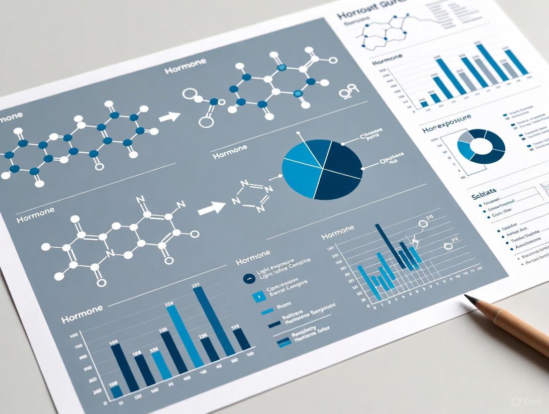
Light Exposure Control in Hormone Sampling: A Research Framework for Circadian Endocrinology and Drug Development
This article provides a comprehensive framework for researchers and drug development professionals on controlling light exposure during hormonal biomarker sampling.
Light Exposure Control in Hormone Sampling: A Research Framework for Circadian Endocrinology and Drug Development
Abstract
This article provides a comprehensive framework for researchers and drug development professionals on controlling light exposure during hormonal biomarker sampling. It explores the foundational science of light's non-visual effects on endocrine physiology, details standardized methodologies for measuring and controlling light in experimental and clinical settings, addresses key troubleshooting and optimization challenges, and presents validation strategies for ensuring data integrity. By synthesizing current evidence and consensus priorities, this guide aims to enhance the reliability and reproducibility of hormone measurements in circadian research and clinical trials, ultimately supporting more accurate assessment of chronotherapeutics and drug efficacy.
The Science of Light-Hormone Interactions: From ipRGCs to Endocrine Disruption
Intrinsically photosensitive Retinal Ganglion Cells (ipRGCs) are a unique class of mammalian photoreceptors that constitute a third class of photoreceptors, in addition to rod and cone cells [1]. These neurons, which make up only about 1-2% of all retinal ganglion cells, are intrinsically photosensitive due to their expression of the photopigment melanopsin (Opn4) [2] [3] [1]. Unlike rods and cones that hyperpolarize in response to light, ipRGCs depolarize and fire action potentials when illuminated, similar to photoreceptors found in invertebrates [2] [3]. The primary function of ipRGCs is non-image-forming vision – they are specialized encoders of ambient light intensity (irradiance) over time, rather than spatial detail or rapid temporal changes [2] [3]. Through the retinohypothalamic tract (RHT), their projections influence diverse physiological functions including circadian photoentrainment, pupillary light reflex, hormonal regulation, and sleep-wake cycles [2] [3] [1].
Frequently Asked Questions (FAQs)
1. What is the peak spectral sensitivity of the melanopsin photopigment, and why does this matter for experimental design? The melanopsin photopigment in ipRGCs has a peak spectral sensitivity (λmax) of approximately 480 nanometers (nm), which falls within the blue-to-cyan portion of the visible spectrum [4] [3] [1]. This is critical for experimental design because using light stimuli at or near this wavelength will most effectively activate the ipRGC pathway. Studies manipulating light quality have shown, for instance, that blue-enriched light (peak ~485 nm) has significantly different physiological and behavioral effects compared to blue-depleted light, even when both are matched for visual brightness [4]. Therefore, controlling and reporting the spectral properties of light sources is essential for interpreting results related to non-image-forming vision.
2. My rodent model lacks all rods and cones. Will it still be able to entrain its circadian rhythms to a light-dark cycle? Yes. A foundational discovery in the field was that mice engineered to entirely lack rods and cones (rodless/coneless) can still entrain their circadian rhythms to a light-dark cycle, suppress melatonin production, and exhibit a pupillary light reflex [2] [3]. The persistence of these functions, which are absent if the eyes are removed, provided key evidence for the existence of a previously unknown ocular photoreceptor: the ipRGC [3]. In your experiments, such models can be used to isolate the functions specifically dependent on melanopsin-mediated photoreception.
3. What are the primary brain regions targeted by ipRGCs, and what functions do they control? IpRGCs innervate dozens of brain regions. The table below summarizes the primary targets and their associated functions [2] [3] [1].
Table 1: Key Brain Targets of ipRGCs and Their Functions
| Brain Region | Abbreviation | Primary Function(s) |
|---|---|---|
| Suprachiasmatic Nucleus | SCN | Master circadian clock; circadian photoentrainment [2] [3] [1] |
| Olivary Pretectal Nucleus | OPN | Control of the pupillary light reflex [2] [3] [1] |
| Supraoptic Nucleus | SON | Fluid homeostasis, social behaviors, appetite [5] |
| Ventrolateral Preoptic Nucleus | VLPO | Sleep regulation [1] |
| Intergeniculate Leaflet | IGL | Circadian entrainment [1] [5] |
| Lateral Geniculate Nucleus | dLGN | Potential role in conscious visual perception (e.g., brightness detection) [2] [1] |
4. How do I measure circadian phase in human studies, and what are the best practices? The gold standard for measuring the endogenous circadian rhythm in humans is the constant routine protocol [6]. In this protocol, participants are kept in constant conditions (low light, semi-recumbent posture, etc.) for at least 24 hours to remove the masking effects of sleep, activity, and posture. For studies requiring a more naturalistic design, rigorous control is still needed. The most reliable circadian phase marker is the timing of the melatonin rhythm [6]. Best practices for measuring melatonin include:
- Maintaining participants in dim light (< 5 lux) before and during sample collection to prevent melatonin suppression [7] [6].
- Using consistent sampling intervals (e.g., hourly) from saliva or plasma.
- Controlling for posture, meal timing, and caffeine/alcohol intake, as these can be confounding variables [6].
5. We observed that bright light exposure at night alters glucose metabolism. Is this finding consistent with the known literature? Yes, this is a documented finding. A study investigating the impact of bright light (>500 lux) at night compared to dim light (<5 lux) found that bright light exposure was associated with significantly higher post-meal plasma glucose and insulin levels, suggestive of acute glucose intolerance and reduced insulin sensitivity [7]. This effect occurred concurrently with a suppression of melatonin, linking light exposure directly to metabolic changes. These findings have significant implications for the health of shift workers and the design of studies involving nighttime hormonal or metabolic sampling [7].
Troubleshooting Common Experimental Issues
Table 2: Common Experimental Challenges and Solutions
| Problem | Possible Cause | Solution |
|---|---|---|
| High variability in circadian phase markers (e.g., melatonin) between subjects. | Uncontrolled confounding variables such as caffeine, alcohol, irregular sleep, or prior light exposure [6]. | Implement strict pre-study participant guidelines: 24-hour abstinence from caffeine/alcohol, stable sleep-wake cycle for ≥7 days (verified by actigraphy/sleep diary) [6]. |
| Unexpectedly low or absent response in a behavioral assay like the pupillary light reflex. | The light stimulus is not effectively activating melanopsin (wrong spectrum, too dim, or too brief) [2] [3]. | Use a 480 nm light source. Ensure stimuli are of sufficient intensity and duration, as melanopsin-driven responses are sluggish and sustained [2]. |
| Inability to replicate findings from a knockout mouse model. | Underlying genetic background or specific experimental conditions (e.g., light levels during animal housing) may be influencing the phenotype. | Ensure all animals are on the same genetic background. Control and document light intensity and spectral quality in animal housing and during experiments [8]. |
| Difficulty in interpreting results from a whole-animal knockout model. | Compensatory developmental mechanisms or systemic effects may mask the cell-type-specific role of a gene. | Consider using conditional, cell-specific knockout models (e.g., rod-specific Bmal1 KO) to isolate function in a specific cell population [8]. |
Detailed Experimental Protocols
Protocol 1: Forced Desynchrony for Isolating Circadian Rhythms
The Forced Desynchrony (FD) protocol is a powerful method to separate the contributions of the endogenous circadian system from the masking effects of the sleep-wake cycle and behaviors [9].
Workflow Diagram: Forced Desynchrony Protocol
- Participant Screening: Recruit healthy participants free from major medical conditions, psychiatric history, and recent shift work or time-zone travel. Strict sleep-wake schedules should be maintained for at least 7 days prior [6] [9].
- Pre-Laboratory Baseline: Monitor sleep and circadian rhythms using actigraphy and sleep diaries to confirm a stable routine.
- Laboratory Phase:
- Place participants in an environment free of time cues.
- Impose a sleep-wake cycle that is distinctly different from 24 hours (e.g., a 20-hour or 28-hour "day").
- Maintain light intensity at very low levels (< 5-10 lux) to minimize its phase-resetting effect on the circadian pacemaker [9].
- This non-24-hour cycle forces the sleep-wake episode to occur at all phases of the endogenous circadian cycle over time.
- Data Collection: Regularly collect physiological samples (e.g., plasma/saliva for melatonin, core body temperature) throughout the protocol [6] [9].
- Data Analysis: Use specialized statistical analyses (e.g., non-orthogonal spectral analysis) to separate the component of a rhythm that is dependent on the circadian phase from the component that is dependent on the duration of wakefulness or sleep [9].
Protocol 2: Investigating Acute Metabolic Effects of Light at Night
This protocol is adapted from a study demonstrating that a single session of bright light exposure at night can alter hormonal and metabolic responses to a meal [7].
Workflow Diagram: Acute Night Light Exposure Study
- Study Design: Use a randomized, two-way crossover design where each participant completes both a Bright Light (BL, >500 lux) and a Dim Light (DL, <5 lux) session, separated by a sufficient washout period (e.g., ≥7 days) [7].
- Participant Preparation: Participants should be healthy, have normal sleep patterns, and refrain from caffeine, alcohol, and excessive exercise for 24 hours prior to each session.
- Light Exposure: Begin the controlled light exposure (BL or DL) for a set period prior to the evening meal and continue throughout the sampling period.
- Meal Challenge: Provide a standardized evening meal at a time individually scheduled based on the participant's endogenous melatonin rhythm (e.g., on the rising phase of the rhythm) for physiological relevance [7].
- Sample Collection: Collect serial plasma and saliva samples at specific intervals before and after the meal.
- Key Metrics: Analyze samples for:
- Salivary Melatonin (to confirm light-induced suppression).
- Plasma Glucose, Insulin, and NEFA (non-esterified fatty acids) to assess metabolic response [7].
The Scientist's Toolkit: Research Reagent Solutions
Table 3: Essential Research Materials and Their Applications
| Reagent / Material | Function / Application | Key Considerations |
|---|---|---|
| Melagen Lighting Device (or equivalent) | Precisely controls spectral quality of light stimuli (e.g., blue-enriched vs. blue-depleted) while maintaining constant photopic illuminance [4]. | Critical for isolating melanopsin-mediated effects from rod/cone contributions. |
| Actigraphy System (e.g., AWL, Cambridge Neurotechnology) | Objectively monitors sleep-wake cycles and rest-activity rhythms in human and animal studies outside the lab [7] [6]. | Validates participant compliance with sleep schedules prior to in-lab studies. |
| Melatonin Radioimmunoassay (RIA) / ELISA Kits | Measures melatonin concentration in saliva, plasma, or its metabolite (6-sulfatoxymelatonin) in urine [7] [6]. | The gold-standard marker for circadian phase in humans. Requires strict dim-light conditions during collection. |
| Electroretinography (ERG) System | Measures the electrical responses of various retinal cell types to light stimuli [8]. | Used to functionally assess retinal health and light response in animal models. |
| Cre-Dependent Viral Vectors (e.g., AAV) | For anterograde tracing of ipRGC projections or selective optogenetic manipulation of specific ipRGC subtypes [5]. | Enables cell-type-specific targeting in transgenic mouse lines (e.g., Opn4-Cre, GlyT2-Cre). |
| Opn4 (Melanopsin) Antibodies | Identifies and visualizes ipRGCs in retinal whole mounts or sections via immunohistochemistry [2] [5]. | Confirms ipRGC identity and morphology. |
Advanced Concepts: ipRGC Diversity and Functional Specialization
The initial model of ipRGCs as a uniform population has evolved. We now know there are at least six distinct subtypes (M1-M6) in the mouse, which differ in their morphology, melanopsin expression levels, synaptic inputs, and, crucially, their central brain targets [2] [5]. This diversity underlies a sophisticated functional specialization.
Signaling Pathway and ipRGC Diversity Diagram
For example, a recently discovered subtype of M1 ipRGCs, identified in a GlyT2Cre mouse line, is located exclusively in the dorsal retina [5]. These cells form a tiled mosaic, suggesting they are optimized to sample a specific region of the visual field—specifically, the ground. They project selectively to the supraoptic nucleus (SON), a brain region involved in fluid homeostasis and social behavior, and also send collaterals to the SCN and other areas [5]. This finding challenges the old paradigm that ipRGC dendrites overlap randomly to maximize photon capture. Instead, it indicates that different ipRGC subtypes are organized to monitor specific parts of the environment for distinct non-image-forming functions.
Light is the primary environmental cue that synchronizes our central circadian clock, the suprachiasmatic nucleus (SCN) in the hypothalamus. This synchronization regulates the production and secretion of numerous hormones, creating complex physiological responses that vary by light intensity, duration, spectral quality, and timing of exposure. For researchers investigating chronobiology, endocrinology, and drug development, understanding these precise relationships is critical for designing robust experiments and accurately interpreting hormonal data. This technical support center provides targeted guidance on the key hormones most sensitive to photic manipulation, with a focus on methodological protocols, data interpretation, and troubleshooting common experimental challenges.
Core Hormonal Pathways and Mechanisms
Anatomical and Molecular Pathways
The process of photic hormone regulation begins when light enters the eye. A specialized subset of intrinsically photosensitive retinal ganglion cells (ipRGCs) containing the photopigment melanopsin are particularly sensitive to short-wavelength (blue) light. These ipRGCs project directly to the SCN via the retinohypothalamic tract.
Upon light stimulation, the SCN orchestrates hormonal output through two primary pathways:
- Neural Pathway for Melatonin: The SCN signals the paraventricular nucleus (PVN), which projects to the intermediolateral column of the spinal cord, then to the superior cervical ganglion, and finally to the pineal gland to inhibit melatonin synthesis.
- Endocrine Pathway for Cortisol: The SCN influences the hypothalamus-pituitary-adrenal (HPA) axis through multisynaptic connections, modulating cortisol release from the adrenal cortex.
The following diagram illustrates this signaling cascade:
Key Light-Sensitive Hormones: Melatonin and Cortisol
Melatonin, the "hormone of darkness," is perhaps the most well-studied light-responsive hormone. Its secretion from the pineal gland is potently suppressed by light exposure, especially during the biological night. The degree of suppression depends on light intensity, wavelength, duration, and timing.
Cortisol, a key glucocorticoid hormone involved in stress response and metabolism, demonstrates a more complex relationship with light. Research shows bright light exposure can significantly reduce plasma cortisol levels when administered during both the rising and descending phases of its circadian rhythm [10]. The effects appear dependent on intensity, duration, and circadian timing of exposure.
Experimental Protocols & Data
Quantitative Hormonal Responses to Light
Table 1: Experimental Parameters and Hormonal Responses to Nocturnal Light Exposure
| Light Exposure Parameter | Melatonin Response | Cortisol Response | Key Experimental Findings |
|---|---|---|---|
| Intensity (Nocturnal) | |||
| ~10,000 lux (bright) | ~40% suppression [11] | Significant reduction [10] | Bright light exposure significantly reduced plasma cortisol at both circadian phases studied [10] |
| ~3 lux (dim) | Minimal suppression | Little to no effect [10] | Dim light exposure had little effect on cortisol levels [10] |
| Temporal Dynamics | |||
| Acute exposure (5-15 min) | Rapid suppression (t₁/₂ = ~13 min) [11] | Linear increase during intermittent stimuli [11] | Melatonin suppression occurs rapidly within first 5 min of exposure [11] |
| Prolonged exposure (6.5 h) | Sustained suppression with continuous light [11] | Trimodal changes under continuous light [11] | Cortisol showed trimodal response: activation, inhibition, then recovery [11] |
| Spectral Sensitivity | |||
| Short wavelengths (460-480 nm) | Maximum suppression | Research ongoing | Melanopsin peak sensitivity ~480 nm drives non-visual responses |
| Long wavelengths (>600 nm) | Minimal suppression | Minimal effect | Red light often used as control condition in experiments |
Table 2: Circadian Phase-Dependent Hormonal Responses
| Circadian Phase | Melatonin Response to Light | Cortisol Response to Light | Methodological Considerations |
|---|---|---|---|
| Biological Day | Minimal suppression | Variable: increase in morning [11] | Phase response curves differ for each hormone |
| Early Biological Night | Potent suppression | Reduction with bright light [10] | Critical period for circadian phase shifting |
| Late Biological Night | Potent suppression | Reduction with bright light [10] | Timing relative to temperature minimum crucial |
| Circadian Phase Markers | DLMO/DLMOff for phase assessment [10] | Cortisol awakening response (CAR) | Melatonin phase more reliable than cortisol for circadian timing |
Detailed Experimental Methodology
For researchers replicating key findings on light-hormone interactions, the following methodological details are essential:
Participant Screening and Pre-Study Conditions:
- Recruit healthy participants with no medical, psychiatric, or sleep disorders
- Verify absence of medications and recreational drugs through comprehensive toxicology screening
- Maintain regular 8h:16h sleep:wake schedules for at least 3 weeks pre-study
- Monitor compliance using wrist actigraphy and sleep logs
- Require abstinence from alcohol, nicotine, and caffeine for three weeks prior to study [11]
Laboratory Protocol for Nocturnal Light Exposure Studies:
- Habituation (Days 1-3): Adapt participants to laboratory environment while maintaining habitual sleep-wake times
- Constant Routine (CR1, Day 4-5): Implement 26.2-hour constant routine to assess endogenous circadian phase under dim light (<1.5 lux)
- Light Exposure (Day 5): Randomize participants to experimental light conditions (e.g., continuous bright, intermittent bright, or continuous dim light)
- Blood Sampling: Collect samples every 30 minutes via indwelling intravenous catheter
- Posture Control: Maintain seated position 12 minutes prior to and throughout light exposure sessions
- Light Monitoring: Use calibrated photometers to verify illuminance levels at eye level [11]
Hormonal Assay Specifications:
- Melatonin: Radioimmunoassay using 125I (e.g., DiagnosTech, Osceola, WI); sensitivity: 2.5 pg/mL; intra-assay CV: 5.9%
- Cortisol: Chemiluminescent assay (e.g., Beckman Coulter, Chaska, MN); sensitivity: 0.4 mg/dL; intra-assay CV: 6.4% [10]
The Scientist's Toolkit
Table 3: Essential Research Reagent Solutions
| Reagent/Equipment | Specification/Function | Application Notes |
|---|---|---|
| Light Source Systems | ||
| Broad-spectrum fluorescent lamps | 4100K color temperature, ceiling-mounted | Can be filtered through UV-stable filters (Lextran 9030) [10] |
| LED-based light panels | Tunable spectrum, precise intensity control | Ideal for spectral sensitivity studies |
| Light Measurement | ||
| IL1400 radiometer | With SEL-033/Y/W and SEL-033/F/W detectors | Measures illuminance and irradiance [10] |
| Spectroradiometer | Full spectral analysis | Essential for characterizing custom light sources |
| Hormonal Assays | ||
| 125I RIA for melatonin | Sensitivity: 2.5 pg/mL; intra-assay CV: 5.9% | Gold standard for melatonin measurement [10] |
| Chemiluminescent cortisol assay | Sensitivity: 0.4 mg/dL; intra-assay CV: 6.4% | High-throughput option for cortisol [10] |
| Salivary collection kits | Non-invasive cortisol and melatonin sampling | Useful for field studies and repeated measures |
| Circadian Phase Assessment | ||
| Dim Light Melatonin Onset (DLMO) | 25% threshold of 3-harmonic peak-to-trough amplitude | Reliable circadian phase marker [10] |
| Core body temperature monitoring | Telemetric temperature sensors | Complementary circadian phase marker |
Troubleshooting Common Experimental Challenges
FAQ 1: Why are we observing inconsistent cortisol responses to light across participants?
Issue: High inter-individual variability in cortisol responses to light exposure.
Solution:
- Control for Circadian Phase: Time light exposures relative to individual circadian phase (using DLMO) rather than clock time [10]
- Standardize Posture: Control participant posture as changes can acutely alter cortisol levels [10]
- Minimize Stress: Ensure comfortable, low-stress environment as anxiety can elevate cortisol independent of light
- Consider Oral Contraceptives: In female participants, document contraceptive use as it can affect cortisol binding globulin
- Increase Sample Size: Account for expected variability in power calculations
FAQ 2: How can we accurately characterize the temporal dynamics of hormonal responses?
Issue: Inadequate sampling frequency missing rapid hormonal changes.
Solution:
- High-Frequency Sampling: For melatonin dynamics, sample at least every 15-30 minutes [11]
- Extended Monitoring: Continue sampling for sufficient duration to capture recovery (melatonin recovery t₁/₂ = ~46 minutes) [11]
- Multiple Hormone Assessment: Measure both melatonin and cortisol from same samples to compare dynamics
- Modeling Approaches: Use appropriate kinetic models (e.g., half-life calculations) to characterize suppression and recovery
FAQ 3: What is the optimal approach for controlling light history in participants?
Issue: Prior light exposure influences subsequent light sensitivity.
Solution:
- Stabilization Period: Implement 3 baseline days with controlled <150 lux maximum light exposure [10]
- Pre-Study Monitoring: Use ambulatory actigraphy with light sensors for 1-2 weeks pre-study
- Standardized Pre-Study Routine: Provide participants with specific light exposure guidelines before laboratory admission
- Dim Light Adaptation: Maintain dim light (<3 lux) for several hours before experimental light exposures [10]
Advanced Technical Considerations
Signaling Pathways and Experimental Workflows
The experimental workflow for comprehensive light-hormone studies involves multiple coordinated stages, as shown in the following diagram:
Emerging Research and Unexplored Mechanisms
Recent evidence suggests additional hormonal systems may be influenced by light exposure, though these pathways are less characterized:
- Reproductive Hormones: Light deprivation disrupts Leydig cell maturation and testosterone production in animal models [12]
- Metabolic Hormones: Artificial light at night associates with increased risk of metabolic diseases including diabetes and obesity [13] [14]
- Growth Hormone: Sleep architecture alterations from light exposure may impact growth hormone secretion patterns [15]
Future research should employ precise spectral control, high-temporal resolution sampling, and multivariate approaches to fully characterize these relationships. Particular attention should be paid to individual differences in photic sensitivity, which may be substantial and clinically relevant.
Frequently Asked Questions
FAQ 1: What is the fundamental difference between how blue and red light exposure affects melatonin secretion?
Blue light is a potent suppressor of nocturnal melatonin, while red light has a minimal effect. A 2025 study exposed participants to three hours of either blue (464 nm) or red (631 nm) LED light. While both lights initially suppressed melatonin after one hour, significant differences emerged by the second hour: melatonin levels under blue light remained low at 7.5 pg/mL, whereas under red light, they recovered to 26.0 pg/mL. This pattern persisted, confirming that blue light has a stronger and more sustained disruptive effect on circadian physiology compared to red light [16].
FAQ 2: What is the biological mechanism behind this spectral sensitivity?
The effect is primarily mediated by a specialized photoreceptor in the eye. Intrinsically photosensitive retinal ganglion cells (ipRGCs) contain the photopigment melanopsin, which exhibits peak sensitivity in the short-wavelength (blue) region of the visible spectrum [16]. When activated by light, particularly blue light, these cells send signals via the retinohypothalamic tract to the suprachiasmatic nucleus (SCN)—the brain's "master clock." The SCN then inhibits the pineal gland's production of melatonin, the hormone that promotes sleep [16] [17]. Red light has a much weaker effect on this specific pathway [18].
FAQ 3: How do factors like age and sex influence an individual's sensitivity to light at night?
Research indicates that sensitivity to the melatonin-suppressing effects of light is not uniform across populations.
- Sex Variations: A 2025 study found that female participants exhibited greater melatonin suppression (+4.69%) in response to moderate light exposure compared to males, though they showed a lower alerting response [19].
- Age Variations: The same study that highlighted blue light's potency also noted that its suppressive effects were more pronounced in younger participants and men [16]. Another study suggested that older adults may require higher light intensities to achieve the same non-visual effects as younger adults, indicating a potential age-related decline in sensitivity [16].
FAQ 4: Does my participants' light exposure during the day affect their sensitivity to light in my evening experiments?
Yes, prior light history is a critical factor. A growing body of evidence suggests that the amount of bright light a person receives during the day can modulate their circadian system's sensitivity to light at night [20]. Studies in adults have shown that increased prior daytime light exposure can decrease the effects of subsequent night light on melatonin secretion and phase-shifting [20]. This underscores the importance of monitoring and, if possible, standardizing participants' light exposure in the days leading up to laboratory experiments.
FAQ 5: What are the recommended light levels for nighttime to minimize circadian disruption?
International expert recommendations propose that during the three hours preceding bedtime, light exposure should not exceed a melanopic Equivalent Daylight Illuminance (mEDI) of 10 lux at the eye. During sleep, it should be as dark as possible, not exceeding 1 mEDI [16]. For context, a mere eight lux—exceeded by most table lamps—can have a measurable effect on melatonin and circadian rhythms [17].
Table 1: Experimental Outcomes of Nocturnal Light Exposure
| Metric | Blue Light (464 nm) | Red Light (631 nm) | Significance & Context |
|---|---|---|---|
| Melatonin Level (2 hours post-exposure) | 7.5 pg/mL [16] | 26.0 pg/mL [16] | p=0.019; Red light allows significant recovery [16] |
| Melatonin Suppression | Stronger suppression [16] | Weaker suppression [16] | Effects are more potent in younger participants and men [16] |
| Relative Melatonin Suppression (by Sex) | Female: +4.69% greater suppression than male [19] | Information Not Specified | Under moderate light exposure from a screen [19] |
| Impact on Circadian Phase | Shifts rhythms significantly (3 hrs in one study) [17] | Minimal phase-shifting effect [18] | Blue light is twice as effective as green light at shifting rhythms [17] |
| Subjective Alertness | Potently increases alertness [17] [20] | Less alerting effect [18] | Female participants showed a lower alerting response to moderate light [19] |
Table 2: Key Experimental Parameters from Cited Studies
| Parameter | Blue Light Condition | Red Light Condition | Measurement & Standards |
|---|---|---|---|
| Peak Wavelength | 464 nm [16] | 631 nm [16] | Full width at half maximum (FWHM): Blue=24 nm, Red=18 nm [16] |
| Corneal Illuminance | 80 lux (common level) [16] [20] | 80 lux (common level) [16] | Controlled using a calibrated luxmeter [16] |
| Exposure Duration | 3 hours (9 pm-midnight) [16] | 3 hours (9 pm-midnight) [16] | Saliva samples collected hourly [16] |
| Melatonin Assay | Enzyme-linked immunosorbent assay (ELISA) [16] | Enzyme-linked immunosorbent assay (ELISA) [16] | Considered the gold standard for salivary biomarkers [16] |
| Key Metric | Melanopic EDI (mEDI) [16] | Melanopic EDI (mEDI) [16] | Standardized by CIE S 026:2018 for non-visual effects [16] [21] |
Detailed Experimental Protocols
Protocol 1: Comparative Effects of Red and Blue LED Light
This protocol is adapted from the 2025 study that directly compared the effects of narrowband red and blue light on melatonin secretion [16].
1. Participant Preparation and Screening:
- Recruit healthy participants (e.g., n=12, age range 19-55). Maintain a controlled, within-subject design.
- Screen for factors affecting circadian sensitivity: extreme chronotypes, poor sleep quality, recent shift work or transmeridian travel, ophthalmological issues, and smoking [19].
- Instruct participants to maintain a consistent sleep-wake schedule for at least one week prior to the laboratory sessions, verified by wrist actigraphy. Restrict alcohol, irregular caffeine, and certain fruits (e.g., bananas, citrus) that can influence salivary melatonin on the day of the session [19].
2. Laboratory Setup and Light Source Calibration:
- Light Sources: Use custom-made luminaires with narrowband LEDs (e.g., Blue: 464 nm, Red: 631 nm).
- Calibration: Characterize the spectral power distribution (SPD) and total irradiance (W·m⁻²) of each LED using a calibrated spectroradiometer.
- Exposure Control: Position the light source to deliver a specific photopic illuminance (e.g., 80 lux) at the corneal plane of the participant. The distance will vary by LED intensity (e.g., 55 cm for blue, 40 cm for red) [16].
- Condition Design: Employ a counterbalanced crossover design where each participant is exposed to both red and blue light conditions on separate nights, with a sufficient washout period in between.
3. Experimental Procedure:
- Timing: Begin light exposure three hours before the participant's habitual bedtime (e.g., 9:00 p.m. to midnight) and conduct sessions in a dedicated, dark laboratory.
- Saliva Sampling: Collect saliva samples hourly (e.g., at 9:00 p.m., 10:00 p.m., 11:00 p.m., and midnight) using appropriate salivettes.
- Sample Handling: Immediately freeze saliva samples at -20°C or lower after collection to preserve melatonin integrity.
4. Data Analysis:
- Melatonin Assay: Analyze salivary melatonin concentrations using a commercially available ELISA kit, following the manufacturer's instructions.
- Statistical Analysis: Compare melatonin levels between conditions at each time point using repeated-measures ANOVA or linear mixed models, with time and light condition as fixed effects.
Protocol 2: Assessing the Impact of Prior Light History
This protocol focuses on how afternoon light exposure modulates the response to evening light, adapted from a 2025 adolescent study [20].
1. Pre-Study Light Monitoring:
- Equipment: Equip participants with wearable light dosimeters (e.g., the Light-Dosimeter "lido") for at least 5-7 days before the lab session. The device should be worn at eye level (e.g., on glasses) to accurately measure near-corneal light exposure [21].
- Metrics: Calculate summary metrics from the data, such as the time spent above a bright light threshold (e.g., ≥ 500 lux melanopic EDI) during the afternoon.
2. Laboratory Intervention and Testing:
- Afternoon Intervention: Expose participants to different light levels in the afternoon-early evening (AEE), several hours before the habitual bedtime (e.g., 4.5 hours of dim: 6.5 lx, moderate: 130 lx, or bright: 2500 lx light, in a counterbalanced order).
- Evening Challenge: Later in the evening, expose all participants to a standard, moderate light level (e.g., 130 lx).
- Primary Outcome: Collect saliva samples during this evening challenge period to calculate the area under the curve (AUC) for melatonin.
- Secondary Outcomes: Measure subjective sleepiness (e.g., Karolinska Sleepiness Scale), vigilance (e.g., Psychomotor Vigilance Task), and distal-to-proximal skin temperature gradient.
Troubleshooting Common Experimental Issues
Issue 1: High Variability in Melatonin Data Between Participants
- Potential Cause: Uncontrolled prior light history and differing circadian phenotypes (chronotypes).
- Solution:
- Standardize Light History: Use wearable dosimeters to monitor and, if possible, instruct participants to seek bright light in the morning and avoid bright light before the experiment to reduce baseline variability [20] [22].
- Stratify by Chronotype: Screen participants using the Munich ChronoType Questionnaire (MCTQ) and either stratify your groups or include chronotype as a covariate in your statistical model [19].
Issue 2: Inaccurate Measurement of Biologically Relevant Light Dose
- Potential Cause: Relying on photopic lux alone, which does not accurately represent the light's impact on the circadian system.
- Solution:
- Use Correct Metrics: Quantify light exposure using circadian-relevant metrics like melanopic EDI (Equivalent Daylight Illuminance) as defined by the CIE S 026:2018 standard [16] [21].
- Calibrate Equipment: Use spectroradiometers to fully characterize your light sources and ensure your wearable dosimeters are calibrated to report these modern metrics [21].
Issue 3: Confounding Effects from Sample Collection and Handling
- Potential Cause: Degradation of melatonin in saliva samples due to improper handling.
- Solution:
- Immediate Processing: Centrifuge salivettes promptly after collection to separate saliva from the cotton swab.
- Proper Storage: Freeze samples at -20°C or -80°C immediately after processing. Avoid repeated freeze-thaw cycles [16].
Visualizing the Mechanistic Pathway
The Scientist's Toolkit: Essential Research Reagents & Materials
Table 3: Key Materials and Equipment for Circadian Light Research
| Item | Function & Application | Key Specifications |
|---|---|---|
| Narrowband LED Light Source | Provides precise spectral stimuli for controlled light exposure. | Peak wavelengths: Blue ~464-470 nm, Red ~630-660 nm [16]. Capable of adjustable intensity. |
| Spectroradiometer | The gold standard for calibrating light sources. Measures the absolute spectral power distribution (SPD). | Wavelength range: ~380-780 nm. Calibrated against NIST-traceable standards [16]. |
| Wearable Light Dosimeter | Measures personal, time-stamped light exposure in the near-corneal plane in real-world conditions. | Reports melanopic EDI; worn on glasses for eye-level measurement; multi-day battery life [21] [22]. |
| Saliva Collection Kit (Salivette) | Non-invasive collection of saliva samples for melatonin assay. | Polyester swab and plastic centrifuge tube. Must be RNase/DNase-free. |
| ELISA Kit for Melatonin | Quantifies melatonin concentration in saliva samples. | Validated for saliva/salivary melatonin; high sensitivity (pg/mL range) [16]. |
| Actigraph | Objectively monitors sleep-wake patterns and physical activity during ambulatory phases. | Worn on the wrist; contains accelerometer and light sensor (though less accurate than a dedicated dosimeter). |
Troubleshooting Guide: Phase Response Curve (PRC) Experiments
This guide addresses common challenges researchers face when designing and interpreting Phase Response Curve (PRC) experiments in the context of light exposure control and hormone sampling research.
Frequently Asked Questions (FAQs)
1. Why did my light stimulus produce a much smaller phase shift than expected? The magnitude of a phase shift depends critically on the timing of the light stimulus relative to the individual's internal circadian phase. A light pulse administered during the "dead zone" of the PRC, typically between a few hours after usual wake-up time and two hours before usual bedtime, will produce little to no phase shift [23]. Ensure you have accurately determined the subject's circadian phase marker (e.g., DLMO or CBTmin) before scheduling the stimulus. The intensity, duration, and wavelength of light are also critical factors [24].
2. How can I accurately measure circadian phase in my subjects? The gold standard method involves assessing the timing of the Dim Light Melatonin Onset (DLMO) or the core body temperature minimum (CBTmin) under controlled conditions, such as during a Constant Routine (CR) protocol [25]. The CR protocol entails continuous enforced wakefulness in a semi-recumbent posture with even distributions of caloric intake and dim light exposure (< 10 lx) to eliminate "masking" effects on circadian phase markers [25].
3. What could cause inconsistent phase shift results among subjects receiving the same light stimulus? Significant inter-individual variation is a well-documented characteristic of PRCs [24]. The published curves are usually aggregates of a test population, and individuals can show mild or significant variation in their response [23]. Factors such as an individual's intrinsic circadian period, age, or the presence of a circadian rhythm sleep disorder can lead to atypical responses [23] [24].
4. Is it possible for a light stimulus to cause a phase delay when we expected an advance? Yes, this can occur if the stimulus is timed incorrectly. The human PRC for light shows a critical phase, often near the CBTmin, where the effect abruptly changes from a phase delay to a phase advance [23] [25]. If your estimate of this critical phase is inaccurate by even a small amount, administering light just before the estimated CBTmin intending to cause a delay might instead occur after the true CBTmin, resulting in an unexpected, smaller advance or no shift at all.
5. Can we combine different stimuli to achieve a larger phase shift? Yes, research has shown that the phase-shifting effects of concurrent treatments can be additive. For example, a combination of morning bright light and afternoon melatonin, both timed to cause a phase advance according to their respective PRCs, can produce a larger phase advance shift than bright light alone [23].
Table 1: Summary of Phase Shifts from Different Light Stimuli
| Stimulus Type | Duration | Intensity | Timing (Relative to CBTmin) | Average Phase Shift | Reference |
|---|---|---|---|---|---|
| White Light | 6.7 hours | ~5,000-10,000 lux | Centered ~4 hours after | ~2 hour Advance | [24] |
| White Light | 1 hour | 8,000 lux | Start ~11 hours after DLMO | ~2 hour Delay | [24] |
| White Light | 1 hour | 8,000 lux | Start ~1 hour after DLMO | ~15 minute Advance | [24] |
| Blue Light | 1.5 hours (over 3 days) | 185 lux | 0-3 hours after DLMO | ~1.5 hour Advance (total) | [24] |
Table 2: Phase Response Curve Key Parameters
| Parameter | Light PRC | Melatonin PRC |
|---|---|---|
| Delay Zone | Evening, before CBTmin [23] | Morning, around wake-up time [23] |
| Advance Zone | Morning, after CBTmin [23] | Afternoon and early evening [23] |
| Transition Point | Near the core body temperature minimum (CBTmin) [23] | ~8 hours after wake-up time [23] |
| Dead Zone/No Effect | Mid-day (~2h after wake to ~2h before bed) [23] | From usual bedtime until wake-up time [23] |
Detailed Experimental Protocols
Protocol 1: Constant Routine (CR) for Circadian Phase Assessment
This protocol is designed to unmask the endogenous circadian phase by minimizing external influences [25].
- Objective: To accurately determine circadian phase markers (DLMO, CBTmin) pre- and post-stimulus.
- Key Steps:
- Subject Preparation: Subjects maintain a strict 8-hour sleep schedule for at least two weeks prior to the lab segment. Compliance is verified via time-stamped voicemails and actigraphy [25].
- Laboratory Admission: Subjects enter a private, time-cue-free environment.
- Pre-Stimulus CR: Following three baseline sleep episodes, subjects begin the CR. They remain awake in a semi-recumbent posture for an extended period (e.g., 27-65 hours). Lighting is maintained at dim levels (< 10 lx). Hourly isocaloric snacks are provided [25].
- Sample Collection: Blood or saliva samples are collected regularly (e.g., hourly) to assay for melatonin, allowing determination of DLMO. Core body temperature is monitored continuously to determine CBTmin.
- Applications: Used as a gold-standard baseline measurement before and after a phase-shifting stimulus to calculate the true magnitude of the phase shift.
Protocol 2: Single Pulse Bright Light PRC Determination
This protocol outlines the methodology for constructing a Phase Response Curve to a single bright light pulse [25].
- Objective: To characterize the phase-dependent phase-shifting effects of a bright light stimulus across the entire circadian cycle.
- Key Steps:
- Pre-Stimulus Phase Assessment: Conduct a Constant Routine (Protocol 1) to determine the initial circadian phase of the subject.
- Light Stimulus Administration: Following the pre-stimulus CR and a recovery sleep episode, subjects are exposed to a bright light pulse (e.g., 6.7 hours of ~5,000-10,000 lux). The timing of this stimulus is systematically varied across different subjects to cover all circadian phases [25].
- Stimulus Control: The light exposure may consist of alternating periods of fixed gaze and free gaze to ensure retinal exposure. The use of ceiling-mounted fixtures can provide more uniform illumination [25] [24].
- Post-Stimulus Phase Assessment: A second Constant Routine is implemented after the light stimulus and another recovery sleep episode to determine the new circadian phase.
- Data Analysis: The phase shift is calculated as the difference in the timing of the circadian phase marker (e.g., melatonin midpoint) between the post- and pre-stimulus CRs. These shifts are then plotted against the circadian phase at which the stimulus was administered to generate the PRC [25].
Experimental Workflow and Signaling Pathway
Light Entrainment Signaling Pathway
PRC Determination Workflow
The Scientist's Toolkit: Research Reagent Solutions
Table 3: Essential Materials for Circadian Light Exposure Studies
| Item | Function/Description | Application Notes |
|---|---|---|
| Constant Routine Setup | A controlled lab environment for unmasking endogenous circadian rhythms by standardizing posture, activity, light (<10 lx), and feeding [25]. | Critical for obtaining accurate pre- and post-stimulus phase measurements (DLMO, CBTmin). |
| Melatonin Assay Kits | For measuring melatonin concentrations in plasma, saliva, or urine. Dim Light Melatonin Onset (DLMO) is a key phase marker [23] [25]. | Requires collection under dim light conditions. Used to establish the timing of the circadian clock. |
| Core Body Temperature Sensor | A precision device for continuous monitoring of core body temperature. The CBTmin is a classic circadian phase marker [25]. | Often measured rectally or via ingestible telemetric pills for high-fidelity data. |
| Calibrated Light Source | A light source with known intensity (lux), spectral distribution (nm), and capable of delivering high irradiance (e.g., up to 10,000 lux) [23] [25]. | Ceiling-mounted panels provide uniform field. Blue-enriched (~460-480nm) sources are more potent [23]. |
| Actigraphy System | A wearable device that monitors motor activity to estimate sleep-wake patterns and verify compliance with pre-study schedules [25]. | Used during the screening phase to confirm stable sleep-wake cycles before lab admission. |
Prior Light History as a Modulating Factor in Circadian Photosensitivity
What is "Prior Light History" and why is it critical for circadian research?
Prior light history refers to the pattern and intensity of light exposure an individual or experimental subject has experienced in the days preceding a circadian photic stimulus. The human circadian system demonstrates dynamic plasticity, meaning its sensitivity to light is not constant but is modulated by recent photic experience [26]. This adaptive mechanism has profound implications for experimental design and data interpretation in chronobiology.
The core principle is that the circadian system's response to a light stimulus is context-dependent. Exposure to dim light conditions prior to an experiment can sensitize the circadian system, leading to an amplified response to a subsequent light stimulus. Conversely, recent exposure to bright light can induce a form of de-sensitization [26] [27].
Key Evidence from Foundational Research
A controlled, in-patient study demonstrated this effect conclusively. When subjects were exposed to 3 days of very dim light (~1 lux) prior to a 6.5-hour light exposure at night, their phase-shifting response to that light was 60–70% greater and acute melatonin suppression was substantially larger, compared to when the same light exposure was preceded by 3 days of typical room light (~90 lux) [26] [28]. This provides direct evidence that prior dim light history sensitizes the human biological clock.
Troubleshooting Guides & FAQs
Experimental Design and Protocol
FAQ: My study results show high variability in circadian phase shifts between subjects. Could prior light history be a factor?
Answer: Yes, unaccounted-for individual light history is a major source of inter-individual variability. Participants enter studies with vastly different light exposure patterns based on their lifestyle, occupation, and environment [27]. This individual light history dictates the sensitivity of their intrinsically photosensitive retinal ganglion cells (ipRGCs) and the ultimate magnitude and direction of their circadian phase shift in response to an experimental stimulus [27].
- Solution: Implement a pre-study monitoring period.
- Action: Require participants to wear calibrated light loggers for at least 3 days (preferably longer) before the in-lab phase of the study [29].
- Goal: Capture individual melanopic light exposure patterns. This data can be used as a covariate in your analysis to explain variability or to stratify participants into groups with similar photic history.
FAQ: What is the minimum duration for controlling prior light history in a laboratory study?
Answer: Research indicates that a period of 3 days under controlled lighting conditions is sufficient to observe significant modulation of circadian photosensitivity [26]. While longer stabilization periods may be beneficial, a 3-day protocol has been empirically validated to establish a defined photic history and reduce noise introduced by participants' free-living light environments.
Measurement and Methodology
FAQ: I am measuring light in lux, but my colleague says this is insufficient for circadian research. What is the recommended metric?
Answer: Your colleague is correct. Measuring total light intensity in photopic lux is a common mistake, as it does not reflect the spectral sensitivity of the circadian system [30] [31]. The circadian system is most sensitive to short-wavelength (blue) light around 480 nm, mediated by the melanopsin photopigment in ipRGCs.
- Solution: Adopt the CI S 026:2018 international standard.
- Action: Use spectrally resolved light measurements and report light exposure levels as Melanopic Equivalent Daylight Illuminance (Melanopic EDI) [31].
- Benefit: Melanopic EDI quantifies the effective radiation for the melanopsin photopigment, providing a physiologically relevant measure of the light's potential impact on circadian physiology, melatonin suppression, and alertness [31].
FAQ: Besides melatonin, what other physiological markers can help track circadian phase shifts?
Answer: While Dim-Light Melatonin Onset (DLMO) is the gold standard, other non-invasive markers can provide valuable data.
- Core Body Temperature: The daily minimum of core body temperature is a reliable circadian phase marker, though it is invasive to measure continuously.
- Wrist Skin Temperature: Shows a predictable rhythm, rising before sleep onset, and can be measured easily with wearable devices, serving as a practical proxy [30].
- Rest-Activity Rhythms: Measured by actigraphy, the timing of rest (L5) and activity (M10) periods can serve as behavioral proxies for circadian phase, especially when analyzed to determine the midpoint of sleep [27].
Data Analysis and Interpretation
FAQ: How does seasonal light history impact a study conducted across different times of the year?
Answer: Long-term seasonal light history can be a significant confounding variable. An individual's circadian system adapts to the gradual changes in photoperiod over months [27]. A study taking place in winter, following months of shorter days, may yield different baseline circadian sensitivity compared to a summer study.
- Solution: Account for seasonality in your experimental design.
- Action: If possible, run all experimental conditions for a given subject in the same season. If the study spans multiple seasons, document the date of participation and include "season" as a factor in your statistical models.
- Rationale: This controls for the slow, adaptive changes in the circadian system driven by the natural light-dark cycle.
Table 1: Summary of Key Quantitative Findings from Seminal Photic History Study [26]
| Parameter | Dim Light History (1 lux) | Typical Room Light History (90 lux) | Effect of Dim History |
|---|---|---|---|
| Phase-Shifting Response | Significantly larger | Smaller baseline response | 60-70% amplification |
| Acute Melatonin Suppression | Substantially greater | Less suppression | Significantly enhanced |
| Prior Light Exposure Duration | 3 days | 3 days | Controlled protocol |
| Test Light Stimulus | 90 lux for 6.5 hours | 90 lux for 6.5 hours | Sub-saturating intensity |
Table 2: Recommended Melanopic EDI Light Levels for Healthy Indoor Environments [31]
| Time of Day | Recommended Minimum Melanopic EDI | Rationale and Physiological Target |
|---|---|---|
| Daytime | 250 lux | Promotes circadian entrainment, alertness, and cognitive performance. |
| Evening (3 hours before bedtime) | < 10 lux | Minimizes circadian phase delay and melatonin suppression. |
| Nighttime (during sleep) | As low as feasible | Prevents disruption of sleep architecture and metabolic processes. |
Experimental Protocol: Controlling for Photic History
Detailed Methodology for a 3-Day Prior Light History Protocol [26]
This protocol is designed to standardize participants' photic history before administering a circadian light stimulus, such as for DLMO assessment or a phase-shifting experiment.
1. Participant Preparation and Screening:
- Recruitment: Enroll healthy adults (e.g., 18-30 years) with no history of medical, psychological, or sleep disorders. Require them to maintain a regular 8-hour sleep schedule for 3 weeks prior to the in-patient phase, verified by actigraphy and sleep diaries.
- Pre-study Restrictions: Participants must refrain from caffeine, nicotine, and alcohol for 3 weeks before and during the study, verified by toxicological tests.
2. In-Patient Controlled Environment:
- Setting: A time-isolation suite with no external time cues (no windows or clocks).
- Baseline Stabilization: Begin with a 3-day alignment segment under moderately bright, stable ambient room light (e.g., ~445 lux) to establish a consistent baseline circadian phase for all participants.
3. Prior Light History Manipulation (3 Days):
- Intervention: Randomly assign participants to one of two prior light history conditions during all waking hours:
- Dim Light History: < 1 lux illuminance.
- Typical Room Light History: ~90 lux illuminance.
- Consistency: Maintain strict control over light levels during all waking activities.
4. Experimental Light Stimulus:
- Timing: Administer a 6.5-hour light exposure session starting at the beginning of the subjective night to induce maximal phase delays.
- Intensity: Use a sub-saturating light level (e.g., 90 lux) to allow sensitivity differences to emerge.
- Control Sessions: Include control sessions with very dim light (< 1-3 lux) to establish baseline melatonin rhythms and confirm that any phase shifts are due to the experimental light stimulus.
5. Data Collection and Phase Assessment:
- DLMO: Collect salivary or plasma melatonin samples in dim light (< 1-3 lux) to determine the circadian phase before and after the light stimulus.
- Actigraphy: Continuously monitor rest-activity patterns.
- Light Monitoring: Use calibrated light loggers at the corneal plane to verify personal light exposure throughout the protocol.
Visualizing the Workflow and Mechanism
Mechanism of Photic History Modulation
Experimental Workflow for Photic History Studies
Troubleshooting Common Experimental Issues
The Scientist's Toolkit: Research Reagent Solutions
Table 3: Essential Materials and Tools for Circadian Light Research
| Item | Function & Application | Technical Notes |
|---|---|---|
| Spectroradiometer | Measures the spectral power distribution of light sources. Essential for calculating α-opic quantities like Melanopic EDI. | Critical for verifying experimental light stimuli and calibrating light loggers. |
| Calibrated Wearable Light Loggers | Measures personal light exposure in free-living conditions and controlled labs. | Should be calibrated to output melanopic EDI. Placement (wrist, chest, spectacles) affects data [29]. |
| Actigraph with Light Sensor | Simultaneously tracks rest-activity rhythms and ambient light exposure. | A multi-sensor device that also measures skin temperature provides a more comprehensive physiological picture [30]. |
| Salivary Melatonin Collection Kit | For non-invasive, frequent sampling to determine Dim-Light Melatonin Onset (DLMO), the gold standard circadian phase marker. | Must be collected under dim light (< 1-3 lux). Requires a specific protocol for handling and assay. |
| Controlled Light Exposure Chamber | Provides a standardized environment for administering light stimuli of precise intensity, spectrum, and duration. | Allows for the presentation of sub-saturating light stimuli to probe system sensitivity. |
| Melanopic EDI Calculation Tool | Software or online calculator that converts raw spectroradiometric data into the CIE S 026 α-opic quantities. | Freely available tools (e.g., from CIE) facilitate the adoption of the standard in research and practice [31]. |
FAQs: Population Vulnerabilities in Research
Q1: What defines a "vulnerable population" in clinical research? A vulnerable population is a group of individuals who are at an increased risk for health problems and health disparities due to social, economic, and/or environmental disadvantages [32]. Their vulnerability can be physical, psychological, or social, and they often experience greater obstacles to health [32] [33].
Q2: Why must researchers give special consideration to vulnerable groups in studies on light exposure? Special consideration is necessary to ensure that the burdens and benefits of research are distributed equitably and to protect individuals who might be unduly influenced to participate due to their compromised position. Furthermore, factors that make a population vulnerable (e.g., chronic illness, age) can also alter physiological responses to environmental factors like artificial light, making it a critical variable in study design [32] [13] [34].
Q3: What are common health domains for classifying vulnerabilities? Vulnerability is often categorized into three overlapping domains [33]:
- Physical: Includes high-risk mothers and infants, the chronically ill and disabled, and persons living with HIV/AIDS.
- Psychological: Includes those with chronic mental conditions, a history of substance abuse, or who are suicidal.
- Social: Includes those living in abusive families, the homeless, immigrants, and refugees.
Q4: Which vulnerable populations are most likely to be affected by artificial light at night (ALAN)? Research indicates that ALAN can disproportionately affect vulnerable groups, including [13]:
- The elderly and those with pre-existing chronic conditions, who may have less resilience to circadian disruption.
- Individuals with mental health disorders, whose conditions may be exacerbated by sleep disturbances.
- Socioeconomically disadvantaged populations, who may live in areas with higher levels of light pollution.
Q5: What is a major ethical consideration when enrolling individuals from vulnerable populations? A primary ethical consideration is obtaining truly informed, voluntary consent. Researchers must ensure that potential participants, or their legally authorized representatives, comprehend the research and are not agreeing to participate due to coercion or undue influence. This process is closely monitored by an Institutional Review Board (IRB) [35].
Troubleshooting Guides for Research Involving Vulnerable Populations
Guide 1: Managing Participant Attrition in Long-Term Studies with Elderly Cohorts
- Problem: High dropout rates in a multi-year study on light exposure and sleep patterns in older adults.
- Investigation: Check if attrition is linked to burden of study visits, worsening health, transportation issues, or lack of engagement.
- Solution: Implement flexible scheduling, provide transportation assistance, simplify protocols where possible, and maintain regular, friendly communication to keep participants engaged.
Guide 2: Addressing Communication Barriers with Participants with Cognitive Impairments
- Problem: Inability to obtain valid informed consent or reliable self-reported data from participants with dementia.
- Investigation: Confirm the participant's level of comprehension and identify a legally authorized representative.
- Solution: Utilize simplified consent forms and assent processes. Engage the participant's representative for formal consent while continually seeking the participant's assent. Rely more on objective data measures (e.g., actigraphy) alongside caregiver reports.
Guide 3: Controlling for Comorbidities in a Cohort with Chronic Illness
- Problem: Difficulty isolating the effect of the experimental light intervention due to participants' multiple pre-existing health conditions and medications.
- Investigation: Review participant health records to document all comorbidities and medications during the screening phase.
- Solution: Use stringent inclusion/exclusion criteria to create a more homogeneous sample, or use statistical methods like multivariate regression to control for confounding variables during data analysis.
Guide 4: Low Recruitment Rates Among Economically Disadvantaged Groups
- Problem: Failure to enroll a representative sample of low-income participants.
- Investigation: Identify barriers such as lack of transportation, inability to take time off work, or mistrust of research institutions.
- Solution: Offer compensation for time and travel, conduct study visits outside of typical working hours, and partner with community leaders to build trust.
Quantitative Data on Vulnerable Populations
| Population Group | Example Health Disparities & Challenges |
|---|---|
| The Very Young & Very Old | Unique age-specific health needs; increased risk of neglect and abuse; elderly often have multiple chronic illnesses [32]. |
| Individuals with Chronic Illness/Disabilities | More mental distress; challenges accessing coordinated care; social stigma for certain conditions (e.g., mental illness, HIV) [32]. |
| Racial & Ethnic Minorities | Experience significant health disparities in life expectancy, morbidity, and mortality [32]. |
| Veterans | Higher risks for mental health disorders, PTSD, traumatic brain injury, and suicide compared to civilian counterparts [32]. |
| LGBTQ Population | High rates of mental health disorders, substance abuse, suicide, and experiences of violence/victimization [32]. |
| The Economically Disadvantaged | More likely to be in fair/poor health; less likely to use many types of healthcare; greater financial strain from out-of-pocket costs [33]. |
| Rural Residents | Encounter significant barriers to accessing healthcare services [33]. |
| Health Outcome | Associated Impact of ALAN Exposure |
|---|---|
| Metabolic Health | Contributes to an increased risk of obesity, hypertension, and Type 2 Diabetes Mellitus (T2DM). |
| Mental Health | Raises the risk of mental disorders, including depression and anxiety. |
| Overall Mechanism | Acts as a significant environmental factor causing chronodisruption, which impacts both metabolic and psychological health. |
Experimental Protocol: Assessing ALAN Impact on Hormonal Profiles
Objective: To evaluate the effects of controlled artificial light at night exposure on cortisol and melatonin rhythms in a vulnerable population (e.g., older adults with insomnia).
Materials:
- Dim-light melatonin onset (DLMO) testing suite.
- Actigraph devices for monitoring sleep/wake cycles.
- Salivary hormone collection kits (salivettes).
- Controlled light exposure chamber with tunable intensity and color temperature.
- Enzyme-linked immunosorbent assay (ELISA) kits for cortisol and melatonin analysis.
- Standardized psychological assessment questionnaires (e.g., PHQ-9 for depression, GAD-7 for anxiety).
Methodology:
- Screening & Informed Consent: Recruit participants meeting criteria. Obtain full informed consent, approved by an IRB.
- Baseline Period (1 week): Participants wear actigraphs and maintain sleep logs. Collect baseline salivary hormones at home at designated times.
- Laboratory Session: Participants adapt to the lab environment. In the evening, they are exposed to a standardized ALAN condition (e.g., 100 lux of blue-enriched light) or a control dim light (<5 lux) for 4 hours.
- Hormone Sampling: Collect salivary samples at hourly intervals during the light exposure and for one hour afterward to capture cortisol decline and melatonin onset.
- Post-Exposure Assessment: Administer psychological questionnaires and a sleep latency test.
- Data Analysis: Compare hormone profiles (timing and amplitude), sleep parameters, and psychological scores between the ALAN and control conditions using paired t-tests or ANOVA.
Research Reagent Solutions
Table 3: Essential Materials for Light Exposure and Hormone Sampling Research
| Item | Function/Brief Explanation |
|---|---|
| Salivary Collection Kit (e.g., Salivette) | For non-invasive, repeated sampling of cortisol and melatonin. Essential for measuring circadian hormone rhythms in free-living or lab settings. |
| ELISA Kits (Melatonin & Cortisol) | Immunoassays used to quantitatively measure the concentration of specific hormones in saliva samples. |
| Actigraph Device | A wrist-worn accelerometer that objectively estimates sleep-wake patterns and physical activity levels over extended periods in a participant's natural environment. |
| Tunable Light Emitting Diodes (LEDs) | Allow precise control over the intensity (lux) and spectral composition (color temperature) of light exposures in a laboratory setting. |
| Light Meter (Spectroradiometer) | A calibrated device used to measure and verify the intensity and wavelength of the experimental light stimulus. |
| Validated Mood/Sleep Questionnaires | Standardized tools (e.g., PSQI, PHQ-9) to collect reliable subjective data on participants' psychological state and sleep quality. |
Signaling Pathways and Experimental Workflow Diagrams
Diagram 1: ALAN Disruption of Circadian Pathways
Diagram 2: Experimental Workflow for ALAN Study
Standardized Protocols for Light Control in Hormone Sampling and Biomarker Studies
Troubleshooting Guides
Guide 1: Resolving Discrepancies Between Circadian Metric Predictions
Problem: Your experimental results on melatonin suppression do not align with predictions from either the melanopic EDI or CS model, or the models provide conflicting predictions for the same light stimulus.
Solution: Follow this systematic workflow to identify the source of discrepancy.
Detailed Steps:
Verify Measurement Protocol: Confirm that light measurements are taken as vertical illuminance at corneal height (approximately 1.2 meters for seated subjects) rather than horizontal illuminance at the work plane [36] [37]. Use a calibrated spectroradiometer to capture the complete spectral power distribution (SPD) of your light stimulus.
Standardize Exposure Parameters: Ensure exposure duration is appropriate for your chosen metric. The CS model was originally developed using a 1-hour exposure duration [38]. Precisely document and control the timing of light exposure relative to subjects' circadian phase (e.g., DLMO).
Apply Contrast-Spectra Analysis: If investigating fundamental differences between models, employ the contrrast-spectra pairs method [38]. This involves designing light spectra that produce equivalent values for one metric while differing in the other, thereby isolating their unique characteristics.
Align Model with Research Context: A recent 2024 comparative analysis found that despite its simpler formulation based solely on ipRGC activation, melanopic EDI demonstrated data-fitting accuracy that did not surpass that of the more intricate CS model across all exposure durations [38] [39]. Consider whether your experimental context requires the CS model's incorporation of rod/cone contributions.
Guide 2: Addressing Low Melatonin Suppression Effect Sizes
Problem: Your study detects smaller-than-expected effect sizes for acute melatonin suppression or other hormonal changes (e.g., cortisol, insulin) in response to circadian light exposure.
Solution: Troubleshoot these critical experimental factors.
Table: Factors Affecting Circadian Light Efficacy and Solutions
| Factor | Common Issue | Solution | Supporting Evidence |
|---|---|---|---|
| Light Intensity | Insufficient melanopic EDI at cornea | Daytime: Achieve ≥ 250 lux vertical melanopic EDI (melanopic lux equivalent). Nighttime: Minimize exposure [40] [37]. | Field studies show values <100 lux provide weak circadian stimulus [40]. |
| Spectral Quality | Relying solely on CCT for spectrum control | Use melanopic DER or Melanopic Ratio to characterize biological potency. CCT is an inadequate proxy [41]. | At 5000 K and 300 lx, mel-EDI can vary from 196-319 lx based on spectrum alone [41]. |
| Spatial Distribution | Measuring horizontal rather than vertical light | Measure and report vertical illuminance at the eye in the primary direction of gaze [36] [42]. | The retina is not uniformly sensitive; vertical light is more relevant for circadian entrainment [36]. |
| Individual Variability | Not accounting for participant differences | Control for age, chronotype, and prior light history. Consider pre-study actigraphy and dim light melatonin onset (DLMO) screening [7] [37]. | A 2017 study found significant inter-individual variation in metabolic and hormonal responses to light at night [7]. |
Frequently Asked Questions (FAQs)
FAQ 1: Which metric should I use for my study—melanopic EDI or Circadian Stimulus?
The choice depends on your research goals and practical constraints. The table below compares their core characteristics.
Table: Comparison of Melanopic EDI and Circadian Stimulus Models
| Characteristic | Melanopic EDI / EML | Circadian Stimulus (CS) |
|---|---|---|
| Basis | Activation of ipRGCs (melanopsin) only [38] | Integrated response of ipRGCs, rods, and cones, including color-opponency [38] |
| Output | Equivalent daylight (D65) illuminance (lux) | Percentage of melatonin suppression (0-70%) [38] |
| Complexity | Relatively simple calculation | Complex model with multiple steps and photoreceptor interactions [38] |
| Key Advantage | Simplicity, standardization (CIE S026), industry adoption (WELL Building Standard) [43] | Accounts for known photoreceptor contributions beyond just melanopsin [38] |
| Key Limitation | Does not incorporate rod/cone interactions | Model complexity without clear accuracy improvement over melanopic EDI in recent analyses [38] [39] |
| Recommended Use | General application, lighting design, standardization | Specific investigation of photoreceptor interactions or color-opponency processes |
FAQ 2: How do I properly measure and report light for circadian studies?
Follow the "VITALS" framework, a industry-recommended set of criteria for human-centric lighting design [36]:
- Vertical: Measure light at the cornea (vertical plane), not the task plane.
- Intensity: Ensure sufficient melanopic EDI (>250 lx for daytime stimulation).
- Timing: Control and report the duration and circadian timing of exposure.
- Appearance: Record the SPD and CCT, but do not rely on CCT alone.
- Location: Consider the spatial distribution of light in the field of view.
- Spectrum: Fully characterize the spectral power distribution.
FAQ 3: Why do my results show high variability in hormonal responses between subjects?
High inter-individual variability is a common challenge in circadian photobiology, influenced by several factors:
- Chronotype: Morningness/eveningness preference affects circadian phase and light sensitivity [37].
- Age: The crystalline lens yellows with age, reducing short-wavelength light transmission to the retina [37].
- Prior Light History: Recent light exposure can influence subsequent circadian responses to light.
- Genetic Factors: Individual differences in the melanopsin gene (Opn4) can affect intrinsic ipRGC sensitivity [37].
- Hormonal State: For hormone sampling research, consider the participant's endocrine status (e.g., menstrual cycle phase, contraceptive use) as these can interact with circadian outputs [7] [44].
FAQ 4: We are finding significant metabolic effects (e.g., on glucose) but weak melatonin suppression. Is this possible?
Yes. A 2017 laboratory study demonstrated that a single night of bright light exposure (>500 lux) significantly increased post-meal plasma glucose and insulin levels compared to dim light, while also suppressing melatonin [7]. This suggests that light can directly influence metabolic hormones, potentially through pathways that are not exclusively dependent on melatonin suppression. It is crucial to measure multiple endocrine endpoints to build a complete picture.
The Scientist's Toolkit
Table: Essential Reagents and Materials for Circadian Light Research
| Item | Function/Justification | Example Application/Protocol Note |
|---|---|---|
| Calibrated Spectroradiometer | Accurately measures the absolute spectral power distribution (SPD) of light sources. Fundamental for calculating melanopic EDI and CS. | Use to characterize experimental light stimuli and verify ambient lighting conditions in the lab. |
| Melanopic EDI Calculation Tool | Software or script that implements the CIE S026:2018 standard to convert SPDs to melanopic EDI and melanopic DER. | The CIE provides a freely available toolbox. Essential for standardizing reporting. |
| CS Calculation Algorithm | Code that implements the Rea et al. CLA/CS model equations, which are complex and require spectral integration. | Needed if the CS metric is central to the hypothesis. Ensure you are using the correct model version (e.g., CS 1.0 vs. 2.0). |
| Saliva/Blood Collection Kits | For measuring hormonal endpoints like melatonin (saliva/plasma), cortisol (saliva), insulin (plasma), and glucose (plasma). | In a 2017 study, plasma samples were used to measure NEFA, glucose, and insulin; saliva was used for melatonin [7]. |
| Actigraphs | Worn by participants before and during the study to monitor sleep-wake cycles and activity, providing context for circadian phase and light history. | Critical for screening and as a covariate to account for individual variability in circadian phase. |
| Standardized Light Exposure Chamber | A controlled environment where light spectrum, intensity, and spatial distribution can be precisely delivered and maintained. | Allows for the application of the "contrast-spectra" method to test model-specific hypotheses [38]. |
Wearable Sensor Technology for Personal Light Exposure Monitoring
Technical Troubleshooting Guides
Troubleshooting Light Sensor Data Anomalies
Problem: Unexpected fluctuations or periodic noise in light exposure data.
| Problem Cause | Diagnostic Test | Solution |
|---|---|---|
| Artificial Light Flicker (e.g., from fluorescent lamps) [45] | Set sampling to 1000 points/second for 0.1 sec; look for variations with 60/120 Hz period (50/100 Hz outside North America) [45]. | Eliminate all artificial light sources; use battery-powered light sources for controlled experiments [45]. |
| Inappropriate Sampling Rate [45] | Review your current sampling rate setting. | Avoid sample rates that are a factor of 60 (e.g., 30, 20, 10 samples/s). Use rates like 17, 23, or 27 samples/s instead [45]. |
| Poor Skin Contact/Sensor Placement [46] | Check for inconsistent readings when the device moves. | Ensure the device is worn correctly and securely, maintaining consistent skin contact and alignment [46]. |
| Low Battery [46] | Check device battery level. | Fully charge the device before data collection sessions using the manufacturer-recommended charger [46]. |
Troubleshooting General Hardware & Connectivity Issues
Problem: Device won't connect, sync, or charge properly.
| Problem Cause | Diagnostic Test | Solution |
|---|---|---|
| Device Not Connecting to PC Software [47] | Check for a constant red light or no light on the device. | Give the device a full 3-hour charge, remove it from the cradle overnight, and give it a second full charge the next day [47]. |
| Bluetooth Connectivity Issues [46] | Check if the device is discoverable but won't pair, or has intermittent connections. | Update device firmware and app; ensure the device is within range and away from interference; restart and re-pair the device [46]. |
| Data Latency in Synchronized Systems [48] | Compare timestamps from multiple devices. | Note that Bluetooth latency can be variable, with a mode of 25ms and maximum values up to 100ms; account for this in analysis [48]. |
Frequently Asked Questions (FAQs)
Device Specifications & Calibration
Q1: What are the typical specifications for a light sensor in research wearables? While specs vary, a representative light sensor may have a wavelength range of 400–800 nm (visible spectrum) and a resolution that varies with intensity, for example, 0.2 lux between 0–600 lux, 2 lux up to 6000 lux, and 50 lux up to 150,000 lux [45].
Q2: How do I calibrate my light sensor for accurate data collection? Some sensors are pre-calibrated before shipping. If calibration is needed, one method requires a calibrated light meter, while another uses the sensor's known sensitivity without extra equipment. Refer to your device's specific manual for the recommended procedure [45].
Q3: Is the device safe to wear for participants with medical implants? Research devices like the GENEActiv do not contain magnets and are generally safe to wear alongside other medical equipment, such as pacemakers and blood pressure monitors. However, you should always remove the device for an MRI scan [47].
Data Collection & Management
Q4: How can I synchronize data from multiple wearable sensors? Synchronization methods depend on the firmware. One option is to set the Real-Time Clock (RTC) on each sensor to a common PC clock before starting. Another method uses a master/slave mode where slaves synchronize their timestamps to a master device, though this can introduce Bluetooth latency and higher power consumption [48].
Q5: What is the maximum sampling frequency I can use? The maximum frequency can be 2048Hz or higher for some devices, but enabling more sensors or streaming data via Bluetooth can lead to packet loss at high rates. Always refer to the documentation for recommended sample rates for your specific signals [48].
Q6: How long does the battery last during continuous monitoring? Battery life is highly dependent on the measurement frequency. For example, one device (GENEActiv) can last for approximately 7 days when recording at 100 Hz, and about 30 days when recording at 20 Hz [47].
Device Maintenance & Care
Q7: How should I clean and disinfect the wearable sensor between participants? Wipe the device with a cloth using warm soapy water or a mild detergent. You can also use alcohol wipes or mild sterilizing solutions. For disinfection, use a clinical-grade wipe, thoroughly wet all surfaces, and allow the device to air dry completely before next use [47].
Q8: Is the wearable device waterproof? Many research-grade wearables are waterproof. For instance, the GENEActiv is rated to be waterproof up to 10 meters, allowing participants to shower and swim while wearing it. However, it should be removed before entering a sauna [47].
Q9: Why is my device not holding a charge? Battery problems can be caused by using faulty or incompatible chargers, extreme temperatures, or physical damage. To maintain battery health, charge the device every two months for three hours, even when not in use, and always use the manufacturer-recommended charger [46] [47].
Experimental Protocols for Hormone Research
Protocol: Investigating Acute Light Exposure Effects on Hormonal and Metabolic Response
This protocol is based on a published study that found bright light exposure at night significantly increased post-meal plasma glucose and insulin levels compared to dim light, while suppressing melatonin [7].
1. Pre-Experimental Phase
- Participant Screening: Recruit healthy participants. Exclude smokers, those on medication (except mild analgesics/oral contraceptives), and those with recent shift work or time zone changes. Assess chronotype using questionnaires like the Munich Chronotype Questionnaire (MCTQ) [7].
- Pre-Laboratory Conditions: Participants maintain a standard sleep-wake cycle for at least 7 days before the study, confirmed by actigraphy and sleep diaries. For 24 hours prior to the lab session, participants should refrain from caffeine, alcohol, and excessive exercise [7].
- Baseline Hormone Assessment: Collect 48-hour sequential urine samples to measure 6-sulfatoxymelatonin (αMT6s), the major metabolite of melatonin, to determine the acrophase of each participant's melatonin rhythm [7].
2. Experimental Design & Setup
- Design: A randomized, two-way cross-over design. Each participant completes both a Dim Light (DL) session (<5 lux) and a Bright Light (BL) session (>500 lux), separated by a washout period of at least 7 days [7].
- Timing & Meal: The laboratory sessions run from approximately 18:00 h to 06:00 h the next day. A standard evening meal is individually scheduled to occur on the rising phase of each participant's endogenous melatonin rhythm, as determined from the pre-lab urinary αMT6s acrophase [7].
3. Data Collection Workflow The following diagram outlines the key stages of the experimental protocol.
4. Sample Collection & Analysis
- Biological Samples: Collect saliva and plasma samples at specific intervals before and after the standard evening meal [7].
- Key Assays:
Pathway: Light-Induced Metabolic Hormone Disruption
The following diagram illustrates the proposed biological pathway through which light exposure at night can disrupt metabolic hormones, based on the findings of the cited study [7].
The Scientist's Toolkit
Research Reagent & Material Solutions
| Item | Function & Application in Research |
|---|---|
| Actigraphy Device (e.g., GENEActiv) [47] | Used for pre-study screening to confirm participants maintain a regular sleep-wake cycle and to collect objective data on physical activity and sleep patterns. |
| Salivary Melatonin Kits [7] | For non-invasive, repeated sampling of melatonin levels as a primary phase marker of the circadian rhythm during laboratory sessions. |
| Radioimmunoassay (RIA) for 6-sulfatoxymelatonin (αMT6s) [7] | To analyze urinary melatonin metabolites from 48-hour collections for determining individual circadian phase before the experimental session. |
| Plasma Glucose & Insulin Assays [7] | Standard enzymatic/immunoassay kits to measure key metabolic response variables (glucose and insulin) from plasma samples collected post-meal. |
| Validated Questionnaires (e.g., MCTQ, PSQI, HÖ) [7] | To screen participants for chronotype (MCTQ), sleep quality (PSQI), and morningness-eveningness preference (Horne-Östberg). |
| Calibrated Light Meter [45] | To verify and maintain the light intensity levels (e.g., <5 lux for DL, >500 lux for BL) throughout the laboratory sessions, ensuring experimental consistency. |
Calibration Procedures for Light Measurement Devices and Spectroradiometers
In research investigating how light exposure controls hormone sampling, the accuracy of your light measurements is not just a technical detail—it is a foundational element of experimental validity. Precise calibration of light measurement devices ensures that the light doses you administer or record are accurate and reproducible. This is critical because even minor inaccuracies in light intensity or spectral composition can lead to significant misinterpretations of hormonal responses, such as melatonin suppression or cortisol rhythm disruptions [20] [49]. This guide provides detailed calibration procedures and troubleshooting FAQs to support researchers in maintaining the highest standards in their photobiological research.
Core Calibration Concepts and Standards
Calibration is the process of comparing the readings of your instrument against a known reference standard to quantify and correct for any errors. In light measurement, this ensures that your data is traceable to international standards.
- NIST Traceability: For the highest accuracy, calibration should be traceable to the National Institute of Standards and Technology (NIST). Metrology labs use calibrated transfer standard detectors, such as a specific Hamamatsu S1337-1010BQ photodiode, which is sourced directly from NIST. This photodiode is mounted behind a precision aperture to define a known active area, and its responsivity is characterized at multiple wavelengths (e.g., every 5 nm) [50].
- Uncertainty: It is important to understand the limits of your measurements. An overall uncertainty of 10% or less is considered very good for most radiometry equipment, while achieving 1% uncertainty is state-of-the-art and typically only attainable by national metrology institutes like NIST itself [50].
Table: Key Performance Metrics for Calibrated Light Measurement Devices
| Metric | Description | Typical Target for Research |
|---|---|---|
| Absolute Uncertainty | The overall margin of error in the measurement, relative to a primary standard. | ≤ 10% [50] |
| Spectral Accuracy | The correctness of the measured wavelength values and spectral responsivity. | Calibrated at peak wavelength and/or across a spectrum [50] |
| Traceability | The unbroken chain of comparisons linking the instrument's calibration to a national metrology institute. | NIST-traceable calibration [50] |
Detailed Calibration Procedures
A comprehensive calibration protocol addresses all aspects of the measurement system. The following procedures are essential for obtaining reliable data.
Wavelength Calibration
Wavelength calibration ensures that your spectrometer accurately assigns wavelengths to the features in a spectrum.
- Method: Use a calibration source that emits or reflects light at known, discrete wavelengths. These can be spectral lamps (e.g., mercury-argon lamps) or lasers.
- Procedure: Measure the emission spectrum of the calibration source. The software will identify the peaks and compare their positions to the known wavelengths. A calibration function is then applied to correct any offset or scaling errors in the wavelength axis [51].
- Application in Hormone Research: Accurate wavelength calibration is crucial for studying the action spectrum of melanopsin in ipRGCs (intrinsically photosensitive retinal ganglion cells), which peak in sensitivity around 480 nm and drive many non-visual light responses, including hormonal changes [49].
Radiometric Responsivity Calibration
This calibration corrects for the system's sensitivity to different intensities of light.
- Method: Use a standard lamp with a known spectral output profile, calibrated by a national lab. Alternatively, a previously calibrated reference detector can be used.
- Procedure: Illuminate your device with the standard lamp or use the reference detector to measure a stable light source. The measured spectrum is compared to the known reference values, and a correction factor is generated and applied across the wavelength range [51] [52].
- Context: This tells your instrument how to convert raw signal counts into accurate physical units like µW/cm²/nm, which is essential for quantifying light doses in experimental interventions.
Dark Measurement and Noise Correction
Electronic detectors generate a signal even in complete darkness. This "dark signal" must be measured and subtracted from your light measurements.
- Procedure: With the input to the sensor completely blocked (e.g., using a lens cap), take a measurement. This records the system's background noise and offset. This "dark scan" is then automatically subtracted from subsequent light measurements by the instrument's software [51] [53].
- Troubleshooting Tip: The dark signal is temperature-dependent and can drift over time. A new dark scan should be taken whenever the ambient temperature changes significantly or if the integration time is altered [53].
Optical Alignment and Stray Light Check
Misalignment or stray light can cause significant errors, such as reduced signal or spectral contamination.
- Procedure: Follow the manufacturer's guidelines for optical alignment. To check for stray light, use a source with a known, sharp spectral feature (like a laser or a sharp cut-on filter). If light is detected at wavelengths where the source emits none, it indicates stray light within the instrument [51] [52].
- Best Practice: Regularly inspect and clean optical components like lenses, diffusers, and optical fibers according to the manufacturer's instructions to prevent performance degradation.
Experimental Protocol: Calibrating Light for a Hormone Study
This protocol outlines the steps for setting up and verifying a light exposure experiment designed to investigate its effects on melatonin levels.
Objective: To ensure the light stimulus delivered to participants has the precise intensity and spectral composition defined in the study design (e.g., 130 lx of a specific white light spectrum) [20].
Materials:
- Spectroradiometer (e.g., from the Apogee PS or SS series) [53]
- NIST-traceable calibration certificate for the spectroradiometer
- Standard lamp or reference light source for system validation
- The light source(s) to be used in the experiment
- A dark cap for the sensor
Procedure:
- Pre-Calibration: Power on the spectroradiometer and allow it to warm up for the time specified in the user manual to ensure electronic stability.
- Dark Measurement: Cover the sensor with the dark cap and take a dark scan. The software will store this for automatic subtraction [53].
- Wavelength Verification: Use a spectral line source (e.g., a low-pressure sodium lamp) to verify the wavelength accuracy of the system. Confirm that the measured peaks align with their known positions (e.g., 589 nm). If a calibration file is out of date, this step will reveal it.
- Intensity Verification: Illuminate the spectroradiometer with your standard lamp at a specified distance. Measure the output and compare it to the lamp's calibration certificate. The values should agree within the combined uncertainty of your device and the standard lamp.
- Measure Experimental Source: Place the spectroradiometer at the location where a study participant's eyes would be (e.g., in a chair facing the light source). Measure the spectral irradiance of your experimental light source.
- Data Recording: Record the full spectral data (e.g., from 300 to 850 nm) and key photometric/radiometric quantities like illuminance (lx), melanopic EDI, and irradiance within specific bands of interest [20].
Diagram 1: Spectroradiometer Calibration and Verification Workflow for Hormone Research.
Troubleshooting Guides and FAQs
Q: My spectrometer's readings are fluctuating erratically. What could be the cause? A: Erratic readings can stem from several issues. First, ensure the integration time is correctly set—if it's too short, the signal may be weak and noisy; if too long, the detector can saturate [53]. Second, verify that a fresh dark scan has been taken, especially if the ambient temperature has changed. Third, check all physical connections, especially if using a fiber optic cable, to ensure they are secure.
Q: How often should I recalibrate my spectroradiometer? A: Recalibration is not a one-time event. Due to wear and tear, aging of components, and environmental factors, the accuracy of all spectroradiometers drifts over time. A common industry practice is annual recalibration. However, the frequency should be based on the instrument's usage intensity, the criticality of your measurements, and your lab's quality assurance protocols. Regular performance verification with a standard source between formal calibrations is highly recommended [51].
Q: I am measuring light for a melatonin suppression study. What specific calibration should I prioritize? A: For melatonin research, which is heavily influenced by the melanopic response of ipRGCs, spectral accuracy is paramount. You must ensure your spectrometer is accurately calibrated across the entire visible range and into the near-infrared (approximately 380-650 nm), with particular attention to the ~480 nm blue region where melanopsin sensitivity peaks. Accurate radiometric calibration is also essential to deliver the correct light dose (illuminance) as defined in your protocol [20] [49].
Q: My measured values are consistently lower than expected. What should I check? A: Begin by performing a new dark measurement and verifying the integration time. If the problem persists, inspect the optical path for obstructions or dirt. Clean the sensor's diffuser, lens, or optical fiber input carefully according to the manufacturer's instructions. Finally, check the calibration file in your software to ensure it is loaded and current.
The Scientist's Toolkit: Essential Research Reagents and Materials
Table: Key Equipment for Controlled Light and Hormone Research
| Item | Function in Research | Application Example |
|---|---|---|
| Array Spectroradiometer | Measures the spectral power distribution of a light source; essential for quantifying experimental stimuli. | Precisely characterizing the spectrum and intensity of light used to suppress evening melatonin in adolescents [20] [52]. |
| NIST-Traceable Calibration Source | A reference lamp or detector used to calibrate or verify the accuracy of a spectroradiometer. | Ensuring that a reported illuminance of 130 lx in a protocol is accurate and comparable across different labs [50]. |
| Salivary Melatonin Kit | A reagent kit for collecting and analyzing saliva samples to measure melatonin concentration. | Determining the area under the curve (AUC) of melatonin production under different prior light history conditions [20]. |
| Controlled Light Exposure System | An enclosure or room with programmable light sources capable of specific intensities and spectral profiles. | Administering controlled afternoon-early evening bright light (e.g., 2500 lx) versus dim light (6.5 lx) interventions [20]. |
| Fiber Optic Cable | Transmits light from a source to a spectrometer with minimal loss or spectral alteration; useful for remote sensing. | Measuring light reflectance from a sample or delivering light in a specific configuration for transmittance/absorbance studies [53]. |
Diagram 2: Simplified Pathway from Light Stimulus to Hormone Sampling.
Troubleshooting Guides
Troubleshooting Guide: Common Experimental Challenges
| Problem | Possible Causes | Solutions | Prevention Tips |
|---|---|---|---|
| Low Melatonin Signal | 1. Uncontrolled ambient light contamination [54].2. Insufficient control for prior light history [20].3. Sample degradation due to improper handling [55]. | 1. Measure and document ambient light at the participant's cornea [54].2. Standardize and record participants' light exposure for 24+ hours before the study [20].3. Implement strict cold chain protocols for sample processing [55]. | Use calibrated spectroradiometers; pre-qualify participants based on habitual light exposure. |
| High Participant Variability | 1. Individual differences in circadian sensitivity (age, health) [16].2. Unrecorded deviations from pre-study protocols [20]. | 1. Stratify participants by age and sex during recruitment [16].2. Use wearable light loggers to objectively monitor compliance with pre-study instructions. | Include a pre-study screening questionnaire for health, medication, and sleep habits. |
| Non-Commutable Sample Matrix | 1. Matrix alterations from improper blood collection or processing [55].2. Use of inappropriate collection tubes or storage vials. | 1. Adhere to validated protocols for "off-the-clot" serum preparation [55].2. Use polypropylene containers; avoid glass for safety and certain plastics for leaching risk [55]. | Test sample containers for adsorption of key analytes before full study initiation. |
| Conflicting Results Between Lab and Field | 1. Artificial lab environment affecting behavior (demand characteristics) [56] [57].2. Uncontrolled extraneous variables in the field reducing internal validity [56]. | 1. For lab studies, use double-blind procedures where possible to minimize participant bias [57].2. For field studies, implement IoT-based intelligent systems to maximize control [54]. | Adopt a hybrid approach: use lab findings to design hypotheses for rigorous field validation [54] [58]. |
Troubleshooting Guide: Implementing Light Interventions
| Problem | Possible Causes | Solutions | Prevention Tips |
|---|---|---|---|
| Unable to Replicate Published Light Effects | 1. Differences in spectral measurement or reporting (e.g., photopic lux vs. melanopic EDI) [16].2. Variations in the timing of light exposure relative to participants' individual circadian phase [20]. | 1. Recalculate light stimulus using standard metrics like melanopic EDI or EML [16].2. Measure individual Dim Light Melatonin Onset (DLMO) to personalize light timing [54]. | In publications, fully report light conditions using CIE S 026 standard α-opic quantities [16]. |
| Participant Discomfort from Light Stimulus | 1. Glare from high-intensity light sources.2. Spectral composition causing aversive sensations. | 1. Diffuse light sources and ensure fixtures are outside the direct line of sight [16].2. For long-duration exposures, consider dynamic spectra that reduce short-wavelength content over time [54]. | Pilot-test lighting conditions with a small group and use questionnaires to assess comfort. |
Frequently Asked Questions (FAQs)
General Experimental Design
Q1: When should I choose a laboratory setting over a real-world field study for light-hormone research?
The choice depends on your research question and what you are trying to prove.
- Choose a Lab Setting when your goal is to establish causality and mechanism. The high degree of control allows you to isolate the effect of a specific light characteristic (e.g., wavelength, intensity) on a hormone level by eliminating confounding variables [56] [57]. It is also more efficient for collecting precise, high-frequency physiological data [59].
- Choose a Field Setting when your goal is to test ecological validity and practical application. It reveals how a lighting intervention performs in the complex, real-world environments where people live and work, which is crucial for developing feasible health interventions [54] [57].
Q2: How can I bridge the gap between controlled lab findings and real-world applications?
The most fruitful approach is to use both methods iteratively [56] [58].
- Use controlled lab experiments to discover fundamental mechanisms and generate hypotheses.
- Test these hypotheses in real-world settings using intelligent, adaptable systems (e.g., IoT-based lighting platforms) to see if they hold up [54].
- Use the field observations to refine your understanding and generate new hypotheses to take back to the lab.
This cycle ensures your research is both scientifically rigorous and practically relevant.
Protocol & Methodology
Q3: What are the critical steps for collecting and handling hormone samples like melatonin to ensure data integrity?
Proper handling is paramount to avoid matrix alterations that make your samples non-commutable across different measurement procedures [55].
- Collection: Use sterile containers and aseptic techniques to minimize microbial contamination [55].
- Processing: Keep samples cold throughout processing to slow enzymatic reactions (e.g., ammonia production). Separate serum from cells as quickly as possible [55].
- Storage: Aliquot and freeze samples at -70°C to -80°C as quickly as possible. This temperature completely immobilizes water molecules, preserving sample integrity for long-term storage [55].
Q4: My study requires a precise light spectrum. How do I properly characterize and report my light stimulus?
Simply reporting illuminance in lux is insufficient for circadian research [16].
- Measure: Use a calibrated spectroradiometer to obtain the Spectral Power Distribution (SPD) of your light source.
- Calculate: Apply the SPD to standard spectral weighting functions to report melanopic Equivalent Daylight Illuminance (mEDI) or Equivalent Melanopic Lux (EML). These metrics quantify light's biological potency [16].
- Report: Always state the metric used (e.g., mEDI), the value, and the position of measurement (e.g., at the cornea). This allows for accurate replication and comparison between studies.
Data & Analysis
Q5: Why do I see such large individual differences in hormonal responses (e.g., melatonin suppression) to the same light stimulus?
Individual variability is a well-known challenge driven by several factors [16]:
- Age: Circadian light sensitivity declines with age [16].
- Prior Light History: Exposure to bright light during the day can decrease sensitivity to light in the evening, and vice versa [20].
- Circadian Phase: The same light will have a different effect if administered in the early morning (phase-advancing) versus late evening (phase-delaying) [20].
- Sex and General Sensitivity: Some individuals are simply more sensitive to light than others [16].
Q6: In a field study, how can I account for the influence of natural daylight on my results?
You cannot eliminate it, so you must measure and account for it.
- Objective Monitoring: Equip participants with wearable light loggers that capture their 24-hour light exposure, including time outdoors. This allows you to quantify and statistically control for "prior light history" [20].
- Study Design: Conduct experiments in seasons with stable natural photoperiods or statistically adjust for daily variations in daylight [54].
Table: Quantitative Effects of Different Light Interventions on Melatonin
This table summarizes key quantitative findings from recent research, providing a benchmark for expected effect sizes.
| Lighting Condition / Pattern | Key Experimental Parameter | Effect on Melatonin (vs. Baseline/Control) | Study Context & Participants |
|---|---|---|---|
| Forward Lighting Pattern (FLP) [54] | Dynamic, blue-enriched morning light | ∼1.5-fold increase in AUC (Area Under the Curve); Δ ≈ 21.7 pg/ml·h ± 15.3 | Real-world office field experiment (4 weeks, n=15) |
| Backward Lighting Pattern (BLP) [54] | Dynamic, blue-enriched evening light | ∼3.7-fold decrease in AUC; Δ ≈ 30.5 pg/ml·h ± 22.1 | Real-world office field experiment (4 weeks, n=15) |
| Blue LED Light [16] | 464 nm, 80 lux at cornea for 3 hours | Significant suppression; levels at 7.5 pg/mL after 2 hours | Controlled lab study (n=12, 19-55 years) |
| Red LED Light [16] | 631 nm, 80 lux at cornea for 3 hours | Allowed recovery; levels at 26.0 pg/mL after 2 hours (p=0.019 vs. blue) | Controlled lab study (n=12, 19-55 years) |
| Afternoon-Early Evening Bright Light [20] | 2500 lx for 4.5 hours prior to testing | Decreased melatonin levels during subsequent evening exposure | Controlled lab crossover study (n=22 adolescents) |
The Scientist's Toolkit: Essential Research Reagents & Materials
| Item | Function / Application in Light-Hormone Research |
|---|---|
| Calibrated Spectroradiometer | Measures the Spectral Power Distribution (SPD) of a light source, which is the foundational data for calculating all circadian light metrics (mEDI, EML, CS) [16]. |
| melanopic EDI / EML Calculation Tool | Software that takes an SPD as input and calculates standardized metrics for circadian-effective light, as defined by CIE S 026 and WELL v2 standards [16]. |
| Salivary Melatonin ELISA Kit | A antibody-based assay used to quantify melatonin concentration in saliva samples. Considered a gold standard for non-invasive circadian phase assessment (DLMO) [16]. |
| Off-the-Clot Serum Collection Materials | Specific protocols and sterile materials (e.g., plastic blood bags, polypropylene vials) for collecting and processing human serum with minimal matrix alterations, ensuring commutability for hormone assays [55]. |
| IoT-Based Intelligent Lighting System | A programmable lighting system used in field studies to implement and automate dynamic lighting patterns (e.g., spectra and intensity shifts) in real-world environments like offices [54]. |
| Wearable Light Logger | A portable device worn by participants to objectively monitor their personal light exposure (timing, intensity, duration) in the 24-48 hours before a lab session or during a field study [20]. |
Experimental Workflows & Signaling Pathways
Circadian Light Signaling Pathway
Experimental Workflow for Light-Hormone Studies
Integrating Light Exposure Documentation into Electronic Health Records
Technical Support Center
This technical support center provides troubleshooting guides and FAQs for researchers integrating personal light exposure data into Electronic Health Records (EHRs) for hormone sampling research. These resources address common technical and methodological challenges in environmental health studies.
Troubleshooting Guides
Guide 1: Resolving EHR Interoperability and Data Mapping Issues
Problem: Light exposure data fails to import correctly into EHR fields or displays inconsistently across systems.
| Symptoms | Potential Causes | Solutions |
|---|---|---|
| Data fields are blank or incorrect after import [60]. | Inconsistent data formats between the light sensor output and EHR structure [61] [62]. | Adopt HL7 FHIR standards to structure light exposure data (e.g., as an Observation resource) for seamless data exchange [63] [62]. |
| Light data is present but not usable for analysis [61]. | Lack of standardized metrics and descriptors for light exposure in clinical data models [64]. | Map light data to existing EHR fields (e.g., "Environmental Observation") using controlled vocabularies and include metadata on measurement metrics [61] [64]. |
| Data flows in one test environment but not in production. | Proprietary vendor restrictions or "vendor lock-in" on EHR APIs [62]. | Use API-based integration and middleware solutions designed for healthcare data (e.g., Redox, Mirth Connect) to bridge system gaps [63] [60]. |
Guide 2: Addressing Data Quality and Measurement Problems
Problem: Collected light exposure data is inaccurate, incomplete, or inconsistent, compromising research validity.
| Symptoms | Potential Causes | Solutions |
|---|---|---|
| Inconsistent readings between identical sensor models [64]. | Lack of device standardization and validation against traceable calibration standards [64]. | Implement a validation protocol before study start: calibrate all sensors against a reference light source; use devices with full spectral sensitivity (including melanopic EDI) [64]. |
| Large volumes of missing data points [64]. | Poor wearability of devices, participant non-compliance, or device failure [64]. | Create standardized wearing protocols; use smaller, more wearable loggers; implement automated data checks for gaps or outliers [64]. |
| Inability to replicate findings or pool data with other studies [64]. | Use of different light metrics (e.g., lux, irradiance) across studies without justification [64]. | Adopt a consensus framework for light descriptors. Report metrics like intensity, spectrum, timing, and duration for every data series [64]. |
Frequently Asked Questions (FAQs)
Q1: What are the most significant barriers to integrating light exposure data into EHRs for large-scale studies? The primary challenges are technical interoperability and a lack of standardized data infrastructure [64]. EHRs often use varying data formats, and current health data standards lack defined models for personal light exposure. There is also an absence of tools for estimating light exposure at scale using proxies from EHRs, job-exposure matrices, or geospatial data [64].
Q2: Our research team is facing staff resistance from clinical IT departments. How can we overcome this? Resistance often stems from workflow disruption and technical complexity [62]. To mitigate this:
- Engage cross-functional teams early, including IT, clinicians, and data administrators [62].
- Use role-based training to show the specific value of the integrated data for different stakeholders [62].
- Propose a phased rollout (e.g., a pilot in one clinic) to demonstrate value and identify issues before organization-wide implementation [60] [62].
Q3: What security and compliance considerations are critical when handling light data linked to patient health information? Any data integrated into or linked with an EHR is subject to regulations like HIPAA [65] [62]. Key measures include:
- Data Encryption: Use AES-256 encryption for data both in transit and at rest [62].
- Access Management: Implement role-based permissions and multi-factor authentication to ensure only authorized researchers can access the data [62].
- Audit Logging: Maintain logs of who accesses the data and when [62].
Q4: Which specific EHR standards should we use for integration, and why?
The HL7 Fast Healthcare Interoperability Resources (FHIR) standard is mandated for certified EHRs and is the most future-proof choice [63] [62]. Represent light exposure data using the FHIR Observation resource, which is designed for clinical measurements and findings. Using FHIR ensures consistency, reduces custom development work, and aligns with initiatives like the US Core Data for Interoperability (USCDI) [66].
Q5: How can we handle the high cost and long development timelines associated with EHR integration? To control costs and timelines:
- Leverage AI-powered middleware and automation tools that can reduce integration time from months to weeks by automating data mapping [63].
- Prioritize API-first integration platforms (e.g., Google Cloud Healthcare API) that reduce custom code [63].
- Start with a minimal viable integration—importing only the essential light data fields—rather than building a comprehensive system initially [60].
Experimental Protocols & Data Standards
Standardized Light Exposure Metrics for Hormone Research
For research on light's impact on hormones like cortisol, consistent metrics are essential. The table below summarizes the core parameters that must be documented in the EHR for robust analysis [64].
| Metric Category | Specific Parameters | Data Format | Research Rationale |
|---|---|---|---|
| Intensity & Spectrum | Melanopic EDI (mel-EDI), Photopic Illuminance (lux) | Numerical (log10 scale recommended) | Melanopic EDI is the standard metric for non-visual light responses; lux provides visual context [64]. |
| Timing | 24-hour time series, Timing relative to midsleep | ISO 8601 DateTime, Continuous data | Circadian phase determines the physiological effect of light (e.g., phase-shifting, melatonin suppression) [64]. |
| Duration | Exposure duration per epoch (e.g., 30-second bins) | Numerical (minutes/seconds) | Critical for establishing dose-response relationships in hormone sampling [64]. |
| Spatial Distribution | Field of view of the sensor, Device placement on body | Categorical (e.g., "wrist", "chest") | Influences the amount and pattern of light reaching the retina; necessary for data interpretation [64]. |
Detailed Methodology: Linking Light Exposure to Hormonal Stress Response
This protocol is based on field experiment methodologies used to investigate the relationship between light exposure and physiological stress markers [67].
Objective: To quantify the effect of controlled exposure to different light correlated colour temperatures (CCT) on the cortisol stress response in human participants.
Materials:
- Light Exposure Monitors: Wearable spectrophotometers calibrated to measure melanopic EDI.
- Hormone Sampling Kits: Salivary cortisol sample collection tubes.
- EHR Integration Tool: A FHIR-based application for logging light data and linking it to participant IDs.
- Controlled Light Environments: Spaces with tunable LED lighting systems (CCTs: 2700K, 4000K, 6500K).
Procedure:
- Participant Recruitment & Baseline: Recruit consenting participants. Record baseline demographics and health status in the EHR.
- Randomization: Randomly allocate participants to a light treatment group using computer-generated randomization [67].
- Exposure Protocol: Expose participants to their assigned light CCT for a predetermined period in the evening.
- Biological Sampling: Collect salivary cortisol samples from each participant immediately before and after the light exposure period [67].
- Data Collection & Integration:
- Continuously record personal light exposure (melanopic EDI) during the trial.
- Log the precise timing of cortisol samples.
- Link the light exposure time-series data and cortisol sample timestamps to the participant's EHR record using the FHIR
Observationresource.
Analysis:
- Calculate the change in cortisol concentration from pre- to post-exposure.
- Use statistical models (e.g., ANOVA) to test for significant differences in cortisol reduction between the light CCT groups [67].
Workflow Visualization
The Scientist's Toolkit: Research Reagent Solutions
| Item | Function / Research Purpose |
|---|---|
| Wearable Spectrophotometer | Measures personal, time-stamped light exposure across the full photobiologically relevant spectrum (including melanopic EDI) in real-world settings [64]. |
| Salivary Cortisol Collection Kit | Enables non-invasive, frequent sampling of free cortisol levels to assess physiological stress response to light exposure [67]. |
| HL7 FHIR-Compatible Data Platform | A middleware or API-based platform that structures and transmits light and hormone data as standardized FHIR "Observation" resources for EHR integration [63] [66] [62]. |
| Tunable LED Lighting System | Provides precise control over light correlated colour temperature (CCT) and intensity in a laboratory setting for controlled-exposure experiments [67]. |
| Job-Exposure Matrix (JEM) for Light | A tool for epidemiological studies that estimates typical light exposure levels for different occupations when direct measurement is not feasible [64]. |
Troubleshooting Common Sampling Issues
Q1: My hormone assay results are inconsistent, and I suspect improper sample handling is to blame. What are the critical steps I might be missing?
- A: Inconsistent results often stem from pre-analytical errors. Ensure you are controlling for these key factors:
- Timing: Adhere strictly to the participant's circadian time, not just the clock time. The circadian phase can vary significantly between individuals [20].
- Light History: Document and, if possible, control the participant's light exposure for 24-32 hours before sampling. Recent bright light exposure can alter hormonal responses during testing [20].
- Sample Matrix Choice: Select the matrix based on your research question. Serum provides total hormone levels, while saliva measures the free, biologically active fraction. Note that their absolute concentrations are not interchangeable [68].
- Immediate Processing: For serum/plasma, centrifuge blood samples promptly after clotting to prevent hormone degradation. Saliva can be stable at room temperature for shorter periods, but consistent freezing at -80°C is recommended for long-term storage [68].
Q2: I am designing a study on light exposure and need to minimize the number of blood draws. How can I reliably estimate 24-hour hormone output?
- A: Research indicates that strategic sparse sampling combined with mathematical interpolation can be effective. One validated protocol involves:
- Sampling: Collect serum samples every 2 hours or salivary cortisol every 2-4 hours over a 24-hour period [68].
- Modeling: Fit a second or third-degree polynomial regression model to the collected data points.
- Interpolation: Use the model to interpolate and estimate the total 24-hour hormone output (Area Under the Curve). This approach has been shown to produce estimates statistically equivalent to those from hourly serum sampling [68].
Frequently Asked Questions (FAQs)
Q3: Why is the time of day so critical for sampling hormones like cortisol and melatonin?
- A: Hormones such as cortisol and melatonin are under strong control of the body's central circadian clock in the suprachiasmatic nucleus (SCN). Their levels follow a robust diurnal rhythm—cortisol typically peaks in the morning and declines throughout the day, while melatonin is produced during the biological night. Sampling at an inconsistent time can introduce large, non-treatment-related variations, obscuring true effects [7] [44] [20].
Q4: How quickly does artificial light at night affect these hormones?
- A: The effects are remarkably fast. Studies show that a single exposure to bright light (>500 lux) at night can acutely suppress melatonin production and increase insulin resistance and plasma glucose levels compared to dim light conditions (<5 lux) [7]. These findings highlight the need to control light exposure during and prior to nighttime sampling.
Q5: What is the best biological matrix for measuring light-sensitive hormones?
- A: The "best" matrix depends on your specific goal. The table below compares common options.
Table: Comparison of Sampling Matrices for Light-Sensitive Hormones
| Matrix | Key Advantages | Key Disadvantages | Ideal For |
|---|---|---|---|
| Serum/Plasma | Gold standard for total hormone concentration; required for some assays. | Invasive; requires clinical setup and immediate processing [68]. | Precise quantification; diagnostic applications; novel hormone validation. |
| Saliva | Non-invasive; measures free, bioavailable hormone; suitable for frequent at-home sampling [68]. | Lower concentrations; can be affected by oral contaminants. | Diurnal rhythm studies, stress research, field studies, pediatric populations. |
| Urine | Provides integrated hormone output over several hours. | Does not capture rapid fluctuations or pulsatile secretion. | Measuring overall hormone production (e.g., 6-sulfatoxymelatonin, a melatonin metabolite). |
Experimental Protocols & Workflows
Detailed Protocol: Assessing 24-Hour Cortisol Rhythm
This protocol is adapted from methods used to validate interpolation models for cortisol [68].
- Participant Preparation: Participants should maintain a regular sleep-wake cycle for at least 7 days prior to the laboratory admission, verified by actigraphy and sleep diaries.
- Laboratory Admission: Participants are admitted for a 24-hour period. An intravenous catheter is placed for serial blood collection.
- Standardized Conditions: Control meal timing, macronutrient composition, and physical activity. For studies on light, rigorously control light intensity and spectrum using calibrated light sources.
- Serial Sampling:
- Serum: Collect blood every 60 minutes for 24 hours (25 samples total).
- Saliva: Collect saliva via passive drool every 120 minutes for 24 hours (13 samples total).
- Sample Handling: Centrifuge blood samples for 12 minutes at 3000 g, aliquot serum, and store all samples at -80°C until assayed.
- Analysis: Use commercial ELISA or other immunoassays for quantification.
Workflow: Controlled Light Exposure Study
The following diagram illustrates a crossover study design to test the impact of light on hormonal response, a common and robust experimental approach.
The Scientist's Toolkit: Essential Reagents & Materials
Table: Key Research Reagents and Solutions for Hormone Sampling
| Item | Function / Application | Technical Notes |
|---|---|---|
| Enzyme-Linked Immunosorbent Assay (ELISA) Kits | Quantitative measurement of specific hormones (e.g., cortisol, melatonin) in serum, plasma, or saliva. | Choose kits validated for your specific sample matrix. Batch-to-batch variation can occur; test a single batch for a full study [68]. |
| Passive Drool Saliva Collection Kit | Non-invasive collection of whole saliva for free hormone analysis. | Includes straws and cryogenic vials. Ensures sample integrity and is easy for participants to use [68]. |
| Serum Separator Tubes (SSTs) | For clean serum collection from whole blood. | Allows for clotting and contains a gel separator for easy serum isolation during centrifugation [68]. |
| Programmable Light Exposure System | Precisely control light intensity (illuminance) and spectrum (CCT) during experimental sessions. | Critical for studies on ALAN. Systems should be calibrated to deliver specific melanopic EDI [20]. |
| Wearable Light Loggers | Measure personal light exposure (illuminance and spectrum) in free-living conditions. | Worn on the wrist, as a pendant, or on glasses to approximate corneal light exposure and calculate 24-hour light history [29]. |
| -80°C Freezer | Long-term storage of biological samples to preserve hormone stability. | Essential for maintaining sample integrity for future analysis. |
Solving Common Challenges in Light-Controlled Endocrine Research
Frequently Asked Questions (FAQs)
- Q: Our hormone assay results (e.g., melatonin) are inconsistent between experiments. What could be causing this?
- A: A common cause is inconsistent lighting conditions during sample collection or processing. Light, particularly at night, can acutely suppress melatonin and alter glucose metabolism [7]. Ensure sample collection uses dim red or <5 lux dim light, standardize the time of day for collection, and document the illuminance and correlated color temperature (CCT) of the lab environment.
- Q: What is the minimum color contrast required for labels on laboratory equipment interfaces or data presentation charts?
- A: For standard text, a contrast ratio of at least 4.5:1 between the text and background is required. For large-scale text (approximately 18pt or 14pt bold), a ratio of at least 3:1 is sufficient [69]. This ensures readability for users with low vision or contrast sensitivity.
- Q: How can we standardize light exposure measurements across different studies?
- A: Use a standardized metric like Equivalent Melanopic Lux (EML) to quantify the circadian-effective light exposure, as it is based on the spectral sensitivity of the ipRGCs that regulate circadian rhythms [54]. This provides a more biologically relevant measure than illuminance (lux) alone.
- Q: We are implementing a dynamic lighting system in our lab. What is a proven effective lighting pattern?
- A: A Forward Lighting Pattern (FLP), which provides high circadian-effective light in the morning and reduces it in the evening, has been shown to be highly effective. One study found it increased average melatonin secretion by approximately 1.5-fold compared to static lighting [54].
Troubleshooting Guides
Problem: Inconsistent Melatonin Measurements in a Nocturnal Sampling Protocol
This guide helps diagnose and resolve issues leading to variable melatonin data.
1. Understand the Problem
- Ask: What is the exact time of sample collection? What is the lighting condition (<5 lux dim light vs. >500 lux bright light) in the lab and during sample processing? Are all researchers trained on the protocol? [70]
- Gather: Saliva or plasma samples from participants, light intensity logs, sample timing logs.
2. Isolate the Issue
- Remove Complexity: Simplify the problem by checking one variable at a time [70].
- Change One Thing at a Time:
- Test Lighting: Reproduce the protocol under strictly controlled dim light (<5 lux) and compare results to a session with standard room light. Bright light at night (>500 lux) is known to significantly suppress melatonin and elevate post-meal glucose and insulin [7].
- Test Timing: Ensure samples are collected at consistent clock times relative to each participant's dim light melatonin onset (DLMO).
- Test Sample Handling: Verify that all technicians follow the same centrifugation, freezing, and assay procedures.
3. Find a Fix or Workaround
- Solution: Implement and document a Standard Operating Procedure (SOP) for nocturnal sampling.
- Workaround: If environmental control is imperfect, consider implementing a cross-over study design where each participant serves as their own control under different lighting conditions, as this can help account for inter-individual variability [7].
Problem: Discrepancies in Reported "Circadian-Effective Light" Values
This guide addresses inconsistencies when quantifying light for circadian research.
1. Understand the Problem
- Ask: Which metric is being used (EML, CS, illuminance)? What tool or sensor is used for measurement? Is the spectral power distribution of the light source known? [54]
- Gather: Light measurement data, specifications of the light source and measurement device.
2. Isolate the Issue
- Compare to a Working Version: Compare your measurement method against a gold-standard reference, such as a calibrated spectroradiometer [70].
- Change One Thing at a Time:
- Test the Metric: Re-calculate your data using a different, but standardized, metric (e.g., EML vs. CS) to see if the discrepancy persists.
- Test the Tool: Use a different, validated light measurement device to see if the issue is with the sensor.
3. Find a Fix or Workaround
- Solution: Adopt a single, internationally recognized metric like Equivalent Melanopic Lux (EML) for all reporting and calibrate measurement tools regularly [54].
- Fix for the Future: Document the specific metric, tool, and calculation method in the methods section of all research protocols and publications to ensure consistency and reproducibility [71].
Experimental Protocols for Cited Studies
Protocol 1: Acute Effects of Light at Night on Hormonal and Metabolic Response
This methodology is adapted from a study investigating how light exposure before and after an evening meal alters plasma hormones and metabolites [7].
- 1. Study Design: A two-way, randomized cross-over design.
- 2. Participants: Healthy adults (e.g., n=17), matched for age and BMI, maintaining a regular sleep-wake cycle.
- 3. Lighting Conditions:
- Dim Light (DL) Session: <5 lux for the entire session.
- Bright Light (BL) Session: >500 lux between 18:00 h and 06:00 h.
- 4. Experimental Procedure:
- Participants are randomized to the order of DL and BL sessions, separated by a washout period (e.g., ≥7 days).
- Following an adaptation period under the assigned light condition, a standard evening meal is provided.
- Saliva and plasma samples are collected at specific intervals before and after the meal.
- 5. Data Collection:
- Primary Outcomes: Plasma glucose, insulin, non-esterified fatty acids (NEFA), triglycerides (TAG); salivary melatonin.
Protocol 2: Long-Term Circadian Lighting Intervention in an Office Setting
This methodology is adapted from a real-world field experiment evaluating dynamic lighting strategies [54].
- 1. Study Design: A field experiment with multiple lighting patterns over several weeks (e.g., 4 weeks).
- 2. Lighting Patterns:
- Static Lighting Pattern (SLP): Constant light intensity and spectrum.
- Backward Lighting Pattern (BLP): High circadian-effective light in the evening.
- Forward Lighting Pattern (FLP): High circadian-effective light in the morning.
- Dynamic Lighting Pattern (DLP): Light spectrum and intensity shifting to mimic natural daylight.
- 3. Experimental Procedure:
- An IoT-based intelligent lighting system is installed in the real-world environment (e.g., an office).
- Participants (e.g., n=15) are exposed to each lighting pattern for a set duration.
- Physiological parameters and questionnaire data are collected throughout the study.
- 4. Data Collection:
- Primary Outcomes: Dim Light Melatonin Onset (DLMO), core body temperature rhythm, sleep quality questionnaires.
Table 1: Metabolic and Hormonal Responses to Light at Night (Cross-Over Study) [7]
| Parameter | Dim Light (DL) Session | Bright Light (BL) Session | P-value |
|---|---|---|---|
| Pre-meal NEFA (mmol/L) | Significantly higher | Significantly lower | < 0.01 |
| Post-meal Glucose | Lower | Significantly greater | 0.02 |
| Post-meal Insulin | Lower | Significantly greater | 0.001 |
| Salivary Melatonin | Significantly higher | Significantly lower | 0.005 |
| Triglycerides (TAG) | No significant difference | No significant difference | Not Significant |
Table 2: Effects of Long-Term Dynamic Lighting Patterns on Melatonin Secretion [54]
| Lighting Pattern | Effect on Melatonin Secretion | Effect on Sleep Quality |
|---|---|---|
| Static (SLP) | Baseline | Baseline |
| Backward (BLP) | ∼3.7-fold decrease | Decreased |
| Forward (FLP) | ∼1.5-fold increase | Improved |
| Dynamic (DLP) | Increased (less than FLP) | Improved |
The Scientist's Toolkit: Essential Research Reagents and Materials
Table 3: Key Materials for Light Exposure and Hormone Sampling Research
| Item | Function / Description |
|---|---|
| Spectroradiometer | Measures the spectral power distribution of a light source, essential for calculating EML and other circadian-relevant metrics. |
| Salivette Collection Device | A standardized device for the hygienic and convenient collection of saliva samples for hormone (e.g., melatonin) assays. |
| Melatonin ELISA Kit | A commercially available enzyme-linked immunosorbent assay (ELISA) kit for quantitatively measuring melatonin levels in saliva or plasma. |
| Dim Red Light Source | A light source with a long wavelength (e.g., >650 nm) that minimally suppresses nocturnal melatonin secretion, used for safe illumination during night-time procedures [7]. |
| Validated Light Meter | A device calibrated to measure illuminance (in lux), crucial for verifying that lighting conditions in the lab meet experimental protocols (e.g., <5 lux for dim light conditions) [7]. |
| Programmable LED Lighting System | A spectrally tunable LED system capable of dynamically adjusting intensity and spectral composition to implement various circadian lighting patterns (e.g., FLP, DLP) [54]. |
� Experimental Workflow and Signaling Pathways
Light Exposure Research Workflow and Signaling Pathway
Mechanism of Light-Induced Metabolic Change
For researchers investigating how light exposure control influences hormone sampling, the Melanopic Daylight Filtering Density (mDFD) has emerged in 2025 as a critical, standardized metric. This tool addresses a significant methodological challenge: the wide variability in efficacy of commercially available "blue-blocking" glasses used to control photic input in circadian and endocrine studies. The mDFD quantifies a filter's capacity to decrease melanopic input, providing researchers with an evidence-based alternative to ad-hoc measures for selecting and validating light-filtering interventions in experimental protocols [72].
The scientific foundation of this approach rests on the well-established pathway through which light influences circadian physiology and hormone production. Light, particularly in the short-wavelength (blue) region between 450-495 nm, is detected by intrinsically photosensitive retinal ganglion cells (ipRGCs) containing the photopigment melanopsin [72] [73]. These ipRGCs project directly to the suprachiasmatic nucleus (SCN), the master circadian clock, which regulates neuroendocrine function including melatonin secretion [72] [74]. Evening blue light exposure suppresses melatonin production, delays circadian phase, and can alter other hormones like leptin, thereby confounding experimental outcomes in hormone sampling research [74] [75]. Blue-blocking glasses serve as an experimental tool to create "virtual darkness" during evening hours, preserving natural hormone rhythms without completely eliminating light exposure [74].
mDFD Conceptual Framework and Validation
Understanding the mDFD Metric
The mDFD metric represents a significant methodological advancement for researchers requiring precise control of light stimuli in hormonal studies. Unlike simple blue-blocking percentage claims, mDFD specifically quantifies how effectively an optical filter reduces light input to the melanopsin-containing ipRGCs that mediate circadian and neuroendocrine responses [72]. This precision is vital for studies examining the impact of light exposure on hormone rhythms, as it directly correlates with a filter's capacity to mitigate the non-visual, biological effects of light.
The metric is grounded in the consensus-based international standard for measuring melanopic light effects, providing a reproducible framework across laboratories [72]. A filter's mDFD value indicates its effectiveness at decreasing melanopic irradiance under standard daylight (D65) conditions. For experimental applications, filters with an mDFD ≥ 1 are considered to provide sufficient reduction in melanopic input to justify the "blue-blocking" classification for circadian research purposes [72]. This threshold provides researchers with a clear cutoff for selecting appropriate optical filters for protocols requiring circadian protection during evening hormone sampling.
mDFD Validation Protocol
To ensure methodological rigor in light exposure control studies, researchers should implement the following validation protocol for characterizing blue-blocking filters:
Experimental Apparatus Setup:
- Spectrophotometer with integrating sphere for precise transmission measurements
- Standardized D65 illuminant to simulate daylight conditions
- Calibrated light source with adjustable spectral composition
- Secure mounting apparatus for consistent filter positioning
Measurement Procedure:
- Measure spectral transmittance of the filter across 380-780 nm range at 1 nm intervals
- Calculate melanopic transmittance using the standard melanopic weighting function
- Compute mDFD as -log₁₀(melanopic transmittance)
- Repeat measurements across three filter samples from the same manufacturer to assess consistency
- Document performance under multiple lighting conditions (e.g., LED, fluorescent, incandescent) if relevant to study design [76]
Data Interpretation Criteria:
- mDFD < 0.5: Insufficient for circadian protection in hormone studies
- mDFD 0.5-0.9: Moderate protection; may be adequate for low-intensity lighting
- mDFD ≥ 1.0: Recommended threshold for effective circadian protection in evening hormone research [72]
This validation protocol ensures consistent characterization of optical filters across research settings, enabling direct comparison of intervention efficacy between studies and improving reproducibility in light exposure control research.
Experimental Implementation Guidelines
Research Reagent Solutions
Table 1: Essential Materials for mDFD-Based Light Control Studies
| Item | Specifications | Research Application |
|---|---|---|
| Spectrophotometer | Integrating sphere attachment, 380-780 nm range | Quantify spectral transmittance of filters for mDFD calculation [72] |
| mDFD-Validated Glasses | mDFD ≥ 1.0 (high efficacy), Orange-tinted lenses | Experimental intervention for circadian protection during evening hormone sampling [72] [76] |
| Actigraphy System | Wrist-worn devices (e.g., Readiband) validated against PSG | Objective measurement of sleep timing and quality as covariates in hormone studies [74] [75] |
| Saliva Collection Kits | Polyethylene tubes, cold storage at -20°C | Assess melatonin, leptin, and other hormone rhythms in response to light interventions [75] |
| Calibrated Light Source | Adjustable intensity and spectral composition | Standardize light exposure conditions across experimental participants [72] |
Experimental Design Considerations
When incorporating mDFd-validated blue-blocking interventions into hormone sampling research, several methodological considerations are critical:
Timing and Duration Protocols:
- Implement minimum 2-hour intervention periods before scheduled bedtime or hormone sampling
- Maintain consistent wear-time across participants with monitoring compliance
- For phase-response curve studies, apply interventions during circadian-sensitive evening periods (typically 2-3 hours before habitual sleep onset) [72] [17]
Control Condition Design:
- Utilize clear-lens glasses with identical appearance as placebo control
- Ensure participants are blinded to intervention condition through identical framing
- Counterbalance treatment order in crossover designs to account for carryover effects [74]
Covariate Assessment and Standardization:
- Record ambient light exposure (lux and spectral composition) throughout experimental period
- Standardize meal timing and composition, particularly for leptin and metabolic hormone studies [75]
- Document prior light exposure history (time outdoors) for 3 days before experimental sessions
Hormone Sampling Protocols:
- For melatonin: collect frequent samples (hourly or more frequent) in dim light (<5 lux) conditions
- For leptin: standardize timing relative to meals and collect pre- and post-intervention samples [75]
- Consider individual differences in circadian phase when scheduling hormone collections
Diagram 1: Experimental workflow for mDFD intervention studies illustrating the crossover design with appropriate washout periods between conditions.
Technical Support: Troubleshooting Guides
Common Experimental Challenges and Solutions
Table 2: Troubleshooting Common Methodology Issues in Blue-Blocking Research
| Problem | Potential Causes | Solutions |
|---|---|---|
| Inconsistent hormone measurements | Variable compliance with wearing protocols; uncontrolled ambient light | Implement wear-time compliance monitoring; measure and record ambient light conditions at participant eye level [72] [75] |
| Lack of significant intervention effects | Insufficient filtering capacity (mDFD < 1); incorrect timing of intervention | Validate mDFD of all experimental glasses; ensure intervention occurs during circadian-sensitive evening period (2-3h before bedtime) [72] |
| High participant dropout | Discomfort with orange-tinted lenses; burden of intensive sampling | Use lighter tints with verified mDFD ≥ 1; balance sampling frequency with participant burden; provide adequate compensation [76] |
| Poor actigraphy data quality | Improper device fitting; failure to synchronize devices | Standardize device placement protocols; synchronize all devices to coordinated universal time before deployment [74] |
| Confounding by individual differences | Variable circadian phase; different baseline light exposure patterns | Measure dim-light melatonin onset (DLMO) for phase assessment; record light exposure history for 3 days pre-study [72] |
mDFD Measurement and Validation Issues
Challenge: Discrepancies in reported mDFD values for commercially sourced blue-blocking glasses.
Troubleshooting Protocol:
- Verify Spectrophotometer Calibration
- Use NIST-traceable standards before measurements
- Confirm proper integration sphere alignment
- Validate system with known filter standards
Standardize Measurement Conditions
- Maintain consistent filter positioning in light path
- Use standardized D65 illuminant for all measurements
- Measure multiple samples from same production batch
Cross-Validate with Biological Assays
- Correlate mDFD values with melatonin suppression assays
- Test subset of filters in controlled light exposure protocol
- Establish internal laboratory reference standards [72]
Documentation Requirements:
- Record complete spectral transmittance curves, not just mDFD values
- Document measurement conditions (instrument model, settings, ambient conditions)
- Report inter-sample variability for each filter type
Frequently Asked Questions (FAQ)
Q1: What is the minimum mDFD threshold recommended for circadian protection in hormone studies?
For research requiring reliable circadian protection during evening hormone sampling, an mDFD ≥ 1.0 is recommended as this threshold provides sufficient reduction in melanopic input to meaningfully impact non-visual physiological responses. Filters with mDFD values below 0.5 provide negligible circadian protection, while those between 0.5-0.9 offer intermediate effects that may be insufficient for robust experimental control of light input [72].
Q2: How does mDFD differ from simply reporting the percentage of blue light blocked?
Unlike simple blue-blocking percentages, mDFD is specifically weighted to the melanopic sensitivity spectrum that drives non-visual responses including melatonin suppression and circadian phase shifting. This biological relevance makes it superior for research applications focused on endocrine outcomes. Two filters with identical blue-blocking percentages may have substantially different mDFD values depending on which specific wavelengths they filter [72].
Q3: Are orange-tinted lenses necessary for adequate circadian protection in research settings?
Evidence indicates that orange-tinted lenses typically provide the highest mDFD values and most reliable protection for circadian and endocrine research applications. Clear lenses marketed as blue-blocking typically achieve mDFD values below 0.5, which are insufficient for robust experimental control. The optimal lens tint depends on the specific research question and required balance between circadian protection and visual function [76].
Q4: What control condition is recommended for studies testing blue-blocking interventions?
The methodologically optimal control condition is clear-lens glasses that are visually indistinguishable from the active intervention glasses. This approach controls for placebo effects and the non-specific impacts of wearing glasses. Studies comparing active versus control conditions should use a randomized, double-blind, crossover design with appropriate washout periods between conditions [74].
Q5: How long before hormone sampling should participants wear blue-blocking glasses?
Interventions should typically begin 2-3 hours before planned bedtime or evening hormone sampling, as this corresponds to the circadian-sensitive period when light exposure has maximal impact on melatonin secretion and circadian phase. Shorter wearing periods may provide incomplete protection, particularly in high-light environments [72] [17].
Q6: What factors beyond mDFD should be considered when designing light control studies?
Critical additional factors include: light intensity (lux) and spectral composition of ambient lighting, duration and timing of light exposure, individual differences in circadian phase and light sensitivity, and participant compliance with wearing protocols. These variables should be measured and controlled in rigorous study designs [72].
Diagram 2: Biological pathway through which light affects hormone secretion, showing the intervention point for blue-blocking glasses in experimental protocols.
Frequently Asked Questions (FAQs)
FAQ 1: What are the most common environmental confounders in light exposure and hormone sampling studies? The most significant confounders include uncontrolled screen time (both active and passive), inappropriate timing of light exposure, inconsistent personal light exposure patterns due to profession or lifestyle, and inadequate measurement of light's spectral composition. Recent studies show that even afternoon-to-early evening bright light exposure can significantly reduce later melatonin production, highlighting the importance of controlling light timing beyond just evening hours [20]. Furthermore, parental screen habits can indirectly affect study outcomes by reducing quality parent-child interactions, which is crucial in developmental studies [77].
FAQ 2: How can I standardize light exposure measurement across a multi-site study? Implement a standardized protocol using wearable light loggers at multiple body sites (near-corneal plane, chest-worn pendant, and wrist-worn) as recommended by recent international multi-centre studies. Key steps include: using devices validated against traceable calibration standards, establishing standardized wearing protocols, implementing quality control guidelines, and collecting rich contextual data through experience sampling methods. This approach has been successfully deployed across six countries with varying geographical and sociocultural contexts [29]. The field currently lacks standardized measurement tools, making protocol consistency critical [64].
FAQ 3: What strategies effectively mitigate screen time confounders in adolescent hormone research? Based on recent adolescent studies, implement these strategies: (1) Control for afternoon-to-early evening light exposure (AEE) which significantly impacts evening melatonin levels; (2) Establish bright light exposure thresholds and timing windows; (3) Record and account for participants' 32-hour light history prior to laboratory entry; (4) For developmental studies, measure and control for parental screen time as it correlates with reduced interaction quality. Contrary to initial hypotheses, bright AEE light exposure actually decreases evening melatonin rather than increasing it, highlighting the need for careful timing controls [20].
Troubleshooting Guides
Problem: Inconsistent Melatonin Measurements Despite Controlled Laboratory Lighting
Symptoms: High variability in melatonin assays between participants with similar laboratory light exposure profiles; Unexpected phase shifts in melatonin rhythm; Inconsistent responses to experimental light interventions.
Solution: Implement a comprehensive pre-laboratory light exposure control protocol.
Step 1: Characterize participants' natural light exposure patterns for 7 days prior to laboratory testing using a multi-site light logger system (near-corneal, chest, and wrist positions) [29].
Step 2: Control for afternoon-to-early evening (AEE) light exposure specifically, as recent evidence shows bright light during this period (4.5 hours before habitual bedtime) significantly reduces later melatonin production during evening light exposure [20].
Step 3: Account for participants' "bright light history" in the 32 hours preceding laboratory assessment, as this correlates with higher evening melatonin levels and sleepiness ratings [20].
Step 4: Implement standardized pre-study guidelines for participants covering screen time limitations, outdoor light exposure, and sleep-wake consistency, particularly focusing on the 48-hour period before hormone sampling.
Problem: Poor Compliance with Screen Time Restrictions in Longitudinal Studies
Symptoms: Self-reported screen time logs inconsistent with objective measures; High dropout rates in studies with strict screen restrictions; Difficulty enforcing screen time limits in real-world settings.
Solution: Deploy a multi-method compliance assurance system.
Step 1: Utilize the Child and Family Experiences (CAFE) tool framework incorporating screen use diaries, passive sensing applications installed on family mobile devices, and periodic validation checks [77].
Step 2: For Android and iOS devices, implement validated passive sensing applications that monitor usage metrics in 5-minute intervals, including total screen time, app-specific usage, and frequency of access [77].
Step 3: Define compliance thresholds (e.g., valid screen time data recorded for at least 6 out of 7 days) and implement regular compliance monitoring with timely follow-up [77].
Step 4: For studies involving children, measure and address parental screen time simultaneously, as fathers and mothers in groups with developmental delays spent significantly more time on screens daily (+0.34h and +0.32h respectively) [77].
Experimental Protocols & Methodologies
Protocol 1: Standardized Personal Light Exposure Assessment
Application: Baseline characterization of participants' real-world light exposure patterns for confounder identification and control.
Materials: Three calibrated light loggers (near-corneal plane mounted on spectacles, neck-worn pendant, wrist-worn device), experience sampling smartphone application, demographic and lifestyle questionnaire.
Procedure:
- Device Calibration: Calibrate all light loggers against traceable standards before deployment, ensuring measurement across photobiologically relevant wavelengths [64].
- Participant Instruction: Provide standardized instructions for device wearing: spectacles logger during waking hours, pendant logger consistently positioned on chest, wrist logger worn continuously.
- Data Collection: Collect continuous light exposure data over 7 consecutive days, capturing full weekly patterns including weekdays and weekends [29].
- Contextual Data: Implement experience sampling 3-5 times daily using smartphone prompts to capture activity, location, and subjective sleepiness (Karolinska Sleepiness Scale) concurrent with light measurements [29].
- Data Validation: Check for device compliance (≥80% of waking hours) and exclude participants falling below threshold [29].
Table 1: Key Light Exposure Metrics for Confounder Analysis
| Metric | Calculation Method | Biological Significance |
|---|---|---|
| Melanopic Equivalent Daylight Illuminance | Derived from spectral measurements weighted by melanopic action spectrum | Primary driver for non-visual effects including melatonin suppression [29] |
| Time Above Threshold (TAT) | Duration spent above specific light intensity thresholds | Quantifies exposure to biologically effective light levels [29] |
| Mean Light Timing (MLiT) | Variability of light timing across measurement period | Indicates consistency of light exposure patterns relative to circadian phase [29] |
| Afternoon-Early Evening (AEE) Exposure | Average melanopic EDI between 7.5-3 hours before habitual bedtime | Predictor of subsequent evening melatonin production [20] |
Protocol 2: Controlled Laboratory Light Exposure with Hormone Sampling
Application: Standardized assessment of light-induced melatonin suppression while controlling for prior light history confounders.
Materials: Spectrally controllable light source, calibrated melanopic EDI measurement system, saliva collection kits for melatonin assay, Karolinska Sleepiness Scale (KSS), Psychomotor Vigilance Task (PVT), skin temperature sensors.
Procedure:
- Pre-laboratory Control: Implement 32-hour controlled light history prior to laboratory testing, standardized across participants [20].
- Afternoon-Early Evening Intervention: Apply controlled light conditions (e.g., dim: 6.5 lx, moderate: 130 lx, bright: 2500 lx) for 4.5 hours during the 7.5-3 hour window before each participant's habitual bedtime [20].
- Evening Testing Protocol: Expose all participants to standardized evening light exposure (130 lx) during the 3 hours before until 1.5 hours after habitual bedtime while collecting outcome measures [20].
- Hormone Sampling: Collect saliva samples at 30-minute intervals during evening testing period for melatonin assay using appropriate collection kits and storage protocols.
- Secondary Measures: Administer KSS for subjective sleepiness, PVT for vigilance assessment, and measure distal-to-proximal skin temperature gradient throughout evening testing [20].
Table 2: Quantitative Effects of Light Exposure on Developmental and Cognitive Outcomes
| Exposure Type | Population | Outcome Measures | Key Findings |
|---|---|---|---|
| Excessive Screen Time (>2h/day) | Preschool Children (2-5 years) | School Readiness (ECD12030) | 52% lower odds of school readiness [78] |
| Parental Screen Time | Children with Language Delay | Parent-Child Interaction Quality | LDD group had >12 minutes more parental entertainment time and lower interaction frequency (16.81% vs 30.19%) [77] |
| Active/Passive Screen Time | Preschool Children (5-6 years) | Executive Functions (NEPSY-II) | Weak negative correlations with cognitive flexibility and verbal working memory [79] |
| Afternoon-Early Evening Bright Light | Adolescents (14-17 years) | Evening Melatonin Production | Significant decrease after bright AEE exposure (2500 lx vs 6.5 lx) [20] |
Research Reagent Solutions
Table 3: Essential Materials for Light Exposure and Hormone Sampling Research
| Item | Specification/Function | Application Notes |
|---|---|---|
| Wearable Light Loggers | Multi-site deployment (near-corneal, chest, wrist) with melanopic EDI capability | Capture personal light exposure in real-world settings; Ensure spectral sensitivity across biologically relevant wavelengths [29] |
| Spectrally Controllable Light Source | Adjustable intensity (1-3000 lx) and spectral composition | Laboratory-based light interventions; Should provide precise melanopic EDI control [20] |
| Salivary Melatonin Collection Kits | Non-invasive hormone sampling with appropriate preservatives | Evening sampling at 30-minute intervals; Store at -20°C until assay [20] |
| Experience Sampling Software | Smartphone-based ecological momentary assessment | Capture contextual factors (activity, location, sleepiness) concurrent with light exposure [29] |
| Psychomotor Vigilance Task (PVT) | Objective measure of vigilance and alertness | Administer during light interventions to assess acute alerting effects [20] |
| Karolinska Sleepiness Scale (KSS) | Subjective sleepiness assessment | 9-point scale administered repeatedly during experimental sessions [29] [20] |
Experimental Workflows and Signaling Pathways
Non-Visual Light Signaling Pathway - This diagram illustrates the biological pathways through which environmental light exposure influences hormonal outputs, highlighting key confounders that can disrupt experimental outcomes.
Experimental Workflow for Controlled Studies - This workflow details the sequential steps for conducting controlled light exposure studies with proper confounder mitigation, from participant screening through data analysis.
Troubleshooting Guides and FAQs
Participant Recruitment and Retention
Q: How can we improve the recruitment and retention of elderly participants in studies involving frequent clinic visits? A: Traditional clinical trials often underrepresent older adults due to logistical barriers. To address this, consider implementing decentralized or remote trial elements. A 2019 feasibility study demonstrated that older participants (aged 60-76) could successfully self-administer cognitive tests at home with over 85% compliance. Key strategies include [80]:
- Remote Participation: Utilize at-home testing kits and virtual visits to reduce travel burden.
- Age-Friendly Facilities: Ensure clinical sites are accessible, with comfortable seating, clear signage, and minimal physical barriers.
- Transportation Support: Offer solutions or reimbursements for travel costs.
- Combating Ageism: Train staff to interact with older participants respectfully and patiently.
Q: What specific barriers hinder the participation of older adults in clinical research, particularly in low-resource settings? A: Beyond general recruitment challenges, studies in low- and middle-income countries (LMICs) highlight additional barriers that require tailored solutions [81]:
- Financial Constraints: Poor funding for age-specific research initiatives.
- Infrastructure Gaps: A lack of age-friendly clinical facilities and transportation networks.
- Societal Factors: Pervasive ageism and a need for culturally sensitive assessment tools.
Experimental Protocol Adaptation
Q: Our research on light exposure and hormonal response involves overnight sampling. What specific metabolic alterations should we anticipate in participants whose circadian rhythms are disrupted, such as shift workers? A: A 2017 crossover study provides a direct reference. When healthy young participants were exposed to bright light (>500 lux) at night versus dim light (<5 lux), significant acute metabolic and hormonal changes were observed [7]. The table below summarizes the key findings from this study:
| Metabolic/Hormonal Parameter | Condition (Bright Light vs. Dim Light) | Change |
|---|---|---|
| Plasma Glucose (post-meal) | Bright Light | Significantly Increased [7] |
| Plasma Insulin (post-meal) | Bright Light | Significantly Increased [7] |
| Salivary Melatonin | Bright Light | Significantly Suppressed [7] |
| Pre-meal NEFA (Non-esterified fatty acids) | Bright Light | Significantly Lower [7] |
These findings suggest that light exposure at night is associated with acute glucose intolerance, insulin insensitivity, and suppression of melatonin. Researchers should account for these shifts in their experimental designs and data interpretation, especially for protocols involving shift workers [7].
Q: How should we adapt hormone sampling protocols for adolescent populations to ensure safety and ethical compliance, especially when studying sensitive endpoints like suicidality? A: Research with transgender and non-binary (TNB) adolescents offers critical insights. A 2025 follow-up study (N=432) found that hormone therapy (HT) was associated with a significant reduction in suicidality [82]. The data demonstrates the importance of monitoring mental health endpoints:
| Outcome Metric | Baseline (Start of HT) | Follow-up (After HT) |
|---|---|---|
| Participants Endorsing Suicidality | 92 (21.3%) | 32 (7.4%) |
| Reported Recent Suicide Attempts | 13 (3.0%) | 2 (0.5%) |
For your protocols [82]:
- Prioritize Mental Health: Integrate standardized, validated mental health screenings (e.g., for suicidality, depression) at baseline and throughout the study.
- Ensure Collaborative Care: Do not work in isolation. Have a clear pathway for referral to mental health professionals if a participant endorses suicidality.
- Incorporate Patient Values: Frame the research within a model of informed consent and support, ensuring participants are making values-aligned decisions.
Data Analysis and Generalization
Q: How should we stratify data analysis for studies involving a wide age range of participants, including the elderly? A: To ensure that findings are applicable across age groups, proactive analytical planning is essential [80]:
- Age-Based Stratification: During the design phase, plan to stratify data by age groups (e.g., <65, 65-74, 75-84, >85). This allows for the assessment of treatment effect consistency and safety profiles across these groups.
- Analysis Populations: Consider using a modified Intent-to-Treat (mITT) population for the primary analysis (e.g., participants under 75) and a full ITT analysis for supplementary safety and efficacy information in all enrolled patients. This provides robust data for both younger and older subgroups.
Experimental Protocol: Light Exposure and Nighttime Metabolism
The following detailed methodology is adapted from a published study investigating the impact of light at night on hormonal and metabolic responses [7].
1. Study Design
- Type: Randomized, two-way crossover design.
- Sessions: Dim Light (DL) session (<5 lux) and Bright Light (BL) session (>500 lux).
- Washout Period: Sessions are separated by at least seven days to minimize carryover effects.
2. Participant Preparation & Screening
- Participants: Recruit healthy adults (e.g., n=17, matched for age and BMI).
- Pre-Study Monitoring: Participants maintain a standard sleep-wake cycle for at least 7 days before the lab sessions, verified by actigraphy and sleep diaries.
- Pre-Lab Restrictions: 24 hours prior to sessions, participants refrain from caffeine, alcohol, excessive exercise, and medication.
- Circadian Timing (Advanced): Perform a 48-hour sequential urine collection to measure 6-sulfatoxymelatonin (aMT6s). Use cosinor analysis to determine the acrophase (peak) and schedule the evening meal on the rising phase of each participant's endogenous melatonin rhythm for precise timing.
3. Laboratory Protocol
- Location: A clinical investigation unit with full overhead light control.
- Light Exposure: From 18:00 h to 06:00 the next day, participants are exposed to either DL or BL conditions based on their randomized group.
- Meal Test: A standard evening meal is provided at the individually calculated time.
- Sample Collection:
- Saliva & Plasma Samples: Collected at specific intervals before and after the evening meal.
- Analytes: Salivary melatonin, plasma glucose, insulin, non-esterified fatty acids (NEFA), and triglycerides (TAG).
4. Data Analysis
- Statistical Comparison: Use paired statistical tests (e.g., paired t-tests or linear mixed models) to compare hormone and metabolite levels between the DL and BL sessions at each time point.
Signaling Pathway: Light-Induced Circadian-Metabolic Disruption
The following diagram illustrates the proposed pathway by which light exposure at night disrupts circadian rhythms and leads to acute metabolic alterations, as suggested by the research [7].
The Scientist's Toolkit: Research Reagent Solutions
| Item | Function & Application |
|---|---|
| Actigraphy Device | Objectively monitors sleep-wake cycles and physical activity for 7+ days prior to lab sessions to verify participant compliance with a regular schedule. [7] |
| Radioimmunoassay (RIA) for aMT6s | Measures the major urinary metabolite of melatonin (6-sulfatoxymelatonin) in sequential urine collections to precisely determine an individual's circadian phase. [7] |
| Controlled Light Environment | A clinical unit with overhead lighting capable of maintaining precise illuminance levels (e.g., <5 lux for dim, >500 lux for bright) for extended periods. [7] |
| Salivary Melatonin Kits | For non-invasive, repeated sampling of melatonin levels to assess the impact of light intervention on the core circadian hormone. [7] |
| Plasma Glucose & Insulin Assays | Standardized kits for measuring plasma glucose and insulin levels to assess postprandial metabolic function and insulin sensitivity under different light conditions. [7] |
| CANTAB Cognitive Battery | A computerized suite of neuropsychological tests (e.g., RTI, PAL, SWM) used to assess cognitive function remotely or in-clinic, particularly relevant for elderly or CNS drug studies. [80] |
| Validated Mental Health Scales | Standardized questionnaires for screening suicidality, depression, and anxiety, which are critical for safeguarding adolescent and other vulnerable participants in hormone-related studies. [82] |
Balancing Experimental Control with Ecological Validity in Study Design
FAQs: Navigating Experimental Design
What is ecological validity and why is it a concern in my research?
Ecological validity refers to the extent to which your study's findings can be generalized to real-world settings and situations [83]. In the context of light exposure and hormone sampling research, a study with high ecological validity would produce results that accurately predict how these systems function in natural, everyday environments, as opposed to highly artificial laboratory conditions [84].
The core concern is a fundamental trade-off: highly controlled laboratory experiments are excellent for establishing clear cause-and-effect relationships (high internal validity) but often do so by creating conditions that are artificial and stripped of the complexity found in the real world. This can make it difficult to apply your findings meaningfully outside the lab [85] [86].
How can I enhance ecological validity without completely sacrificing experimental control?
You do not have to choose entirely between the lab and the real world. Several methodologies allow for a balance:
- Utilize Simulated Environments: Technologies like Virtual Reality (VR) can create digitally simulated real-world settings. This allows participants to experience emotionally engaging and contextually rich scenarios while you maintain precise control over stimulus presentation and automated logging of responses [87].
- Adopt a Function-Led Approach: When designing your study, start with the real-world behavior you want to understand (the function) and work backward to create your experimental tasks. This is opposed to a purely "construct-driven" approach that uses abstract tests not directly linked to everyday activities [87]. For example, instead of only using a standard cognitive test, design a task that requires participants to make decisions in a simulated day/night cycle relevant to your hormone of interest.
- Incorporate Real-World Elements: You can enhance ecological validity by using naturally occurring stimuli (e.g., realistic light spectra and intensities), ensuring behavioral responses are natural (e.g., using a steering wheel in a simulator rather than a computer mouse), and designing test environments that mimic crucial features of real-world settings [83].
What are veridicality and verisimilitude?
These are two key methods for establishing and assessing ecological validity in research [83]:
- Verisimilitude: This focuses on the appearance of your test. It is the degree to which the tasks performed during testing resemble those performed in daily life. In your research, this could involve having participants complete typical daily routines under different lighting conditions rather than performing abstract lab tasks.
- Veridicality: This focuses on the predictive power of your test. It is the degree to which scores from your laboratory measures successfully correlate with or predict measures of real-world functioning. For instance, it assesses whether a specific hormone response pattern observed in your lab accurately predicts that individual's hormone rhythm in their home environment.
My study has high internal validity but low ecological validity. Are the findings useless?
No, the findings are not useless. Studies with high internal validity are crucial for isolating causal mechanisms and understanding the fundamental effects of an intervention, such as how a specific wavelength of light directly impacts melatonin secretion [88].
The key is to understand the purpose and limitations of your study. A finding with high internal validity tells you that an effect can occur under specific, controlled conditions. The subsequent step is to conduct further research to see if and how this effect manifests in more complex, real-world situations [89] [86]. A robust research program often includes both highly controlled studies and those with greater ecological validity.
Troubleshooting Guides
Problem: Inability to Generalize Lab Findings to Real-World Contexts
Solution: Systematically assess and enhance the key dimensions of your experimental design.
| Dimension | Low Ecological Validity Approach | High Ecological Validity Approach | Application to Light/Hormone Research |
|---|---|---|---|
| Test Environment [83] [90] | Artificial, controlled lab; minimized distractions | Natural or semi-natural setting; features familiar to participant | Use a living lab setup (e.g., a controlled apartment) instead of a bare clinical room |
| Stimuli [83] [90] | Abstract, artificial, repetitive stimuli | Naturally occurring, dynamic stimuli | Use dynamic, real-world light sources (e.g., sunlight, room lights) vs. static, monochromatic LED |
| Behavioral Response [83] | Artificial response (e.g., button press) disconnected from real-world action | Natural response that approximates real-world behavior | Measure ability to maintain alertness on a simulated task vs. a simple reaction time test |
Problem: Conflicts Between Control and Realism in Protocol Design
Solution: Intentionally select a study design that matches your primary research goal. The table below outlines how the priority shifts based on your investigation's purpose [89].
| Research Goal | Priority | Rationale | Recommended Design Strategy |
|---|---|---|---|
| Testing a Causal Hypothesis (e.g., Light A causes Hormone change B) | Higher Internal Validity | Essential to isolate the causal variable and eliminate confounds. | Randomized Controlled Trial (RCT) in a tightly controlled lab environment [91] [88]. |
| Exploring Behavioral Regularities (e.g., How do people interact with light in homes?) | Higher Ecological Validity | Need to observe behavior in its natural context to discover real-world patterns. | Quasi-experimental or observational study in field settings [91]. |
| Model Validation (e.g., Validating a predictive model of circadian phase) | Balance of Both | Need realistic data for validation while maintaining some control for accurate measurement. | Use simulated environments (VR) or living labs that blend control with realistic context [87]. |
Problem: Participant Behavior in the Lab Does Not Reflect Real-Life Behavior
Solution: Address the factors that make lab behavior artificial.
- Mask the Experiment: Where ethically possible, mask part or all of the participants' perception that an experiment is taking place to reduce the "guinea pig" effect [83].
- Use Engaging, Meaningful Tasks: Incorporate tasks that are inherently engaging and relevant to the participant. Gamification of tasks can help maintain attention and elicit more naturalistic responses [90].
- Allow for Self-Pacing and Natural Movement: Avoid overly rigid protocols. Where possible, allow participants to control the pace of the experiment or interact with the environment in a natural way, rather than following a strict, scripted sequence [85].
The Scientist's Toolkit: Research Reagent Solutions
| Item | Function in Light Exposure / Hormone Research |
|---|---|
| Actigraphs | Wearable devices that monitor activity and rest cycles, providing objective measures of sleep-wake patterns in real-world settings to correlate with hormone data. |
| Portable Hormone Sampling Kits | Allows for the collection of saliva or capillary blood (dried blood spots) by participants in their home environment, enabling hormone sampling in ecologically valid contexts. |
| Programmable Light Systems | LED systems capable of mimicking natural daylight spectra and intensities, used to create realistic lighting conditions in a laboratory or living lab setting. |
| Virtual Reality (VR) Headsets | Used to create immersive, controlled, yet realistic environments for testing participant responses to light and other cues without the cost and complexity of building physical spaces. |
| Data Logging Wearables | Devices (e.g., light sensors, heart rate monitors) that participants wear in their daily lives to collect continuous, real-world data on environmental exposure and physiological states. |
Troubleshooting Guide: Managing Missing Hormone Data
Problem: Incomplete data in hormone sampling datasets, leading to biased analysis and unreliable research conclusions. [92]
Why it Happens:
- Sample Collection Errors: Participant non-adherence to sampling protocols, equipment failure, or improper sample handling. [92]
- Processing & Storage Issues: Sample degradation, mislabeling, or data entry mistakes during logging. [92]
- Inconsistent Protocols: Variations in sampling times or conditions across participants or study sites. [92]
Impact on Research: Missing hormone data can skew the understanding of circadian rhythms, compromise statistical power, and lead to incorrect conclusions about the effect of light exposure on hormonal responses like cortisol and melatonin. [92] [93]
Resolution Protocol:
- Assess & Classify: First, determine the pattern and extent of the missing data. Is it random or systematic? [92]
- Data Validation Rules: Implement automated checks to flag records where required hormone levels (e.g., cortisol, melatonin) are absent. [92] [94]
- Root Cause Analysis: Investigate the source. Was it a specific collection kit, a particular time of day, or a single research participant? [92]
- Appropriate Imputation: Select a data imputation method based on the classification from step 1.
- For data missing completely at random, statistical imputation (e.g., mean/median of present values) may be suitable.
- For data with a predictable pattern (e.g., a known circadian rhythm), more advanced techniques like time-series forecasting or regression imputation should be used. [92]
- Documentation: Meticulously document all instances of missing data and the imputation methods applied for transparency and reproducibility. [92] [94]
Troubleshooting Guide: Detecting and Correcting Calibration Drift
Problem: A prediction model's outputs (e.g., estimated hormone levels from a biosensor) become less accurate over time compared to new, observed data due to changing conditions. [95]
Why it Happens:
- Population Drift: The characteristics of the study population change over time (e.g., seasonal variations, new recruitment criteria). [95]
- Covariate Shift: The distribution of input variables (e.g., baseline light exposure, participant activity levels) changes between the model's training data and current data. [95]
- Concept Drift: The fundamental relationship between the input variables and the target outcome (e.g., cortisol level) evolves. [95]
Impact on Research: Calibration drift leads to inaccurate hormone predictions, misrepresents the true effect of light interventions, and reduces the validity and safety of clinical predictions derived from the models. [95]
Resolution Protocol:
- Continuous Monitoring: Implement a system to prospectively and iteratively assess model calibration as new data arrives. This can be done using Dynamic Calibration Curves, which use online stochastic gradient descent to maintain an evolving assessment of model performance. [95]
- Drift Detection: Use an Adaptive Sliding Window (Adwin) algorithm to monitor the calibration error from the dynamic curves. This method triggers an alert when a statistically significant increase in error is detected, signaling drift. [95]
- Model Updating: Upon drift detection, use the window of recent data identified by the detection system (which is free of further significant drift) to recalibrate or refit the model. [95] This approach is more efficient than scheduled, periodic refitting. [95]
Frequently Asked Questions (FAQs)
Q1: What are the most critical data quality dimensions to monitor in hormone sampling research? A: The most critical dimensions are: [94]
- Completeness: Ensuring all required samples and data points are collected.
- Accuracy: Verifying that the measured hormone values correctly reflect the true physiological state.
- Consistency: Ensuring uniform data formats and sampling procedures across the entire study.
- Freshness/Timeliness: Using data that is up-to-date and relevant, which is crucial for capturing circadian rhythms.
Q2: Our biosensor data for cortisol seems inconsistent with saliva assays. How can we validate what's correct? A: This is a data inconsistency problem. [92]
- Establish a "Golden Source": Designate the most reliable method (e.g., laboratory-based saliva or blood serum assays) as the single source of truth for validation purposes. [92] [93]
- Cross-Validation: Periodically collect paired samples (biosensor and gold-standard) from a subset of participants. [93]
- Root Cause Analysis: Investigate the biosensor's performance, including its calibration status, potential environmental interference, and the algorithm used to convert sensor signals into concentration values. [93] [96]
Q3: We suspect our light exposure data is outdated because participant habits changed. How do we handle this? A: This is a problem of outdated data. [92]
- Implement Regular Audits: Schedule checks to detect stale or incorrect data. [92] [94]
- Data Update Policy: Establish a policy to define when data becomes outdated and should be refreshed. [92]
- Automated Monitoring: Use tools to track data freshness metrics and flag data that hasn't been updated within a predefined period. [92] [94]
Q4: What is the best way to visually communicate data quality issues to our research team? A: Use strategic color coding in dashboards and reports. [97]
- Use a Single Color in a gradient to show the severity of an issue (e.g., light to dark red for number of missing data points). [97]
- Use Contrasting Colors to highlight comparisons (e.g., red for problematic data sources, green for validated ones). [97]
- Avoid using too many colors or colors that are not easily distinguishable to prevent confusion. [97]
Hormone Detection Methods for Circadian Research
This table summarizes common methods for detecting key hormones like cortisol in circadian rhythm studies, helping you select the appropriate methodology based on your research needs. [93]
| Method | Sample Type | Key Characteristics | Primary Use in Circadian Research |
|---|---|---|---|
| ELISA | Saliva, Blood Serum, Urine | High sensitivity, widely available, measures total cortisol. | Suitable for 24h monitoring of diurnal rhythm; requires multiple samples. [93] |
| LC-MS/MS | Blood Serum, Saliva | High specificity, gold standard for accuracy, measures free cortisol. | High-precision profiling of ultradian and diurnal patterns. [93] |
| Point-of-Care (POC) Biosensors | Sweat, Interstitial Fluid | Emerging technology, potential for real-time, continuous monitoring. | Future potential for unobtrusive, dense longitudinal data collection. [93] |
| Hair Analysis | Hair | Measures cumulative cortisol exposure over weeks/months. | Identifying chronic changes and prolonged elevations in cortisol levels; not for acute rhythms. [93] |
The Scientist's Toolkit: Essential Research Reagents & Materials
| Item | Function in Experiment |
|---|---|
| Salivary Cortisol/ Melatonin ELISA Kit | Quantifies hormone concentrations in saliva samples; essential for establishing diurnal profiles and validating other methods. [93] |
| Portible Polysomnography (PSG) Device | Gold-standard for measuring sleep architecture; provides context for interpreting nocturnal hormone secretion. [96] |
| Research-Grade Actigraph | Measures rest-activity cycles and light exposure; used to estimate circadian phase and stability in naturalistic settings. [96] |
| Controlled Light Exposure System | Precisely delivers light stimuli of specific intensities and colour temperatures; the key intervention tool for studying light's impact on hormones. [98] [13] |
| Adwin Algorithm Implementation | A core computational tool for detecting changes in data streams; used to identify calibration drift in predictive models. [95] |
Experimental Workflow for Data Quality Assurance
This diagram outlines the core process for maintaining data quality, from collection through to monitoring and issue resolution, specifically for hormone sampling studies.
Calibration Drift Detection System
This diagram illustrates the automated system for detecting calibration drift in predictive models, a key concern for long-term studies.
Validation Frameworks and Comparative Analysis of Light Exposure Methodologies
Validating Wearable Sensors Against Gold-Standard Spectroradiometry
Experimental Protocols & Validation Data
The following table summarizes the key performance metrics achieved in recent validation studies for wearable light sensors.
Table 1: Performance Metrics for Validated Wearable Light Sensors
| Sensor Type / Model | Parameter Validated | Validation Method | Key Performance Metric | Reference |
|---|---|---|---|---|
| Wearable Lighting Sensor (Study by Wang et al.) | Photopic Lux | Laboratory calibration vs. spectrophotometer | Adjusted R² = 0.858 | [99] [100] |
| Wearable Lighting Sensor (Study by Wang et al.) | Correlated Color Temperature (CCT) | Laboratory calibration vs. spectrophotometer | Adjusted R² = 0.982 | [99] [100] |
| Wearable Lighting Sensor (Study by Wang et al.) | Circadian Stimulus (CS) | Predictive Model (Random Forest) | Adjusted R² = 0.915; Cross-validation R² = 0.857 | [99] [100] |
| Clouclip (Spectacle-mounted) | Illuminance | Field comparison vs. daily logs and other sensors | Systematically higher readings than wrist-worn devices | [101] |
| Actiwatch (Wrist-worn) | Illuminance | Field comparison vs. daily logs and other sensors | Systematically lower readings than spectacle-mounted devices | [101] |
Detailed Validation Protocol
A robust validation protocol involves a two-stage process: an initial controlled laboratory calibration followed by field validation. The methodology below is synthesized from established research practices [99] [102] [100].
Objective: To develop and validate calibration and predictive models that enable wearable sensors to accurately measure personal circadian lighting exposure, using professional spectroradiometer measurements as ground truth.
Materials:
- Wearable light sensors (measuring CCT, photopic lux, RGB irradiance).
- Gold-standard spectroradiometer.
- Controlled light laboratory with variable electrical lighting systems.
- Data collection and analysis software (e.g., Python/R for machine learning).
Procedure:
- Laboratory Calibration:
- Setup: Place the wearable sensor and the spectroradiometer in the same location under a controlled light source.
- Data Collection: Systematically vary the lighting conditions (intensity and spectral composition) across the expected operational range. Simultaneously record data from both the wearable sensor and the spectroradiometer.
- Model Development: Use the collected data to develop calibration models that convert the raw sensor signals (e.g., RGB values) into standardized photometric and colorimetric data (photopic lux, CCT). Regression analysis is typically used here [99] [100].
Predictive Model Development for Circadian Metrics:
- Inputs: Use the calibrated photopic lux and CCT values from the wearable sensors.
- Outputs: Predict circadian metrics such as circadian stimulus (CS) or melanopic equivalent daylight illuminance.
- Algorithm Training: Train machine learning models (e.g., Random Forest) on a dataset where the circadian metrics have been calculated from the gold-standard spectroradiometric data. Compare the performance of machine learning against simple linear regression [99] [100].
Field Implementation and Validation:
- Deployment: Implement the calibrated sensors and validated predictive models in a real-world setting (e.g., a nursing home for a light therapy study).
- Validation: Collect long-term data and spot-check with the spectroradiometer to ensure ongoing accuracy. Analyze the data for individual variation in light exposure [99] [100].
Troubleshooting Guides & FAQs
Frequently Asked Questions (FAQs)
Q1: Our wearable sensors show significant drift in readings over a multi-week study. What could be the cause and how can we correct for it? A: Drift is a common challenge where a sensor's output gradually changes over time or due to environmental factors like temperature and humidity [103]. To address this:
- Pre-Study: Choose sensors known for high stability and conduct a pre-study unit calibration check across the expected measurement range [102] [103].
- During Study: If possible, perform periodic recalibration using a portable light source with known properties. For chemical or complex optical sensors, this may require returning to the lab [103].
- Data Processing: Implement algorithms in your data processing pipeline that can detect and correct for baseline drift, especially if a pre- and post-study calibration check is performed.
Q2: We are getting inconsistent light exposure readings between participants. Is this an instrument error or a real phenomenon? A: This is most likely a real and significant finding. Individual behaviors (e.g., time spent outdoors, proximity to windows, use of artificial light) create substantial variation in personal light exposure, which is often missed by ambient room sensors [99] [100] [101]. To confirm:
- Validate Sensor Consistency: Ensure all your units are calibrated against the same gold standard to rule out inter-instrument variability [102].
- Cross-Check with Logs: Have participants maintain a simple activity log. The objective sensor data should correlate with reported behaviors (e.g., high illuminance readings during logged outdoor time) [101].
Q3: The circadian light values predicted by our model are inaccurate when participants are under monochromatic light sources. Why? A: This is a limitation of using standard RGB sensors and models based on broad-spectrum lights. The predictive models for circadian metrics (like CS) are often trained on specific, common light sources [99] [100] [104].
- Solution: The most robust solution is to use a spectrometer for such specific lighting conditions. If wearables are necessary, ensure your calibration and predictive models were trained under a wide variety of light sources, including the monochromatic ones relevant to your study. Machine learning models generally handle this spectral diversity better than simple regression [99].
Troubleshooting Common Hardware Issues
Table 2: Common Sensor Hardware Issues and Solutions
| Issue | Potential Cause | Corrective Action |
|---|---|---|
| Low Battery Life/Device Shutdown [46] | Faulty chargers, excessive use, old battery. | Use manufacturer-specified chargers. Establish a charging protocol for participants. Check battery health before study initiation. |
| Inaccurate/Inconsistent Readings [103] [46] | Sensor drift, improper calibration, software bugs, incorrect placement on body. | Re-calibrate sensors. Update device firmware. Ensure participants wear the device as instructed (e.g., snug on wrist, correct orientation on glasses) [101]. |
| Connectivity & Sync Errors [46] | Low battery, Bluetooth interference, software glitches, out-of-range. | Fully charge devices before syncing. Ensure companion app/software is up-to-date. Restart devices and re-pair if necessary. |
| Physical Damage (Screen, Casing) [46] | Accidental drops, impacts, water exposure. | Use protective cases and screen protectors. Provide clear instructions to participants on device care. |
The Scientist's Toolkit: Essential Research Reagents & Materials
Table 3: Key Materials for Wearable Sensor Validation Studies
| Item | Function / Application |
|---|---|
| Spectroradiometer | The gold-standard instrument for measuring the spectral power distribution (SPD) of light. Used as the reference for calibrating all wearable sensors [99] [100]. |
| Calibrated Wearable Light Sensors | The devices under test. Must be capable of measuring key parameters like photopic lux and Correlated Color Temperature (CCT) at a minimum [99] [100]. |
| Controlled Light Laboratory | A space with programmable, variable electric lighting systems. Essential for the initial calibration stage to generate precise and repeatable light conditions [99]. |
| Data Analysis Software (Python/R) | Used for statistical analysis, developing calibration curves, and training machine learning models (e.g., Random Forest) to predict circadian metrics [99] [100]. |
| Actiwatch or Similar Actigraphy Device | A wrist-worn device that measures illuminance and activity. Useful for cross-validation and studying patterns of light exposure and sleep [101]. |
| Clouclip or Similar Spectacle-Mounted Sensor | A device mounted on glasses to measure viewing distance and illuminance at the eye level. Provides a more accurate measure of light entering the eye than wrist-worn devices [101]. |
Comparative Analysis of Indirect Exposure Assessment Methods
Indirect exposure assessment is a methodological approach used to quantify exposure by estimating the amount of a substance contacted and the frequency/duration of contact, subsequently linking these together to estimate exposure or dose [105]. This approach relies on developing exposure scenarios—sets of facts, assumptions, and inferences about how exposure takes place—in contrast to point-of-contact approaches that directly measure exposures or doses [105].
In the context of light exposure research, indirect methods are particularly valuable for estimating historical exposures, predicting future exposure scenarios, and assessing exposures across large population studies where direct measurement is impractical. For hormone sampling research, understanding these exposure pathways is crucial for interpreting biological measurements and establishing causal relationships between light exposure and endocrine outcomes.
Core Methodologies in Indirect Exposure Assessment
Scenario Evaluation Approach
The scenario evaluation method represents a fundamental indirect estimation technique that quantifies exposure through a structured framework [105]. This approach requires developing comprehensive exposure scenarios containing specific components:
- Exposure Setting: The physical environment where exposure occurs, including geographic boundaries and spatial characteristics
- Stressor Characterization: Identification and properties of the stressor (e.g., light wavelength, intensity, spectral composition)
- Exposure Pathways: Complete routes from source to receptor, including environmental fate and transport mechanisms
- Exposed Population: Identification of exposed individuals or populations with relevant receptor characteristics
- Intake and Uptake Rates: Quantification of stressor transfer across biological boundaries [105]
Modeling Approaches for Exposure Estimation
Various modeling frameworks have been developed for indirect exposure assessment, each with distinct strengths and applications:
Table 1: Comparison of Exposure Modeling Approaches
| Model Type | Spatial Scale | Primary Application | Key Input Parameters |
|---|---|---|---|
| Far-field Models (e.g., RAIDAR, USEtox) | Regional to national | Indirect exposures from environmental sources | Emission rates, physicochemical properties, degradation half-lives |
| Near-field Models (e.g., PRoTEGE) | Microenvironments (homes, vehicles) | Direct exposures from consumer products | Product use patterns, release rates, microenvironment concentrations |
| Dispersion Models | Local to regional | Air pollution exposure assessment | Emission sources, meteorological data, land use characteristics |
| Land-Use Regression (LUR) | Local | Air pollution exposure assessment | Monitoring data, geographic variables, traffic patterns |
The selection of an appropriate model depends on the exposure scenario, with far-field models showing closer agreement when emission compartments are consistent, while near-field and far-field models often diverge due to different exposure drivers and assumptions [106].
Application to Light Exposure and Hormone Sampling Research
Methodological Framework for Light Exposure Assessment
In light exposure research, indirect assessment methods typically involve:
Geospatial Light Mapping: Utilizing satellite-derived data (e.g., DMSP-OLS, SNPP-VIIRS) to estimate outdoor artificial light at night (ALAN) exposures based on participant residential addresses [14]. This approach was successfully implemented in a study of 11,729 participants where annual mean LAN values were matched to individual addresses using GIS software [14].
Personal Exposure Modeling: Combining time-activity patterns with spatially-resolved light data to estimate personal exposure profiles. This method accounts for individual mobility across different light environments.
Temporal Exposure Characterization: Assessing exposure timing (e.g., morning, afternoon, evening) to account for circadian phase-dependent effects of light exposure on hormonal responses [107].
Integration with Hormone Sampling Protocols
Effective integration of indirect light exposure assessment with hormone sampling requires:
- Temporal Alignment: Synchronizing exposure assessment windows with biological sampling times to account for lagged effects
- Exposure Metrics: Developing biologically relevant exposure metrics (e.g., circadian timing, intensity thresholds, spectral characteristics)
- Confounder Control: Accounting for potential confounders including individual characteristics, seasonal variations, and other environmental exposures
Experimental Protocols for Light Exposure Assessment
Protocol: Satellite-Based Light Exposure Assessment
Objective: To estimate long-term outdoor artificial light at night exposure for epidemiological studies [14]
Materials:
- Defense Meteorological Satellite Program Operational Linescan System (DMSP-OLS) or similar nighttime light remote sensing data
- Geographic Information System (GIS) software (e.g., ArcGIS)
- Participant residential location data
- Calibrated annual light exposure data
Methodology:
- Obtain calibrated annual satellite light data for the study region
- Geocode participant residential addresses to geographic coordinates
- Extract mean annual light values for each participant's location using spatial analysis tools
- Categorize exposures (e.g., quartiles) or use continuous measures for analysis
- Validate exposure estimates with ground-based measurements where feasible
Application: This method was successfully applied in the CHARLS study involving 11,729 participants to investigate associations between ALAN and metabolic diseases [14].
Protocol: Controlled Light Intervention Study
Objective: To investigate the effects of afternoon-early evening light exposure on subsequent melatonin production [20]
Materials:
- Controlled light exposure environment with adjustable intensity and spectral properties
- Salivary melatonin sampling kits
- Psychomotor Vigilance Task (PVT) equipment
- Karolinska Sleepiness Scale (KSS) questionnaires
- Skin temperature monitoring equipment
Methodology:
- Implement a counterbalanced crossover design with different light conditions
- Apply afternoon-early evening (AEE) light interventions (e.g., 130 lx, 2500 lx) compared to dim control conditions (6.5 lx illuminance)
- Maintain consistent spectral distributions across conditions while varying intensity
- Measure salivary melatonin levels during subsequent evening light exposure
- Assess subjective sleepiness, vigilance performance, and physiological parameters
- Analyze data using linear mixed models to account for repeated measures
Key Findings: Contrary to hypotheses, bright AEE light exposure decreased evening melatonin levels, suggesting interference with circadian rhythms rather than protective effects [20].
Technical Support Center: FAQs and Troubleshooting
Frequently Asked Questions
Q: What are the key differences between direct and indirect exposure assessment methods? A: Direct methods involve biological sampling or personal monitoring to measure internal dose or exposure at the point of contact. Indirect methods use scenario evaluation, modeling, and questionnaires to estimate exposure based on environmental concentrations and contact patterns. Direct methods provide greater accuracy for individuals, while indirect methods are more practical for large populations and historical exposure assessment [108].
Q: How do I select an appropriate exposure model for light research? A: Model selection depends on your research question, spatial scale, and exposure pathways. For outdoor light exposure assessment, geospatial models using satellite data are appropriate. For indoor or personal exposure, microenvironmental models incorporating time-activity patterns are preferable. Consider whether you need far-field (environmental) or near-field (microenvironmental) approaches [106].
Q: What are the major sources of uncertainty in indirect light exposure assessment? A: Key uncertainties include: (1) spatial misalignment between exposure estimates and actual locations, (2) temporal variability in light exposure patterns, (3) differences between outdoor estimates and personal exposure, (4) individual behavioral factors affecting actual exposure, and (5) instrument detection limits for low light levels [109].
Q: How can I validate indirect exposure assessment methods? A: Validation approaches include: (1) comparison with direct measurement subsets, (2) prediction of known gradients, (3) sensitivity analysis of model parameters, (4) comparison with alternative models, and (5) assessment of dose-response relationships with health outcomes [109].
Troubleshooting Guide
Table 2: Common Methodological Issues and Solutions
| Problem | Potential Causes | Solutions |
|---|---|---|
| High variability in exposure estimates | Inadequate spatial or temporal resolution | Increase monitoring density; incorporate time-activity data; use higher resolution satellite data |
| Poor correlation between modeled and measured exposures | Incorrect exposure parameters; model misspecification | Validate model assumptions; calibrate with ground measurements; consider alternative exposure metrics |
| Inconsistent effects across studies | Different exposure assessment methods | Standardize exposure metrics; report method details thoroughly; conduct sensitivity analyses |
| Unable to detect expected exposure-response relationships | Exposure misclassification; inadequate range of exposure | Improve exposure assessment precision; include wider exposure range; increase sample size |
| Discrepancies between far-field and near-field exposures | Different exposure pathways and drivers | Integrate both approaches; develop integrated models; account for all relevant exposure sources |
Signaling Pathways and Experimental Workflows
Light-Induced Neuroendocrine Signaling Pathway
Diagram 1: Light-Induced Neuroendocrine Signaling Pathway
Indirect Exposure Assessment Workflow
Diagram 2: Indirect Exposure Assessment Workflow
Researcher's Toolkit: Essential Materials and Reagents
Table 3: Research Reagent Solutions for Light Exposure and Hormone Studies
| Item | Specifications | Application | Key Considerations |
|---|---|---|---|
| Satellite Light Data | DMSP-OLS, VIIRS annual composites | Geospatial exposure assessment | Calibration for sensor differences; spatial resolution limitations |
| GIS Software | ArcGIS, QGIS, R packages | Spatial analysis and exposure mapping | Coordinate system consistency; address geocoding accuracy |
| Light Measurement Instruments | Spectroradiometers, lux meters | Ground truth validation | Calibration traceability; spectral sensitivity matching |
| Salivary Collection Kits | Salivette, passive drool | Melatonin sampling | Collection timing; storage conditions; interference minimization |
| Hormone Assay Kits | ELISA, RIA kits | Melatonin, cortisol quantification | Sensitivity; cross-reactivity; validation against gold standards |
| Statistical Software | R, SAS, SPSS | Exposure modeling and data analysis | Appropriate mixed models; multiple comparison adjustments |
Comparative Data Analysis
Method Performance Comparison
Table 4: Quantitative Comparison of Exposure Assessment Methods in Mortality Studies
| Exposure Assessment Method | Pollutant | Hazard Ratio Range | Correlation Between Methods | Key Advantages |
|---|---|---|---|---|
| Land-Use Regression (LUR) | BC, NO₂, UFP, PM₂.₅ | 1.01-1.09 per 1 μg/m³ | High (0.8-0.9 between methods) | High spatial resolution; incorporation of local variables |
| Dispersion Models | BC, NO₂, UFP, PM₂.₅ | 1.03-1.07 per 1 μg/m³ | High (0.8-0.9 between methods) | Source attribution; temporal variability |
| Fixed-site Monitoring | BC, NO₂, UFP, PM₂.₅ | 1.02-1.06 per 1 μg/m³ | Moderate to high | Direct measurement; regulatory compliance |
| Mobile Monitoring | BC, NO₂, UFP, PM₂.₅ | 1.04-1.08 per 1 μg/m³ | Moderate to high | High spatial density; route characterization |
Note: Adapted from a comparison of 8 exposure assessment methods applied to a Dutch cohort of 10.7 million adults [109]. All methods showed consistent positive associations with mortality, though effect estimates differed substantially between methods.
Impact of Assessment Method on Effect Estimates
Research demonstrates that while different exposure assessment methods generally show consistent directions of effect, the magnitude of effect estimates can vary substantially. In air pollution mortality studies, hazard ratios for black carbon ranged from 1.01 to 1.09 depending on the exposure assessment method used, representing meaningful differences in risk interpretation [109]. These findings highlight the importance of method selection and transparency in reporting exposure assessment approaches.
The consistency in correlation between different modeling approaches over a 10-year period suggests that exposure assessment methods maintain relative ranking of individuals within a population, even if absolute exposure estimates differ [109]. This property is particularly valuable for epidemiological studies focusing on relative risks rather than absolute dose-response relationships.
Cross-Validation of Hormone Assays Under Different Lighting Conditions
Welcome to the Technical Support Center for researchers investigating the interplay between light exposure and endocrine function. This resource provides essential troubleshooting guides, experimental protocols, and FAQs to support your work in cross-validating hormone assays under controlled lighting conditions. The content is framed within the broader context of thesis research on how light exposure controls hormone sampling, addressing the critical methodological considerations for obtaining reliable, reproducible data in this specialized field.
Core Concepts: Light-Hormone Interactions
Key Physiological Relationships
Understanding the fundamental relationships between light exposure and endocrine function provides the foundation for appropriate experimental design:
- Circadian Regulation: Light is the primary zeitgeber (time-giver) for the human circadian system, directly influencing the timing and amplitude of hormone secretion [20].
- Sex Differences: Recent evidence demonstrates that women exhibit significantly greater melatonin suppression than men under bright light conditions (400 lux and 2000 lux), though no differences are observed at lower light levels (10-200 lux) [110].
- Metabolic Consequences: Artificial Light at Night (ALAN) has been associated with disrupted circadian rhythms and adverse metabolic outcomes including obesity, hypertension, and type 2 diabetes, highlighting the importance of controlled light conditions in metabolic hormone research [13] [14].
- Hormone Measurement Challenges: Accurate hormone assessment requires careful method selection, as immunoassays may suffer from cross-reactivity and matrix effects, while LC-MS/MS methods offer superior specificity but require significant expertise [111].
Experimental Lighting Parameters
Table 1: Key Lighting Parameters for Hormone Research
| Parameter | Physiological Significance | Research Considerations |
|---|---|---|
| Illuminance (lux) | Determines strength of non-visual effects; differential sensitivity by sex at >400 lux [110] | Standardize using photopic lux measurements; consider sex-specific responses |
| Spectral Composition | Melanopic EDI drives non-visual responses via ipRGCs [20] | Report melanopic EDI in addition to photopic lux; control for spectral variations |
| Timing/Duration | Phase response curve determines direction and magnitude of phase shifts [20] | Reference to individual circadian phase (DLMO); document prior light history |
| Light History | Prior exposure modulates subsequent circadian photosensitivity [20] | Control and document at least 24-48 hours of prior light exposure |
Experimental Protocols
Protocol 1: Dose-Response Curve Generation for Circadian Light Sensitivity
Objective: To characterize individual-level dose-response curves for light-induced melatonin suppression [110].
Materials:
- Controlled light exposure system capable of precise illuminance levels (10-2000 lux)
- Salivary or plasma melatonin collection materials
- Radioimmunoassay or LC-MS/MS for melatonin quantification
- Actigraphs for monitoring sleep-wake cycles prior to testing
Methodology:
- Participant Preparation: Maintain participants on a fixed 8-hour sleep/16-hour wake schedule for at least 1 week before each laboratory session, verified by actigraphy and sleep diaries [110].
- Light Exposure Protocol:
- Implement a within-subjects design with weekly laboratory sessions
- Begin with a dim control condition (<1 lux)
- Expose participants to varying illuminances (10, 30, 50, 100, 200, 400, and 2000 lux) in counterbalanced order
- Administer light exposures from 4 hours before habitual bedtime until 1 hour after habitual bedtime
- Sample Collection:
- Collect salivary melatonin samples at regular intervals (e.g., hourly)
- For plasma melatonin, insert intravenous catheter for serial sampling
- Data Analysis:
- Calculate melatonin suppression relative to dim control condition
- Generate individual dose-response curves
- Analyze sex differences and hormone correlations
Protocol 2: Cross-Validation of Hormone Assay Methods
Objective: To compare the performance of immunoassay versus LC-MS/MS methods for hormone quantification under different lighting conditions [111].
Materials:
- Paired serum/plasma samples from light intervention studies
- Commercial immunoassay kits for target hormones
- LC-MS/MS system with validated analytical methods
- Quality control materials at low, medium, and high concentrations
Methodology:
- Sample Collection and Storage:
- Collect samples under standardized lighting conditions
- Process samples promptly and aliquot for multiple assays
- Store at -80°C with limited freeze-thaw cycles
- Parallel Analysis:
- Analyze all samples using both immunoassay and LC-MS/MS methods
- Run samples in duplicate with appropriate quality controls
- Include standard reference materials when available
- Method Comparison:
- Calculate correlation coefficients between methods
- Assess bias using Bland-Altman analysis
- Evaluate clinical concordance at decision thresholds
Troubleshooting Guides
Hormone Assay Performance Issues
Table 2: Troubleshooting Common Hormone Assay Problems
| Problem | Potential Causes | Solutions |
|---|---|---|
| Weak or No Signal | Reagents not at room temperature; incorrect storage; expired reagents; insufficient detector antibody [112] | Allow reagents to equilibrate 15-20 min at RT; verify storage conditions; check expiration dates; optimize antibody concentrations |
| High Background | Insufficient washing; substrate exposure to light; prolonged incubation times [112] | Implement rigorous washing protocols; protect substrate from light; adhere to recommended incubation times |
| Poor Replicate Data | Inconsistent pipetting; plate sealer issues; uneven temperature distribution [112] | Verify pipette calibration; use fresh plate sealers; ensure even incubator temperature |
| Inconsistent Results Between Assays | Lot-to-lot reagent variation; temperature fluctuations; improper standard curve preparation [112] [111] | Use same reagent lots within studies; control incubation temperature; verify dilution calculations |
Lighting-Specific Methodological Issues
Problem: Inconsistent melatonin suppression results across lighting conditions.
Potential Causes:
- Uncontrolled prior light history affecting circadian photosensitivity [20]
- Sex differences in bright light sensitivity not accounted for in analysis [110]
- Variations in spectral composition between light sources
- Individual differences in circadian phase at time of light exposure
Solutions:
- Standardize and document at least 24 hours of prior light exposure for all participants
- Implement sex-stratified analysis for bright light conditions (>400 lux)
- Measure and report melanopic EDI in addition to photopic lux
- Reference light timing to individual dim light melatonin onset (DLMO)
Problem: Discrepant hormone results between assay methods.
Potential Causes:
- Cross-reactivity in immunoassays, particularly for steroid hormones [111]
- Matrix effects due to binding protein variations [111] [113]
- Interference from light-induced changes in binding proteins
- Differential recognition of hormone variants [111]
Solutions:
- Validate immunoassays against LC-MS/MS reference methods [111]
- Use ID-LC-MS/MS for steroid hormones when possible [111]
- Consider binding protein concentrations in data interpretation
- Implement thorough assay verification using study-specific samples [111]
Frequently Asked Questions (FAQs)
Q1: Why is prior light history important in hormone studies, and how should we control for it?
A1: Prior light history significantly modulates circadian photosensitivity, with studies showing that recent bright light exposure can reduce subsequent melatonin suppression [20]. To control for this:
- Document and standardize light exposure for at least 24-48 hours before laboratory sessions
- Use actigraphs with light sensors to quantify personal light exposure
- Consider implementing a pre-study light stabilization protocol
- Include light history as a covariate in statistical analyses
Q2: What are the key differences between immunoassays and LC-MS/MS for hormone measurement in light studies?
A2: The choice of method involves important trade-offs:
Table 3: Comparison of Hormone Assay Methods
| Parameter | Immunoassays | LC-MS/MS |
|---|---|---|
| Specificity | Subject to cross-reactivity, especially for steroids [111] | High specificity with proper method development |
| Throughput | High, suitable for large sample numbers | Lower throughput, but improving with automation |
| Sample Volume | Generally low | May require larger volumes, depending on analytes |
| Multiplexing | Limited without specialized panels | Can measure multiple hormones in single run [111] |
| Binding Protein Interference | Susceptible to matrix effects [111] [113] | Less affected after proper sample extraction |
| Cost | Lower equipment costs | Higher equipment and expertise requirements |
Q3: How do we account for sex differences in designing light-hormone studies?
A3: The recent finding that women show greater melatonin suppression than men only at brighter light levels (>400 lux) has important design implications [110]:
- For dim to moderate light studies (≤200 lux), sex may have minimal impact on light sensitivity
- For bright light studies, implement sex-stratified randomization and analysis
- In mixed-sex studies, ensure balanced representation across experimental conditions
- Consider menstrual phase in premenopausal women, though evidence suggests minimal effect on melatonin suppression [110]
Q4: What are the best practices for measuring melatonin in light exposure studies?
A4: For reliable melatonin assessment:
- Reference collection times to individual dim light melatonin onset (DLMO) when possible
- Use salivary melatonin for frequent sampling with minimal invasiveness
- For plasma melatonin, standardize catheter placement and sampling protocols
- Consider using area under the curve (AUC) analysis for summary measures [20]
- Protect samples from light during collection and processing
- Validate assay sensitivity for the expected concentration range
Q5: How does artificial light at night (ALAN) affect metabolic hormones, and what are the research implications?
A5: Epidemiological studies show that ALAN exposure is associated with increased risk of metabolic diseases including obesity, diabetes, and dyslipidemia [13] [14]. Research implications include:
- Consider measuring metabolic hormones (insulin, leptin, adiponectin) in ALAN studies
- Account for potential ALAN exposure in community-based studies
- Control for ALAN in residential environments when studying metabolic hormones
- Explore mechanisms linking circadian disruption to metabolic hormone regulation
The Scientist's Toolkit: Essential Research Reagents and Materials
Table 4: Key Research Reagents and Materials for Light-Hormone Studies
| Item | Function | Application Notes |
|---|---|---|
| Controlled Light Source | Precisely calibrated light delivery | Should specify illuminance, spectral composition, and spatial distribution |
| Melatonin Assay Kits | Quantification of melatonin levels | Choose between salivary, plasma, or urinary formats; validate against reference method |
| LC-MS/MS System | Gold-standard for steroid hormone analysis | Essential for cross-validation of immunoassays; requires significant expertise [111] |
| Actigraphs with Light Sensors | Objective monitoring of activity and light exposure | Critical for documenting compliance with pre-study protocols and light history [110] |
| Standard Reference Materials | Assay calibration and quality control | Particularly important for method cross-validation and longitudinal studies |
| Binding Protein Assays | Assessment of SHBG, CBG, Albumin | Essential for interpreting total hormone measurements and calculating free fractions [113] |
| RNA/DNA Collection Kits | Molecular analysis of circadian gene expression | For mechanistic studies linking light exposure to hormonal outcomes |
Quality Assurance Framework
Pre-Analytical Considerations
- Sample Collection Timing: Reference to circadian phase and experimental light exposure
- Storage Conditions: Minimal freeze-thaw cycles, consistent temperature monitoring
- Light Exposure Documentation: Detailed records of illuminance, spectrum, duration, and timing
Analytical Considerations
- Assay Verification: Conduct thorough verification for each new assay lot [111]
- Quality Controls: Include independent controls spanning expected concentration range
- Batch Analysis: Analyze samples from all experimental conditions together to minimize batch effects
Post-Analytical Considerations
- Method Comparison Statistics: Implement correlation, Bland-Altman, and clinical concordance analysis
- Covariate Adjustment: Include relevant variables (sex, age, BMI, circadian phase) in statistical models
- Data Transparency: Report complete methodological details including assay coefficients of variation
This Technical Support Center provides a foundation for rigorous research examining hormone assays under different lighting conditions. As the field evolves, continued attention to methodological details will enhance data reliability and reproducibility, ultimately advancing our understanding of light-hormone interactions.
Machine Learning Approaches for Predicting Circadian Light Exposure
This technical support center provides essential resources for researchers conducting studies on circadian light exposure and its impact on hormone sampling. The accurate prediction of personal circadian light exposure is crucial for investigations into how light influences hormonal rhythms such as melatonin and cortisol, which are central to numerous physiological processes and drug development research. Machine learning (ML) approaches have emerged as powerful tools for translating ambulatory light sensor data into accurate predictions of circadian phase markers, enabling more precise and personalized research outcomes [114] [115].
The following guides and FAQs address specific technical challenges you might encounter when implementing these ML approaches in your experimental workflows.
Frequently Asked Questions (FAQs)
FAQ 1: What are the most accurate machine learning models for predicting circadian phase from wearable sensor data?
Random forest models have demonstrated superior performance for predicting circadian metrics such as circadian stimulus. In one validation study, a random forest model achieved an adjusted R² of 0.915 and a cross-validation R² of 0.857, significantly outperforming simple linear regression models [114]. Other effective approaches include:
- Dynamic Models (Jewett-Kronauer model): Root Mean Square Error (RMSE) of 68 minutes for predicting Dim Light Melatonin Onset (DLMO), accurate to within ±1 hour in 58% of participants [116].
- Statistical Regression Models: RMSE of 57 minutes for predicting DLMO, accurate to within ±1 hour in 75% of participants [116].
- Decision Tree Classifiers: Effective for discriminating between sleep disorders like primary insomnia and Delayed Sleep-Wake Phase Disorder (DSWPD) using ambulatory circadian monitoring data, with accuracy and sensitivity >85% [117].
FAQ 2: How can I improve the accuracy of my circadian light exposure predictions?
Several strategies can enhance prediction accuracy:
- Sensor Calibration: Implement robust calibration models for photopic lux and correlated color temperature. Research shows calibrated sensors can achieve an adjusted R² of 0.858 and 0.982, respectively [114].
- Feature Engineering: Incorporate light exposure during specific phase response curve regions (delay and advance portions), which significantly improves explained variance (R² = 0.61 vs. R² = 0.49 using sleep timing alone) [116].
- Multi-Modal Data Integration: Combine light data with other circadian signals such as wrist temperature, motor activity, and body position to improve model robustness [117].
- Individualized Model Training: Account for individual differences in light sensitivity and circadian period, as population-level models often regress extreme phase individuals toward the mean [116] [115].
FAQ 3: My model performs well in the lab but poorly in field conditions. How can I improve generalizability?
This common challenge arises due to uncontrolled environmental factors in field settings. Solutions include:
- Limit-Cycle Oscillator Models: These dynamical systems models have demonstrated better generalization under challenging conditions like circadian misalignment compared to statistical models [115].
- Data Augmentation: Expose models to diverse lighting conditions and participant behaviors during training to improve robustness [114].
- Adaptive Algorithms: Implement models that continuously learn from individual data streams to account for personal habits and environmental changes [114].
- Comprehensive Input Signals: Utilize Ambulatory Circadian Monitoring (ACM) that captures temperature, activity, position, and light (TAPL) for more resilient predictions [117].
FAQ 4: What are the common sources of error in circadian light sensing experiments, and how can I troubleshoot them?
Table: Common Data Collection Issues and Solutions
| Error Source | Impact on Data | Troubleshooting Solution |
|---|---|---|
| Sensor Placement | Inaccurate personal light exposure measurement | Ensure consistent wear on non-dominant wrist; document non-compliance |
| Variable Compliance | Data gaps; incomplete time series | Use wearable design optimized for comfort; implement compliance reminders [114] |
| Missing Data | Reduced model performance; biased predictions | Use algorithms allowing ≤2 hours missing data; impute with mean of previous 2 hours [116] |
| Improper Calibration | Systematic measurement error | Develop lab-specific calibration models using professional spectrophotometer as ground truth [114] |
| Light Sampling Frequency | Oversimplified light exposure profile | Bin light in 60-minute windows using maximum value within bin for optimal performance [116] |
FAQ 5: Which circadian phase markers should I use for model validation in hormone-focused research?
The choice of validation marker depends on your specific research question and population:
- Dim Light Melatonin Onset (DLMO): Gold standard for central circadian timing; ideal for studies focusing on sleep-onset and phase-shifting interventions [116] [115].
- Urinary aMT6s Acrophase: Practical for 24-48 hour sampling in free-living conditions; suitable for populations with highly variable sleep timing [115].
- Wrist Temperature Rhythm: Reliable non-invasive circadian marker; correlates with DLMO and useful for continuous monitoring [117].
- Core Body Temperature Minimum: Historically significant but requires more controlled measurement conditions [115].
For hormone sampling research, DLMO is often the most relevant as it directly measures the onset of melatonin secretion, a key hormone regulated by the circadian system.
Experimental Protocols & Data
Key Experimental Methodology: Wearable Sensor Validation for Circadian Lighting Assessment
This protocol outlines the development and validation of ML models for predicting personal circadian light exposure, suitable for integration with hormone sampling studies [114].
1. Sensor Selection and Calibration
- Equipment: Wearable light sensors with capability for continuous data logging.
- Calibration Procedure:
- Conduct controlled laboratory experiments covering range of light intensities and color temperatures.
- Use professional spectrophotometer measurements as ground truth.
- Develop calibration models for photopic lux and correlated color temperature (CCT).
- Validate models to achieve target accuracy (adjusted R² >0.85 for lux, >0.98 for CCT).
2. Data Collection in Real-World Settings
- Participant Population: Target specific cohorts relevant to your research (e.g., nursing home residents, shift workers, clinical populations).
- Monitoring Period: Minimum 5-7 days of continuous monitoring to capture daily patterns.
- Additional Measures: Collect sleep diaries, hormone sampling times, and environmental light surveys.
- Sample Size: Aim for sufficient participants to train and test ML models (e.g., N=150+ for robust validation) [116].
3. Ground Truth Circadian Phase Assessment
- DLMO Measurement:
- Collect saliva samples every 30-60 minutes under dim light conditions (<10 lux).
- Begin sampling ~5 hours before and continue ~2 hours after habitual bedtime.
- Assay for melatonin concentration to determine onset time.
- Alternative Markers: Consider urinary aMT6s rhythm for longer sampling periods or wrist temperature for continuous phase estimation [117] [115].
4. Feature Engineering and Model Training
- Light Exposure Features:
- Calculate light intensity during phase delay and advance regions of phase response curve.
- Compute timing, duration, and intensity of light exposure.
- Derive variability metrics across multiple days.
- Model Development:
- Compare multiple approaches: random forest, linear regression, dynamic models.
- Use k-fold cross-validation to prevent overfitting.
- Optimize hyperparameters for your specific dataset.
5. Model Validation and Performance Assessment
- Metrics: Calculate Root Mean Square Error (RMSE), Mean Absolute Error (MAE), and variance explained (R²).
- Benchmarks:
- Target RMSE <60 minutes for DLMO prediction [116].
- Aim for >75% of predictions within ±1 hour of actual DLMO.
- Compare against simple benchmarks like fixed phase angle from sleep timing.
Quantitative Performance of Circadian Phase Prediction Models
Table: Comparison of Model Performance for DLMO Prediction
| Model Type | RMSE (minutes) | % Within ±1 Hour | Key Advantages | Best Use Cases |
|---|---|---|---|---|
| Random Forest | N/A | N/A | Handles non-linear relationships; high accuracy (R²=0.915) [114] | Complex light exposure patterns; multiple input variables |
| Dynamic Model | 68 | 58% | Based on circadian physiology; generalizes across conditions [116] [115] | Shift work studies; circadian misalignment protocols |
| Statistical Regression | 57 | 75% | Computational efficiency; interpretable coefficients [116] | Large-scale studies; initial exploratory analysis |
| Sleep Timing Proxy | 129 | ~40% | Extreme simplicity; no specialized equipment [116] | Population-level estimates; low-resource settings |
The Scientist's Toolkit
Essential Research Reagent Solutions
Table: Key Materials for Circadian Light Exposure Research
| Item | Function | Example Application | Technical Notes |
|---|---|---|---|
| Wearable Light Sensors | Continuous personal light exposure monitoring | Field studies of light exposure patterns | Select models with calibrated output and wide dynamic range [114] |
| Professional Spectrophotometer | Ground truth light measurement for calibration | Laboratory validation of wearable sensors | Essential for establishing laboratory-specific calibration curves [114] |
| Salivary Melatonin Kits | DLMO assessment for model validation | Determining gold-standard circadian phase | Require dim-light conditions (<10 lux) during collection [116] [115] |
| Ambulatory Monitoring Devices | Multi-parameter data collection (TAPL) | Comprehensive circadian rhythm assessment | Captures temperature, activity, position, light simultaneously [117] |
| Light Therapy Lamps | Controlled light interventions | Phase-shifting experiments; calibration | Enable standardized light exposure for protocol development [118] |
Workflow Visualization
Circadian Prediction Experimental Workflow
ML Approach Selection Guide
Advanced Troubleshooting Guide
Problem: Model predictions are inaccurate for extreme chronotypes
- Cause: Most models regress toward population mean, underestimating early and late types [116].
- Solution: Implement individualized circadian period (τ) estimation and incorporate chronotype questionnaires to adjust predictions.
Problem: Discrepancy between predicted phase and hormone measurements
- Cause: Individual differences in phase relationship between DLMO and hormone rhythms (e.g., cortisol).
- Solution: Establish laboratory-specific phase angles for your population rather than relying on literature values.
Problem: Participant compliance declines during long-term monitoring
- Cause: Wearable sensor discomfort or burden of continuous wear [114].
- Solution: Optimize sensor design for wearability, implement compliance monitoring, and provide clear participant instructions.
For additional support in implementing these approaches in your specific hormone sampling research, consult your institutional chronobiology experts or refer to the validated protocols cited in this guide.
Reproducibility Assessment Across Laboratories and Study Populations
Troubleshooting Guides
Guide 1: Addressing Inconsistent Hormonal Assay Results Across Batches
Problem: Researchers observe significant variation in melatonin or cortisol measurements when the same experiment is repeated at different times.
Solution: This is a classic reproducibility challenge often caused by uncontrolled environmental and temporal variables.
- Question: Why do my hormone assay results vary when I repeat the exact same experiment months later?
Answer: Even highly standardized animal studies are vulnerable to "batch effects" – subtle variations in uncontrolled environmental factors that change over time. These can include temperature fluctuations, personnel changes, seasonal light variations, and microbiota shifts [119]. Implement a "mini-experiment" design where your total study population is split into several smaller cohorts tested at different time points a few weeks apart [119]. This systematically introduces heterogeneity into your study population, enhancing external validity and making your findings more robust and reproducible.
Question: How can I implement this "mini-experiment" approach for light exposure studies?
- Answer: Rather than testing all animals simultaneously, divide them into multiple mini-experiments conducted at different times. For example, if your total sample size requires 36 animals, test 12 animals in three separate batches over several weeks. Keep conditions constant within each mini-experiment but allow normal environmental variations between them [119]. This approach mimics the beneficial heterogeneity of multi-laboratory studies while remaining practical for a single laboratory.
Guide 2: Managing Variable Responses to Light Exposure Interventions
Problem: Experimental subjects show inconsistent physiological responses to identical light exposure protocols.
Solution: Control for prior light history and implement rigorous monitoring of environmental conditions.
- Question: Why do participants in my bright light intervention show different melatonin suppression despite identical exposure protocols?
Answer: Individual responses to light depend significantly on prior light history. Recent exposure to bright light can alter circadian photosensitivity [20]. Standardize and document participants' light exposure for 24-48 hours before laboratory testing. Use light monitors to quantify this exposure, and consider it as a covariate in your analysis.
Question: How does the timing of light exposure affect my hormone sampling results?
- Answer: The circadian system exhibits phase-dependent responses to light. Early evening bright light exposure (4.5 hours before habitual bedtime) can significantly reduce later melatonin production, interfering with accurate hormone sampling [20]. Carefully time your interventions relative to participants' individual circadian phases, and maintain consistent timing across all experimental replications.
Guide 3: Ensuring Consistent Environmental Conditions Across Replications
Problem: Difficulty maintaining identical laboratory conditions for long-term or multi-site studies.
Solution: Implement systematic monitoring and controlled variation strategies.
- Question: What environmental factors most significantly impact hormone measurements in light exposure studies?
Answer: Beyond light itself, factors like temperature, noise, personnel routines, and testing equipment calibration can introduce variability [119]. These factors often covary with time, creating batch effects that compromise reproducibility between studies conducted at different times.
Question: How can I document environmental conditions effectively?
- Answer: Create standardized checklists for all environmental variables and testing procedures. Use automated data logging for temperature and humidity. Document any personnel changes, equipment maintenance, or reagent lot changes. This documentation helps identify potential sources of variation when troubleshooting inconsistent results.
Frequently Asked Questions (FAQs)
Q1: What is the most critical factor in improving reproducibility for hormone sampling research? Systematically introducing controlled heterogeneity through mini-experiment designs has been empirically shown to improve reproducibility in about half of all strain comparisons in animal research [119]. This approach enhances external validity without the logistical challenges of multi-laboratory studies.
Q2: How does artificial light at night (ALAN) specifically affect metabolic hormone measurements? Higher levels of ALAN exposure show significant positive correlations with metabolic diseases including diabetes, metabolic syndrome, and dyslipidemia [13] [14]. In human studies, each interquartile range increase in LAN exposure was associated with 3-8% higher odds of various metabolic conditions, potentially confounding hormone measurements in related research [14].
Q3: What experimental design effectively tests light exposure impacts on hormonal pathways? Counterbalanced crossover studies with multiple light intensity conditions (e.g., dim: 6.5 lx, moderate: 130 lx, bright: 2500 lx) applied for several hours in the afternoon-early evening period effectively capture non-visual light effects on melatonin production [20]. Such designs should control for prior light history and individual circadian timing.
Q4: Why would restricting smartphone use before bedtime be insufficient for controlling light exposure in adolescents? Evening restrictions alone are challenging to enforce and may not address the complex effects of afternoon light exposure. Research shows that afternoon-early evening bright light exposure itself can reduce later melatonin production, independent of immediate pre-bedlight exposure [20]. Comprehensive light management throughout the day is more effective.
Data Tables
Table 1: Association Between Outdoor Artificial Light at Night and Metabolic Disease Risk
Data from cross-sectional study of 11,729 participants from the CHARLS survey [14]
| Metabolic Disease | Odds Ratio (Highest vs. Lowest LAN Quartile) | 95% Confidence Interval |
|---|---|---|
| Diabetes | 1.03 | 1.01, 1.05 |
| Metabolic Syndrome | 1.04 | 1.02, 1.06 |
| Overweight | 1.08 | 1.06, 1.11 |
| Obesity | 1.03 | 1.01, 1.05 |
| Dyslipidemia | 1.03 | 1.01, 1.05 |
Table 2: Experimental Light Conditions and Effects on Evening Melatonin
Data from counterbalanced crossover study with 22 adolescents [20]
| Afternoon-Early Evening Light Condition | Illuminance Level | Duration | Effect on Later Evening Melatonin |
|---|---|---|---|
| Dim light | 6.5 lx | 4.5 hours | Reference condition |
| Moderate light | 130 lx | 4.5 hours | Decreased compared to dim |
| Bright light | 2500 lx | 4.5 hours | Significantly decreased compared to dim |
Experimental Protocols
Protocol 1: Mini-Experiment Design for Improved Reproducibility
Methodology: Split total study population into several 'mini-experiments' conducted at different time points spaced weeks apart [119].
- Population Splitting: Divide animals into multiple cohorts (e.g., 3 animals per strain per mini-experiment instead of 9)
- Temporal Spacing: Conduct testing in 3-4 separate mini-experiments over several weeks or months
- Environmental Monitoring: Allow normal environmental variations between mini-experiments while maintaining strict standardization within each
- Statistical Analysis: Apply linear mixed models (LMM) accounting for 'strain-by-replicate experiment' interaction effects
Application: This methodology improved reproducibility in approximately 50% of strain comparisons in behavioral and physiological testing [119].
Protocol 2: Assessing Light Exposure Impacts on Hormonal Secretion
Methodology: Counterbalanced crossover study measuring physiological responses to different light intensities [20].
- Participant Selection: Recruit target population (e.g., 22 adolescents aged 14-17, balanced by sex)
- Light Interventions: Apply different light conditions (dim: 6.5 lx, moderate: 130 lx, bright: 2500 lx) for 4.5 hours in afternoon-early evening
- Hormone Sampling: Collect salivary melatonin samples during subsequent evening light exposure (130 lx)
- Additional Measures: Record subjective sleepiness, vigilance performance, and skin temperature
- Statistical Analysis: Calculate area under the curve (AUC) for melatonin and use linear mixed models
Research Reagent Solutions
Table 3: Essential Materials for Light Exposure and Hormone Sampling Research
| Item | Function | Application Notes |
|---|---|---|
| Calibrated DMSP-OLS-like night light data | Quantifies outdoor artificial light at night exposure [14] | Satellite data processed through GIS systems |
| Melanopic Equivalent Daylight Illuminance metrics | Standardizes light exposure by biological effectiveness [20] | Accounts for spectral sensitivity of ipRGCs |
| Salivary melatonin assay kits | Measures circadian hormone levels non-invasively | Collection timing critical relative to individual bedtime |
| Light monitoring devices | Documents personal light exposure history | Should be worn for 24-48h pre-testing |
| Linear Mixed Model (LMM) statistical software | Analyzes data with multiple random effects | Essential for mini-experiment designs [119] |
Experimental Workflows and Signaling Pathways
Light Exposure Hormone Research Workflow
Light-Induced Circadian Disruption Pathway
For researchers in chronobiology and drug development, precise control and measurement of light are critical for studies investigating light's impact on hormonal pathways, such as melatonin and cortisol secretion. The CIE S026:2018 standard provides the foundational metrology for quantifying light's non-visual effects, while the WELL Building Standard translates this science into practical application for creating healthy built environments. This technical support center addresses the specific challenges scientists face when aligning experimental protocols with these benchmarks to ensure reliable, reproducible results in hormone sampling research.
Frequently Asked Questions (FAQs)
1. What is the critical difference between photopic lux and melanopic EDI, and why does it matter for hormone research?
- Photopic Lux measures illuminance based on the sensitivity of the cone cells (M and L cones) in the eye that are primarily responsible for visual perception under well-lit conditions [120].
- Melanopic EDI (Equivalent Daylight Illuminance) measures the irradiance that effectively stimulates the intrinsically photosensitive retinal ganglion cells (ipRGCs). These cells are the primary drivers of non-visual responses, including circadian entrainment and the suppression of melatonin secretion [121] [120]. For hormone research focused on circadian rhythms, melanopic EDI is the physiologically relevant metric because it directly correlates with the light-driven signals sent to the suprachiasmatic nucleus (SCN), the body's master clock [121].
2. Our laboratory is WELL Certified. How do the WELL v2 requirements for circadian light (L03) support controlled hormone studies?
The WELL v2 standard for Circadian Lighting Design (L03) mandates minimum vertical melanopic EDI levels at the eye. Tier 1 requires 136 melanopic EDI and Tier 2 requires 250 melanopic EDI [120]. A laboratory adhering to these standards provides a consistent, quantified baseline of circadian-effective light exposure for study participants during daytime hours. This reduces uncontrolled circadian disruption from the environment itself, thereby decreasing background noise and enhancing the signal-to-noise ratio when testing specific light interventions in your experiments.
3. According to CIE S026, which photoreceptors are involved in non-visual responses?
The CIE S026:2018 standard defines spectral sensitivity functions for five distinct retinal photoreceptor classes that contribute to non-visual effects [121]:
- S-cone (short-wavelength-sensitive)
- M-cone (medium-wavelength-sensitive)
- L-cone (long-wavelength-sensitive)
- Rod
- Melanopsin (in ipRGCs) Evidence indicates that melanopsin-based photoreception is a primary driver for many non-visual responses to medium and long-duration light exposures, such as circadian phase shifting and melatonin suppression [121].
4. What are common pitfalls when measuring light for circadian research, and how can they be avoided?
- Measuring only horizontal illuminance: Circadian light exposure is received at the cornea. Always measure vertical illuminance at eye level.
- Relying on photopic lux alone: Use a spectrometer capable of calculating melanopic EDI based on the spectral power distribution of the light source.
- Ignoring light history: A participant's light exposure in the days prior to an experiment can influence their subsequent circadian response [22]. Standardize and record participant light history when possible.
- Insufficient sample size: Field studies using wearable light loggers require careful power analysis. For certain metrics, sample sizes can range from as few as 3 to over 50 participants, depending on the variability of the data and the specific metric being analyzed [22].
Troubleshooting Guides
Problem: Inconsistent Melatonin Suppression Results Across Participants
Possible Causes and Solutions:
Cause 1: Unaccounted for Individual Variations in Light Sensitivity.
- Solution: Incorporate a dim-light melatonin onset (DLMO) protocol to establish individual circadian phase at the start of the study. Screen for and document factors like age and chronotype, which can influence sensitivity.
Cause 2: Inconsistent Light Exposure Prior to Testing (Light History).
- Solution: Implement a pre-study protocol where participants maintain a stable sleep-wake schedule and avoid extreme light exposure (e.g., bright sunlight or late-night screen use) for at least three days prior to hormone sampling. Use wearable light loggers to verify compliance [22].
Cause 3: Improper Light Measurement Methodology.
- Solution: Ensure light dosing is calibrated and reported in melanopic EDI rather than photopic lux. Use calibrated spectrometers to measure vertical illuminance at the participant's eye level throughout the intervention [120].
Problem: Failure to Achieve Circadian Phase Shifts with a Light Intervention
Possible Causes and Solutions:
Cause 1: Incorrect Timing of Light Exposure.
- Solution: The phase-response curve (PRC) to light dictates that light before the core body temperature minimum (CBTmin) causes phase delays, and light after CBTmin causes phase advances [121]. Time light interventions relative to individual DLMO or CBTmin, not clock time.
Cause 2: Insufficient Melanopic EDI Dose.
- Solution: Increase the intensity and/or duration of the light intervention. Refer to the WELL Building Standard's Tier 2 level (250 melanopic EDI) as a robust daytime benchmark, but note that higher doses may be required for therapeutic phase-shifting [120].
Cause 3: Spectral Composition of Light Source is Ineffective.
- Solution: Verify the spectral output of your light source. Melanopsin has a peak sensitivity at ~490 nm (blue-shifted). Use light sources with a high melanopic-to-photopic (M/P) ratio and characterize them using the CIE S026 toolset [121].
Experimental Protocols & Data Presentation
Table 1: Influences of light exposure behaviors on circadian phase, sleep, and cognition, based on partial least square structural equation modeling (PLS-SEM) results from a study of 301 adults [122].
| Light Exposure Behavior | Circadian Phase Change | Impact on Sleep Quality | Impact on Memory & Concentration | Impact on Mood (Positive Affect) |
|---|---|---|---|---|
| Increased time spent outdoors | Phase advancement (Rising time: 0.14, Peak time: 0.20, Retiring time: 0.17) | Not specified | Not specified | Direct increase (0.33) |
| Increased use of mobile phone before sleep | Phase delay (Retiring time: -0.25; Rising time: -0.23; Peak time: -0.22) | Reduced quality (Direct effect: 0.13) | Increased trouble (Total effect: 0.20 and 0.23) | Not specified |
| Use of tunable/LED light in morning/daytime | Phase advancement (Peak time: 0.15; Retiring time: 0.15) | Improved quality (Direct effect: -0.16) | Not specified | Not specified |
| Less use of blue filters outdoors (day) | Earlier peak time (Direct effect: -0.25) | Not specified | Not specified | Not specified |
Protocol: Measuring and Implementing a Circadian-Effective Light Intervention
Objective: To administer a controlled light exposure that induces a phase advance in melatonin rhythm.
Materials:
- Light source with tunable spectrum and intensity, capable of delivering at least 250 melanopic EDI.
- Spectrometer calibrated per CIE S026 (e.g., UPRtek MK350S Premium) [120].
- Saliva or blood collection kits for melatonin sampling.
- Wearable light loggers (for pre-study monitoring).
Methodology:
- Pre-Study Stabilization: For seven days prior, participants maintain a fixed sleep-wake schedule. Wearable light loggers monitor ambient light exposure to establish light history [22].
- Baseline Phase Assessment: On the evening prior to intervention, perform a Dim Light Melatonin Onset (DLMO) assessment by collecting saliva/blood samples every 30-60 minutes in dim light (< 5 lux melanopic EDI) until melatonin levels clearly rise.
- Light Intervention: For the next three mornings, participants are exposed to the intervention light for a predetermined duration (e.g., 2 hours), starting at their habitual wake time. The light should be measured as vertical melanopic EDI at eye level.
- Post-Intervention Phase Assessment: Repeat the DLMO protocol on the final evening to measure the shift in melatonin timing compared to baseline.
Data Analysis: The phase shift is calculated as the difference in clock time of the DLMO before and after the intervention. Statistical analysis (e.g., paired t-test) can determine the significance of the phase advance.
Signaling Pathways and Workflows
Diagram: Non-Visual Light Signaling Pathway
Non-Visual Light Signaling Pathway
Diagram: Experimental Workflow for Light-Hormone Research
Light-Hormone Research Workflow
The Scientist's Toolkit
Table 2: Essential Research Reagents and Materials for Circadian Light Research
| Item | Function/Application | Key Specifications |
|---|---|---|
| Spectroradiometer | Measures the spectral power distribution of a light source. Fundamental for calculating melanopic EDI. | CIE S026:2018 compliance; Capability to output melanopic EDI and other α-opic quantities [121] [120]. |
| Wearable Light Loggers | Continuously monitors personal light exposure in field studies to quantify light history and compliance. | Measures vertical illuminance; Robust data logging; Capable of capturing high dynamic range of light levels [22]. |
| Hormone Assay Kits | Quantifies concentrations of melatonin (e.g., from saliva/blood) or cortisol in participant samples. | High sensitivity and specificity (e.g., for detecting low, dim-light melatonin levels); ELISA or RIA. |
| Controlled Light Source | Provides the calibrated light intervention in laboratory studies. | Tunable intensity and spectrum; Capable of achieving high melanopic EDI levels (e.g., > 250 lux); Diffuse source to minimize glare. |
| CIE S026 Toolbox | Software that calculates α-opic quantities from spectral power distribution data. | Enables researchers to convert raw spectrometer data into the physiologically relevant metrics defined by CIE S026 [121]. |
Conclusion
Controlling light exposure is not merely an experimental consideration but a fundamental requirement for valid hormone sampling in circadian research and drug development. The integration of standardized light measurement, consistent application of melanopic metrics, and appropriate timing of interventions forms the foundation for reliable endocrine data. Future directions must address critical gaps in standardized methodologies, develop scalable exposure estimation tools, and establish outcome standards specifically for light-controlled endocrine research. As chronotherapeutics advances, precise light exposure control will become increasingly crucial for accurately assessing drug efficacy, understanding treatment timing effects, and developing personalized medicine approaches that account for circadian biology. The research community must prioritize building consensus frameworks and data infrastructure to support these advancements.
References
- 1. Intrinsically photosensitive retinal ganglion cell [https://en.wikipedia.org/wiki/Intrinsically_photosensitive_retinal_ganglion_cell]
- 2. MELANOPSIN AND THE INTRINSICALLY PHOTOSENSITIVE ... [https://pmc.ncbi.nlm.nih.gov/articles/PMC6944442/]
- 3. Intrinsically Photosensitive Retinal Ganglion Cells - PMC [https://pmc.ncbi.nlm.nih.gov/articles/PMC4374737/]
- 4. Circadian photoreception influences loss aversion [https://www.nature.com/articles/s41598-025-97370-z]
- 5. A melanopsin ganglion cell subtype forms a dorsal retinal ... [https://www.nature.com/articles/s41467-023-36955-6]
- 6. Human circadian rhythm studies: Practical guidelines for ... [https://pmc.ncbi.nlm.nih.gov/articles/PMC9382328/]
- 7. A single night light exposure acutely alters hormonal and ... [https://pmc.ncbi.nlm.nih.gov/articles/PMC5424773/]
- 8. Circadian clock disruption promotes retinal photoreceptor ... [https://pmc.ncbi.nlm.nih.gov/articles/PMC11962828/]
- 9. Desynchronizing the sleep–wake cycle from circadian timing ... [https://sleep.hms.harvard.edu/news/desynchronizing-sleep-wake-cycle-circadian-timing-assess-their-separate-contributions]
- 10. Acute Effects of Bright Light Exposure on Cortisol Levels - PMC [https://pmc.ncbi.nlm.nih.gov/articles/PMC3686562/]
- 11. Characterizing the temporal Dynamics of Melatonin and ... [https://www.nature.com/articles/s41598-019-54806-7]
- 12. Light is Life: The Inseparable Link Between Light Exposure ... [https://www.evolutamente.it/en/light-and-life-the-inseparable-link-between-light-exposure-and-hormonal-health/]
- 13. The health impacts of artificial nighttime light on metabolic ... [https://www.sciencedirect.com/science/article/pii/S2666558125000260]
- 14. Association between outdoor artificial light at night and ... [https://www.frontiersin.org/journals/public-health/articles/10.3389/fpubh.2025.1515597/full]
- 15. Effects mediated by melatonin and cortisol of artificial light ... [https://www.explorationpub.com/Journals/en/Article/100657]
- 16. Comparative Effects of Red and Blue LED Light on Melatonin ... [https://pmc.ncbi.nlm.nih.gov/articles/PMC12113466/]
- 17. Blue light has a dark side [https://www.health.harvard.edu/staying-healthy/blue-light-has-a-dark-side]
- 18. Is Red Light Good for Sleep? [https://www.sleepfoundation.org/bedroom-environment/is-red-light-good-for-sleep]
- 19. Sex and Seasonal Variations in Melatonin Suppression and ... [https://pmc.ncbi.nlm.nih.gov/articles/PMC12603368/]
- 20. Afternoon to early evening bright light exposure reduces ... [https://www.nature.com/articles/s44323-025-00040-6]
- 21. The Light-Dosimeter: A new device to help advance ... [https://pmc.ncbi.nlm.nih.gov/articles/PMC10353031/]
- 22. Power analysis for personal light exposure measurements ... [https://journals.plos.org/plosone/article?id=10.1371/journal.pone.0308768]
- 23. Phase response curve [https://en.wikipedia.org/wiki/Phase_response_curve]
- 24. Phase Response Curve (PRC) [https://www.circadiansleepdisorders.org/info/PRC.php]
- 25. A phase response curve to single bright light pulses in ... [https://pmc.ncbi.nlm.nih.gov/articles/PMC2342968/]
- 26. The human circadian system adapts to prior photic history [https://pmc.ncbi.nlm.nih.gov/articles/PMC3060589/]
- 27. Individual light history matters to deal with the Antarctic ... [https://www.nature.com/articles/s41598-023-39315-y]
- 28. The human circadian system adapts to prior photic history [https://pubmed.ncbi.nlm.nih.gov/21224217/]
- 29. Protocol for a prospective, multicentre, cross-sectional cohort ... [https://bmcpublichealth.biomedcentral.com/articles/10.1186/s12889-024-20206-4]
- 30. Common Mistakes in Sleep & Circadian Research [https://web.fibion.com/articles/sleep-circadian-research-mistakes/]
- 31. Recommendations for daytime, evening, and nighttime indoor ... [https://pmc.ncbi.nlm.nih.gov/articles/PMC8929548/]
- 32. Chapter 17 Vulnerable Populations - Nursing - NCBI - NIH [https://www.ncbi.nlm.nih.gov/books/NBK590046/]
- 33. Vulnerable Populations: Who Are They? [https://www.ajmc.com/view/nov06-2390ps348-s352]
- 34. Populations & Vulnerabilities | Tracking Program [https://www.cdc.gov/environmental-health-tracking/php/data-research/populations-vulnerabilities.html]
- 35. Frequently Asked Questions on Drug Development and ... [https://www.fda.gov/drugs/cder-small-business-industry-assistance-sbia/small-business-assistance-frequently-asked-questions-drug-development-and-investigational-new-drug]
- 36. Human-Centric Lighting Design: A Framework for ... [https://www.mdpi.com/2075-5309/14/4/1125]
- 37. Human-Centric Lighting: Foundational Considerations and ... [https://www.frontiersin.org/journals/neurology/articles/10.3389/fneur.2021.630553/full]
- 38. Comparative analysis of circadian lighting models [https://opg.optica.org/oe/abstract.cfm?uri=oe-32-17-29494]
- 39. melanopic illuminance vs. circadian stimulus [https://pubmed.ncbi.nlm.nih.gov/39573138/]
- 40. Effects of dynamic natural lighting variables on melanopic ... [https://www.sciencedirect.com/science/article/abs/pii/S2352710225003602]
- 41. Correlated color temperature is not a suitable proxy for the ... [https://www.nature.com/articles/s41598-022-21755-7]
- 42. Measurement of Circadian Effectiveness in Lighting for ... [https://www.mdpi.com/2076-3417/11/15/6936]
- 43. Biologically Relevant Lighting: An Industry Perspective - PMC [https://pmc.ncbi.nlm.nih.gov/articles/PMC8215265/]
- 44. Endocrine effects of exposure to artificial light at night [https://www.sciencedirect.com/science/article/abs/pii/S0303720723000783]
- 45. Light Sensor Troubleshooting and FAQs [https://www.vernier.com/til/1419?srsltid=AfmBOop6UdGD5zcOcoFajBI7jfajXILfiUnTG5uOqLKhTtmK6wG6cL5o]
- 46. What are the most common hardware issues with wearable ... [https://www.linkedin.com/advice/0/what-most-common-hardware-issues-wearable-technology-uaxgc]
- 47. FAQs [https://activinsights.com/resources/faqs/]
- 48. FAQs - Shimmer Wearable Sensor ... [https://www.shimmersensing.com/support/wireless-sensor-networks-faqs/]
- 49. Hormonally mediated effects of artificial light at night on ... [https://pmc.ncbi.nlm.nih.gov/articles/PMC5897701/]
- 50. Light Measurement Calibration - Chapter 14 [https://internationallight.com/light-measurement-calibration-chapter-14-light-measurement-tutorial?srsltid=AfmBOoqrKT5OXbXfSt6fyp6EPQ0PghvX_3UOxjqvsrmgXYguUG5StZbU]
- 51. Spectrometer Calibration: Ensuring Accuracy in Spectral ... [https://www.lisungroup.com/news/technology-news/spectrometer-calibration-ensuring-accuracy-in-spectral-measurements.html?srsltid=AfmBOoqsyqqOP6Q9QpgybHm2kYNxymG31rAfK0tg568O9IdRBV73clNm]
- 52. Calibration, Characterization and Use of Array ... [https://cie.co.at/publications/calibration-characterization-and-use-array-spectroradiometers]
- 53. Lab Spectroradiometer FAQs [https://www.apogeeinstruments.com/lab-spectroradiometer-faqs/]
- 54. A long-term evidence-based study of circadian rhythm ... [https://www.sciencedirect.com/science/article/abs/pii/S0360132325010844]
- 55. An updated protocol based on CLSI document C37 for ... [https://pmc.ncbi.nlm.nih.gov/articles/PMC7153737/]
- 56. Comparison between Field Research and Controlled ... [http://www.fortunejournals.com/articles/comparison-between-field-research-and-controlled-laboratory-research.html]
- 57. Field vs. Lab: Choosing the Right Environment for Your ... [https://research-rebels.com/blogs/rebelsblog/field-vs-lab-choosing-the-right-environment-for-your-research]
- 58. When Are Controlled Test Conditions Better Than Field ... [https://agrithority.com/when-are-controlled-conditions-better-than-field-environments/]
- 59. Lab vs. field instruments—when to use both [https://environmentalbiophysics.org/lab-vs-field-instruments/]
- 60. 7 EHR Integration Challenges In 2025 (+ Solutions) [https://murphi.ai/ehr-integration-challenges/]
- 61. Challenges and Recommendations for Electronic Health ... [https://www.jmir.org/2025/1/e73987]
- 62. Solving Common EHR Integration Challenges in 2025 [https://hart.com/blog/solving-common-ehr-integration-challenges-in-2025]
- 63. EHR Integration Guide for 2025 [https://www.specode.ai/blog/ehr-integration-guide]
- 64. Ocular light and optical radiation exposure as a modifiable ... [https://www.medrxiv.org/content/10.1101/2025.08.27.25333976v1.full-text]
- 65. Obtaining Data From Electronic Health Records - - NCBI - NIH [https://www.ncbi.nlm.nih.gov/books/NBK551878/]
- 66. How to Integrate a Health App with Epic EHR/EMR [https://topflightapps.com/ideas/how-integrate-health-app-with-epic-ehr-emr/]
- 67. Randomised trial reveals a mismatch between preferences for ... [https://journals.plos.org/plosone/article?id=10.1371/journal.pone.0327843]
- 68. Optimization of Serum and Salivary Cortisol Interpolation ... [https://pmc.ncbi.nlm.nih.gov/articles/PMC12030809/]
- 69. Understanding Success Criterion 1.4.3: Contrast (Minimum) [https://www.w3.org/WAI/WCAG21/Understanding/contrast-minimum.html]
- 70. The Art of Troubleshooting for Customer Support ... [https://www.helpscout.com/helpu/art-of-troubleshooting-support/]
- 71. Challenges to Global Standardization of Outcome Measures [https://pmc.ncbi.nlm.nih.gov/articles/PMC8378612/]
- 72. Optimizing the Potential Utility of Blue-Blocking Glasses for ... [https://pmc.ncbi.nlm.nih.gov/articles/PMC12315928/]
- 73. Can blue light-blocking glasses improve your sleep? [https://www.health.harvard.edu/blog/can-blue-light-blocking-glasses-improve-your-sleep-202110262625]
- 74. Efficacy of blue-light blocking glasses on actigraphic sleep ... [https://www.frontiersin.org/journals/neurology/articles/10.3389/fneur.2025.1699303/full]
- 75. Hunger hormone and sleep responses to the built-in blue ... [https://pmc.ncbi.nlm.nih.gov/articles/PMC6932843/]
- 76. Spectrophotometric properties of commercially available blue ... [https://www.semanticscholar.org/paper/Spectrophotometric-properties-of-commercially-blue-Mason-Tubbs/61fea2a04d70ad652f0706172d60cd6b6b5cdf52]
- 77. Analysis of the impact of parents' electronic screen time ... [https://pmc.ncbi.nlm.nih.gov/articles/PMC12423064/]
- 78. Associations Between Screen Exposure, Home Media ... [https://pmc.ncbi.nlm.nih.gov/articles/PMC12220927/]
- 79. Screen time exposure and executive functions in preschool ... [https://www.nature.com/articles/s41598-024-79290-6]
- 80. Guidance for including elderly patients in clinical trials [https://cambridgecognition.com/guidance-for-including-elderly-patients-in-clinical-trials/]
- 81. Enhancing the methodology of clinical trials in older people [https://www.sciencedirect.com/science/article/pii/S1279770725001071]
- 82. Hormone Therapy & Suicidality: What The 2025 Follow-Up ... [https://lukeallenphd.com/blog/ht-improvements-suicidality]
- 83. Ecological validity | Definition & Examples | Britannica [https://www.britannica.com/science/ecological-validity]
- 84. Internal, External, and Ecological Validity in Research ... [https://pmc.ncbi.nlm.nih.gov/articles/PMC6149308/]
- 85. Ecological Validity - an overview | ScienceDirect Topics [https://www.sciencedirect.com/topics/nursing-and-health-professions/ecological-validity]
- 86. The 'Real-World Approach' and Its Problems: A Critique of ... [https://pmc.ncbi.nlm.nih.gov/articles/PMC7204431/]
- 87. Virtual Reality for Enhanced Ecological Validity and ... [https://pmc.ncbi.nlm.nih.gov/articles/PMC4675850/]
- 88. Internal vs External Validity: Key Research Differences [https://treatmh.com/mental-health/balancing-internal-and-external-validity-the-key-differences/]
- 89. The notion of validity in experimental crowd dynamics [https://www.sciencedirect.com/science/article/pii/S2212420923002303]
- 90. Towards enhancing ecological validity in user studies: a systematic review of guidelines and implications for QoE research | Quality and User Experience [https://link.springer.com/article/10.1007/s41233-023-00059-2]
- 91. How to Balance Control and Ecological Validity in ... [https://www.linkedin.com/advice/3/how-can-you-balance-control-ecological-validity-experimental-enw5c]
- 92. Data Quality Problems? 8 Ways to Fix Them in 2025 [https://atlan.com/data-quality-problems/]
- 93. Cortisol Detection Methods and the Hormone's Role in ... [https://www.mdpi.com/1422-0067/26/18/9141]
- 94. Data Quality Management in 2025 - Process, Challenges ... [https://www.getcollate.io/learning-center/data-quality-management]
- 95. Detection of Calibration Drift in Clinical Prediction Models ... [https://pmc.ncbi.nlm.nih.gov/articles/PMC8627243/]
- 96. Wearable Technology in Circadian Rhythm Research [https://www.chronobiologyinmedicine.org/journal/view.php?number=196]
- 97. Your Data Visualization Color Guide: 7 Best Practices | Sigma [https://www.sigmacomputing.com/blog/7-best-practices-for-using-color-in-data-visualizations]
- 98. Randomised trial reveals a mismatch between preferences for ... [https://pmc.ncbi.nlm.nih.gov/articles/PMC12338827/]
- 99. Using Wearable Sensors to Measure and Predict Personal ... [https://aging.jmir.org/2025/1/e72338]
- 100. Using Wearable Sensors to Measure and Predict Personal ... [https://pmc.ncbi.nlm.nih.gov/articles/PMC12501904/]
- 101. Wearable Sensors for Measurement of Viewing Behavior ... [https://pmc.ncbi.nlm.nih.gov/articles/PMC8587946/]
- 102. CALIBRATION AND VALIDATION OF WEARABLE ... [https://pmc.ncbi.nlm.nih.gov/articles/PMC3273335/]
- 103. Strategies to Improve Sensor Accuracy and Reliability [https://www.volersystems.com/blog/device-design-strategies-to-improve-sensor-accuracy-and-reliability]
- 104. Wearable Sensors: Modalities, Challenges, and Prospects [https://pmc.ncbi.nlm.nih.gov/articles/PMC5771841/]
- 105. Exposure Assessment Tools by Approaches - Indirect ... [https://www.epa.gov/expobox/exposure-assessment-tools-approaches-indirect-estimation-scenario-evaluation]
- 106. Comparison of modeling approaches to prioritize ... [https://www.sciencedirect.com/science/article/abs/pii/S0048969713004828]
- 107. The role of sunlight in sleep regulation: analysis of morning ... [https://bmcpublichealth.biomedcentral.com/articles/10.1186/s12889-025-24618-8]
- 108. A comparison of the direct and indirect methods of human ... [https://pubmed.ncbi.nlm.nih.gov/1956938/]
- 109. Comparison of air pollution mortality effect estimates using ... [https://www.sciencedirect.com/science/article/pii/S0013935125010837]
- 110. Greater sensitivity of the circadian system of women to bright ... [https://pmc.ncbi.nlm.nih.gov/articles/PMC10909465/]
- 111. Improving Science by Overcoming Laboratory Pitfalls With ... [https://pmc.ncbi.nlm.nih.gov/articles/PMC7993596/]
- 112. ELISA Troubleshooting Guide [https://www.thermofisher.com/us/en/home/life-science/protein-biology/protein-biology-learning-center/protein-biology-resource-library/pierce-protein-methods/overview-elisa/elisa-troubleshooting-guide.html]
- 113. Principles and pitfalls of free hormone measurements [https://www.sciencedirect.com/science/article/abs/pii/S1521690X13000936]
- 114. Using Wearable Sensors to Measure and Predict Personal ... [https://pubmed.ncbi.nlm.nih.gov/41055091/?utm_source=FeedFetcher&utm_medium=rss&utm_campaign=None&utm_content=1ZSp7ZOQTWbbf6WadgNBQJn_Xo53to4Ur2OwE_PtYNHa9DuYAJ&fc=None&ff=20251023221611&v=2.18.0.post22+67771e2]
- 115. Computational approaches for individual circadian phase ... [https://www.sciencedirect.com/science/article/abs/pii/S2452310020300160]
- 116. Light-based methods for predicting circadian phase in ... [https://www.nature.com/articles/s41598-021-89924-8]
- 117. Application of Machine Learning Methods to Ambulatory ... [https://www.frontiersin.org/journals/neuroscience/articles/10.3389/fnins.2019.01318/full]
- 118. How to Fix Your Circadian Rhythm: 6 Easy Steps [https://www.sleepfoundation.org/circadian-rhythm/can-you-change-your-circadian-rhythm]
- 119. Improving reproducibility in animal research by splitting the study ... [https://www.nature.com/articles/s41598-020-73503-4?error=cookies_not_supported&code=f2b33102-5839-4e51-86a8-31681b2e937d]
- 120. How WELL Certification Enhances Building Environments ... [https://www.uprtek.com/en/blogs/how-well-certification-enhances-building-environments-for-better-health-and-well-being]
- 121. The Lighting Environment, Its Metrology, and Non-visual ... [https://pmc.ncbi.nlm.nih.gov/articles/PMC7970181/]
- 122. Light exposure behaviors predict mood, memory and sleep ... [https://pmc.ncbi.nlm.nih.gov/articles/PMC10394000/]