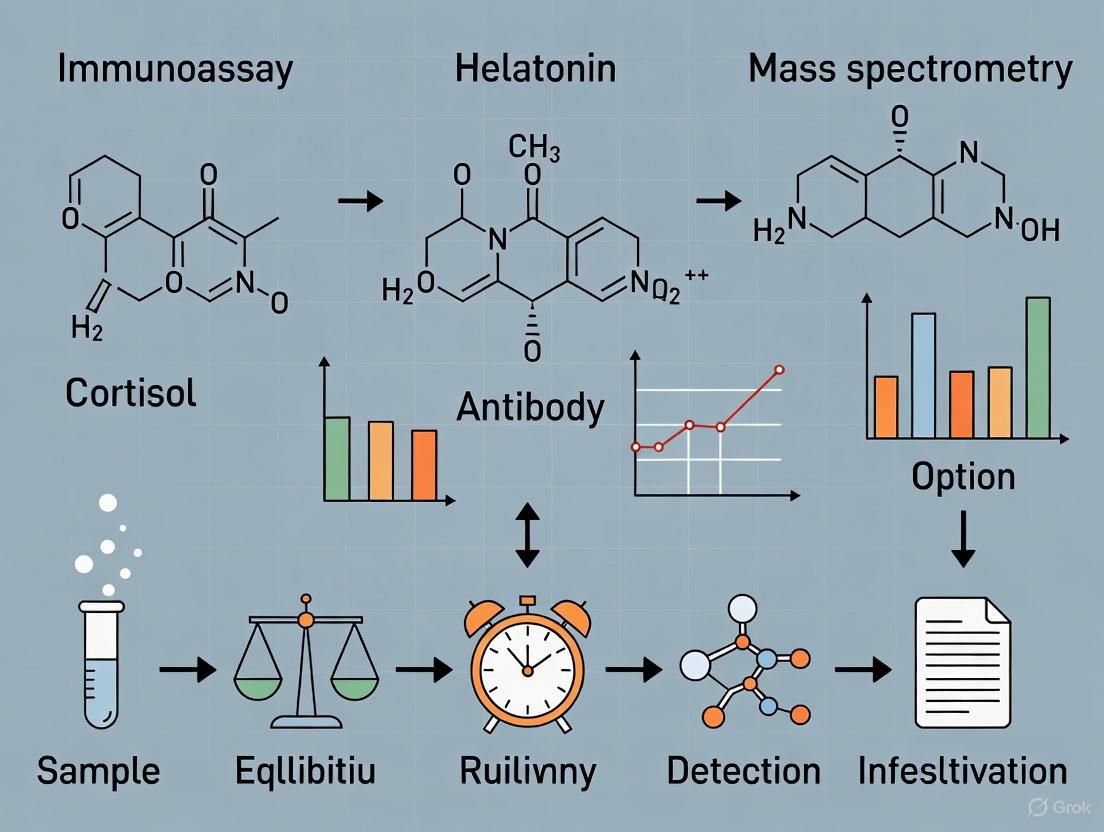
Immunoassay vs. Mass Spectrometry: A Critical Validation for Circadian Biomarker Analysis
Accurate measurement of circadian biomarkers like melatonin and cortisol is paramount for diagnosing sleep disorders, optimizing chronotherapy, and understanding the role of circadian disruption in diseases ranging from cancer to...
Immunoassay vs. Mass Spectrometry: A Critical Validation for Circadian Biomarker Analysis
Abstract
Accurate measurement of circadian biomarkers like melatonin and cortisol is paramount for diagnosing sleep disorders, optimizing chronotherapy, and understanding the role of circadian disruption in diseases ranging from cancer to metabolic syndrome. This article provides a comprehensive comparison of immunoassay and liquid chromatography-tandem mass spectrometry (LC-MS/MS) methodologies for circadian rhythm validation. Tailored for researchers and drug development professionals, we explore the foundational principles of key circadian markers, detail methodological protocols and applications, address critical troubleshooting and optimization strategies and present a rigorous validation and comparative analysis of the two techniques. The synthesis underscores LC-MS/MS as the gold standard for sensitivity and specificity while guiding the selection of the appropriate analytical platform based on research objectives and logistical constraints.
The Pillars of Circadian Biology: Core Biomarkers and Their Clinical Significance
The Central Pacemaker and Core Molecular Clock
In mammals, the suprachiasmatic nucleus (SCN) of the hypothalamus acts as the master circadian pacemaker, coordinating 24-hour rhythms in physiology and behavior. This central clock synchronizes cellular oscillators found in nearly every tissue throughout the body [1]. At its core, the circadian clock mechanism consists of interlocking transcriptional-translational feedback loops (TTFLs) involving a set of core clock genes and their protein products: CLOCK, BMAL1, PER, and CRY [1] [2].
The primary feedback loop begins when CLOCK and BMAL1 proteins form a heterodimer that binds to E-box enhancer elements in the promoters of target genes, including the Period (Per1, Per2, Per3) and Cryptochrome (Cry1, Cry2) genes [1]. After translation, PER and CRY proteins form complexes in the cytoplasm that translocate back to the nucleus to repress CLOCK-BMAL1 transcriptional activity, thereby completing a cycle that takes approximately 24 hours [1]. A stabilizing secondary loop involves nuclear receptors REV-ERBα/β and RORα/γ, which rhythmically regulate Bmal1 expression through RRE elements [2].
Table 1: Core Components of the Mammalian Circadian Clock
| Component | Gene Symbol | Primary Function in Clockwork |
|---|---|---|
| BMAL1 | Arntl | Forms heterodimer with CLOCK; primary transcriptional activator |
| CLOCK | Clock | Forms heterodimer with BMAL1; histone acetyltransferase activity |
| Period 1/2 | Per1, Per2 | Forms repressor complex with CRY; inhibits CLOCK-BMAL1 activity |
| Cryptochrome 1/2 | Cry1, Cry2 | Forms repressor complex with PER; inhibits CLOCK-BMAL1 activity |
| REV-ERBα/β | Nr1d1/2 | Represses Bmal1 transcription through RRE elements |
| RORα/γ | Rora, Rorc | Activates Bmal1 transcription through RRE elements |
Experimental Approaches for Circadian Rhythm Monitoring
Real-time Bioluminescence Imaging of SCN Slices
Protocol Overview: This technique utilizes transgenic mice expressing luciferase reporters fused to circadian clock genes (e.g., PER2::LUC) to visualize molecular oscillations in the SCN with single-cell resolution [3].
Detailed Methodology:
- Animal Preparation: Use homozygous PER2::LUC knock-in mice entrained to a standard light-dark cycle.
- SCN Slice Preparation: Sacrifice animals and extract brains. Prepare coronal SCN slices (200-400 μm thickness) using a vibratome.
- Culture Conditions: Place SCN slices on culture membranes in sealed dishes containing serum-free explant medium supplemented with luciferin (1 mM).
- Data Acquisition: Transfer cultures to an inverted microscope stage equipped with a photomultiplier tube or cooled CCD camera. Record bioluminescence with 30-minute temporal resolution for 3-5 days continuously [3].
- Data Analysis: Apply rhythm analysis algorithms (e.g., Cosinor analysis) to determine period, phase, and amplitude of PER2::LUC rhythms at both tissue and single-cell levels.
Cellular Circadian Reporter Assays
Protocol Overview: Fibroblast cell lines (e.g., NIH3T3) transfected with clock gene promoter-driven luciferase reporters enable high-throughput screening of clock components and chemical modifiers [4] [2].
Detailed Methodology:
- Cell Culture: Maintain NIH3T3 cells in DMEM supplemented with 10% FBS and antibiotics.
- Transfection: Co-transfect cells with E-box-driven luciferase reporter constructs (e.g., Per1-luc, Per2-luc) and Renilla luciferase control vector using lipid-based transfection reagents.
- Serum Shock Synchronization: Treat cells with 50% horse serum for 2 hours to synchronize cellular clocks.
- Bioluminescence Monitoring: Transfer cultures to photomultiplier assembly in medium containing luciferin (0.1 mM). Measure light emissions integrated for 1 minute at 15-minute intervals for multiple days [4].
- Pharmacological Manipulation: Apply kinase inhibitors (e.g., erbstatin analog) or other compounds to investigate post-translational regulation mechanisms [4].
Figure 1: Core Mammalian Circadian Clock Mechanism. The diagram illustrates the transcriptional-translational feedback loop involving CLOCK-BMAL1 heterodimer binding to E-box elements to activate Per and Cry transcription. After translation, PER-CRY complexes inhibit CLOCK-BMAL1 activity. A secondary stabilizing loop involves RRE-mediated regulation of Bmal1 by REV-ERB and ROR proteins [1] [2].
Comparative Analysis of Circadian Biomarker Measurement Techniques
Accurate assessment of circadian phase in humans relies on measuring endocrine biomarkers, primarily melatonin and cortisol. The choice between immunoassays and mass spectrometry represents a critical methodological consideration with significant implications for data quality and clinical interpretation [5] [6].
Melatonin Phase Assessment: Dim Light Melatonin Onset (DLMO)
Melatonin secretion from the pineal gland begins 2-3 hours before habitual sleep time, with its onset under dim light conditions (DLMO) considered the gold standard marker of circadian phase [5]. DLMO assessment typically requires sampling over a 4-6 hour window before bedtime.
Table 2: Methodological Comparison for Melatonin Measurement
| Parameter | Immunoassays (ELISA) | Liquid Chromatography-Tandem Mass Spectrometry (LC-MS/MS) |
|---|---|---|
| Analytical Principle | Antibody-antigen binding with enzymatic or fluorescent detection | Separation by chromatography with mass-based detection |
| Sample Matrix | Serum, saliva, urine | Serum, saliva, urine |
| Sensitivity | Moderate (functional sensitivity ~1-3 pg/mL in saliva) | High (detection limits <1 pg/mL) |
| Specificity | Subject to cross-reactivity with melatonin metabolites | High specificity for target analyte |
| Throughput | High | Moderate |
| Cost per Sample | Lower | Higher |
| Technical Expertise | Moderate | Extensive |
| Recommended DLMO Threshold | 3-4 pg/mL in saliva | 2 pg/mL in plasma for low producers |
Cortisol Rhythm Assessment
Cortisol exhibits a diurnal rhythm opposite to melatonin, peaking in the morning and reaching its nadir around midnight. The cortisol awakening response (CAR) and late-night salivary cortisol are key circadian markers [5] [6].
Table 3: Methodological Comparison for Cortisol Measurement
| Parameter | Immunoassays | Liquid Chromatography-Tandem Mass Spectrometry (LC-MS/MS) |
|---|---|---|
| Analytical Principle | Antibody-based detection | Mass-based detection after chromatographic separation |
| Sample Matrix | Serum, saliva, urine | Serum, saliva, urine |
| Specificity Issues | Cross-reactivity with cortisol metabolites (e.g., cortisone, prednisolone) | Minimal cross-reactivity |
| Urinary Free Cortisol | Requires pre-extraction to remove conjugated metabolites; results ~2× higher than LC-MS/MS | Direct measurement without extraction; considered true UFC |
| Diagnostic Accuracy (Cushing's) | AUC: 0.953-0.969 [7] | Reference method |
| Standardization | Poor across laboratories | Excellent standardization |
| Simultaneous Analysis | Single analyte | Multiple steroids simultaneously |
The Scientist's Toolkit: Essential Research Reagents and Materials
Table 4: Key Research Reagents for Circadian Rhythm Investigation
| Reagent/Material | Application | Function/Utility |
|---|---|---|
| PER2::LUC Knock-in Mice | SCN slice bioluminescence imaging | Endogenous reporting of PER2 protein oscillations [3] |
| Luciferin | Bioluminescence assays | Substrate for luciferase enzyme; enables light emission detection |
| Serum-Free Explant Medium | SCN slice culture | Maintains tissue viability during longitudinal imaging [3] |
| E-box Luciferase Reporters | Cellular circadian assays | Monitoring CLOCK-BMAL1 transcriptional activity [4] |
| Casein Kinase Inhibitors (e.g., PF-670462) | Post-translational regulation studies | Investigating phosphorylation-dependent clock protein degradation [4] |
| Cortisol/Melatonin Antibodies | Immunoassays | Detection and quantification of circadian biomarkers [5] |
| Mass Spectrometry Internal Standards (e.g., deuterated cortisol) | LC-MS/MS biomarker analysis | Precision quantification through isotope dilution [5] [6] |
Molecular Interactions and Functional Relationships Among Clock Components
CRY-PER Interactions: Beyond Simple Repression
While PER and CRY traditionally function as collaborative repressors of CLOCK-BMAL1 activity, emerging evidence reveals a more complex regulatory relationship. Studies indicate that PER proteins can counteract CRY-mediated transcriptional repression through physical interaction, potentially blocking CRY recruitment to the CLOCK-BMAL1 complex [8]. This anti-CRY activity appears specific to PER1 and PER2, with PER3 lacking this function [8]. This buffering effect on CRY repression may help prolong the circadian period and create distinct transcriptional phases.
Phosphorylation-Dependent Regulation
Post-translational modifications, particularly phosphorylation, critically regulate clock protein stability and activity. Casein kinase 1δ/ε (CK1δ/ε) phosphorylates PER proteins, marking them for ubiquitination and proteasomal degradation [1]. Conversely, PER can protect CLOCK phosphorylation against CRY, with PER1 and PER2 demonstrating protective activity while PER3 does not [4]. This regulatory mechanism fine-tunes the timing and amplitude of circadian oscillations.
Figure 2: Circadian Rhythm Assessment Workflow. The diagram outlines the pathway from environmental entrainment to biomarker measurement, highlighting the central role of the SCN in coordinating physiological rhythms and the methodological options for quantifying circadian phase markers [5] [1] [6].
Implications for Experimental Design and Data Interpretation
The choice between immunoassays and mass spectrometry for circadian biomarker assessment involves important trade-offs. While immunoassays offer practical advantages of lower cost and higher throughput, LC-MS/MS provides superior specificity and accuracy, particularly for low-concentration analytes like salivary melatonin [5]. Method selection should align with research objectives, with mass spectrometry preferred for definitive phase assessment and immunoassays potentially adequate for large-scale screening when properly validated.
Understanding the complex interactions among molecular clock components is essential for interpreting genetic and pharmacological manipulations. The compensatory capacity of the SCN network [3], functional redundancies among paralogs (e.g., PER1/2 vs. PER3, CRY1 vs. CRY2) [8], and tissue-specific differences in clock gene expression [2] all contribute to the remarkable robustness of circadian timekeeping despite molecular perturbations.
Circadian rhythms are intrinsic, near-24-hour oscillations that coordinate fundamental physiological processes, from sleep-wake cycles to hormone secretion and metabolism [5]. The accurate assessment of an individual's circadian phase is crucial for both basic research and clinical practice, particularly in diagnosing and treating circadian rhythm sleep-wake disorders (CRSDs) [9]. Among the available markers, the dim light melatonin onset (DLMO) has emerged as the gold-standard marker of the central circadian clock in humans when measured under dim light conditions from plasma or saliva [5] [10]. Melatonin, a neurohormone produced by the pineal gland, exhibits a distinct daily rhythm, with secretion beginning approximately 2-3 hours before habitual bedtime and peaking during the night [5] [11]. The DLMO specifically refers to the time when melatonin concentrations first rise significantly under dim light conditions, thereby signaling the onset of the "biological night" [5] [12]. Its precision is superior to other circadian markers; melatonin allows for suprachiasmatic nucleus (SCN) phase determination with a standard deviation of 14-21 minutes, compared to approximately 40 minutes for cortisol-based methods [5].
The critical importance of DLMO measurement is particularly evident in clinical contexts. Treatment of CRSDs with melatonin, light therapy, or chronotherapy is most effective when timed according to the individual's endogenous circadian phase [9]. Administering these treatments without knowledge of the DLMO can render them ineffective or even produce contrary effects [9]. Furthermore, DLMO measurement enables differential diagnosis of CRSDs from other sleep disorders like insomnia, ensuring patients receive appropriate and effective interventions [11] [9]. The following sections provide a comprehensive comparison of the analytical methods used to quantify melatonin for DLMO determination, detail standardized experimental protocols, and present emerging technologies that are shaping the future of circadian biomarker assessment.
Analytical Face-Off: Immunoassay vs. Mass Spectrometry
The reliable quantification of melatonin, particularly at the low concentrations found in saliva, presents significant analytical challenges. The two primary methodological approaches are immunoassays (e.g., ELISA, RIA) and liquid chromatography-tandem mass spectrometry (LC-MS/MS), each with distinct advantages and limitations.
Performance Comparison
Table 1: Comparison of Melatonin Quantification Methods for DLMO Assessment
| Method | Sensitivity | Specificity | Sample Throughput | Sample Volume | Cost & Accessibility | Key Applications |
|---|---|---|---|---|---|---|
| LC-MS/MS | ~1.0 pg/mL [13] | High (minimal cross-reactivity) [5] | Moderate | 50-100 µL [11] | High equipment cost, requires skilled personnel [5] [12] | Gold-standard validation, research requiring highest accuracy [5] |
| ELISA | Varies by kit; e.g., 1.35 pg/mL (Salimetrics) [11] | Moderate (potential for cross-reactivity) [5] [12] | High | 100 µL per well [11] | Lower cost, widely accessible [5] | High-throughput screening, clinical studies [5] |
| Radioimmunoassay (RIA) | <0.3 pg/mL [13] | Moderate (potential for cross-reactivity) [12] | High | ~1 mL [13] | Moderate cost, requires radioisotope handling [12] | High-sensitivity research applications |
| Novel Aptamer (ELAA) | ~0.57 pg/mL [12] | High (low cross-reactivity) [12] | High (potentially) | Not Specified | Developing technology | Potential for high-sensitivity, high-throughput point-of-care |
Immunoassays, including enzyme-linked immunosorbent assays (ELISAs) and radioimmunoassays (RIAs), have been widely used due to their relatively low cost and high throughput [5]. However, they can suffer from cross-reactivity with structurally similar molecules, such as serotonin or tryptophan metabolites, which can compromise specificity and lead to overestimation of melatonin concentrations, particularly at low levels [5] [12]. For example, many ELISA kits rely on antibodies raised against melatonin chemically linked to an immunogenic molecule, a process that is complicated and not fully understood, often contributing to reported poor specificity [12].
In contrast, LC-MS/MS is increasingly recognized as the superior analytical technique due to its exceptional sensitivity and specificity [5] [13]. It eliminates cross-reactivity by separating melatonin from other compounds chromatographically before detection based on its precise mass-to-charge ratio [5]. This technique allows for the simultaneous analysis of multiple hormones, such as cortisol and melatonin, without additional cost or time, providing a more comprehensive insight into circadian interactions [5]. The primary limitations of LC-MS/MS are its high initial equipment costs, complex sample preparation requiring skilled personnel, and lower throughput compared to immunoassays [5] [12].
Practical Considerations for DLMO Determination
The choice of analytical method directly impacts the reliability of the DLMO calculation. The most common methods for determining DLMO from a series of samples are the fixed threshold and variable threshold approaches.
- Fixed Threshold Method: DLMO is defined as the time when interpolated melatonin concentrations cross an absolute value, typically 3 pg/mL or 4 pg/mL in saliva [5] [14]. This method is straightforward but risks missing the DLMO in individuals who are low melatonin producers (e.g., the elderly or those with certain pathologies) whose levels may never reach this threshold [5] [11].
- Variable Threshold Method ("3k Method"): The threshold is calculated individually as the mean of the first three low daytime samples plus two standard deviations [5] [11]. This method is more adaptable for low producers and accounts for individual baseline variations, though it can be unreliable with inconsistent baselines [5].
For both methods, the superior specificity of LC-MS/MS provides more accurate baseline and rising-phase melatonin values, leading to a more reliable DLMO calculation. Immunoassays, with their potential for cross-reactivity, may overestimate low concentrations, potentially leading to an earlier and less accurate DLMO estimate [5] [12].
Experimental Protocols for DLMO Assessment
A standardized protocol is vital for obtaining reliable and reproducible DLMO measurements, whether in a clinical or research setting.
Sample Collection Workflow
The following diagram illustrates the key stages in a typical salivary DLMO assessment protocol.
Detailed Methodologies
Pre-Assessment Conditions and Sample Collection
- Participant Preparation: Participants should maintain a fixed sleep schedule for at least one week before sampling to stabilize their circadian rhythm [14]. The use of medications that suppress (e.g., NSAIDs, beta-blockers) or elevate (e.g., antidepressants, contraceptives) melatonin should be recorded and considered, as they can confound results [5].
- Sampling Environment: Sampling must occur under dim light conditions (< 20 lux) to prevent the suppressive effect of light on melatonin secretion [14] [10]. Participants should also avoid activities that can interfere with saliva composition, such as eating, drinking caffeinated beverages, or brushing teeth, in the 30 minutes before each sample [11].
- Sampling Protocol: The recommended sampling window is typically 6 hours, starting 5 hours before habitual bedtime and ending 1 hour after bedtime [5] [14]. Samples can be collected every 30 or 60 minutes. Research in adolescents has shown that 60-minute sampling provides DLMO estimates within ±1 hour of 30-minute sampling when using a fixed threshold, offering a cost-effective and less burdensome protocol [14]. Saliva is most commonly collected via passive drool into polypropylene tubes or using specialized devices like Salivettes [11] [9]. After collection, samples should be stored frozen (≤ -20°C) until analysis [11].
Laboratory Analysis Procedures
Table 2: Key Research Reagent Solutions for Melatonin Quantification
| Reagent / Material | Function / Role in Experiment | Example Specification / Note |
|---|---|---|
| Saliva Collection Device | Non-invasive collection of salivary melatonin | e.g., Salivette (Sarstedt) or passive drool into polypropylene tubes [9] |
| Melatonin Antibody | Biorecognition element in ELISA; binds melatonin | Critical for assay specificity and sensitivity; potential for cross-reactivity [5] [12] |
| Melatonin Aptamer | Novel biorecognition element in ELAA; binds melatonin | Chemically synthesized DNA/RNA; high specificity and batch uniformity [12] |
| LC-MS/MS Mobile Phase | Chromatographic separation of melatonin | e.g., Solvent A: 0.1% formic acid in water; Solvent B: methanol [15] |
| Melatonin Reference Standard | Calibration and quantification | Essential for both LC-MS and immunoassay methods; purity is critical [15] |
| Stable Isotope-Labeled Melatonin | Internal standard for LC-MS/MS | Corrects for matrix effects and losses during sample prep (e.g., d4-melatonin) [15] |
For LC-MS/MS Analysis: The protocol typically involves a solid-phase or liquid-liquid extraction step to purify and concentrate melatonin from the saliva sample [13]. The extract is then injected into the LC-MS/MS system. Melatonin is separated chromatographically (e.g., using a C18 column with a methanol/water gradient) and detected using multiple reaction monitoring (MRM), which offers high specificity [5] [13]. The use of a stable isotope-labeled internal standard (e.g., d4-melatonin) is critical for achieving high accuracy by correcting for matrix effects and preparation inefficiencies [15].
For ELISA Analysis: Saliva samples are typically centrifuged to remove particulate matter and then added directly to the assay plate without extraction [11]. The competitive ELISA procedure involves incubating the sample with an enzyme-labeled melatonin conjugate and an anti-melatonin antibody. After a washing step, a substrate is added, and the color development is measured spectrophotometrically. The intensity of the signal is inversely proportional to the concentration of melatonin in the sample [11].
Emerging Technologies and Future Directions
The field of circadian biomarker assessment continues to evolve with the development of new technologies aimed at increasing sensitivity, reducing cost, and enabling point-of-care applications.
- Aptamer-Based Assays: A novel competitive enzyme-linked aptamer-based assay (ELAA) has been developed as a promising alternative to antibody-based methods [12]. Aptamers are single-stranded DNA or RNA molecules that bind to targets with high affinity and specificity. This emerging technology has demonstrated a detection limit of ~0.57 pg/mL for salivary melatonin, which is superior to many existing ELISAs and approaches the sensitivity of LC-MS/MS [12]. Aptamers offer advantages including reproducible batch synthesis and ease of modification, potentially leading to more robust and cost-effective assays in the future [12].
- Non-Invasive Phase Prediction Models: To circumvent the need for frequent saliva sampling, computational models are being developed to predict DLMO from non-invasive data. One study used a statistical model based on light exposure, sleep timing, and demographic variables to predict DLMO in patients with Delayed Sleep-Wake Phase Disorder (DSWPD) with a root mean square error of 57 minutes, with predictions accurate to within ±1 hour in 75% of participants [16]. While not a replacement for direct biochemical measurement, these models show significant promise for screening and clinical applications where direct DLMO assessment is not feasible [16].
The accurate determination of DLMO is fundamental to advancing the field of circadian medicine. While salivary melatonin has made this biomarker more accessible, the choice of analytical method remains critical. LC-MS/MS stands as the gold standard for researchers requiring the highest level of accuracy and specificity for validation purposes. However, well-validated immunoassays provide a practical and high-throughput alternative for many clinical and research studies. The ongoing development of novel technologies, such as aptamer-based assays and computational prediction models, promises to further refine the assessment of circadian phase, ultimately improving the diagnosis and treatment of circadian rhythm disorders and associated health conditions. For researchers and clinicians, the decision between immunoassay and mass spectrometry must balance the demands of analytical rigor with practical constraints of cost, throughput, and accessibility.
The Cortisol Awakening Response (CAR) is a distinct neuroendocrine phenomenon characterized by a sharp increase in cortisol secretion during the first 30-45 minutes after awakening, typically increasing by 38% to 75% from awakening levels [17]. This response represents a crucial point of reference within the healthy cortisol circadian rhythm and serves as a key biomarker of hypothalamic-pituitary-adrenal (HPA) axis activity [17]. The CAR is theorized to serve an adaptive function, preparing individuals for anticipated energy demands and challenges of the forthcoming day and potentially providing an "allostatic boost" for upcoming emotional and cognitive tasks [18] [19] [20]. As research into circadian biomarkers advances, the accurate measurement of CAR has become increasingly important for both research and clinical applications, particularly in the context of stress-related disorders, neurodegenerative diseases, and metabolic conditions [5]. This comparison guide examines the two primary analytical methodologies—immunoassay and mass spectrometry—for quantifying CAR, providing researchers with experimental data and protocols to inform their circadian validation research.
Analytical Methodologies: Technical Comparison of Immunoassay vs. LC-MS/MS
The reliable quantification of cortisol concentrations presents significant analytical challenges, with methodological choices directly impacting data quality and clinical interpretations. The two predominant techniques—immunoassay and liquid chromatography-tandem mass spectrometry (LC-MS/MS)—differ substantially in their technical principles and performance characteristics.
Table 1: Core Principle Comparison of Immunoassay and LC-MS/MS for Cortisol Measurement
| Feature | Immunoassay | LC-MS/MS |
|---|---|---|
| Analytical Principle | Antibody-antigen binding with colorimetric, chemiluminescent, or fluorescent detection | Physical separation followed by mass-to-charge ratio detection |
| Specificity | Susceptible to cross-reactivity with structurally similar steroids (e.g., 11-deoxycortisol, cortisone) [21] | High specificity; distinguishes cortisol from isobaric compounds and metabolites |
| Sample Throughput | High (automated platforms) | Moderate to high (modern systems) |
| Sample Volume | Low (typically 10-100 µL) | Low to moderate (50-200 µL) |
| Sample Preparation | Minimal (often dilution only) | Extensive (protein precipitation, liquid-liquid, or solid-phase extraction) |
| Capital Equipment Cost | Lower | Significantly higher |
Table 2: Performance Characteristics for Cortisol Measurement
| Performance Parameter | Immunoassay | LC-MS/MS |
|---|---|---|
| Sensitivity (LLOQ) | Varies by platform; generally sufficient for salivary cortisol | Superior; can reach sub-nanomolar levels [22] |
| Accuracy in Challenging Matrices | Compromised by metabolites and interfering substances [21] | High accuracy due to separation and selective detection |
| Precision (%CV) | Typically 5-10% | Typically 3-8% |
| Multiplexing Capability | Limited to single or few analytes per run | High; simultaneous quantification of multiple steroids |
Immunoassays employ antibody-based recognition, which creates vulnerability to cross-reactivity with structurally related steroids. This limitation becomes clinically significant in conditions where steroid metabolism is altered. For instance, in patients receiving metyrapone (an 11β-hydroxylase inhibitor), immunoassays demonstrated a positive bias of 23% (59 nmol/L) compared to LC-MS/MS due to cross-reactivity with accumulating 11-deoxycortisol [21]. This discrepancy can directly impact clinical decisions, potentially leading to over-treatment and unrecognized hypoadrenalism.
LC-MS/MS methods overcome these specificity issues through physical separation of analytes prior to detection. The technology provides high mass accuracy and resolving power, enabling distinction between cortisol and its metabolites or synthetic analogs [5] [23]. While traditionally considered laborious, modern UPLC-HRMS (Ultra-Performance Liquid Chromatography-High Resolution Mass Spectrometry) methods have improved efficiency with analysis times as short as 6 minutes per sample [23].
Experimental Protocols for CAR Assessment
Sample Collection Protocols
Salivary Cortisol Collection: Saliva sampling has gained popularity for CAR assessment due to its non-invasive nature and suitability for repeated, ambulatory measurements in home settings [5]. The standard protocol involves:
- Sample Timing: Collect samples immediately upon awakening (S1), then at 30 (S2), and 45 (S3) minutes post-awakening. Some protocols extend to 60 minutes (S4) with additional samples throughout the day for diurnal profiling [19].
- Collection Devices: Use Salivettes or similar specialized collection devices.
- Participant Instructions: Participants should avoid eating, drinking, brushing teeth, or smoking during the sampling period. They should record exact awakening and sampling times to monitor protocol adherence [18].
- Storage: Freeze samples at -20°C or lower until analysis.
Serum/Plasma Collection: For higher analyte concentrations and potentially better reliability, venous blood sampling can be employed:
- Sample Timing: Serial sampling at similar intervals to salivary protocols, though less practical for home settings.
- Collection Tubes: Serum separator tubes or EDTA plasma tubes.
- Processing: Centrifuge within 2 hours of collection; aliquot and freeze plasma/serum at -20°C or lower.
Innovative Approaches: Recent studies have utilized in vivo microdialysis to measure tissue-free cortisol levels in interstitial fluid continuously over 24-hour periods, allowing assessment of pre- and post-awakening cortisol dynamics in naturalistic environments [18].
Laboratory Analysis Protocols
Immunoassay Protocol (Representative Chemiluminescent Assay):
- Principle: Competitive binding between labeled cortisol and sample cortisol for antibody binding sites.
- Procedure:
- Pipette 50 µL of calibrators, controls, and samples into appropriate wells.
- Add 100 µL of cortisol-alkaline phosphatase conjugate.
- Add 100 µL of anti-cortisol antibody.
- Incubate for 30 minutes at room temperature.
- Wash plates to remove unbound materials.
- Add chemiluminescent substrate and measure light emission.
- Calculation: Generate standard curve from calibrators to determine sample concentrations.
LC-MS/MS Protocol (Representative):
- Sample Preparation:
- Add 50 µL of internal standard solution (e.g., cortisol-d4) to 100 µL of sample.
- Precipitate proteins with 400 µL of acetonitrile or methanol.
- Vortex mix and centrifuge at 11,290 × g for 5 minutes at 4°C.
- Transfer supernatant and evaporate to dryness under nitrogen or vacuum.
- Reconstitute in 100 µL mobile phase (e.g., 45:55 acetonitrile:2mM ammonium acetate with 0.1% formic acid).
- Chromatography:
- Column: Acquity UPLC BEH C18 (50 × 2.1 mm, 1.7 µm)
- Mobile Phase: A: 2mM ammonium acetate with 0.1% formic acid; B: 0.1% formic acid in acetonitrile
- Gradient: 45% B to 50% B at 1.0 min; to 80% B at 2.5 min; to 100% B at 3.0 min
- Flow Rate: 0.3 mL/min; Injection Volume: 5 µL
- Mass Spectrometry:
- Ionization: Electrospray ionization positive mode
- Scan Type: Multiple reaction monitoring (MRM)
- Cortisol transition: 363.2 → 121.2 (quantifier) and 363.2 → 97.1 (qualifier)
- Internal standard transition: 367.2 → 121.2
Comparative Experimental Data
Table 3: Method Comparison Studies for Cortisol Measurement
| Study Context | Immunoassay Platform | LC-MS/MS Comparison | Bias/Agreement | Clinical Implications |
|---|---|---|---|---|
| General Population [24] | Beckman Access Cortisol & Abbott Architect | Xevo-TQ-S (Waters) | 8.38% & 8.78% positive bias vs. LC-MS/MS | Both immunoassays showed close agreement with LC-MS/MS, deemed suitable for routine determination |
| Metyrapone Therapy Patients [21] | Not specified | LC-MS/MS | 23% positive bias (59 nmol/L) in immunoassay | Potential for erroneous clinical decisions concerning dose titration |
| Urinary Free Cortisol in Cushing's [7] | Four new direct immunoassays (Autobio, Mindray, Snibe, Roche) | Laboratory-developed LC-MS/MS | Strong correlations (r=0.950-0.998) with proportional positive bias | High diagnostic accuracy (AUC>0.95) for Cushing's syndrome with method-specific cut-offs |
| Cyclosporine A Monitoring [23] | Chemiluminescent Microparticle Immunoassay (CMIA) | UPLC-Orbitrap-MS | CMIA significantly higher (85.70±48.99 vs. 67.06±34.56 ng/mL, P<0.0001) | Positive bias in immunoassay due to metabolite cross-reactivity |
The data consistently demonstrate that while immunoassays generally show good correlation with LC-MS/MS, they frequently exhibit positive biases, particularly in clinically challenging scenarios where steroid metabolism is altered. The proportional nature of these biases suggests that absolute cortisol concentrations derived from different methodologies may not be directly interchangeable in longitudinal studies or when applying universal clinical decision limits.
Signaling Pathways and Experimental Workflows
Diagram 1: CAR Assessment Workflow from Biology to Quantification
This diagram illustrates the complete pathway from cortisol regulation to analytical quantification, highlighting parallel methodologies. The suprachiasmatic nucleus (SCN) coordinates HPA axis activity, leading to cortisol secretion from the adrenal cortex with its characteristic CAR pattern. Biological samples are then collected and analyzed through either immunoassay or mass spectrometry pathways to generate quantitative data.
The Scientist's Toolkit: Essential Research Reagent Solutions
Table 4: Essential Research Reagents for Cortisol Circadian Studies
| Reagent/Material | Function/Application | Technical Considerations |
|---|---|---|
| Salivary Collection Devices (e.g., Salivettes) | Non-invasive sample collection for CAR assessment | Use cotton or polyester synthetic swabs; avoid citric acid-treated devices which can stimulate saliva flow |
| Cortisol Immunoassay Kits | Quantification of cortisol in biological fluids | Select kits with validated performance for matrix of interest; verify cross-reactivity profile |
| Stable Isotope-Labeled Internal Standards (e.g., cortisol-d4) | Normalization in LC-MS/MS analysis | Essential for correcting matrix effects and recovery variations during sample preparation |
| Solid Phase Extraction (SPE) Cartridges | Sample clean-up and analyte concentration for LC-MS/MS | C18 or mixed-mode phases provide optimal recovery of corticosteroids |
| LC-MS/MS Mobile Phase Additives | Chromatographic separation optimization | Ammonium acetate/formate with 0.1% formic acid enhances ionization efficiency |
| Quality Control Materials | Method validation and daily performance monitoring | Use pooled human serum/saliva at low, medium, and high cortisol concentrations |
The methodological comparison between immunoassay and LC-MS/MS for CAR assessment reveals a nuanced landscape where practical considerations must be balanced with analytical rigor. Immunoassays offer practical advantages for high-throughput studies where relative changes in CAR are sufficient, while LC-MS/MS provides superior specificity essential for absolute concentration measurements, particularly in research involving pharmacological interventions or pathological conditions that alter steroid metabolism.
Recent research utilizing innovative continuous sampling methodologies has challenged traditional interpretations of CAR, suggesting that the rate of cortisol increase may not change at awakening compared to the preceding hour, and highlighting extraordinary intersubject variability partly explained by sleep duration and wake time consistency [18]. These findings underscore the importance of methodological precision in advancing our understanding of HPA axis dynamics.
For circadian validation research, the choice between immunoassay and mass spectrometry should be guided by study objectives, sample volume availability, required specificity, and resources. As the field progresses toward standardized protocols and reference methods, the combination of rigorous sampling protocols with specific analytical technologies will enhance data comparability across studies and strengthen the validity of CAR as a biomarker in both research and clinical applications.
Emerging research underscores circadian disruption as a fundamental biological mechanism linking a spectrum of human diseases. This review synthesizes evidence connecting dysregulated circadian rhythms to the pathogenesis of neurodegenerative diseases, psychiatric disorders, and metabolic conditions. We further dissect the critical methodological framework of immunoassay versus mass spectrometry for validating circadian biomarkers, providing a comparative analysis of their performance in quantifying key hormonal rhythms. By integrating findings from molecular studies, clinical epidemiology, and advanced biomarker analytics, this article provides a foundational guide for researchers and drug development professionals navigating the intersection of chronobiology and human disease.
Circadian rhythms are intrinsic, approximately 24-hour cycles that govern a vast array of physiological processes, from sleep-wake patterns and hormone secretion to cellular metabolism and immune function [25] [26]. These rhythms are orchestrated by a master clock in the suprachiasmatic nucleus (SCN) of the hypothalamus, which synchronizes peripheral clocks in virtually every tissue and organ system [27]. At the molecular level, the circadian machinery is driven by transcriptional-translational feedback loops (TTFLs) involving core clock genes such as CLOCK, BMAL1, PER, and CRY [25] [27].
The disruption of these precise rhythms—through genetic, environmental, or behavioral means—is increasingly recognized as a hallmark of and contributor to a diverse range of disorders. Circadian dysfunction is not merely a symptom but can be a causal factor in disease pathogenesis, sometimes manifesting before overt clinical symptoms appear [25]. This article explores the mechanistic links between circadian disruption and three major disease categories: neurodegenerative, psychiatric, and metabolic disorders. Furthermore, in the context of a growing focus on circadian medicine, we critically evaluate the experimental protocols and analytical techniques essential for robust circadian research, with a specific focus on the comparative validation of immunoassay and mass spectrometry methods for measuring core circadian biomarkers.
Circadian Disruption in Neurodegenerative Diseases
The connection between a disrupted circadian system and neurodegeneration is supported by substantial evidence. Disruptions in sleep-wake cycles, core body temperature, and hormonal rhythms are common features in Alzheimer's disease (AD), Parkinson's disease (PD), and Huntington's disease (HD) [25]. These disruptions often occur early in the disease process, suggesting a potential role as early-stage molecular biomarkers [27].
Key Pathophysiological Links
The relationship is bidirectional: neurodegenerative pathology can damage the SCN, while circadian disruption can accelerate disease processes.
- Molecular Pathway Disruptions: Core clock genes regulate critical processes commonly disrupted in neurodegeneration, including redox balance, mitochondrial function, and neuroinflammation [25]. For instance, BMAL1 deficiency has been shown to impair synaptic vesicle cycling and increase susceptibility to oxidative damage and inflammation [27].
- Sleep and Protein Aggregation: Sleep deprivation aggravates key pathological processes, such as the accumulation of Aβ plaques and tau protein tangles in the brain, which are critical in AD pathogenesis [25]. The sleep-wake cycle is crucial for the glymphatic system's clearance of these waste products from the brain.
- Melatoninergic Signaling: The neurohormone melatonin, a key output of the circadian system, modulates clock gene expression, mitochondrial stability, and inflammatory responses. Its secretion is often suppressed in neurodegenerative conditions, reducing its neuroprotective effects [27].
Figure 1: Bidirectional Pathways Between Circadian Disruption and Neurodegeneration. Circadian disruption drives cellular pathologies that accelerate neurodegeneration, while neurodegenerative damage to the SCN further disrupts circadian rhythms, creating a vicious cycle [25] [27].
The Psychiatric and Metabolic Dimensions of Circadian Dysregulation
The impact of circadian disruption extends profoundly into mental and metabolic health, with large-scale epidemiological and real-world digital studies strengthening the evidence base.
Psychiatric Disorders: The Light-Darkness Connection
Light at night (LAN) is a potent disruptor of circadian rhythms, and meta-analyses have confirmed its association with several psychiatric conditions. A systematic review and meta-analysis of 19 studies found that LAN exposure was significantly associated with increased odds of depression prevalence (OR: 1.18), bipolar disorder (OR: 1.19), and anxiety (OR: 1.10) [28]. The association was stronger for directly measured indoor LAN exposure than for satellite-measured outdoor LAN.
Large-scale digital phenotyping studies using wearables have provided real-world validation. Research involving over 800 first-year physicians—a cohort exposed to significant circadian challenge from shift work—demonstrated that misalignment between the central circadian rhythm and the sleep-wake cycle was most predictive of next-day negative mood [29]. These digital markers offer a scalable, non-invasive method for assessing circadian disruption and its psychiatric impact.
Metabolic Disease: The Muscle Clock Link
Disruption of peripheral clocks, particularly in metabolic tissues like skeletal muscle, is a key contributor to metabolic disease. Investigators found that mice lacking the core clock gene BMAL1 specifically in their muscles developed accelerated glucose intolerance when placed on a high-fat, high-carbohydrate diet, despite no differences in weight gain [30].
The mechanism involves a critical link between the circadian clock and nutrient-sensing pathways. BMAL1 works together with the hypoxia-inducible factor (HIF) pathway to adapt muscle metabolism to nutrient stress. When the muscle clock is disrupted, this connection is lost, leading to impaired glucose utilization in the early stages of glycolysis [30]. This finding highlights how circadian disruption in peripheral tissues can predispose to insulin resistance and diabetes, independent of central obesity.
Figure 2: Converging Pathways from Disruption to Disease. Environmental disruptors like LAN or irregular schedules trigger divergent pathological pathways in psychiatric and metabolic disorders through shared mechanisms of central and peripheral clock misalignment.
Analytical Core: Immunoassay vs. Mass Spectrometry in Circadian Biomarker Validation
Accurate measurement of circadian phase is foundational to this field. The hormones melatonin and cortisol serve as primary peripheral biomarkers for the central clock's phase. Their quantification, however, presents significant analytical challenges, centering on the comparison between immunoassays and liquid chromatography-tandem mass spectrometry (LC-MS/MS) [26].
Comparative Performance of Analytical Techniques
Table 1: Method Comparison for Circadian Biomarker Analysis [26] [7]
| Parameter | Immunoassay | Liquid Chromatography-Tandem Mass Spectrometry (LC-MS/MS) |
|---|---|---|
| Principle | Antibody-based recognition | Physical separation by mass/charge ratio |
| Throughput | High | Moderate to High |
| Cost per Sample | Lower | Higher |
| Specificity | Subject to cross-reactivity | High specificity |
| Sensitivity | Variable; can be sufficient for saliva | Excellent, especially for low concentrations in saliva |
| Multiplexing | Limited (usually single analyte) | High (can measure multiple analytes simultaneously) |
| Best Use Case | High-throughput screening, clinical diagnostics | Gold-standard validation, research requiring high precision |
Immunoassays, while high-throughput and widely available, can suffer from cross-reactivity with structurally similar molecules, leading to inaccurate results, particularly at the low concentrations found in saliva [26]. In contrast, LC-MS/MS provides superior specificity, sensitivity, and reproducibility, making it the preferred method for research requiring high accuracy, though it requires more specialized equipment and expertise [26].
A recent comparative study on urinary free cortisol measurement for Cushing's syndrome diagnosis demonstrated that while modern immunoassays show strong correlation with LC-MS/MS (Spearman coefficient r > 0.95), they consistently exhibited a proportional positive bias [7]. This underscores the necessity of using method-specific reference intervals in both clinical practice and research.
Key Circadian Biomarkers and Their Measurement
- Melatonin and Dim Light Melatonin Onset (DLMO): The DLMO, measured under dim light conditions, is the gold standard marker for assessing the phase of the endogenous circadian pacemaker [26]. It is typically determined from serial saliva or plasma samples collected over a 4-6 hour window before habitual bedtime. DLMO can be calculated using a fixed threshold (e.g., 3-4 pg/mL in saliva) or a variable threshold based on baseline values [26].
- Cortisol and the Cortisol Awakening Response (CAR): Cortisol exhibits a robust diurnal rhythm with a sharp peak 30-45 minutes after waking. The CAR serves as an index of hypothalamic-pituitary-adrenal (HPA) axis activity and is influenced by circadian timing, sleep, and stress [26]. While less precise than DLMO for phase assessment (standard deviation of ~40 minutes vs. 14-21 minutes for melatonin), it remains a valuable and easily measured circadian marker [26].
Experimental Protocols for Circadian Biomarker Assessment
Robust measurement of circadian parameters requires stringent, standardized protocols to minimize confounding variables.
- Participant Preparation: Participants should avoid alcohol, caffeine, and strenuous exercise for 24 hours prior. They should not have undertaken recent transmeridian travel or shift work. The use of certain medications (e.g., beta-blockers, NSAIDs, antidepressants) may need to be controlled, as they can suppress or artificially elevate melatonin.
- Sampling Environment: Sampling must occur under dim light conditions (<10-30 lux) to prevent melatonin suppression. A dedicated, light-controlled room or the participant's home with all bright lights and screens off is required.
- Sample Collection: Serial saliva samples are collected every 30-60 minutes over a 4-6 hour window ending 1 hour after habitual bedtime. Saliva is typically collected using passive drool or salivette kits. Participants must remain awake and in a seated or relaxed posture.
- Sample Handling: Samples should be stored immediately at -20°C or below until analysis.
- Analytical Method: Analysis via LC-MS/MS is recommended for its superior specificity for low salivary melatonin concentrations. If using an immunoassay, its performance characteristics for salivary melatonin must be rigorously validated.
- DLMO Calculation: The time of DLMO is determined by interpolating the time at which melatonin concentration crosses a predefined threshold (e.g., 3 pg/mL or 2 standard deviations above the mean of baseline samples).
- Sampling Schedule: Participants collect saliva samples immediately upon waking, and then at 30, 45, and 60 minutes post-awakening. The exact clock time of each sample must be recorded.
- Participant Control: Participants should not eat, drink (except water), smoke, or brush their teeth until after the final sample is collected to avoid contamination or stimulation of cortisol release.
- Compliance Monitoring: Self-reported compliance is often unreliable. Use of electronic medication monitors (e.g., TrackCap) to timestamp sample tubes can significantly improve data quality.
- Analytical Method: Both immunoassays and LC-MS/MS are used. LC-MS/MS is favored for multi-analyte profiling and avoiding immunoassay cross-reactivity.
- Data Analysis: The CAR is typically calculated as the area under the curve with respect to increase (AUCi) or the mean increase in cortisol concentration from waking to the peak sample.
Table 2: The Scientist's Toolkit: Essential Reagents and Materials for Circadian Biomarker Studies
| Item | Function/Best Practice |
|---|---|
| Salivette or similar saliva collection kit | Standardized, non-invasive collection of saliva samples for melatonin and cortisol analysis. |
| LC-MS/MS system | Gold-standard platform for high-specificity, multiplexed analysis of low-concentration hormones in saliva and serum. |
| Dim red light source | Provides illumination for sample collection during DLMO protocols without suppressing melatonin production. |
| Portable lux meter | Crucial for verifying that ambient light levels remain below the melatonin suppression threshold during DLMO studies. |
| Electronic compliance monitor (e.g., TrackCap) | Objectively documents participant adherence to sampling times, critical for reliable CAR and DLMO data. |
| -80°C Freezer | For long-term stability of hormone samples prior to batch analysis. |
Discussion and Future Directions in Circadian Validation Research
The evidence linking circadian disruption to a spectrum of chronic diseases is compelling and points to circadian biology as a promising frontier for therapeutic intervention. Strategies such as timed bright light therapy, melatonin supplementation, and time-restricted feeding are being evaluated for their potential to stabilize rhythms and mitigate disease progression [25].
A critical advancement is the recognition that a substantial portion of the human plasma proteome—over 26% of proteins—exhibits significant diurnal variation [31]. This includes 36 clinically utilized biomarkers, such as albumin, amylase, and cystatin C. This finding has profound implications for diagnostic precision, suggesting that the time-of-day of blood sampling may significantly influence biomarker levels and clinical interpretation.
Future research must focus on several key areas:
- Establishing standardized, validated protocols for circadian biomarker assessment that are feasible for large-scale clinical and epidemiological studies.
- Further elucidating the bidirectional causality between circadian disruption and disease pathology through longitudinal and interventional studies.
- Integrating digital circadian markers from wearables with traditional biochemical biomarkers to create a more comprehensive picture of an individual's circadian health [29].
- Developing time-sensitive reference intervals for clinical biomarkers to improve diagnostic accuracy.
The choice between immunoassay and LC-MS/MS remains central to these endeavors. While immunoassays offer practical advantages for screening, LC-MS/MS provides the analytical rigor required for definitive validation research. As the field of circadian medicine advances, the precise and accurate measurement of circadian phase will be indispensable for diagnosing circadian dysfunction, personalizing chronotherapeutic interventions, and developing novel circadian-targeting drugs.
The selection of an appropriate biological matrix is a critical foundational step in bioanalysis, therapeutic drug monitoring (TDM), and clinical diagnostics. This choice directly influences analytical accuracy, workflow efficiency, patient compliance, and ultimately, the success of scientific research and clinical outcomes. Serum, saliva, and urine represent the most commonly used matrices, each possessing distinct characteristics that make them suitable for specific applications. Within the context of immunoassay versus mass spectrometry circadian validation research, the matrix selection becomes particularly significant due to the profound impact on assay specificity, sensitivity, and the reliability of circadian rhythm assessments.
This guide provides a comprehensive, data-driven comparison of these three biological matrices, focusing on their practical advantages, methodological challenges, and performance characteristics when analyzed with immunoassay and liquid chromatography-tandem mass spectrometry (LC-MS/MS) platforms. By synthesizing recent experimental data and validation studies, we aim to equip researchers and drug development professionals with the evidence needed to make informed decisions for their specific analytical requirements.
Comparative Analysis of Biological Matrices
The table below summarizes the core characteristics, advantages, and challenges of serum, saliva, and urine as biological matrices, providing a quick reference for researchers.
Table 1: Core Characteristics of Serum, Saliva, and Urine Matrices
| Characteristic | Serum/Plasma | Saliva/Oral Fluid | Urine |
|---|---|---|---|
| Invasiveness of Collection | High (venipuncture) | Low (non-invasive) | Low (non-invasive) |
| Patient Compliance | Lower, requires trained staff | High, suitable for self-sampling | High |
| Matrix Complexity | High (proteins, lipids) | Lower, but mucins can interfere | High (metabolites, salts) |
| Primary Applications | TDM, clinical chemistry, endocrinology | TDM, drugs of abuse, stress hormone testing | Drugs of abuse, metabolic studies, urinary free cortisol |
| Reflects | Total circulating concentration | Free, pharmacologically active fraction | Cumulative excretion/metabolites |
| Key Challenge | Invasive collection; requires processing | Variable composition; potential for contamination | Requires normalization (e.g., creatinine); collection accuracy |
Analytical Performance in Immunoassay vs. Mass Spectrometry
The choice of analytical platform is inextricably linked to matrix selection. Immunoassays are widely used in clinical laboratories due to their high throughput and ease of use, but they can suffer from cross-reactivity. Mass spectrometry, particularly LC-MS/MS, is recognized for its high specificity and sensitivity and is often considered a reference method. The following table compares the performance of these platforms across the different matrices, using cortisol measurement as a key example.
Table 2: Platform-Specific Analytical Performance by Matrix
| Matrix & Analyte | Platform | Key Performance Findings | Reference |
|---|---|---|---|
| Urine: Free Cortisol | Immunoassay (Autobio, Mindray, Snibe, Roche) | Strong correlation with LC-MS/MS (r=0.950-0.998); positive bias due to cross-reactivity. AUC for Cushing's diagnosis: 0.953-0.969. | [32] |
| Urine: Free Cortisol | LC-MS/MS | Reference method; avoids cross-reactivity with cortisol metabolites. Immunoassays can overestimate results by ~twofold. | [6] |
| Saliva: Allopregnanolone | ELISA (Adapted from serum) | One validated ELISA showed suitable range/sensitivity for pregnant women; the other showed significant matrix effects. | [33] |
| Saliva: Allopregnanolone | LC-MS | Concentrations below the lower limit of quantification (<1.0 ng/mL) in the same saliva samples. | [33] |
| Saliva: 37 Drugs of Abuse | LC-MS/MS (SALLE) | High sensitivity achieved (LOD: 0.001-0.03 ng/mL); suitable for high-throughput routine analysis (>1000 samples/month). | [34] |
| Serum: Flualprazolam & Isotonitazene | LC-MS/MS (SPE) | High sensitivity and linearity (1–100 ng/mL); LODs of 0.192-0.608 ng/mL. Essential for detecting low-concentration NPS. | [35] |
Experimental Protocols and Methodologies
Protocol: Validation of Urinary Free Cortisol Immunoassays
A 2025 study directly compared four new direct immunoassays for urinary free cortisol (UFC) with a laboratory-developed LC-MS/MS method. The protocol serves as a model for rigorous method comparison [32].
- Sample Collection: Residual 24-hour urine samples from 337 patients (94 with Cushing's syndrome, 243 non-CS) were used. Urine was refrigerated during collection and frozen upon receipt at the laboratory.
- Reference Method (LC-MS/MS): Urine specimens were diluted 20-fold with pure water. An internal standard (cortisol-d4) was added, followed by centrifugation. The supernatant was injected into a SCIEX Triple Quad 6500+ mass spectrometer. Separation was achieved on a UPLC BEH C8 column with a water/methanol mobile phase [32].
- Immunoassay Methods: UFC was measured using direct methods on four automated platforms: Autobio A6200, Mindray CL-1200i, Snibe MAGLUMI X8, and Roche 8000 e801, following manufacturers' instructions without extraction.
- Statistical Analysis: Passing-Bablok regression and Bland-Altman plots were used for method comparison. Diagnostic performance for identifying Cushing's syndrome was evaluated using ROC curve analysis, with cut-off values determined via Youden's index [32].
Protocol: High-Throughput LC-MS/MS Analysis of Drugs in Saliva
A 2025 study developed a fully automated, high-throughput method for 37 drugs in oral fluid, showcasing advanced sample preparation for a complex matrix [34].
- Sample Collection: Oral fluid was collected using Greiner Bio-ONE or Quantisal collection devices.
- Automated Sample Prep (SALLE): The procedure was based on salting-out assisted liquid-liquid extraction (SALLE). This technique uses a salt (e.g., ammonium acetate) to induce phase separation between the aqueous saliva matrix and a water-miscible organic solvent (e.g., acetonitrile), efficiently extracting analytes with varied properties.
- LC-MS/MS Analysis: A novel feature of this method enabled the direct injection of the saturated organic solvent extract into the LC-MS/MS system without an evaporation/reconstitution step, significantly increasing throughput.
- Validation: The method was fully validated for sensitivity, precision, accuracy, and matrix effects. It demonstrated a limit of quantification (LOQ) between 0.02 and 0.09 ng/mL for all compounds, confirming high sensitivity suitable for this matrix [34].
Workflow and Decision Pathway
The following diagram illustrates the key decision-making process for selecting and processing a biological matrix, integrating the platforms and considerations discussed.
Diagram 1: Matrix and Platform Selection Workflow
The Scientist's Toolkit: Essential Research Reagents and Materials
Successful bioanalysis requires careful selection of not only the matrix and platform but also the associated materials and reagents. The following table details key solutions used in the experiments cited in this guide.
Table 3: Key Research Reagent Solutions and Materials
| Item | Function/Application | Example Use-Case |
|---|---|---|
| Salivette Collection System (Sarstedt) | Device for standardized saliva collection using a cotton swab and centrifuge tube. | Used in multiple pediatric and adult studies for collecting saliva for TDM and hormone analysis [36]. |
| Quantisal/Greiner Bio-ONE Oral Fluid Collection Device | Device that collects and stabilizes a defined volume of oral fluid for forensic and clinical testing. | Used for high-throughput collection of oral fluid for drugs of abuse testing prior to SALLE and LC-MS/MS analysis [34]. |
| Oasis HLB Solid Phase Extraction (SPE) Cartridges (Waters) | Mixed-mode sorbent cartridges for cleaning up and concentrating analytes from complex biological matrices. | Used for the extraction of flualprazolam and isotonitazene from serum samples prior to LC-MS/MS analysis [35]. |
| Deuterated Internal Standards (e.g., Cortisol-d4) | Stable isotope-labeled versions of the target analyte used in LC-MS/MS to correct for matrix effects and loss during sample preparation. | Added to urine samples in the LC-MS/MS reference method for urinary free cortisol to improve quantification accuracy [32]. |
| Ammonium Formate/Bicarbonate Buffers | Salts used in Salting-Out Assisted Liquid-Liquid Extraction (SALLE) to induce phase separation and improve extraction efficiency. | Critical components in the automated SALLE method for extracting 37 drugs from oral fluid [34]. |
Analytical Techniques in Practice: From Immunoassay Protocols to LC-MS/MS Workflows
Immunoassays are foundational tools in biomedical research and clinical diagnostics, enabling the sensitive detection of analytes through antigen-antibody interactions. Among these, the Enzyme-Linked Immunosorbent Assay (ELISA) and Radioimmunoassay (RIA) have become cornerstone methodologies across diverse fields, including the rapidly advancing area of circadian rhythm research [37] [26]. These techniques allow researchers to quantify crucial circadian biomarkers such as melatonin and cortisol, thereby facilitating the assessment of circadian phase markers like the Dim Light Melatonin Onset (DLMO) and the Cortisol Awakening Response (CAR) [26].
However, as the demand for precision in biomarker measurement grows—particularly in chronobiology and drug development—significant challenges associated with immunoassays have emerged. Cross-reactivity remains a fundamental limitation, where antibodies bind to structurally similar molecules other than the target analyte, potentially compromising assay specificity and leading to inaccurate results [38] [39]. This challenge is especially critical when measuring low-concentration hormones in saliva for circadian assessment, where immunoassays may demonstrate substantial bias compared to more definitive methods [40] [41].
This guide provides a comprehensive comparison of ELISA and RIA methodologies, examines their performance relative to the gold standard of liquid chromatography-tandem mass spectrometry (LC-MS/MS), and details experimental protocols for rigorous assay validation within circadian research contexts.
Fundamental Principles of Immunoassays
Core Principles and Historical Context
Immunoassays are based on the specific molecular recognition between an antibody and its target antigen. RIA, first developed in the 1960s, utilizes radioactive isotopes for detection and was among the first immunoassay techniques to achieve widespread adoption for hormone measurement [37] [42]. ELISA evolved subsequently, replacing radioactive labels with enzyme-based colorimetric, fluorescent, or chemiluminescent detection systems, thereby improving safety and accessibility [37].
The fundamental principle shared by both techniques involves detecting the signal generated from antigen-antibody complexes. In competitive formats (common for small molecules like steroids), the target analyte in a sample competes with a labeled version for a limited number of antibody binding sites [37] [39]. In sandwich formats (for larger proteins), the analyte is captured between two antibodies, providing enhanced specificity [37].
Key Immunoassay Formats
The diagram above illustrates the primary immunoassay formats. Competitive assays, including direct and indirect ELISA, are typically used for measuring small molecules like cortisol and melatonin, where the target analyte and a labeled version compete for limited antibody binding sites [37]. In contrast, sandwich immunoassays utilize two antibodies for superior specificity and are preferred for larger proteins but are generally unsuitable for small molecules [37].
The Cross-Reactivity Challenge
Mechanisms and Impact of Cross-Reactivity
Cross-reactivity occurs when antibodies bind to structurally similar compounds rather than exclusively to the target analyte. This phenomenon represents a significant limitation in immunoassays, particularly for drug testing and hormone assessment [38]. The molecular similarity between the target compound and cross-reactants determines the likelihood of this interference, which can lead to false-positive results or overestimation of analyte concentrations [38] [39].
In circadian rhythm research, this challenge is particularly acute when measuring low-abundance biomarkers like melatonin in saliva, where cross-reactivity with metabolites or structurally similar compounds can significantly distort results [40] [26]. For drug of abuse and toxicology (DOA/Tox) screening, cross-reactivity can cause misinterpretation of a patient's pharmacological status, with serious clinical implications [38].
Factors Influencing Cross-Reactivity
Cross-reactivity is not an immutable property of the antibodies themselves but is influenced by multiple assay conditions [39]. Key factors include:
- Assay Format and Reagent Concentrations: The same antibody can demonstrate different cross-reactivity profiles in different assay formats (e.g., FPIA vs. ELISA) due to variations in reagent concentrations and detection methods [39].
- Antibody Type: While monoclonal antibodies generally offer higher specificity, they may fail to detect related compounds within a desired class. Polyclonal antibodies typically have broader reactivity but may exhibit increased cross-reactivity with unrelated compounds [38].
- Reaction Conditions: Factors such as pH, incubation time, and temperature can all influence antibody binding specificity [39].
Research has demonstrated that shifting to lower concentrations of immunoreagents can decrease cross-reactivities by up to five-fold, highlighting the potential to modulate assay selectivity through optimization of reaction conditions [39].
Methodology Comparison: ELISA vs. RIA vs. LC-MS/MS
Performance Characteristics in Circadian Biomarker Assessment
Table 1: Method Comparison for Salivary Hormone Quantification
| Parameter | ELISA | RIA | LC-MS/MS |
|---|---|---|---|
| Sensitivity | Moderate | Moderate | High (LLOQ: 2.15 pmol/L melatonin, 0.14 nmol/L cortisol) [40] |
| Specificity | Subject to cross-reactivity [38] [39] | Subject to cross-reactivity | High (measures exact molecular mass) [40] |
| Throughput | High | Moderate | High after setup |
| Cost | Low to moderate | Moderate | High initial investment |
| Technical Demand | Low | Moderate (radioactive handling) | High |
| Multiplexing | Single analyte per assay | Single analyte per assay | Simultaneous measurement of multiple analytes [43] |
| Sample Volume | Moderate | Moderate | Low (e.g., 250 μL saliva) [43] |
Experimental Data Comparing Method Performance
Table 2: Experimental Comparison of Method Performance
| Study Focus | Key Experimental Findings | Implications |
|---|---|---|
| Salivary Melatonin & Cortisol [40] | LC-MS/MS vs. ELISA showed strong correlation (r=0.910 melatonin, r=0.955 cortisol) but significant mean bias: 23.2% for melatonin (range: 54.0-143.7%) and 48.9% for cortisol (range: 59.7-184.7%) | ELISA overestimates concentrations, especially critical for low-level salivary measurements |
| Urinary Estrogens [44] | RIA/ELISA concentrations were 1.6-2.9x higher in premenopausal and 1.4-11.8x higher in postmenopausal women vs. LC-MS/MS (all p<0.0001) | Immunoassay overestimation is more pronounced at lower hormone levels |
| Salivary Cortisol & Testosterone [41] | All methods detected diurnal patterns, but ELISA overestimated values especially at low concentrations and failed to achieve expected male-to-female testosterone ratio of >10:1 (met by LC-MS/MS and RIA) | LC-MS/MS showed highest precision and better recovery across all validity criteria |
Experimental Protocols for Method Validation
Protocol for Salivary Melatonin and Cortisol Using LC-MS/MS
Sample Collection and Preparation [40] [43]:
- Collect saliva into polypropylene tubes by passive drool or chewing on Parafilm. Centrifuge to remove particulate matter.
- Store samples at -20°C or lower until analysis.
- For extraction: Combine 300 μL saliva with stable isotope-labeled internal standards (e.g., melatonin-d4, cortisol-d4).
- Liquid-liquid extraction using methyl tert-butyl ether (1 mL), vortex for 30 minutes, then centrifuge.
- Evaporate organic layer and reconstitute in mobile phase compatible solvent.
- Chromatography: Reversed-phase C18 column (e.g., 2.1×50 mm, 2.6 μm). Mobile phase: (A) 2-mM ammonium acetate in water, (B) 0.1% formic acid in acetonitrile. Gradient elution over 6 minutes at 250 μL/min flow rate.
- Mass Spectrometry: Positive electrospray ionization with multiple reaction monitoring (MRM). Example transitions: melatonin m/z 233→174; cortisol m/z 363→121.
- Quantification: Peak area ratio of analyte to internal standard using a linear calibration curve.
Protocol for Cross-Reactivity Assessment
Experimental Design [38] [39]:
- Test structurally related compounds, common metabolites, and commonly co-administered drugs.
- Prepare serial dilutions of cross-reactants and measure dose-response.
- Calculate cross-reactivity as: (IC50 of target analyte / IC50 of cross-reactant) × 100% [39].
- Lower IC50 ratios indicate higher cross-reactivity.
Minimizing Cross-Reactivity [39]:
- Optimize reagent concentrations: Lower antibody and tracer concentrations generally reduce cross-reactivity.
- Evaluate different assay formats (e.g., FPIA vs. ELISA) with the same antibodies.
- Adjust reaction conditions (pH, ionic strength) to favor specific binding.
The Scientist's Toolkit: Essential Research Reagents
Table 3: Essential Reagents for Immunoassay Development and Validation
| Reagent/Category | Function | Specific Examples |
|---|---|---|
| Solid Phase | Provides surface for antigen/antibody immobilization | 96-well microplates (polystyrene, polyvinyl) [37] |
| Detection Antibodies | Bind specifically to target analyte for detection | Monoclonal or polyclonal antibodies (e.g., anti-human IgG) [37] |
| Enzyme Conjugates | Generate detectable signal upon substrate reaction | Horseradish peroxidase (HRP), Alkaline phosphatase (AP) [37] |
| Substrates | React with enzyme to produce measurable signal | TMB (tetramethylbenzidine), BCIP/NBT [37] |
| Reference Materials | Enable assay calibration and quality control | Pure analyte standards, isotope-labeled internal standards [40] [43] |
| Sample Preparation | Extract and purify analytes from complex matrices | Methyl tert-butyl ether, dissociation reagents for bound hormones [40] [42] |
Circadian Rhythm Validation: Implications for Research and Diagnostics
The choice between immunoassay and LC-MS/MS has profound implications for circadian rhythm research. While immunoassays offer practical advantages for high-throughput screening, LC-MS/MS provides superior specificity and sensitivity for definitive biomarker measurement [26] [43].
For DLMO assessment, which requires precise detection of the evening rise in melatonin, the limited sensitivity and specificity of immunoassays at low concentrations can lead to inaccurate phase determination [40] [26]. Similarly, for CAR measurement, where accurate quantification of cortisol dynamics is essential, the overestimation and cross-reactivity observed with immunoassays may obscure true physiological patterns [40] [41].
The diagram above illustrates how technical limitations of immunoassays can impact circadian rhythm interpretation. These methodological considerations are particularly relevant for chronotherapy applications, where timing medication administration to circadian rhythms can optimize efficacy and minimize side effects [26]. As circadian medicine advances, implementing appropriate analytical methods with understanding of their limitations becomes increasingly critical for both research and clinical applications.
ELISA and RIA remain valuable tools for circadian biomarker assessment, particularly when high-throughput analysis is prioritized and absolute precision is not required. However, their inherent limitations regarding cross-reactivity and accuracy at low concentrations necessitate careful method validation and interpretation of results [40] [41].
For definitive circadian rhythm validation, particularly in research contexts or when establishing reference ranges, LC-MS/MS provides superior specificity, sensitivity, and reliability [40] [43] [41]. The choice between methods should be guided by the specific research question, required precision, available resources, and intended application of the results.
As circadian medicine continues to evolve, researchers and clinicians must maintain critical awareness of the methodological foundations underlying hormone measurement to ensure accurate data interpretation and appropriate clinical applications.
Liquid Chromatography-Tandem Mass Spectrometry (LC-MS/MS) has emerged as a cornerstone analytical technology across scientific disciplines, particularly revolutionizing the quantification of biomarkers and therapeutic drugs. This technique combines the superior separation power of liquid chromatography with the exceptional detection capabilities of tandem mass spectrometry. In the specific context of circadian rhythm research, the accurate quantification of key hormones like melatonin and cortisol is paramount, and LC-MS/MS has demonstrated clear advantages over traditional immunoassays. Its capacity for highly specific, sensitive, and multiplexed analysis makes it an indispensable tool for researchers and drug development professionals who require definitive analytical data. The principles of LC-MS/MS, its sophisticated instrumentation, and its unparalleled multiplexing capabilities position it as the gold standard for validation in complex biological research, offering a level of analytical rigor that immunoassays struggle to achieve [45] [46].
Core Principles and Technological Evolution
The fundamental principle of LC-MS/MS involves two distinct analytical stages. First, liquid chromatography separates the complex mixture of compounds in a sample based on their chemical affinity for a stationary phase versus a mobile phase. This separation is crucial for isolating the analytes of interest from the biological matrix, reducing potential interference. Second, the separated analytes are introduced into the mass spectrometer, where they are ionized, and their mass-to-charge ratios (m/z) are measured in two successive stages. The first mass analyzer (MS1) selects the precursor ion of a specific analyte, which is then fragmented in a collision cell. The second mass analyzer (MS2) then selects a characteristic product ion for detection. This two-stage mass analysis provides a high degree of specificity, as it monitors a defined molecular transition unique to the target compound [45].
The development of LC-MS has been marked by groundbreaking innovations. The technique was first conceptualized in the mid-20th century, with the first commercial systems emerging in the 1970s. A critical turning point came in the 1980s and 1990s with the introduction of soft ionization techniques, primarily electrospray ionization (ESI) and atmospheric pressure chemical ionization (APCI), which enabled the efficient analysis of a wide range of biomolecules without extensive fragmentation. These techniques, for which John B. Fenn and Koichi Tanaka received the 2002 Nobel Prize in Chemistry, facilitated the analysis of large, polar molecules like proteins, peptides, and nucleic acids, marking a revolution for biomolecular research [45]. Continued advancements have since focused on enhancing sensitivity, resolution, and throughput. The integration of ultra-high-pressure liquid chromatography (UHPLC) with sub-2-µm particle columns has drastically reduced analysis times to just 2-5 minutes per sample while improving chromatographic resolution. Meanwhile, mass analyzer technology has evolved to include ion traps (ITs), quadrupoles (Q), Orbitrap, and time-of-flight (TOF) instruments, alongside hybrid systems like triple quadrupole (QQQ), quadrupole-TOF (Q-TOF), and quadrupole-Orbitrap (Q-Orbitrap), which offer high resolution, enhanced sensitivity, and superior mass accuracy [45].
o~~~~~~~~~o~~~~~~~~~o~~~~~~~~~o~~~~~~~~~o~~~~~~~~~o
| LC-MS/MS WORKFLOW |
o~~~~~~~~~o~~~~~~~~~o~~~~~~~~~o~~~~~~~~~o~~~~~~~~~o
| | |
v v v
+----------+ +------------------+ +----------+
| Sample | | Liquid | | Tandem |
| Preparation | Chromatography | | Mass |
| & Injection| ---> | (Separation) |--->| Spectrometry |
| | | | | (Detection) |
+----------+ +------------------+ +----------+
| | |
| Protein | Analytes are | Analytes
| Precipitation, | separated based | are ionized,
| Solid-Phase | on chemical | filtered by
| Extraction | properties | mass (MS1),
| | | fragmented,
| | | & filtered
| | | again (MS2)
o~~~~~~~~~o~~~~~~~~~o~~~~~~~~~o~~~~~~~~~o~~~~~~~~~o
Diagram 1: The core workflow of an LC-MS/MS analysis, from sample preparation to final detection.
Instrumentation and Key Research Reagents
A modern LC-MS/MS system is a sophisticated integration of several key components. The liquid chromatography module includes high-pressure pumps, an autosampler, and a column oven for precise temperature control. The mass spectrometer consists of an ion source (like ESI or APCI), a vacuum system, the mass analyzers (MS1 and MS2), and a detector. For highly multiplexed analyses, software innovations such as time-resolved Selected Reaction Monitoring (SRM)—known as Scheduled MRM or iSRM by various manufacturers—are critical. This functionality allows the instrument to monitor specific SRM transitions only during their expected elution windows, enabling the quantification of hundreds of analytes in a single run while still collecting sufficient data points across sharp chromatographic peaks for reliable quantification [47].
The reliability of LC-MS/MS data is heavily dependent on the use of high-quality reagents and consumables. The following table details essential materials used in a typical LC-MS/MS workflow for circadian biomarker analysis.
Table 1: Key Research Reagent Solutions for LC-MS/MS Analysis
| Item Category | Specific Examples | Function & Importance |
|---|---|---|
| Chromatography Columns | Sub-2-µm particle UHPLC columns; Fused-core columns | Provide high-resolution separation of analytes, reducing matrix effects and improving sensitivity. |
| Sample Preparation Kits | Mixed-mode solid-phase extraction (SPE) kits (e.g., Plexa) | Isolate and concentrate target analytes from complex biological matrices like blood or saliva. |
| Calibrators & Controls | Commercial kits with multi-point calibrators & QC samples (e.g., DOSIMMUNE kits) | Establish the calibration curve and ensure the accuracy and precision of the analytical run. |
| Isotope-Labeled Internal Standards | [¹³C,²H₄]-Tacrolimus; [²H₁₂]-Cyclosporin A | Correct for sample loss during preparation and ion suppression/enhancement in the mass spectrometer. |
| Mass Spec Buffers & Solvents | High-purity mobile phases (e.g., methanol, acetonitrile, ammonium formate) | Form the LC gradient and ensure consistent ionization efficiency and low background noise. |
Multiplexing Capabilities: A Definitive Advantage
Multiplexing—the simultaneous quantification of multiple analytes in a single analytical run—is one of the most powerful features of LC-MS/MS. This capability stands in stark contrast to immunoassays, which are typically limited to measuring a single analyte at a time due to antibody cross-reactivity. The high specificity of monitoring unique mass transitions allows LC-MS/MS to distinguish between structurally similar compounds, such as a parent drug and its metabolites, or different steroid hormones, with minimal interference [48] [49].
A prime example of this power is a validated method for the simultaneous quantification of 19 different steroid hormones in a single 20-minute LC-MS/MS run. This method demonstrated excellent performance, with linearity (R² > 0.992), precision (%CV < 15%), and accuracy (recovery: 91.8–110.7%) across all analytes [48]. In proteomics, highly multiplexed analyses have been used to monitor dozens of proteins in a single experiment. In one case, 43 SRM transitions were used to quantify 25 different plasma proteins, ranging in concentration from 50 mg/mL (albumin) to 100 ng/mL (Insulin-like Growth Factor-I, IGF-I), all within a short analysis cycle [47]. This demonstrates the technique's remarkable dynamic range and multiplexing capacity. The implementation of time-resolved SRM is key to such feats, as it allows the instrument to focus on specific transitions only when the analyte is expected to elute, thereby maintaining a sufficiently short duty cycle to capture narrow chromatographic peaks accurately [47].
o~~~~~~~~~~~o~~~~~~~~~~~o~~~~~~~~~~~o~~~~~~~~~~~o
| MULTIPLEXED VS. SINGLE-PLEX ANALYSIS |
o~~~~~~~~~~~o~~~~~~~~~~~o~~~~~~~~~~~o~~~~~~~~~~~o
| | |
v v v
+------------------+ +------------------+ +------------------+
| LC-MS/MS | | Immunoassay | | Immunoassay |
| (Multiplexed) | | (Single-plex) | | (Single-plex) |
+------------------+ +------------------+ +------------------+
| | | | |
| Melatonin| Cortisol | Melatonin | Cortisol |
| Data | Data | Data | Data |
| Output | Output | Output | Output |
| | | | |
o-----o-----o----------o-------------------o-------------------o
| | |
| One Sample, | Two Separate |
| One Run | Samples, Two Runs |
| | |
o~~~~~~~~~~~o~~~~~~~~~~~o~~~~~~~~~~~o~~~~~~~~~~~o~~~~~~~~~~~o
Diagram 2: Conceptual comparison of multiplexed LC-MS/MS analysis versus single-plex immunoassays, highlighting efficiency gains.
LC-MS/MS vs. Immunoassay: Experimental Data and Comparison
Direct comparative studies consistently highlight the superior performance of LC-MS/MS for quantitative bioanalysis, especially at low concentrations and for structurally similar molecules. In circadian research, the quantification of melatonin and cortisol is a critical application where these differences are pronounced.
Experimental Protocol for Circadian Biomarker Validation
A standard protocol for validating an LC-MS/MS method for steroid hormones involves several key stages [48]:
- Sample Preparation: Protein precipitation combined with solid-phase extraction (SPE) is used to clean up plasma or serum samples and concentrate the analytes.
- Chromatography: Separation is achieved on a UHPLC system using a reversed-phase C18 column and a gradient of water and organic solvent (e.g., methanol or acetonitrile), often with a modifier like ammonium formate.
- Mass Spectrometry: Analysis is performed on a triple quadrupole mass spectrometer operating in positive electrospray ionization (ESI+) mode with Selected Reaction Monitoring (SRM). For example, melatonin and cortisol are identified by monitoring specific precursor ion > product ion transitions.
- Validation Parameters: The method is rigorously validated by assessing:
- Linearity: Across the physiological range using calibration standards.
- Sensitivity: Limit of Detection (LOD) and Lower Limit of Quantification (LLOQ).
- Precision & Accuracy: Intra- and inter-assay percent coefficient of variation (%CV) and percent recovery.
- Specificity: Confirming no interference from other matrix components.
Comparative Performance Data
The following table synthesizes experimental data from comparative studies, showcasing the objective performance differences between LC-MS/MS and immunoassays.
Table 2: Experimental Data Comparison: LC-MS/MS vs. Immunoassay
| Performance Metric | LC-MS/MS Performance | Immunoassay Performance | Context & Implications |
|---|---|---|---|
| Specificity | High specificity via unique MRM transitions. Low cross-reactivity [49]. | Susceptible to cross-reactivity with metabolites and similar steroids [46] [49]. | LC-MS/MS provides more accurate results in complex matrices (e.g., for androstenedione [50]). |
| Sensitivity (LOD) | 0.05–0.5 ng/mL for steroids [48]. Sufficient for salivary melatonin [46]. | Often inadequate for low salivary melatonin, leading to underestimated concentrations [46]. | LC-MS/MS is essential for measuring low-abundance biomarkers in non-invasive samples like saliva. |
| Accuracy (Bias) | Reference method. ~20% lower values for tacrolimus vs. immunoassay [49]. | Proportional bias, overestimating concentrations by ~20-38% [50] [49]. | Immunoassay bias can lead to clinical misinterpretation; LC-MS/MS is preferred for TDM. |
| Multiplexing | Simultaneous quantification of 19 steroids [48] or 25 proteins [47] in one run. | Inherently single-plex. Requires separate tests for each analyte. | LC-MS/MS offers superior efficiency and a more comprehensive profile for circadian studies. |
| Precision (%CV) | Excellent precision (%CV < 15%) for steroids [48]. | Generally higher and more variable %CV [49]. | LC-MS/MS delivers more reliable and reproducible data for longitudinal circadian studies. |
For androstenedione measurement, a 2025 study found that while some automated immunoassays showed excellent correlation with each other, they exhibited significant proportional biases when compared to an isotope-dilution LC-MS/MS (ID-LC-MS/MS) method. One immunoassay (Diasorin) measured concentrations 33-38% higher than the ID-LC-MS/MS reference method, whereas another (IDS) showed close alignment with a mean bias of -0.2% [50]. This underscores the potential for immunoassays to deliver systematically inaccurate results, which is a critical concern for both research and clinical decision-making.
LC-MS/MS stands as a powerful and versatile analytical platform whose principles, advanced instrumentation, and unparalleled multiplexing capabilities make it the definitive choice for rigorous scientific validation. In the specific field of circadian rhythm research, its ability to provide specific, sensitive, and simultaneous quantification of key biomarkers like melatonin and cortisol offers a clear advantage over traditional immunoassays. While immunoassays may offer operational simplicity and speed for some clinical applications, the experimental data consistently demonstrates that LC-MS/MS delivers superior accuracy, precision, and comprehensiveness. As the technology continues to evolve with greater automation and throughput, its role as the gold standard for biomarker validation in research and drug development is firmly established, providing scientists with the reliable data necessary to unravel the complexities of circadian biology and beyond.
In the emerging field of circadian medicine, precise assessment of biological timing has become a critical component for both research and clinical practice. Circadian rhythms, the endogenous near-24-hour oscillations that coordinate physiological functions, are increasingly recognized as key determinants of human health [5]. When these rhythms become misaligned, there is an increased risk for neurodegenerative and psychiatric disorders, metabolic syndrome, sleep disturbances, and even certain cancers [5]. The hormones melatonin and cortisol represent crucial biochemical markers of circadian phase, with Dim Light Melatonin Onset (DLMO) and Cortisol Awakening Response (CAR) serving as two clinically informative markers [5]. This guide examines the standardized protocols for assessing these biomarkers within the context of methodological comparisons between immunoassays and mass spectrometry, providing researchers and drug development professionals with evidence-based recommendations for implementing these assessments in both clinical and research settings.
Analytical Methodologies: Immunoassay vs. Mass Spectrometry
The accurate quantification of melatonin and cortisol presents significant analytical challenges due to their low concentrations in biological matrices like saliva. Two primary analytical platforms dominate this space: immunoassays and liquid chromatography-tandem mass spectrometry (LC-MS/MS). The choice between these methodologies has profound implications for data quality, with each offering distinct advantages and limitations as summarized in Table 1.
Table 1: Comparison of Analytical Methods for Circadian Biomarker Quantification
| Parameter | Immunoassays | LC-MS/MS |
|---|---|---|
| Sensitivity | Variable; may struggle with low salivary concentrations [5] | Superior; LLOQ of 2.15 pmol/L for melatonin and 0.14 nmol/L for cortisol [40] |
| Specificity | Subject to cross-reactivity with structurally similar compounds [5] [40] | High specificity due to chromatographic separation and MRM detection [40] |
| Multiplexing Capability | Separate tests required for each analyte [40] | Simultaneous quantification of multiple hormones in a single run [40] [51] |
| Throughput | High | Moderate to high |
| Cost | Lower per test | Higher initial investment |
| Sample Volume | 100 μL per well for melatonin [11] | 300 μL for simultaneous melatonin and cortisol [40] |
| Correlation with Reference Method | Significant positive bias (23.2% for melatonin, 48.9% for cortisol) [40] | Reference method |
A 2021 method comparison study demonstrated that although immunoassays show strong correlation with LC-MS/MS (Pearson's r=0.910 for melatonin, r=0.955 for cortisol), they exhibit significant mean bias of 23.2% for melatonin and 48.9% for cortisol [40]. This bias is particularly problematic for circadian assessment where precise concentration thresholds determine phase markers like DLMO. The study concluded that LC-MS/MS provides more sensitive and reliable quantification of salivary melatonin and cortisol compared to immunoassays [40].
For research requiring comprehensive circadian profiling, LC-MS/MS offers the distinct advantage of simultaneously quantifying multiple circadian hormones and their metabolites. A 2023 study developed a UPLC-MS/MS method that simultaneously quantified nine endogenous hormones in human overnight urine, including melatonin, its metabolites (6-hydroxymelatonin, 6-sulfatoxymelatonin), cortisol, and related steroid hormones [51]. This approach provides a more comprehensive view of circadian system dynamics than single hormone measurements.
Standardized Sampling Protocols for DLMO
DLMO Sampling Framework
Dim Light Melatonin Onset (DLMO) represents the most reliable marker of internal circadian timing, marking the time when melatonin secretion begins under dim light conditions [5] [52]. Standardized DLMO assessment requires careful control of environmental conditions and sampling procedures:
- Light Control: Samples must be collected under dim light conditions (<10-15 lux) to avoid light-induced melatonin suppression [52] [11]. Participants should remain in dim light for several hours before and during sample collection.
- Sampling Duration: Traditional protocols require sampling over 6-8 hours to reliably capture the melatonin rise [11].
- Sampling Frequency: Samples are typically collected every 30-60 minutes [52] [11]. While half-hourly sampling provides higher precision, hourly sampling often suffices for clinical applications with minimal difference in DLMO estimation [11].
Sampling Window Variations
Research has explored various sampling windows to balance practicality with accuracy:
- Standard Protocol: 7 samples collected hourly beginning 5 hours before habitual bedtime through 1 hour after bedtime [11].
- High-Precision Protocol: 13 samples collected every half hour from 5 hours before to 1 hour after habitual bedtime [11].
- Reduced Protocol for Shift Workers: A 2025 study demonstrated that a targeted 5-hour sampling window (from 3 hours before to 2 hours after estimated DLMO) combined with wearable sleep-wake data successfully identified DLMO in shift workers, reducing experiment time from 24 hours to just 5 hours [53]. This approach successfully identified DLMO for all participants in a study of 19 shift workers, whereas traditional methods failed for more than 40% of participants [53].
DLMO Calculation Methods
Several analytical approaches exist for determining DLMO from melatonin profiles:
- Fixed Threshold Method: DLMO is defined as the time when interpolated melatonin concentrations cross a predetermined threshold, typically 3-4 pg/mL for saliva or 10 pg/mL for plasma [5]. This method works well for typical melatonin producers but may miss DLMO in low producers.
- Variable Threshold Method ("3k Method"): The threshold is set as two standard deviations above the mean of three or more baseline (pre-rise) values [5] [11]. This approach accommodates individual differences in baseline melatonin and is particularly useful for low producers and aging populations [11].
- Hockey-Stick Algorithm: An objective, automated method that estimates the point of change from baseline to rise in melatonin levels, showing better agreement with expert visual assessments than threshold methods [5].
Table 2: Standardized DLMO Sampling Protocols
| Protocol Type | Sampling Window | Frequency | Sample Count | Best Applications |
|---|---|---|---|---|
| Standard DLMO | 5 hours before to 1 hour after bedtime [11] | Hourly | 7 | Most research and clinical cases |
| High-Precision DLMO | 5 hours before to 1 hour after bedtime [11] | Every 30 minutes | 13 | Research requiring high temporal precision |
| Targeted DLMO | 3 hours before to 2 hours after estimated DLMO [53] | Hourly or every 30 minutes | 5-6 | Shift workers, challenging populations |
| Extended DLMO | Variable extended window [5] | Hourly | Variable | Blind individuals, irregular sleep-wake cycles |
Diagram 1: DLMO Assessment Workflow
Standardized Sampling Protocols for CAR
CAR Sampling Framework
The Cortisol Awakening Response (CAR) represents the rapid increase in cortisol levels that occurs within 20-45 minutes after waking, providing an index of hypothalamic-pituitary-adrenal (HPA) axis activity [5]. Unlike DLMO, which reflects circadian phase, CAR is influenced by both circadian timing and situational factors, including stress and sleep quality [5].
Sampling Protocol Specifications
Standardized CAR assessment requires strict adherence to timing and collection conditions:
- Sampling Schedule: Samples are collected immediately upon waking (Sample 1) and at 30, 45, and 60 minutes post-awakening [5]. Some protocols extend to 90 minutes to capture the peak.
- Collection Compliance: Precise timing is critical, as delays of even 10-15 minutes can significantly affect CAR measurements. Participants should record exact sampling times.
- Environmental Controls: Participants should avoid eating, drinking caffeinated beverages, smoking, or brushing teeth before completing sample collection to minimize confounding factors.
Analytical Considerations for CAR
While CAR assessment is methodologically simpler than DLMO, analytical challenges remain:
- Diurnal Rhythm: Cortisol exhibits a characteristic diurnal rhythm with peak levels in the morning and nadir around midnight [5]. The CAR is superimposed on this circadian pattern.
- Methodological Precision: Studies indicate that melatonin allows for SCN phase determination with greater precision (standard deviation of 14-21 minutes) compared to cortisol-based methods (SD of about 40 minutes) [5].
- Simultaneous Assessment: LC-MS/MS enables simultaneous analysis of both cortisol and melatonin without additional cost or time, providing more comprehensive circadian insights [5].
Table 3: Standardized CAR Sampling Protocol
| Time Point | Collection Timing | Critical Controls | Primary Purpose |
|---|---|---|---|
| Sample 1 | Immediately upon waking (within 1-2 minutes) | Record exact wake time; sample before getting out of bed | Establish baseline cortisol level |
| Sample 2 | 30 minutes after waking | Avoid eating, drinking (except water), smoking | Capture rising phase |
| Sample 3 | 45 minutes after waking | Maintain low activity level | Confirm peak response |
| Sample 4 | 60 minutes after waking | Continue controls until all samples collected | Document decline phase |
Experimental Protocols for Circadian Assessment
Laboratory Analytical Protocols
LC-MS/MS Protocol for Simultaneous Melatonin and Cortisol Quantification
A validated LC-MS/MS method for simultaneous measurement of salivary melatonin and cortisol demonstrates the technical requirements for high-quality circadian assessment [40]:
- Sample Preparation: 300 μL of saliva combined with 20 μL of internal standard solution and 1,000 μL of methyl tert-butyl ether for liquid-liquid extraction [40].
- Chromatography: C18 column (2.1×50 mm, 2.6 μm) with gradient elution using 2-mmol/L ammonium acetate in deionized water (mobile phase A) and 0.1% formic acid in acetonitrile (mobile phase B) over 6 minutes [40].
- Mass Spectrometry: Positive ion mode electrospray ionization with multiple reaction monitoring (MRM) for specific detection [40].
- Performance Characteristics: Linear range of 2.15-430 pmol/L for melatonin and 0.14-27.59 nmol/L for cortisol, with LLOQs of 2.15 pmol/L and 0.14 nmol/L, respectively [40].
Immunoassay Protocol for Melatonin
Commercial immunoassays provide a more accessible alternative for laboratories without LC-MS/MS capabilities:
- Salimetrics Melatonin ELISA: Competitive ELISA format requiring 100 μL of saliva per well with no extraction needed, 3.5-hour total assay time, and sensitivity of 1.35 pg/mL [11].
- Sample Collection: Passive drool collection with 0.5 mL sufficient for duplicate measurements, typically collected in polypropylene tubes and stored at -20°C until analysis [11].
At-Home Collection Protocols
For field studies and clinical applications, at-home collection protocols have been validated:
- DLMO Home Collection: Participants collect saliva samples hourly beginning 5 hours before habitual bedtime through 1 hour after bedtime under dim light conditions (<15 lux) [11]. Compliance monitoring is essential, including light exposure logging and exact sampling time recording.
- CAR Home Collection: Participants are provided with pre-labeled collection tubes and timer to ensure precise timing of awakening samples. Compliance verification through electronic monitoring caps or detailed self-reporting is recommended.
The Scientist's Toolkit: Essential Research Reagents and Materials
Table 4: Essential Research Materials for Circadian Assessment
| Item | Specification | Application | Representative Examples |
|---|---|---|---|
| Saliva Collection Devices | Polypropylene tubes, passive drool aids | Sample collection for DLMO and CAR | Salivettes, cryovials [11] |
| Light Meters | Digital lux meters capable of measuring low light levels (<10 lux) | Verification of dim light conditions | Commercial digital lux meters |
| Melatonin Assay Kits | Sensitivity ≤2 pg/mL, no extraction required | Melatonin quantification | Salimetrics ELISA, Bühlmann ELISA [40] [11] |
| LC-MS/MS System | Triple quadrupole mass spectrometer with HPLC | High-sensitivity hormone quantification | Agilent 6490, Sciex 6500+ [40] |
| Internal Standards | Deuterated analogs (melatonin-d4, cortisol-d4) | Isotope dilution for LC-MS/MS | Cambridge Isotopes, CDN Isotopes [40] |
| Solid Phase Extraction Plates | 96-well HLB μElution plates | Sample cleanup for LC-MS/MS | Waters Oasis HLB [51] |
| Portable Freezers | -20°C portable freezers | Sample storage during transport | Commercial portable freezers |
| Electronic Compliance Monitors | Smart caps, temperature loggers | Sample collection verification | Commercial electronic monitoring systems |
Diagram 2: Circadian Signaling Pathways
The standardized assessment of DLMO and CAR provides powerful insights into human circadian function, with applications spanning basic research, clinical diagnosis, and therapeutic monitoring. The comparison between immunoassays and mass spectrometry reveals a clear trade-off between accessibility and analytical performance. While immunoassays offer practical solutions for clinical settings with higher throughput and lower equipment costs, LC-MS/MS provides superior specificity and sensitivity essential for research applications and challenging populations like shift workers or low melatonin producers.
The evolution of sampling protocols toward shorter, targeted windows represents an important advancement for practical circadian assessment, particularly in special populations. The successful implementation of a 5-hour DLMO protocol for shift workers demonstrates how combining wearable data with targeted biomarker sampling can overcome traditional limitations in circadian research [53].
For researchers and clinicians implementing circadian assessment protocols, the critical considerations include: (1) matching analytical methodology to application requirements, (2) strictly controlling environmental conditions during sampling, (3) implementing rigorous compliance monitoring, and (4) selecting appropriate calculation methods for phase determination. As circadian medicine continues to evolve, these standardized protocols for DLMO and CAR assessment will play an increasingly important role in personalized medicine approaches aimed at aligning treatments with individual biological timing.
In circadian biology, the Dim Light Melatonin Onset (DLMO) is established as the most reliable marker of internal circadian timing in humans [26]. It signifies the evening onset of melatonin secretion by the pineal gland, a event controlled by the suprachiasmatic nucleus (SCN) [26]. Accurate determination of DLMO is critical for diagnosing circadian rhythm sleep-wake disorders, optimizing chronotherapy, and for research exploring the links between circadian disruption and pathologies such as neurodegenerative diseases, metabolic syndrome, and cancer [26].
The quantification of DLMO presents a methodological challenge. Unlike simple concentration measurements, it requires a method to pinpoint the time at which melatonin levels begin to rise reliably in the evening. This has led to the development of several calculation methodologies, primarily the fixed threshold, dynamic threshold, and hockey-stick algorithm [26] [54]. The choice of method impacts the resulting phase estimate and, consequently, clinical and research conclusions. Furthermore, the accuracy of any method is intrinsically linked to the analytical technique used for melatonin quantification, such as immunoassay or liquid chromatography-tandem mass spectrometry (LC-MS/MS), with the latter increasingly recognized for its superior sensitivity and specificity [26] [13].
This guide provides a comparative analysis of the primary methodologies for calculating DLMO. It is framed within the context of analytical validation research, underscoring how the choice of both bioanalytical platform and data processing algorithm shapes the precision of circadian phase assessment.
Core Methodologies for DLMO Calculation
The following sections detail the operational principles, applications, and limitations of the three main DLMO calculation methods.
Fixed Threshold Method
The fixed threshold method is one of the most commonly used approaches due to its straightforward application. It defines DLMO as the time when the interpolated melatonin concentration crosses a pre-defined absolute threshold.
- Principle: A universal concentration threshold is applied to all individual melatonin profiles. Common thresholds are 10 pg/mL in serum or 3-4 pg/mL in saliva [26].
- Procedure: Melatonin is sampled every 30-60 minutes under dim-light conditions for 4-6 hours before habitual bedtime. The resulting concentration curve is interpolated, and the clock time at which it crosses the pre-specified threshold is calculated.
- Considerations: This method's major limitation is its failure to account for significant inter-individual variation in melatonin amplitude. For individuals who are "low producers," a fixed threshold may fall above their peak nightly concentration, making it impossible to determine a DLMO. Conversely, in "high producers," the threshold may be crossed earlier relative to the initial rise, potentially leading to a phase advance estimate [26]. To mitigate this, some studies employ a lower threshold (e.g., 2 pg/mL for plasma) for low producers [26].
Dynamic Threshold Method
The dynamic threshold method, also known as the variable threshold method, aims to individualize the threshold based on each participant's baseline melatonin levels.
- Principle: DLMO is defined as the time when melatonin levels exceed a value calculated as two standard deviations above the mean of three or more baseline (pre-rise) daytime values [26].
- Procedure: Samples are collected as in the fixed threshold method. The key additional step is the calculation of a person-specific threshold from the baseline samples collected before the evening rise. The point at which the subsequent rising curve crosses this individualized threshold is the DLMO.
- Considerations: This method addresses the issue of inter-individual amplitude variation. However, its reliability is highly dependent on obtaining a stable and accurate baseline. If too few baseline samples are collected, or if they are inconsistent due to assay noise or a steeply changing curve, the calculated threshold can be unstable and produce inaccurate phase estimates [26]. One study noted that the dynamic method could produce DLMO estimates that were 22-24 minutes earlier than a fixed 3 pg/mL threshold [26].
Hockey-Stick Algorithm
The hockey-stick algorithm represents a more recent, objective, and automated approach that does not rely on an arbitrary threshold.
- Principle: This algorithm fits the evening melatonin profile using a piecewise linear-parabolic function. The curve is modeled as a straight line (the "shaft" of the hockey stick) that switches to the branch of a parabola (the "blade"). The switch point between these two mathematical functions is computationally determined and defined as the DLMO [55].
- Procedure: After sample analysis and data collection, the concentration-time data is processed by a specialized computer program that performs the piecewise regression to identify the switch point.
- Considerations: As an objective method, it eliminates inter-rater variability and errors arising from subjective visual estimation [55]. It is particularly useful because it is independent of absolute concentration thresholds and is designed to be applicable to both salivary and plasma melatonin values [55]. Validation studies have shown it to have excellent agreement with the consensus of expert visual raters [55] [54].
Comparative Analysis of Method Performance
Direct comparative studies provide the most robust evidence for evaluating the performance of these DLMO calculation methods. The following table summarizes key quantitative findings from a recent agreement and repeatability study.
Table 1: Comparative performance of DLMO calculation methods against visual estimation and test-retest repeatability (n=62). Adapted from [54].
| Method | Mean Difference vs. Visual Estimation | Intraclass Correlation Coefficient (ICC) | Key Characteristics |
|---|---|---|---|
| Hockey-Stick | +5 minutes | 0.95 | Objective, automated, no threshold needed; shows superior agreement with expert consensus. |
| Dynamic Threshold | Varies | Not Reported | Individualized threshold; sensitive to unstable baselines. |
| Fixed Threshold | Varies | Not Reported | Simple to apply; fails for low melatonin producers. |
A separate repeatability study (n=31) found that all three methods demonstrated good to perfect test-retest reliability across two nights [54]. However, the agreement study highlights that the hockey-stick method showed equivalent or superior performance compared to the threshold methods when the benchmark was the mean visual estimation of multiple chronobiologists [54].
The relationship between these methods and the broader context of circadian phase assessment is summarized in the workflow below.
Diagram 1: Experimental workflow for DLMO assessment, showing the convergence of analytical techniques and calculation algorithms.
Methodological Protocols in Practice
Standardized Sampling Protocol
Regardless of the chosen calculation method, obtaining a reliable melatonin profile requires a controlled sampling protocol.
- Sampling Window: Collection typically occurs over a 4–6 hour window in the evening, starting 5 hours before and ending 1 hour after habitual bedtime [26].
- Sampling Frequency: Samples (saliva or blood) are collected every 30 to 60 minutes [55] [26].
- Environmental Controls: Sampling must occur under dim light conditions (<8 lux) to prevent light-induced suppression of melatonin secretion [54] [16]. Other factors, such as body posture, sleep deprivation, and medication use (e.g., beta-blockers, NSAIDs), are significant confounders that must be controlled or recorded [26].
The Critical Role of Analytical Technique: Immunoassay vs. Mass Spectrometry
The calculation of DLMO is only as valid as the underlying concentration data. The choice between immunoassays and LC-MS/MS is a key decision in circadian validation research.
- Immunoassays (e.g., RIA): These are traditionally used and can offer high sensitivity, with some kits claiming detection limits below 0.3 pg/mL [13]. However, they can suffer from cross-reactivity with other molecules, leading to potentially inaccurate concentration readings [26].
- Liquid Chromatography-Tandem Mass Spectrometry (LC-MS/MS): This technique is increasingly considered the gold standard for hormone quantification. It provides superior specificity, sensitivity, and reproducibility for both salivary and serum melatonin [26] [13]. It avoids the cross-reactivity issues of immunoassays and can detect melatonin at levels as low as 1 pg/mL in plasma [13]. The enhanced specificity of LC-MS/MS is particularly crucial for detecting the low concentrations typical of salivary melatonin and for studying low-producing individuals.
Table 2: Essential research reagents and materials for DLMO assessment.
| Item | Function/Description | Key Considerations |
|---|---|---|
| Salivette or similar | Device for standardized saliva sample collection. | Non-invasive, suitable for frequent sampling; requires subject compliance. |
| Dim Red Light Source | Provides illumination during sampling while minimizing melatonin suppression. | Light intensity should be verified to be <8 lux. |
| Melatonin Immunoassay Kit | For melatonin quantification via immunoassay. | Check for cross-reactivity with similar molecules; functional sensitivity. |
| LC-MS/MS System | For high-specificity melatonin quantification. | Offers high sensitivity and specificity; requires specialized equipment and expertise. |
| Hockey-Stick Algorithm Software | Computer program to objectively calculate DLMO. | Provides an automated, threshold-free estimate; reduces subjective bias. |
The accurate determination of DLMO is a multi-faceted process reliant on both rigorous experimental protocol and sound data analysis. The fixed and dynamic threshold methods, while historically prevalent, have documented limitations related to inter-individual variation and baseline stability. Evidence from recent comparative studies indicates that the hockey-stick algorithm offers a more reliable and objective estimate of melatonin onset, demonstrating excellent agreement with expert judgement and strong test-retest repeatability [55] [54].
For the circadian research community and drug development professionals, these findings carry significant implications. The move towards objective algorithms like the hockey-stick method enhances the consistency and comparability of data across studies. Furthermore, the integration of these advanced analytical methods with gold-standard quantification techniques like LC-MS/MS creates a powerful framework for circadian biomarker validation. This combined approach ensures that the fundamental data driving DLMO calculations are both accurate and precise, thereby strengthening the foundation for future research in circadian medicine and chronotherapy.
The accurate assessment of circadian rhythms is fundamental to understanding a wide spectrum of physiological processes and disorders. Within the context of immunoassay versus mass spectrometry (MS) validation research for circadian biomarkers, saliva has emerged as a critically important biospecimen. Its non-invasive nature allows for frequent, repeated sampling, which is essential for capturing dynamic circadian patterns such as the dim light melatonin onset (DLMO) and the cortisol awakening response (CAR) [5]. This guide objectively compares two emerging salivary methodologies—Aptamer-Based Enzyme-Linked Assays (ELAA) and Gene Expression Profiling—detailing their experimental protocols, performance metrics, and applicability within circadian and broader clinical research.
Aptamer-Based Enzyme-Linked Assay (ELAA) for Salivary Melatonin
Principle and Workflow
Aptamers are single-stranded DNA or RNA oligonucleotides that bind to target molecules with high specificity and affinity, serving as synthetic alternatives to antibodies in assay design [12] [56]. For small molecules like melatonin, a competitive assay format is employed. The core principle involves competition between immobilized melatonin-antigen conjugates and free melatonin in a saliva sample for binding sites on a biotin-labeled capture aptamer. The resulting signal is inversely proportional to the concentration of melatonin in the sample [12].
Detailed Experimental Protocol for Competitive ELAA
The following workflow outlines the key steps for a salivary melatonin ELAA, as derived from published research [12]:
- Plate Coating: A melatonin-ovalbumin (MLT-OVA) conjugate is passively adsorbed onto a 96-well polystyrene plate using a carbonate-bicarbonate coating buffer (pH 9.6). The plate is then incubated and washed to remove unbound conjugate.
- Sample and Aptamer Incubation: Processed and diluted saliva samples are added to the wells. Simultaneously, a 5’-biotin-modified melatonin DNA aptamer (e.g., the 36-mer sequence: 5’-GTCTTGGGGGTGGTGGGTTTGGCTGGTACTTAGGGC) is introduced.
- Competitive Binding: The free melatonin in the saliva and the immobilized MLT-OVA compete for binding to the limited number of biotinylated aptamer molecules.
- Signal Generation: A streptavidin-enzyme conjugate (e.g., Streptavidin-Horseradish Peroxidase) is added, which binds to the biotin on the aptamers captured on the plate.
- Detection: A chromogenic substrate is added, and the enzymatic reaction produces a color change. The intensity of color, measured spectrophotometrically, is inversely related to the salivary melatonin concentration.
Performance Data and Comparison to Other Platforms
The table below summarizes the analytical performance of ELAA for salivary melatonin detection and compares it with traditional immunoassays and the gold standard, mass spectrometry.
Table 1: Performance Comparison for Salivary Melatonin Detection
| Analytical Parameter | Competitive ELAA | Traditional Immunoassays (ELISA) | Liquid Chromatography-Tandem Mass Spectrometry (LC-MS/MS) |
|---|---|---|---|
| Detection Principle | Aptamer-based binding | Antibody-based binding | Physical separation and mass detection |
| Limit of Detection (LOD) | ~0.57 pg/mL (2.5 × 10⁻¹² M) [12] | Varies; often higher than ELAA or MS [5] | < 1 pg/mL (highest sensitivity) [12] [5] |
| Linear Dynamic Range | 3.9 × 10⁻¹¹ M to 8.62 × 10⁻⁶ M [12] | Limited, may require sample dilution | Exceptionally wide |
| Specificity | High; minimal cross-reactivity with analogues like serotonin [12] | Moderate; potential for cross-reactivity due to antibody issues [12] [5] | Very High; distinguishes based on mass/charge |
| Sample Throughput | High (96-well plate format) | High (96-well plate format) | Low to Moderate |
| Key Advantage | Chemically synthesized aptamers (batch-to-batch consistency); high sensitivity for DLMO | Familiar, established technology | Unbiased detection; gold standard for accuracy and specificity |
| Key Disadvantage | Relatively novel; limited commercial availability | Potential for immunogenicity and cross-reactivity [12] | Complex operation; high cost; requires skilled personnel [12] [5] |
Gene Expression Profiling in Saliva
Principle and Workflow
Saliva contains human RNA, making it possible to analyze gene expression patterns for diagnostic and prognostic purposes [57] [58]. This approach can identify molecular signatures associated with various conditions, including head and neck squamous cell carcinoma (HNSCC) [59], and holds potential for understanding circadian biology at the molecular level. The primary challenge is the overwhelming abundance of bacterial RNA and the low concentration of human RNA in saliva [57].
Detailed Experimental Protocol for Salivary Gene Expression
The protocol below incorporates key modifications to overcome the inherent challenges of salivary RNA, ensuring accurate measurement of human gene expression [57]:
- Sample Collection: Collect unstimulated whole saliva (e.g., ~2 mL) into a specialized stabilizing kit (e.g., ORAgeneRNA). Store samples as per manufacturer's instructions, typically at room temperature overnight before transfer to -20°C or lower.
- RNA Extraction: Isolate total RNA using a combined protocol, such as the ORAgene RNA purification and the mirVana kit (phenol-chloroform extraction followed by silica membrane purification). Include a DNase digestion step to remove genomic DNA.
- RNA Quality and Quantity Control: Measure RNA concentration and purity spectrophotometrically (e.g., NanoDrop). Assess RNA integrity (RIN) using an Agilent Bioanalyzer. Acceptable RIN values average around 6.7 [57].
- cDNA Synthesis (Critical Modification): Use an oligo(dT) primer for reverse transcription. This selectively targets the poly(A)+ tails of eukaryotic mRNA, thereby enriching for human RNA and excluding the majority of bacterial RNA [57].
- Pre-Amplification (Critical Modification): Perform a targeted pre-amplification PCR reaction using a pool of TaqMan assays for the genes of interest. This step increases the copy number of specific human cDNA targets to detectable levels.
- qRT-PCR and Linearity Control (Critical Modification): Perform quantitative PCR. Crucially, for each gene, proof of linear pre-amplification must be established instead of relying solely on manufacturer Ct value thresholds. This ensures quantitative accuracy and increases the number of evaluable samples [57].
Performance Data and Research Applications
Gene expression profiling in saliva is primarily a research tool with demonstrated clinical potential. The table below outlines its performance characteristics and contrasts them with proteomic and aptamer-based methods.
Table 2: Performance Comparison of Salivary Biomarker Platforms
| Analytical Parameter | Salivary Gene Expression Profiling | Aptamer-Based Proteomics (e.g., SOMAscan) | Mass Spectrometry Proteomics |
|---|---|---|---|
| Target Analyte | Human mRNA transcripts | Proteins | Proteins and Peptides |
| Multiplexing Capacity | Medium (10s-100s of targets) | Very High (1000s of targets) [56] | High (1000s of targets) [60] |
| Key Challenge | High bacterial RNA background; low human RNA yield [57] | Potential epitope inaccessibility; protein must be known [60] | Analytical complexity; high cost [60] |
| Key Methodological Solution | Oligo(dT) priming & pre-amplification [57] | Use of trimeric aptamers for higher affinity [61] | Not Applicable (N/A) |
| Detection Specificity | High (sequence-specific primers/probes) | High, but dependent on aptamer affinity and structure [56] [61] | Very High (based on mass/charge) [60] |
| Primary Application | Biomarker discovery for oral/systemic diseases (e.g., HNSCC [59]) | High-throughput protein biomarker discovery and validation [56] | Unbiased, deep proteome coverage; post-translational modification analysis [60] |
| Relation to Circadian Research | Emerging potential for clock gene expression analysis | Direct measurement of circadian hormones (melatonin) and proteins | Gold standard for validating hormone concentrations (melatonin, cortisol) [5] |
The Scientist's Toolkit: Essential Research Reagents and Materials
Successful implementation of these emerging methodologies requires specific, high-quality reagents. The following table details key solutions for the featured ELAA and gene expression protocols.
Table 3: Essential Research Reagents and Materials
| Item | Function / Description | Example from Protocol |
|---|---|---|
| 5'-Biotin Modified DNA Aptamer | Capture probe; binds target analyte (e.g., melatonin). Spacer arms (e.g., TEG) reduce steric hindrance [12]. | 36-mer melatonin aptamer with HPLC purification [12]. |
| Melatonin-Protein Conjugate | Coating antigen; immobilized on plate to compete with free analyte in sample [12]. | Melatonin-Ovalbumin (MLT-OVA) conjugate. |
| Saliva RNA Collection Kit | Stabilizes RNA at point of collection, preventing degradation by RNases for reliable gene expression results [57]. | ORAgeneRNA (RE-100) vial collection kit [57]. |
| Oligo(dT) Primers | For cDNA synthesis; binds poly-A tail of mRNA to selectively reverse transcribe human RNA, excluding bacterial RNA [57]. | Not specified in search results, but a standard molecular biology reagent. |
| Pre-Amplification Master Mix | Amplifies specific cDNA targets prior to qPCR to overcome low abundance of human transcripts in saliva [57]. | Not specified in search results, but available from various PCR reagent suppliers. |
| High-Affinity Trimeric Aptamer | Engineered aptamer structure that significantly improves assay sensitivity and accuracy by increasing binding affinity [61]. | Trimeric SARS-CoV-2 aptamer (Kd ≈ 10 pM) for saliva-based detection [61]. |
The evolution of salivary diagnostics is providing powerful tools for circadian research and beyond. Aptamer-based assays like ELAA offer a robust, sensitive, and specific alternative to traditional immunoassays for quantifying key circadian hormones such as melatonin, addressing a critical need for accurate DLMO assessment [12] [5]. Simultaneously, gene expression profiling from saliva, while methodologically challenging, opens a new dimension for exploring molecular rhythms and disease biomarkers by leveraging a completely different class of analytes [57] [59].
The choice between these methodologies, as well as their validation against gold-standard techniques like LC-MS/MS, should be guided by the specific research question. ELAA excels at high-sensitivity, high-throughput quantification of specific low-abundance proteins, whereas gene expression profiling provides a multiplexed view of transcriptional activity. Together, they significantly expand the utility of saliva as a "mirror of the body" [58], enabling a more comprehensive and non-invasive approach to understanding human physiology and disease.
Navigating Analytical Pitfalls: Confounders, Standardization, and Protocol Optimization
In the field of chronobiology, accurate measurement of circadian rhythms is paramount for both research and clinical diagnostics. These measurements, however, are vulnerable to a range of confounding factors that can obscure the true signal of the endogenous circadian clock. Key among these confounders are ambient light exposure, posture, sleep deprivation, and medication use. The impact of these factors is further complicated by the choice of analytical methodology, particularly the ongoing shift from traditional immunoassays to mass spectrometry-based techniques in the validation of circadian biomarkers. This guide objectively compares the influence of these confounders across different measurement platforms, providing researchers and drug development professionals with the experimental data and protocols needed to design robust circadian studies.
Confounder 1: Ambient Light Exposure
Ambient light is the primary zeitgeber (time cue) for the human circadian system. Its intensity, spectral composition, and timing can directly reset the central pacemaker in the suprachiasmatic nucleus (SCN), thereby masking the true endogenous rhythm.
Experimental Data on Lighting Interventions
Table 1: Impact of Controlled Ambient Light on Circadian and Functional Outcomes
| Lighting Condition | Experimental Context | Key Measured Outcome | Result | Citation |
|---|---|---|---|---|
| Circadian Lighting (≥275 EML, 7 a.m.–12 p.m.) | Hospital ward; retrospective study on fall incidence | Percentage of patients experiencing falls | Intervention group: 7.4% Control group: 15.0% (p=0.018) | [62] |
| Low Light (<5 lux) during wakefulness | Forced desynchrony protocol in lab | Assessment of intrinsic circadian period of plasma melatonin rhythm | Minimized masking effects of light-dark cycle, allowing for clean period measurement (~24.16 h) | [63] |
| Real-world light exposure | Medical interns using wearables | Circadian rhythm in heart rate (CRHR) extracted from heart rate and activity data | Method allowed for estimation of circadian phase in free-living conditions | [64] [65] |
Experimental Protocol: Measuring and Controlling for Light
The equivalent melanopic lux (EML) is a modern metric designed to quantify the effectiveness of light for stimulating the ipRGCs that regulate circadian rhythms [62]. A protocol to control for this confounder involves:
- Measurement: Use a calibrated spectrometer to measure illuminance (lux) and calculate EML at the eye level of participants, particularly during scheduled wake periods.
- Intervention: In controlled settings, implement lighting systems that deliver a specific EML (e.g., ≥275 EML) during the biological day and lower, warmer light in the evening [62].
- Protocol Design: For fundamental period assessment, use protocols like forced desynchrony or constant routine that maintain very low and constant light levels (<5 lux) to minimize the masking effects of light on circadian outputs like melatonin and core body temperature [63].
Figure 1: Signaling pathway for ambient light's influence on circadian phase. The pathway originates in the eye and projects to the central circadian pacemaker.
Confounder 2: Posture and Physical Activity
Postural changes and physical activity directly affect cardiovascular and endocrine physiology, which can mask circadian rhythms in measures like heart rate and hormone concentration.
Impact on Hormone Assay Measurements
- Posture Effect: Transition from upright to supine posture causes fluid shifts that can increase hormone concentrations in blood by 5-10%. This is a critical consideration for frequent sampling protocols [64].
- Activity Effect: Physical activity is a potent driver of heart rate. Wearable-based algorithms that extract the circadian rhythm in heart rate (CRHR) must statistically account for the "direct effects of activity" to isolate the endogenous circadian component [64].
Experimental Protocol: Accounting for Posture and Activity
A method for characterizing daily physiology from wearables uses a statistical model to disentangle these effects [64]:
- Data Collection: Continuously collect heart rate and accelerometer data from a wearable device (e.g., Fitbit, ActiGraph).
- Model Fitting: Employ a linear mixed model that includes the following fixed and random effects:
- Circadian rhythm (as a sinusoidal component).
- Direct effect of activity on heart rate (from accelerometer data).
- Effects of meals, posture, and stress (often proxied by time of day or reported events).
- Output: The model yields an estimate of the underlying circadian rhythm in heart rate (CRHR) that is corrected for the confounding effects of activity and posture.
Confounder 3: Sleep Deprivation and Sleep-Wake Homeostasis
The sleep-wake homeostat, which tracks the pressure for sleep, interacts dynamically with the circadian system. Sleep deprivation can therefore mask circadian rhythms by introducing performance and mood deficits that are not a direct function of circadian time.
Experimental Data on Sleep Deprivation Effects
Table 2: Documented Effects of Sleep Deprivation on Various Outputs
| Output Measure | Experimental Context | Impact of Sleep Deprivation | Citation |
|---|---|---|---|
| Mood (Self-reported score 1-10) | Medical interns in real-world setting | Significantly deteriorated mood (p<0.001) and amplified circadian rhythm of mood | [65] |
| Postural Control (Sway path, COP) | Systematic review of 49 studies | All investigations indicated sleep loss deteriorates postural control, increasing fall risk | [66] |
| Neurobehavioral Performance (PVT attention) | Laboratory chronic sleep restriction | Deficits are most prominent during the circadian night and escalate across days | [67] |
Experimental Protocol: The Forced Desynchrony Protocol
This gold-standard protocol is designed to separate the confounding effects of the sleep-wake cycle from the endogenous circadian rhythm [63] [67].
- Schedule: Participants live on a sleep-wake cycle that is far from 24 hours (e.g., 28-hour "days") for multiple cycles.
- Environment: Maintained in very low, constant light to avoid light masking.
- Outcome: This schedule distributes sleep and wake episodes evenly across all circadian phases, allowing researchers to measure the pure endogenous circadian rhythm (e.g., of plasma melatonin) and the independent effects of time spent awake.
Figure 2: The forced desynchrony protocol logically separates the sleep-wake confounder from the endogenous circadian signal.
Confounder 4: Medication and Substance Use
Various medications and substances can directly alter circadian physiology or interfere with the analytical methods used to measure circadian biomarkers.
Medication as a Physiological Confounder
- Anticonvulsants: Identified as a significant risk factor for falls (aOR = 3.68), which may be linked to circadian disruption or direct side effects like drowsiness [62].
- Exogenous Glucocorticoids: Can cause iatrogenic Cushing's syndrome, directly disrupting the natural cortisol rhythm [6].
Analytical Interference in Immunoassays vs. Mass Spectrometry
The choice of assay is critical when measuring circadian hormones like cortisol. Immunoassays (ELISA) are prone to cross-reactivity with structurally similar molecules, while Liquid Chromatography-Tandem Mass Spectrometry (LC-MS/MS) offers superior specificity [68] [6].
Table 3: Direct Comparison of Immunoassay vs. Mass Spectrometry for Cortisol Measurement
| Characteristic | Enzyme-Linked Immunosorbent Assay (ELISA) | Liquid Chromatography-Tandem Mass Spectrometry (LC-MS/MS) |
|---|---|---|
| Analytical Principle | Antibody-antigen binding | Mass-to-charge ratio of ionized molecules |
| Specificity | Low; cross-reactivity with cortisol metabolites (e.g., dihydrocortisol) leads to positive bias | High; distinguishes cortisol from metabolites with minimal interference |
| Reported Accuracy | Overestimates salivary sex hormones (estradiol, progesterone) compared to LC-MS/MS; poor performance for these hormones [68] | Superior; considered the reference method for true cortisol quantification [68] [6] |
| Pre-analysis Steps | Often requires manual extraction for urine samples to reduce interference [6] | May require minimal sample preparation |
| Impact on Circadian Validation | Can obscure the true amplitude and phase of circadian rhythm due to inaccurate quantification | Provides a more accurate and reliable profile of circadian hormone fluctuation |
Experimental Protocol: Validating Circadian Hormone Assays
For rigorous validation of circadian hormone measurements (e.g., in saliva or urine):
- Sample Collection: Follow standardized protocols (e.g., for late-night salivary cortisol, collect sample in the late evening under ambient, dim light) [6].
- Method Comparison: If using immunoassay, perform a validation study against a mass spectrometry reference method for the same set of samples. This quantifies the bias and cross-reactivity [68].
- Data Interpretation: Establish separate reference ranges for each assay type. Be aware that immunoassay results for urinary free cortisol (UFC) can be approximately double those from mass spectrometry [6].
The Scientist's Toolkit: Essential Research Reagents and Materials
Table 4: Key Materials and Reagents for Circadian Rhythm Research
| Item | Function/Application | Example in Search Results |
|---|---|---|
| ActiGraph GT3X+ | Worn on the wrist to continuously monitor activity and rest cycles (actigraphy). Data is used for non-parametric analysis of circadian rhythm variables like IS, IV, and RA. | [69] |
| Forced Desynchrony Protocol | A laboratory paradigm to disentangle the endogenous circadian pacemaker from the confounding effects of sleep/wake cycles and light. | [63] [67] |
| Lentiviral Reporter Constructs (e.g., Bmal1-luc) | Used to generate fibroblasts that express a luminescent reporter under the control of a circadian gene promoter. Allows for real-time monitoring of circadian period in vitro. | [63] |
| LC-MS/MS System | The gold-standard analytical platform for quantifying specific hormones (cortisol, melatonin, sex hormones) with high specificity, minimizing analytical confounders. | [68] [6] |
| Circadian Lighting System | LED-based lighting that can be programmed to deliver specific spectral qualities and intensities (measured in EML) to entrain or study circadian rhythms in real-world settings. | [62] |
| Posturographic Platform | A force platform that measures center of pressure (CoP) trajectory to quantitatively assess postural control, which is influenced by circadian rhythm and sleep deprivation. | [69] |
| Salivary Cortisol Collection Kit | Used for non-invasive collection of saliva samples for circadian hormone analysis, particularly for measuring the diurnal slope or late-night cortisol. | [6] |
The Critical Role of Controlled Sampling Conditions and Standardized Operating Procedures (SOPs)
In the evolving field of circadian rhythm research, the validation of biomarkers like cortisol and melatonin hinges upon two critical pillars: controlled sampling conditions and rigorously applied Standardized Operating Procedures (SOPs). These elements are non-negotiable for generating reliable, reproducible data that can accurately reflect the endogenous circadian phase. The central methodological debate often involves choosing between the accessibility of immunoassays (IA) and the specificity of liquid chromatography-tandem mass spectrometry (LC-MS/MS). While immunoassays are widely used due to their simplicity and lower cost, emerging comparative studies consistently highlight the superior specificity and accuracy of LC-MS/MS, particularly for low-concentration salivary hormones [70] [5] [71]. This guide objectively compares these analytical techniques within the context of circadian validation, providing researchers and drug development professionals with the experimental data and protocols necessary to inform their methodological choices.
Analytical Technique Comparison: Immunoassay vs. Mass Spectrometry
The choice between immunoassay and mass spectrometry fundamentally influences the validity of circadian hormone measurements. The tables below summarize the core performance characteristics and comparative data of these techniques.
Table 1: Key Characteristics of Immunoassay and Mass Spectrometry Techniques
| Feature | Immunoassay (IA) | Liquid Chromatography-Tandem Mass Spectrometry (LC-MS/MS) |
|---|---|---|
| Principle | Antigen-antibody binding with enzyme, fluorescence, or luminescence detection [72] | Physical separation followed by mass-to-charge ratio detection [5] |
| Throughput | Typically high | Moderate to high |
| Cost | Lower per sample | Higher initial investment and per-sample cost |
| Expertise Required | Moderate | High |
| Specificity | Susceptible to cross-reactivity with similar molecules [5] | Very high; minimizes cross-reactivity [70] [5] |
| Sensitivity | Good for many analytes, but may be insufficient for low salivary concentrations [5] [72] | Excellent; capable of detecting low picogram per milliliter levels [5] [73] |
| Multiplexing Capability | Limited to single or few analytes per test | High; can simultaneously quantify multiple steroids [5] |
Table 2: Experimental Comparison Data for Salivary Hormone Measurement
| Hormone | Technique | Key Comparative Finding | Study Context |
|---|---|---|---|
| Salivary Sex Hormones (Estradiol, Progesterone, Testosterone) | ELISA vs. LC-MS/MS | A strong between-methods relationship was found for testosterone only. LC-MS/MS showed expected physiological differences, and machine-learning classification models performed better with LC-MS/MS data [70]. | Healthy young adults (COC users, naturally cycling women, and men) [70] |
| Salivary Cortisol | IA vs. LC-MS/MS | IA concentrations were consistently higher than LC-MS/MS, showing a systematic bias. However, both could assess dynamic HPA axis changes [71]. | Comparison with serum-free cortisol in 47 participants [71] |
| Melatonin & Cortisol (for Circadian Phase) | IA vs. LC-MS/MS | LC-MS/MS is highlighted as superior due to enhanced specificity, sensitivity, and reproducibility, which is crucial for low-abundance salivary analytes [5]. | Review of circadian biomarker methodologies [5] |
The Critical Role of SOPs in Biomarker Research
Standard Operating Procedures (SOPs) are the backbone of operational consistency, quality control, and regulatory compliance in a laboratory setting [74]. They are detailed, step-by-step instructions that ensure routine tasks are performed consistently and safely, aligning with pre-defined quality standards [75].
Core Benefits of SOPs in the Laboratory:
- Ensuring Consistent Results: SOPs standardize operations, delivering the same level of quality regardless of the researcher, location, or shift [74] [75].
- Decreasing Mistakes: Clear instructions reduce errors and ensure work is performed correctly every time, which is critical for data integrity [74].
- Enhancing Reproducibility: By preserving critical "how-to" knowledge, SOPs protect against expertise loss from employee turnover and allow other labs to replicate studies precisely [74] [76].
- Supporting Compliance and Safety: SOPs help ensure that every action aligns with legal, regulatory, and industry requirements, while also minimizing risks to personnel [74] [75].
The risks of poorly crafted or missing SOPs are significant, leading to errors, quality variability, non-compliance penalties, and wasted resources [74] [75]. For high-complexity techniques like LC-MS/MS, which are considered "volatile" in performance from day to day, a rigorous and dynamic system of series validation is a crucial part of the SOP framework [77].
Experimental Protocols for Circadian Biomarker Analysis
Protocol for Salivary Hormone Sampling (Melatonin/Cortisol)
Salivary sampling is favored for its non-invasive nature, allowing for frequent, ambulatory collection that is essential for capturing circadian rhythms like the Cortisol Awakening Response (CAR) and Dim Light Melatonin Onset (DLMO) [5].
Key Steps:
- Participant Preparation: Instruct participants to avoid confounding substances (e.g., caffeine, nicotine, alcohol) and vigorous exercise prior to sampling. For DLMO assessment, participants must be in dim light (<10-30 lux) from at least 2-3 hours before the first sample until collection is complete [5].
- Sample Collection: Use appropriate, validated saliva collection devices (e.g., Salivettes). For CAR, collect samples immediately upon waking, and then at set intervals (e.g., 15, 30, 45 minutes) post-awakening. For DLMO, collect samples every 30-60 minutes over a 4-6 hour window before habitual bedtime [5].
- Sample Handling: Clearly document the exact sampling time. Centrifuge samples to remove mucins and debris. Store aliquoted samples at ≤ -20°C or -80°C until analysis [5].
Protocol for LC-MS/MS Analysis of Salivary Hormones
LC-MS/MS is the gold standard for specificity in steroid hormone analysis. The following protocol outlines a generalized workflow.
Key Steps:
- Sample Preparation: Thaw samples on ice. Precisely aliquot a defined volume of saliva (e.g., 100-500 µL). Add a stable isotope-labeled internal standard (IS) for each analyte to correct for matrix effects and losses during preparation. Perform protein precipitation, liquid-liquid extraction, or solid-phase extraction to clean up the sample and concentrate the analytes [77] [73].
- Liquid Chromatography (LC): Inject the extracted sample onto a reverse-phase UHPLC column. Use a gradient of water and organic solvent (e.g., methanol, acetonitrile) to separate the target hormones from other isobaric compounds in the matrix, reducing ion suppression [5] [73].
- Mass Spectrometry (MS/MS): The eluent from the LC is ionized (typically by electrospray ionization) and introduced into the mass spectrometer. The first quadrupole (Q1) selects the precursor ion (parent ion) of the hormone. The collision cell (Q2) fragments the precursor ion using an inert gas. The third quadrupole (Q3) selects a specific product ion (fragment ion). This multiple reaction monitoring (MRM) mode provides high specificity [5] [73].
- Data Analysis: The peak area ratio of the analyte to its internal standard is used for quantification against a matrix-matched calibration curve, which is characterized in each analytical series [77].
Diagram 1: LC-MS/MS analytical workflow for hormone detection.
Protocol for Immunoassay Analysis of Salivary Hormones
While simpler to execute, immunoassays require careful validation when applied to saliva.
Key Steps:
- Sample Preparation: Thaw and centrifuge saliva samples to ensure clarity. Samples may need to be diluted with the provided assay buffer to fall within the dynamic range of the kit.
- Assay Procedure: Following the manufacturer's instructions, add samples, standards, and controls to the antibody-coated wells. After an incubation period and washing steps, add the enzyme-conjugate. Following another incubation and wash, add a substrate solution to develop color [72].
- Detection and Analysis: Measure the color intensity (absorbance) using a microplate reader. Generate a standard curve from the calibrators and interpolate the concentration of unknown samples [72].
Essential Research Reagent Solutions and Materials
The following table details key materials and reagents essential for conducting high-quality circadian hormone analyses.
Table 3: Essential Research Reagent Solutions for Hormone Analysis
| Item | Function/Description | Key Considerations |
|---|---|---|
| Stable Isotope-Labeled Internal Standards (e.g., Cortisol-d4, Melatonin-d4) | Added to samples prior to extraction; corrects for variable recovery and matrix effects during LC-MS/MS analysis [77]. | Purity and isotopic enrichment are critical for accurate quantification. |
| Matrix-Matched Calibrators | Calibration standards prepared in a matrix similar to the sample (e.g., hormone-stripped saliva) for LC-MS/MS and IA [77]. | Essential for achieving trueness, as it accounts for matrix-induced signal suppression or enhancement. |
| Quality Control (QC) Materials | Pooled samples with low, medium, and high analyte concentrations; analyzed in every batch to monitor assay performance [77] [78]. | QC data is used to accept or reject an analytical series against pre-defined performance goals. |
| Solid-Phase Extraction (SPE) Plates/Cartridges | Used for sample clean-up and concentration prior to LC-MS/MS analysis, removing interfering salts and proteins [73]. | The chemistry (e.g., C18, mixed-mode) must be optimized for the target hormones. |
| Saliva Collection Devices (e.g., Salivette) | Device for passive drool or absorbent swab collection of saliva [5]. | Device material must not interfere with the assay; validation is required. |
| Immunoassay Kits | Pre-coated plates, antibodies, conjugates, and buffers for hormone measurement [72]. | Must be validated for salivary matrix; check for cross-reactivity with similar steroids. |
Implementing a Robust Quality Assurance System
For laboratories, particularly those using LC-MS/MS, establishing a quality assurance (QA) system that goes beyond initial method validation is paramount. This involves "dynamic validation"—an ongoing process that monitors method performance over its entire lifecycle [77]. A suggested framework includes a 32-item checklist to define series validation rules, covering areas like calibration, quality control, and carry-over [77].
Critical Series Validation Checkpoints:
- Calibration: A conclusive policy must be defined, whether using a full calibration curve in every series or a minimum calibration at defined intervals. Predefined pass criteria for slope, intercept (R²), and back-calculated calibrator accuracy (e.g., ±15%) must be met [77].
- Quality Control (QC): QC samples at multiple levels must be within established limits. These limits are often derived from biological variation or regulatory goals (e.g., CLIA), which for cortisol allow a total analytical error (TEa) of ±20% to ±25% [78].
- Internal Standard (IS) Monitoring: Consistent IS peak area across the series is monitored to identify issues with ionization stability or individual sample preparation failures [77].
Diagram 2: Series validation checklist for accepting analytical runs.
The pursuit of valid and reproducible data in circadian rhythm research demands an integrated approach that marries analytical rigor with operational consistency. While immunoassays offer a practical solution for many applications, the superior specificity and accuracy of LC-MS/MS make it the emerging gold standard for quantifying low-abundance salivary hormones like melatonin, cortisol, and sex steroids [70] [5]. This technical advantage, however, is fully realized only when coupled with meticulously controlled sampling conditions—such as dim light for melatonin assessment—and unwavering adherence to comprehensive SOPs. By implementing robust quality assurance systems, including dynamic series validation, researchers can ensure their findings are not only scientifically sound but also capable of withstanding the scrutiny of replication and regulatory evaluation, ultimately accelerating the translation of circadian biology into clinical applications.
Accurately measuring melatonin is paramount for circadian rhythm research and clinical diagnostics, particularly in elderly or ill populations who are often characterized as "low producers" of the hormone. These individuals exhibit consistently low melatonin levels, presenting a significant challenge for conventional immunoassays whose detection limits may fall within the range of endogenous hormone concentration. The reliable assessment of circadian phase markers, such as the Dim Light Melatonin Onset (DLMO), depends entirely on an assay's ability to detect low, rising hormone concentrations with high sensitivity and specificity. Within the broader context of immunoassay versus mass spectrometry validation research, this guide provides a definitive comparison of these competing technologies, supported by experimental data and detailed methodologies, to equip researchers and drug development professionals with the tools necessary for accurate hormonal assessment in challenging populations.
Analytical Techniques: A Head-to-Head Comparison
Fundamental Principles and Performance Characteristics
Immunoassays (IA) operate on the principle of antibody-antigen recognition. While cost-effective and technically accessible, they are susceptible to cross-reactivity with structurally similar molecules, leading to overestimation of hormone concentrations. Liquid Chromatography-Tandem Mass Spectrometry (LC-MS/MS) separates analytes by liquid chromatography before ionizing and detecting them based on their unique mass-to-charge ratios in a mass spectrometer. This physical separation and mass-based detection provide superior specificity.
Table 1: Analytical Performance Comparison of Melatonin and Cortisol Assays
| Parameter | Immunoassay (IA) | Liquid Chromatography-Tandem Mass Spectrometry (LC-MS/MS) |
|---|---|---|
| Principle | Antibody-antigen binding | Physical separation and mass-based detection |
| Specificity | Moderate (subject to cross-reactivity) | High (minimal cross-reactivity) |
| Sensitivity (LLOQ) | Melatonin: ~6.9 pmol/L [79] | Melatonin: 2.15 pmol/L [40] |
| Cortisol: ~3.0 nmol/L [79] | Cortisol: 0.14 nmol/L [40] | |
| Sample Volume | Higher (200-500 μL per analyte) [79] | Lower (300 μL for simultaneous analysis) [40] |
| Multiplexing | Separate tests required | Simultaneous measurement of melatonin and cortisol |
| Throughput | High | Moderate to High |
| Method Bias | Significant positive bias (Melatonin: 23.2%, Cortisol: 48.9%) [79] | Reference method |
Impact on Data Accuracy and Clinical Interpretation
The analytical bias of immunoassays is not merely a statistical concern; it has direct implications for data integrity. A comparison study using 121 saliva samples found that while IA and LC-MS/MS results were strongly correlated (r=0.910 for melatonin), IA demonstrated a significant mean positive bias of 23.2% for melatonin and 48.9% for cortisol [79]. This systematic overestimation can obscure true low-end concentrations, making it difficult to identify true low producers. Furthermore, immunoassays can produce aberrantly high readings at times when melatonin should be at baseline (e.g., during the day), further confounding phase assessment [79]. In contrast, LC-MS/MS offers high precision and accuracy, even at very low concentrations, which is critical for reliable circadian phase marking.
Experimental Protocols for Circadian Biomarker Assessment
Sample Collection and Handling Protocols
Accurate circadian phase assessment requires stringent control over sampling conditions. The following protocols are consensus-based recommendations for collecting different sample matrices [5] [80].
- Saliva Sampling for DLMO: Saliva collection is a non-invasive method suitable for field and clinical studies. Samples should be collected every 30 to 60 minutes under dim light conditions (< 30 lux) for at least 1 hour before and throughout the expected melatonin rise. Participants must avoid contaminants like food, caffeine, or toothpaste. Samples are typically stored at -20°C until analysis [80]. Salivary melatonin levels are approximately one-third of plasma levels [80].
- Plasma Sampling for High-Resolution Profiles: For the most precise phase and amplitude analysis, blood is drawn via an intravenous catheter. The catheter should be inserted at least 2 hours before sampling to avoid stress-induced artifacts. Sampling at intervals of 20-30 minutes throughout the night (e.g., from afternoon through overnight) provides a high-fidelity profile. This method is invasive and requires a clinical setting but offers the highest sensitivity, especially for individuals with very low melatonin production [80].
- Urinary aMT6s for Global Rhythm Assessment: The primary melatonin metabolite, 6-sulphatoxymelatonin (aMT6s), can be measured in urine collected every 2-8 hours over 24-48 hours. This is less disruptive for sleep and practical for special populations (e.g., dementia patients). Phase is typically estimated from the acrophase of a fitted cosine curve [80].
LC-MS/MS Analysis: A Detailed Workflow
The following validated protocol for simultaneous analysis of salivary melatonin and cortisol via LC-MS/MS highlights the technical rigor of this methodology [40].
- Sample Preparation: To 300 μL of saliva, add 20 μL of internal standard solution (melatonin-d4 and cortisol-d4) and 1,000 μL of methyl tert-butyl ether.
- Extraction: Seal the tubes and vortex for 30 minutes, then centrifuge at 20,600×g for 10 minutes.
- Post-Processing: Transfer 930 μL of the organic supernatant to a deep-well plate and evaporate to dryness under a stream of nitrogen or using a microplate evaporator.
- Reconstitution: Reconstitute the dried extract with 100 μL of 20% (v/v) methanol and mix for 30 minutes.
- LC-MS/MS Analysis:
- Chromatography: Inject 20 μL onto a C18 column (e.g., 2.1×50 mm, 2.6 μm). Use a mobile phase of (A) 2-mmol/L ammonium acetate in water and (B) 0.1% formic acid in acetonitrile with a gradient elution. Total run time is 6 minutes at a flow rate of 250 μL/min.
- Mass Spectrometry: Operate the mass spectrometer in positive electrospray ionization mode with multiple reaction monitoring (MRM). Specific MRM transitions are used for quantitation (e.g., m/z 233.2→174.2 for melatonin; m/z 363.2→121.2 for cortisol) alongside their deuterated internal standards.
This method demonstrated excellent performance, with linear calibration (r>0.99), accuracy of 96.9–107.8%, and inter-assay precision (CV) of 3.5–6.8% for melatonin and 3.7–4.7% for cortisol [40].
LC-MS/MS Workflow for Melatonin and Cortisol
Defining Circadian Phase: Strategies for Low Producers
Determining the DLMO from a partial melatonin profile is the standard for circadian phase assessment. However, the method must be adapted for low producers.
- Fixed Absolute Threshold: DLMO is defined as the time when the interpolated melatonin concentration crosses a pre-set threshold. For plasma, a common threshold is 10 pg/mL (~43 pmol/L); for saliva, it is 3-4 pg/mL (~13-17 pmol/L) [80]. For low producers, a lower threshold (e.g., 2 pg/mL in plasma) may be necessary [5] [80].
- Relative Threshold: DLMO is defined as the time when melatonin levels rise 2 standard deviations above the mean of three or more baseline (pre-rise) samples. This method adapts to an individual's baseline but can be unreliable if baseline values are unstable [5] [80].
- Alternative Algorithmic Methods: The "hockey-stick" algorithm provides an objective, automated estimate of the point of change from baseline to rising levels and has shown good agreement with expert visual assessment, potentially offering a robust solution for low-amplitude profiles [5].
Table 2: DLMO Calculation Methods for Low Melatonin Producers
| Method | Description | Advantages | Limitations for Low Producers |
|---|---|---|---|
| Fixed Threshold | Crosses a pre-defined concentration (e.g., 2-4 pg/mL saliva). | Simple, widely used. | Requires a sensitive assay; may not be reached by true low producers. |
| Relative Threshold | Crosses 2 SD above the mean of baseline samples. | Adapts to individual baseline. | Unreliable with few or fluctuating baseline samples. |
| Hockey-Stick Algorithm | Algorithmically identifies the inflection point. | Objective, automatable, validated against expert judgment [5]. | Requires specialized software or programming. |
The Researcher's Toolkit: Essential Reagents and Materials
Table 3: Key Research Reagent Solutions for Melatonin/Cortisol LC-MS/MS
| Item | Function/Description | Example |
|---|---|---|
| Calibrators | A series of solutions with known analyte concentrations used to construct the calibration curve. | Prepared from pure melatonin and cortisol reference standards in methanol [40]. |
| Deuterated Internal Standards (IS) | Correct for variability in sample preparation and ionization efficiency; crucial for precision. | Melatonin-d4 and Cortisol-d4 [40] [81]. |
| Mass Spectrometry-Compatible Solvents | High-purity solvents for mobile phase and sample preparation to minimize background noise. | LC-MS grade water, acetonitrile, and methanol [40]. |
| Solid-Phase Extraction (SPE) or Liquid-Liquid Extraction (LLE) Kits | Isolate and concentrate analytes from the biological matrix, removing interfering substances. | Methyl tert-butyl ether for LLE [40]. |
| Specific Antibodies | For immunoassay-based methods; key source of cross-reactivity and bias. | Varies by commercial kit (e.g., Bühlmann ELISA) [79]. |
Melatonin Secretion and Measurement Pathway
For circadian rhythm validation research, particularly in elderly or ill populations who are frequently low melatonin producers, the choice of analytical platform is decisive. While immunoassays offer operational simplicity, their inherent lack of sensitivity and significant positive bias render them suboptimal for critical phase assessment in this demographic. LC-MS/MS emerges as the unequivocally superior technology, providing the specificity, sensitivity, and quantitative rigor required to accurately define circadian phase via DLMO, even in the presence of very low hormone concentrations. Adopting LC-MS/MS, coupled with robust sampling protocols and appropriate data analysis strategies for low producers, is essential for generating reliable data that can advance our understanding of circadian biology and inform drug development.
In the realm of bioanalysis, particularly within advanced research domains such as circadian rhythm validation, Liquid Chromatography-Tandem Mass Spectrometry (LC-MS/MS) has emerged as a cornerstone technology due to its superior specificity, sensitivity, and multiplexing capabilities compared to immunoassays [82] [70]. However, the accuracy and reliability of any LC-MS/MS method are fundamentally dependent on the sample preparation step. Inadequately prepared samples can introduce analytical errors through three primary mechanisms: poor extraction recovery, significant matrix effects, and problematic carry-over [83] [84] [85]. These challenges are especially pronounced when analyzing complex biological matrices such as blood, urine, or saliva for circadian biomarker profiling (e.g., cortisol, melatonin) [86] [87]. This guide provides a systematic comparison of sample preparation techniques, supported by experimental data, to help researchers optimize their protocols for robust and reproducible LC-MS/MS analysis.
Key Challenges in LC-MS/MS Sample Preparation
Extraction Recovery
Extraction recovery refers to the efficiency with which an analyte is isolated from its biological matrix during sample preparation. High recovery is essential for achieving optimal method sensitivity and accuracy. Incomplete recovery directly leads to an underestimation of the true analyte concentration.
Matrix Effects
Matrix effects represent the most significant challenge in quantitative LC-MS/MS and are defined as the suppression or enhancement of analyte ionization due to co-eluting compounds from the sample matrix [85]. These effects originate from endogenous compounds such as phospholipids, salts, and metabolites that co-extract with the target analytes. Matrix effects compromise quantitative accuracy, precision, and reproducibility, making them the "Achilles heel" of quantitative LC-MS/MS methods [85].
Carry-Over
Carry-over (or the "memory effect") occurs when a fraction of analytes from a previous sample remains in the LC-MS/MS system and is detected in subsequent runs [83]. This phenomenon is particularly problematic for "sticky" biomolecules like peptides and hydrophobic compounds, which can adsorb to system components such as the autosampler needle, injection valves, and column frits [83]. Carry-over leads to the over-estimation of analyte concentrations and can be severe enough to compromise quantitative analysis, especially when measuring very small quantities in real samples [83].
Comparative Analysis of Sample Preparation Techniques
The following section objectively compares the performance of major sample preparation methodologies based on published experimental data.
Table 1: Comparison of Major Sample Preparation Techniques for LC-MS/MS
| Technique | Mechanism | Optimal Use Cases | Extraction Recovery | Matrix Effect Control | Carry-over Risk |
|---|---|---|---|---|---|
| Protein Precipitation (PPT) | Precipitation of proteins using organic solvents. | High-throughput, non-volatile analyte screening. | Moderate to High | Poor (high phospholipid content) [84] | Low to Moderate |
| PPT with Phospholipid Removal (PLR) | PPT combined with a sorbent to remove phospholipids. | Blood-based matrices (serum, plasma, whole blood) where phospholipids cause significant matrix effects [84]. | High | Good (significant phospholipid reduction) [84] | Low to Moderate |
| Solid Phase Extraction (SPE) | Selective retention and elution based on multiple interactions. | Complex matrices; requires high sample cleanliness and sensitivity [88] [84]. | High | Excellent (superior sample cleanliness) [84] | Low |
| Microelution SPE | SPE with a much smaller sorbent bed mass. | Limited sample volume; sustainable workflows with less solvent [84]. | High | Excellent | Low |
| Dilute-and-Shoot | Simple sample dilution before injection. | High-throughput analysis; thermally stable and soluble analytes. | Very High | Poor to Moderate (highly dependent on dilution factor) [85] | Low |
Specialized Workflow: Nanoflow LC-MS with High Dilution
A specialized but highly effective approach to combat matrix effects involves coupling nanoflow LC-MS with high dilution factors. The extreme sensitivity of nanoflow LC-MS enables high sample dilution (e.g., 1:50 or beyond), which dramatically reduces the concentration of matrix interferents. One study demonstrated that with a 50-fold dilution, matrix effects became negligible across challenging matrices like food extracts, human urine, and wastewater, allowing for accurate quantification using simple external calibration with neat solvents [85].
Experimental Protocols and Performance Data
Case Study: Systematic Troubleshooting of NPY Carry-over
A 2020 study provided a definitive protocol for identifying and mitigating carry-over of Neuropeptide Y (NPY), a "sticky" peptide neurotransmitter [83].
Experimental Protocol:
- System Cleaning: The entire LC-MS system was thoroughly cleaned before experiments to remove pre-existing contaminants [83].
- Carry-over Quantification: A 1 µL aliquot of 1 µM NPY standard was injected, followed by four consecutive blank injections (50% aqueous acetonitrile). Carry-over was calculated as the ratio of the NPY peak area in the blank to the peak area in the standard analysis [83].
- Source Identification: Candidate parts of the LC-MS system (see Table 2) were removed or bypassed one-by-one to isolate the source of carry-over [83].
Table 2: Candidate Sites for Carry-over in an LC-MS System [83]
| System Component | Specific Candidate Parts |
|---|---|
| Auto-sampler | Sampling needle, injection loop, mechanical seals, valves, lines |
| Chromatography | Guard column, analytical column |
| Mass Spectrometer | Ion source (cone, transfer tube, capillary tube) |
Results and Solution:
- The initial carry-over for 1 µL of 1 µM NPY was measured at 4.05%, a level severe enough to impede accurate quantification of low-abundance samples [83].
- Through systematic troubleshooting, the primary sources were identified as the guard column and the consumable seals of the sample needle and high-pressure valves [83].
- Replacing the guard column and the affected seals was the most effective corrective action to reduce carry-over to acceptable levels.
Case Study: SPE Optimization for Micropollutants Using RSM
A 2025 study optimized a multi-residue Solid-Phase Extraction (SPE) method for 36 micropollutants in water using Response Surface Methodology (RSM), a powerful statistical tool for method optimization [88].
Experimental Protocol:
- Experimental Design: An RSM-based Design of Experiments (DoE) was used to efficiently investigate the influence of multiple SPE parameters (e.g., sample pH, sample volume, eluent composition) on extraction efficiency (EE), matrix effect (ME), and absolute recovery (AR) [88].
- Method Validation: The optimized SPE protocol was validated for 32 target micropollutants in surface water matrices [88].
Results:
- The optimized conditions (sample pH of 3-4, sample volume of 375 mL, and 3.5 mL of ethanol as the eluent) yielded an average extraction efficiency of 65%, a matrix effect of 8%, and an absolute recovery of 73% for the 32 target analytes [88].
- This study highlights the effectiveness of RSM in developing robust, multi-analyte SPE methods that balance high recovery with minimal matrix effects.
Case Study: Overcoming Matrix Effects in Complex Matrices
A foundational study systematically evaluated the combination of nanoflow LC-MS and high dilution factors to eliminate matrix effects [85].
Experimental Protocol:
- Sample Preparation: Various complex matrices (human urine, wastewater, food extracts) were simply diluted at factors of 1:20 and 1:50 without any prior extraction [85].
- LC-MS Analysis: Samples were analyzed using a nanoflow LC system coupled to a high-resolution mass spectrometer (Orbitrap) [85].
- Matrix Effect Calculation: Matrix effects were determined by comparing the analyte response in a post-extraction spiked sample to the response in a neat standard solution [85].
Results:
- The combination of nanoflow LC-MS and a 50-fold dilution successfully eliminated matrix effects across all tested matrices, making the use of matrix-matched calibration or isotopically labelled standards unnecessary [85].
- The dramatic increase in sensitivity provided by nanoflow LC-MS was critical to maintaining fit-for-purpose detection limits despite the high dilution factors [85].
The Scientist's Toolkit: Essential Research Reagents and Materials
Table 3: Key Reagent Solutions for LC-MS/MS Sample Preparation
| Item | Function / Description | Example Application |
|---|---|---|
| Phospholipid Removal (PLR) Plates | Specialized sorbents that remove phospholipids from protein-precipitated samples. | Significantly reduces matrix effects in blood-based analyses [84]. |
| Mixed-Mode SPE Sorbents | Polymeric sorbents (e.g., Strata-X) that utilize both reversed-phase and ion-exchange mechanisms. | Provides superior sample cleanliness for a wide range of acidic, basic, and neutral drugs [84]. |
| Microelution SPE Plates | SPE format with a very low sorbent bed mass (e.g., 2 mg), enabling low elution volumes. | Ideal for limited sample volumes; eliminates the need for evaporation and reconstitution [84]. |
| SPE Method Development Plates | Plates containing multiple different SPE chemistries for screening. | Streamlines and accelerates the process of finding the optimal SPE sorbent for new analytes [84]. |
| Core-Shell HPLC Columns | Columns packed with superficially porous particles, offering high efficiency. | Fast, highly efficient separations for complex drug panels (e.g., Kinetex Biphenyl) [84]. |
| Integrated Emitter Nano-LC Columns | Nanoflow LC columns with an integrated electrospray emitter tip. | Enables high-sensitivity analysis and is key to workflows that minimize matrix effects [85]. |
Workflow and Logical Diagrams
The following diagram illustrates the strategic decision-making process for selecting and troubleshooting a sample preparation method for LC-MS/MS.
Optimizing sample preparation is a non-negotiable prerequisite for generating reliable and reproducible data in LC-MS/MS analysis, especially in demanding fields like circadian biomarker research. As demonstrated by the experimental data, each technique—from simple protein precipitation to sophisticated SPE—presents a unique balance of extraction recovery, matrix effect control, and carry-over risk. The choice of method must be guided by the specific analytical requirements, the nature of the sample matrix, and the target analytes. Furthermore, systematic approaches like RSM for SPE optimization and logical troubleshooting protocols for carry-over are invaluable for developing robust analytical methods. By carefully selecting and validating the sample preparation workflow, researchers can significantly enhance the quality of their LC-MS/MS data, ensuring accurate insights into complex biological rhythms and drug interactions.
Immunoassays remain a cornerstone of diagnostic testing and biomedical research, yet their performance for low-abundance analytes is often challenged by limitations in specificity and sensitivity. This guide provides a comprehensive comparison between advanced immunoassay platforms and liquid chromatography-tandem mass spectrometry (LC-MS/MS), with experimental data demonstrating how modern immunoassays are closing historical performance gaps. Within the context of circadian rhythm research—where precise measurement of fluctuating biomarkers like cortisol is paramount—we evaluate methodological approaches for optimizing assay performance. The data reveals that while LC-MS/MS maintains advantages for complex analyses, newly developed immunoassays show remarkable improvements, achieving strong correlation with mass spectrometry while offering greater practicality for routine laboratory use.
Circadian rhythm research presents unique analytical challenges due to the need to accurately quantify biomarkers that fluctuate throughout the 24-hour cycle. Cortisol and melatonin represent crucial biochemical markers of circadian phase, with cortisol exhibiting a characteristic diurnal rhythm that peaks in the morning and reaches its nadir around midnight [5]. The accurate measurement of these hormones is essential for both research and clinical applications, particularly for assessing conditions like Cushing's syndrome or circadian rhythm disorders [32] [5].
The fundamental limitation of traditional immunoassays has been analytical specificity, often compromised by antibody cross-reactivity with structurally similar molecules, and sensitivity constraints when detecting low-abundance analytes [5] [70]. This is especially problematic in circadian studies where precise quantification of temporal fluctuations is critical. As research reveals that approximately 26% of the human plasma proteome exhibits significant diurnal oscillations [89], the demand for robust analytical techniques has never been greater.
Methodological Comparison: Immunoassay vs. Mass Spectrometry
Fundamental Technical Principles
Immunoassays rely on antibody-antigen interactions to detect and quantify analytes. The two primary formats are competitive and sandwich immunoassays, which can employ various detection systems including colorimetric, fluorescent, or chemiluminescent readouts [90]. The antibody affinity and specificity are critical determinants of assay performance, defining the strength of antibody-antigen interaction and precision in target recognition without cross-reactivity [91].
Liquid chromatography-tandem mass spectrometry (LC-MS/MS) separates compounds through liquid chromatography followed by detection and quantification based on mass-to-charge ratios in a mass spectrometer. This technique offers superior specificity by distinguishing compounds based on molecular mass and fragmentation patterns, effectively eliminating cross-reactivity concerns [32] [70].
Experimental Protocol for Method Comparison
A standardized approach for comparing immunoassay and LC-MS/MS performance involves several critical stages:
- Sample Collection: Collect biological samples (serum, plasma, urine, or saliva) under controlled conditions. For circadian studies, implement timed sampling over the 24-hour cycle [5] [89].
- Sample Preparation:
- Analysis:
- Data Analysis: Employ Passing-Bablok regression, Bland-Altman plots, and receiver operating characteristic (ROC) analysis to compare method performance and establish diagnostic accuracy [32].
Circadian Biomarker Case Study: Cortisol Measurement
Recent research directly compares four new immunoassays with LC-MS/MS for measuring urinary free cortisol (UFC) in Cushing's syndrome diagnosis [32] [7]. The study utilized residual 24-hour urine samples from 94 Cushing's syndrome patients and 243 non-CS patients. UFC was measured using Autobio A6200, Mindray CL-1200i, Snibe MAGLUMI X8, and Roche 8000 e801 platforms alongside a laboratory-developed LC-MS/MS method as reference [32].
The experimental protocol emphasized standardized sample handling: all instruments were maintained in good condition, calibration and quality controls provided by manufacturers were used, and operation procedures followed manufacturer instructions strictly. Method comparison was performed using Passing-Bablok regression and Bland-Altman plot analyses, while diagnostic performance was evaluated through ROC analysis [32].
Table 1: Analytical Performance of Four Immunoassays vs. LC-MS/MS for Urinary Free Cortisol
| Platform | Principle | Correlation with LC-MS/MS (Spearman r) | Sensitivity (%) | Specificity (%) | AUC | Cut-off Value (nmol/24h) |
|---|---|---|---|---|---|---|
| Autobio A6200 | Competitive chemiluminescence | 0.950 | 89.66 | 93.33 | 0.953 | 178.5 |
| Mindray CL-1200i | Sandwich chemiluminescence | 0.998 | 93.10 | 96.67 | 0.969 | 272.0 |
| Snibe MAGLUMI X8 | Competitive chemiluminescence | 0.967 | 92.31 | 95.00 | 0.963 | 193.5 |
| Roche 8000 e801 | Competitive electrochemiluminescence | 0.951 | 90.22 | 93.33 | 0.958 | 235.0 |
The data demonstrates that all four immunoassays showed strong correlations with LC-MS/MS, with the Mindray platform exhibiting near-perfect correlation (r = 0.998) [32]. Importantly, these modern immunoassays achieved high diagnostic accuracy without organic solvent extraction, simplifying workflows while maintaining performance. The areas under the curve (AUC) all exceeded 0.95, indicating excellent diagnostic utility for Cushing's syndrome [32] [7].
Strategic Approaches for Enhancing Immunoassay Performance
Antibody Optimization for Improved Specificity
The foundation of any immunoassay is the antibody. Antibody affinity—the strength of interaction between antibody and antigen—directly impacts the ability to detect low-abundance biomarkers. High-affinity antibodies bind more tightly to their target, which is critical for detecting low-concentration analytes [91]. Simultaneously, antibody specificity ensures precise target recognition without cross-reactivity with similar molecules [91].
Strategies for optimizing antibody properties include:
- Phage display libraries for screening high-affinity binders
- Hybridoma technology for monoclonal antibody development
- Recombinant antibody engineering to enhance specificity and reduce cross-reactivity
- Affinity maturation through directed evolution techniques
Signal Amplification Systems for Enhanced Sensitivity
Signal amplification is crucial for detecting low-abundance analytes. Several amplification methods can significantly enhance immunoassay sensitivity:
Table 2: Signal Amplification and Detection Systems
| Amplification Method | Mechanism | Sensitivity | Applications | Limitations |
|---|---|---|---|---|
| Enzyme-Linked (HRP/AP) | Enzyme catalyzes substrate conversion to colored/fluorescent product | Moderate | ELISA, routine diagnostics | Limited by substrate turnover rates |
| Chemiluminescence | Chemical reaction produces light measured by luminometer | High | Clinical diagnostics, low-abundance analytes | Requires specialized equipment |
| Fluorescence | Fluorophore-conjugated antibodies emit light at specific wavelengths | High | Multiplex assays, kinetic studies | Photobleaching, spectral overlap |
| Nanoparticle-Based | Nanoparticles with unique optical properties amplify signal | Ultra-high | Point-of-care diagnostics, lateral flow assays | Complex synthesis, reproducibility challenges |
Protocol Optimization to Minimize Variability
Implementing rigorous procedural controls is essential for reliable results:
- Sample Preparation: Thoroughly thaw, vortex, and centrifuge samples at minimum 10,000 × g to remove debris and lipids that interfere with analysis [92].
- Plate Washing: Use optimized orbital shaker settings (500-800 rpm without splashing) and ensure complete washing with appropriate buffers to reduce background [92].
- Incubation Control: Maintain consistent incubation times and temperatures; for overnight steps in cold rooms, ensure power supply for orbital shakers [92].
- Reagent Handling: Warm all reagents to room temperature before use and vortex thoroughly before adding to plates [92].
Advanced Applications in Circadian Rhythm Research
Circadian research presents particular challenges for immunoassay applications due to the need to detect subtle fluctuations in hormone levels throughout the 24-hour cycle. The cortisol awakening response (CAR)—a rapid increase in cortisol levels within 20-45 minutes after waking—serves as a key index of hypothalamic-pituitary-adrenal (HPA) axis activity and is influenced by circadian timing, sleep quality, and psychological stress [5]. Accurate measurement of these dynamics requires robust analytical methods.
Research comparing immunoassay and LC-MS/MS for salivary sex hormone analysis revealed that while testosterone showed good correlation between methods, estradiol and progesterone measurements were much less valid with ELISA [70]. Machine-learning classification models demonstrated superior performance with LC-MS/MS data, highlighting the importance of methodological selection for circadian hormone profiling [70].
The emerging field of circadian proteomics further illustrates the importance of analytical precision. Recent research utilizing high-throughput mass spectrometry revealed that approximately 26% of the human plasma proteome exhibits significant diurnal oscillations, including clinically utilized biomarkers like albumin, amylase, and cystatin C [89]. These findings suggest that temporal fluctuations may impact diagnostic accuracy, emphasizing the need for standardized sampling times or time-sensitive reference intervals in both research and clinical practice.
The Scientist's Toolkit: Essential Research Reagents and Materials
Table 3: Essential Research Reagents for Immunoassay Optimization
| Reagent Category | Specific Examples | Function | Application Notes |
|---|---|---|---|
| Solid Surfaces | Greiner high-binding plates, Costar EIA/RIA plates | Immobilize capture antibodies or antigens | Choice affects binding capacity and background |
| Coating Buffers | 50 mM sodium bicarbonate (pH 9.6), PBS (pH 8.0) | Facilitate adsorption of biomolecules to solid phase | pH and composition affect orientation and activity |
| Blocking Buffers | 1% BSA, 10% host serum, Casein buffer | Reduce nonspecific binding to minimize background | Must be optimized for specific antibody-antigen pairs |
| Wash Buffers | PBST (0.05% Tween-20), TBST | Remove unbound reagents while maintaining complex stability | Detergent concentration critical for stringency |
| Detection Enzymes | Horseradish peroxidase (HRP), Alkaline phosphatase (AP) | Catalyze signal generation from substrates | Sensitivity varies with substrate selection |
| Enzyme Substrates | TMB (colorimetric), Luminol (chemiluminescent) | Generate measurable signal proportional to analyte | Chemiluminescent offers highest sensitivity |
| Microbeads | MagPlex microspheres, Luminex beads | Solid phase for capture antibodies in multiplex assays | Enable high-throughput multiplexed detection |
The evolving landscape of immunoassay technology demonstrates significant progress in addressing historical limitations in specificity and sensitivity. The direct comparison of four new immunoassays with LC-MS/MS for urinary free cortisol measurement reveals that modern platforms can achieve strong analytical consistency with the reference method while offering practical advantages in workflow simplicity and accessibility [32]. These advancements are particularly relevant for circadian rhythm research, where precise quantification of fluctuating biomarkers is essential.
Future developments in immunoassay technology will likely focus on increased multiplexing capabilities, further sensitivity enhancements through nanotechnology, and integration with automated platforms for high-throughput applications. Additionally, as circadian medicine advances, the establishment of method-specific cut-off values and time-sensitive reference intervals will be crucial for both research and clinical practice [32] [89]. By implementing the optimization strategies outlined in this guide—including antibody engineering, signal amplification, and rigorous protocol standardization—researchers can significantly enhance immunoassay performance for even the most challenging low-abundance analytes in circadian rhythm studies.
Head-to-Head Validation: A Data-Driven Comparison of Immunoassay and LC-MS/MS Performance
In the fields of clinical diagnostics, therapeutic drug monitoring, and circadian rhythm research, the ability to accurately detect and quantify biomarkers or pharmaceuticals at low concentrations is paramount. The Limit of Detection (LOD) and Lower Limit of Quantification (LLOQ) are two fundamental analytical performance parameters that define the sensitivity and utility of any bioanalytical method. The LOD represents the lowest analyte concentration that can be reliably distinguished from a blank sample, while the LOQ is the lowest concentration that can be measured with acceptable precision and accuracy [93] [94]. These parameters become especially critical when monitoring compounds with narrow therapeutic windows, such as cardiac glycosides or chemotherapeutic agents, or when detecting low-abundance biomarkers in circadian rhythm studies [95] [96].
The ongoing methodological debate in bioanalysis centers on the relative performance of immunoassay (IA) platforms versus liquid chromatography-tandem mass spectrometry (LC-MS/MS) technologies. While immunoassays offer practical advantages for routine clinical use, mass spectrometry is increasingly regarded as the gold standard for specificity and sensitivity at low concentrations [97]. This comparison guide provides an objective, data-driven analysis of these competing technologies, focusing specifically on their documented performance in LOD and LLOQ parameters across various applications relevant to researchers, scientists, and drug development professionals.
Conceptual Framework: Defining Sensitivity Parameters
Terminology and Statistical Definitions
Understanding the distinction between different sensitivity parameters is essential for evaluating analytical method performance. The Limit of Blank (LOB) represents the highest apparent analyte concentration expected when replicates of a blank sample are tested, establishing the baseline noise level of the assay [94]. Statistically, it is calculated as:
LoB = meanₘₑₐₙ blank + 1.645(SDₛₜₐₙ𝒹ₐᵣ𝒹 𝒟ₑᵥᵢₐₜᵢₒₙ blank)
The Limit of Detection (LOD) is defined as the lowest analyte concentration that can be reliably distinguished from the LOB, typically with a 95% confidence level. The CLSI EP17 guideline defines it as:
LoD = LoB + 1.645(SDₗₒ𝓌 𝒸ₒₙ𝒸ₑₙₜᵣₐₜᵢₒₙ 𝓈ₐₘₚₗₑ)
The Limit of Quantification (LOQ) represents the lowest concentration at which the analyte can be quantitatively measured with established precision and bias, often defined as the concentration yielding a 20% coefficient of variation (CV) [98] [94]. These parameters establish a concentration continuum: below LOD ("not detected"), between LOD and LOQ ("qualitatively detected"), and above LOQ ("quantitatively measured") [99].
Methodological Approaches for Determination
Multiple approaches exist for determining these critical limits, with the Classical Statistical Method (CSM) and Graphical Validation Strategies (GVS) being most prominent. The CSM relies on statistical parameters derived from blank and low-concentration sample measurements [94]. In contrast, graphical tools like uncertainty profiles and accuracy profiles provide visual validation methods based on tolerance intervals and measurement uncertainty [100]. Research indicates that classical strategies may provide underestimated LOD and LOQ values, while graphical approaches offer more realistic assessments, with uncertainty profiles providing precise estimates of measurement uncertainty [100].
Figure 1: Analytical Sensitivity Framework illustrating the relationship between LOB, LOD, and LOQ, and the concentration regions they define.
Technology Comparison: Immunoassay vs. Mass Spectrometry
Fundamental Methodological Differences
Immunoassays and mass spectrometry employ fundamentally different detection principles that directly impact their sensitivity and specificity characteristics. Immunoassays, including ELISA, EMIT, and EIA formats, rely on antibody-antigen interactions for molecular recognition, followed by various signal amplification and detection strategies (e.g., enzymatic, fluorescent, or chemiluminescent) [101]. The specificity is determined primarily by antibody cross-reactivity profiles, while sensitivity depends on antibody affinity and signal amplification efficiency [99].
In contrast, LC-MS/MS combines physical separation by liquid chromatography with highly specific mass-based detection. This technology identifies compounds by their mass-to-charge ratio and fragmentation patterns, providing orthogonal separation and detection mechanisms that significantly enhance specificity [95] [96]. The sensitivity of MS methods depends on ionization efficiency, detector sensitivity, and sample clean-up procedures that reduce matrix effects [102].
Direct Performance Comparison Data
Multiple studies have directly compared the performance of immunoassay and LC-MS/MS platforms for various analytes, revealing consistent patterns in sensitivity and specificity differences. The following table summarizes key comparative data from recent studies:
Table 1: Direct comparison of LOD and LOQ parameters between immunoassay and LC-MS/MS methods
| Analyte | Matrix | Immunoassay Method | IA LOD/LOQ | LC-MS/MS Method | MS LOD/LOQ | Reference |
|---|---|---|---|---|---|---|
| Digoxin | Plasma | Elecsys (Roche) | LOQ: 0.4 ng/mL | UPLC-MS/MS | LLOQ: 0.25 ng/mL | [95] |
| Methotrexate | Serum | EMIT (Siemens) | LOQ: 0.03 µmol/L | LC-MS/MS | LLOQ: 0.01 µmol/L | [96] |
| Methotrexate | Serum | EIA (ARK) | LOQ: 0.04 µmol/L | LC-MS/MS | LLOQ: 0.01 µmol/L | [96] |
| Estradiol (E2) | Serum | Electrochemiluminescence IA | Not specified (moderate correlation with MS) | GC-MS/MS | Superior sensitivity & specificity | [97] |
| Sotalol | Plasma | Not applicable | HPLC with uncertainty profile | LOD: 2.5 ng/mL, LOQ: 5 ng/mL | [100] |
The data consistently demonstrates that LC-MS/MS methods achieve significantly lower (superior) LOD and LOQ values compared to immunoassays. For methotrexate monitoring, the LC-MS/MS method provided approximately 3-4 times lower LLOQ compared to immunoassay methods [96]. Similarly, for digoxin analysis, the UPLC-MS/MS method demonstrated an LLOQ of 0.25 ng/mL compared to the immunoassay's declared LOQ of 0.4 ng/mL [95].
Beyond absolute sensitivity metrics, LC-MS/MS exhibits superior specificity, particularly important for circadian validation research where accurate low-concentration measurements are critical. The digoxin study found a "relevant negative bias of the UPLC-MS/MS method versus the immunoassay," consistent with "immunoassay overestimation due to cross-reaction events with endogenous digoxin-like substances" [95]. Similarly, methotrexate immunoassays demonstrated significant cross-reactivity with metabolites (DAMPA and 7-OH-MTX), potentially leading to clinical overestimation [96].
Figure 2: Comparative analytical workflows for immunoassay and LC-MS/MS platforms, highlighting the more extensive sample processing and orthogonal separation/detection mechanisms in LC-MS/MS that contribute to its superior specificity.
Experimental Protocols and Methodologies
LC-MS/MS Method for Glycoside Analysis
The development and validation of a UPLC-MS/MS method for digoxin and digitoxin analysis exemplifies rigorous sensitivity optimization [95]. The sample preparation employed a simple liquid-liquid extraction with methyl tert-butyl ether for sample clean-up, enhancing sensitivity by reducing matrix effects. Chromatographic separation utilized an Acquity UHPLC BEH C18 column (2.1 × 50 mm, 1.7 μm) with a gradient elution from 30% to 60% acetonitrile (containing 0.1% formic acid) over 5 minutes. The mass spectrometer operated in positive-ion MRM mode with specific ion transitions: m/z 798.6→651.6 for digoxin and m/z 782.7→635.5 for digitoxin [95].
Method validation followed ICH guidelines, demonstrating "acceptable matrix effects and very good linearity, accuracy, precision, and recovery" across the concentration range of 0.25-5 ng/mL for digoxin and 0.25-50 ng/mL for digitoxin. The application to 220 clinical plasma samples revealed consistent discrepancies with immunoassay results, highlighting the "immunoassay overestimation of digoxin plasmatic levels due to cross-reaction events with endogenous digoxin-like substances" [95].
Immunoassay Protocols and Limitations
Commercial immunoassay platforms typically follow standardized protocols with minimal sample preparation. For example, the ARK Methotrexate EIA assay is based on competition between the analyte and glucose-6-phosphate dehydrogenase (G6PDH) labeled with MTX for antibody binding [96]. The Syva MTX EMIT assay operates on similar principles, with both systems offering practical advantages for clinical laboratories, including the VITROS 5600 and Dimension EXL 200 platforms, respectively [96].
However, these methods face inherent limitations in specificity. The digoxin immunoassay study noted that "digoxin- and digitoxin-like compounds can interfere with the analysis," potentially leading to clinically significant overestimation [95]. Similarly, estradiol immunoassays demonstrate "questionable specificity, especially at lower E2 concentrations," making them "unreliable in postmenopausal women and men" where concentrations are naturally lower [97].
The Scientist's Toolkit: Essential Research Reagents and Materials
Table 2: Key research reagent solutions for sensitivity and specificity optimization in bioanalysis
| Reagent/Material | Function/Purpose | Application Notes |
|---|---|---|
| High-Affinity Antibodies | Molecular recognition in immunoassays | Critical for sensitivity; determines LOD through binding affinity [101] |
| Stable Isotope-Labeled Internal Standards | Normalization in MS quantification | Compensates for matrix effects and recovery variations; essential for accurate LOQ [96] |
| Solid Phase Extraction Cartridges | Sample clean-up and analyte enrichment | Reduces matrix effects; improves MS sensitivity and specificity [95] |
| LC-MS Grade Solvents | Mobile phase preparation | Minimizes background noise; essential for low LOD in MS [95] [96] |
| Calibrators and Quality Controls | Method calibration and validation | Verify assay performance across dynamic range; essential for LOQ determination [101] |
| Matrix-Matched Blank Samples | Specificity assessment | Evaluates matrix effects; critical for accurate LOD/LOQ determination [102] |
Implications for Circadian Validation Research
The methodological differences between immunoassay and LC-MS/MS platforms have profound implications for circadian rhythm research, where precise quantification of low-abundance biomarkers with temporal precision is essential. The superior sensitivity of LC-MS/MS enables detection of ultradian rhythms and low-amplitude oscillations that might be missed by immunoassays. More importantly, the specificity of mass spectrometry prevents cross-reactivity with structurally similar compounds or metabolites that could generate artifactual rhythmic patterns [97].
Research on estradiol measurement exemplifies these concerns, demonstrating that immunoassay techniques correlated only moderately (rS=0.53-0.76) with MS measurements and showed significant interference from C-reactive protein, potentially confounding inflammation-related circadian studies [97]. Similarly, the documented cross-reactivity of digoxin immunoassays with endogenous substances could mask or distort true circadian variations in drug pharmacokinetics [95].
For circadian validation research requiring absolute specificity at low concentrations, LC-MS/MS represents the unequivocal gold standard. However, when processing large sample volumes typical of dense temporal sampling designs, immunoassays may offer practical advantages despite their analytical limitations, provided researchers acknowledge their constraints in sensitivity and specificity [97] [96].
The direct comparison of LOD and LLOQ parameters between immunoassay and LC-MS/MS technologies reveals a consistent pattern: mass spectrometry provides superior sensitivity and specificity across multiple analyte classes and matrices. The documented performance advantages of LC-MS/MS, including lower quantification limits (0.01 µmol/L for methotrexate vs. 0.03-0.04 µmol/L for immunoassays) and minimal cross-reactivity interference, establish it as the preferred methodology for research requiring precise low-concentration measurements [96].
For circadian validation research specifically, where accurate temporal profiling of low-abundance biomarkers is essential, LC-MS/MS offers the specificity necessary to distinguish true rhythmic patterns from analytical artifacts. While immunoassays retain utility for high-throughput applications where absolute specificity is less critical, researchers should prioritize mass spectrometry-based approaches when investigating subtle circadian variations or monitoring compounds with known metabolite interference [95] [97] [96].
The ongoing advancement of both technologies continues to push detection limits lower, but the fundamental specificity advantage of mass spectrometry ensures its position as the gold standard for rigorous circadian validation research and critical therapeutic drug monitoring applications where the precise determination of sensitivity parameters directly impacts scientific and clinical conclusions.
In the evolving field of chronobiology, the accurate assessment of circadian biomarkers is fundamental for both research and clinical diagnostics. Method-comparison studies serve a crucial function in determining whether a new measurement technique can effectively replace an established one without affecting the interpretation of results or subsequent medical decisions [103] [104]. The central clinical question is one of substitution: Can researchers or clinicians measure a specific biomarker using either Method A or Method B and obtain equivalent results? [103] Within circadian validation research, this question frequently manifests in comparisons between immunoassay platforms and mass spectrometry techniques for quantifying key hormonal biomarkers such as melatonin and cortisol.
A fundamental misunderstanding in interpreting these studies is the conflation of correlation with agreement. While correlation measures the strength of a linear relationship between two methods, it cannot detect systematic differences or bias [104]. Two methods can be perfectly correlated yet exhibit significant, clinically relevant differences in their measurements. Proper interpretation of method-comparison studies requires a clear understanding of specific statistical approaches and graphical tools, notably Bland-Altman plots and bias statistics, which are designed to quantify and visualize the agreement between methods [103] [105].
Analytical Performance: Immunoassay vs. Mass Spectrometry
The choice between immunoassay (IA) and liquid chromatography-tandem mass spectrometry (LC-MS/MS) represents a classic trade-off between analytical performance and practical utility. The table below summarizes their comparative characteristics, which are critical for planning circadian research.
Table 1: Technical Comparison of Immunoassay and Mass Spectrometry Platforms
| Characteristic | Immunoassay (IA) | Liquid Chromatography-Tandem Mass Spectrometry (LC-MS/MS) |
|---|---|---|
| Principle of Detection | Antibody-antigen binding | Separation by mass-to-charge ratio and fragmentation patterning |
| Throughput | High | Moderate to High |
| Sample Preparation | Relatively simple, often direct | Complex, requires extraction (e.g., Supported Liquid Extraction) |
| Analytical Specificity | Susceptible to cross-reactivity with structurally similar compounds [5] | High specificity due to physical separation and mass filtering [5] [106] |
| Sensitivity at Low Concentrations | Limited accuracy, especially at low concentrations (e.g., <100 ng/dL for testosterone) [106] | High sensitivity and accuracy across the entire measurable range [5] [106] |
| Trueness/Bias | Often shows positive bias compared to LC-MS/MS [7] [71] | Considered a reference method; high trueness [7] [106] |
Quantitative Performance Data from Comparative Studies
Empirical data from method-comparison studies consistently reveal systematic differences between these platforms. The following table compiles key findings from recent research involving circadian-relevant biomarkers.
Table 2: Summary of Method-Comparison Findings for Key Biomarkers
| Biomarker (Matrix) | Comparison | Key Finding | Correlation (r) | Observed Bias |
|---|---|---|---|---|
| Urinary Free Cortisol [7] | 4 IAs vs. LC-MS/MS | Strong correlation but proportional positive bias for all IAs | 0.950 - 0.998 | Proportional positive bias; IA cut-off values 178.5-272.0 nmol/24h |
| Salivary Cortisol [71] | IA vs. LC-MS/MS | Comparable circadian rhythm pattern, but systematic bias | Robust correlation reported | IA concentrations consistently higher than LC-MS/MS |
| Testosterone (Serum) [106] | Beckman IA vs. LC-MS/MS | Poor agreement at low concentrations | R²=0.403 (below 100 ng/dL) | IA results ~20% lower than LC-MS/MS |
Essential Methodologies for Method-Comparison Studies
Core Experimental Design Principles
A well-designed method-comparison study is the foundation for valid conclusions. Key design considerations include [103] [104]:
- Sample Selection and Number: A minimum of 40, and preferably 100 or more, patient samples should be selected to cover the entire clinically meaningful measurement range. This ensures the experiment can identify unexpected errors and adequately estimate bias across concentrations.
- Simultaneous Measurement: For a comparison to be valid, the two methods must measure the same substance in samples collected at the same time. The definition of "simultaneous" depends on the rate of change of the analyte; for stable markers, measurements within minutes may suffice, while for pulsatile hormones, true simultaneity is critical [103].
- Coverage of Physiological Range: The samples must represent the full spectrum of physiological conditions under which the methods will be used. For instance, a thermometer only validated in the normal range is useless for febrile patients [103].
Statistical Analysis and Interpretation
The analysis phase moves beyond data collection to rigorous statistical evaluation, which involves both visual and quantitative techniques.
- Visual Inspection with Plots: The first analytical step is the visual examination of data patterns using scatter plots and difference plots (Bland-Altman plots) to identify outliers, distribution issues, and the nature of the relationship between methods [103] [104].
- Bias and Precision Statistics:
- Bias: The mean difference between the paired measurements (new method minus established method). It quantifies how much higher (positive bias) or lower (negative bias) values are with the new method [103].
- Precision: The standard deviation (SD) of the individual differences, which measures the variability or scatter between the methods [103].
- Limits of Agreement: Calculated as Bias ± 1.96 SD, these limits define the range within which 95% of the differences between the two methods are expected to fall [103].
Diagram 1: Analysis workflow for method-comparison studies
Application to Circadian Biomarker Validation
The principles of method comparison are directly applicable to the validation of circadian biomarkers. Key protocols include:
Dim Light Melatonin Onset (DLMO) Assessment: DLMO is the gold standard marker for assessing the phase of the endogenous circadian pacemaker [5]. To assess DLMO, sampling typically occurs over a 4–6 hour window, from 5 hours before to 1 hour after habitual bedtime [5]. The most common method for determining DLMO from partial profiles is the fixed threshold method, where DLMO is defined as the time when interpolated melatonin concentrations reach 3–4 pg/mL in saliva or 10 pg/mL in serum. For individuals with low melatonin production, a lower threshold (e.g., 2 pg/mL in plasma) may be applied [5]. Saliva sampling is preferred for its non-invasive nature, enabling repeated measurements in ambulatory settings.
Cortisol Awakening Response (CAR) and Diurnal Rhythm: The diurnal rhythm of cortisol, with its characteristic peak in the morning and the Cortisol Awakening Response (CAR), provides another key circadian readout [5]. Salivary cortisol is often measured as it reflects the biologically active, free fraction of serum cortisol [71]. For CAR, participants provide saliva samples immediately upon waking and at set intervals (e.g., 30, 45 minutes) over the following hour. The area under the curve or the mean increase is used to quantify the response [5].
The Scientist's Toolkit: Key Reagents and Materials
Successful execution of method-comparison studies, particularly those involving mass spectrometry, relies on specific, high-quality reagents and materials.
Table 3: Essential Research Reagent Solutions for Method-Comparison Studies
| Reagent / Material | Function / Application | Example from Literature |
|---|---|---|
| Supported Liquid Extraction (SLE) Plates | Simplifies and automates sample preparation for LC-MS/MS by providing a clean, efficient extraction of analytes from biological matrices. | ISOLUTE SLE+ plates used for testosterone extraction from serum/plasma [106]. |
| Stable Isotope-Labeled Internal Standards | Corrects for sample loss during preparation and ion suppression/enhancement during MS analysis, critical for assay accuracy and precision. | Testosterone-2,3,4-13C used for quantifying native testosterone in serum [106]. |
| Charcoal-Stripped Serum | Serves as a analyte-free matrix for preparing calibration standards to establish the quantitative relationship in LC-MS/MS. | DC Mass Spect Gold human serum used for preparing testosterone calibrators [106]. |
| Certified Reference Materials | Provides the foundational stock for creating a traceable calibration curve, ensuring method accuracy. | Certified testosterone stock solution (e.g., from Cerilliant) diluted to create working calibrators [106]. |
| Quality Control Materials | Monitors the stability and reproducibility of the assay across multiple runs. Commercially available QC materials (e.g., BioRad) are analyzed with each batch. | BioRad Liquicheck Immunoassay plus materials used as QC for testosterone LC-MS/MS [106]. |
Diagram 2: LC-MS/MS workflow for hormone analysis
The choice between immunoassay and mass spectrometry has direct consequences for research interpretation and clinical diagnostics. For circadian rhythm assessment, the higher specificity of LC-MS/MS is particularly advantageous for measuring low melatonin concentrations to determine DLMO accurately and for quantifying the precise dynamics of the cortisol awakening response [5]. Immunoassays, while practical, may overestimate concentrations due to cross-reactivity, potentially leading to a miscalculation of circadian phase [5] [71].
In endocrine diagnostics, the inaccuracy of immunoassays at low concentrations, as demonstrated with testosterone, can lead to misdiagnosis of conditions like hypogonadism in men and hyperandrogenism in women [106]. The diagnostic accuracy for conditions like Cushing's syndrome remains high for both platforms, but it is critical to note that method-specific cut-off values must be established and used; applying a cut-off validated for LC-MS/MS to an immunoassay result can lead to diagnostic error [7].
In conclusion, a robust method-comparison study transcends the calculation of a correlation coefficient. It requires a carefully designed experiment, appropriate statistical analysis focusing on bias and limits of agreement, and a clinical judgment of whether the observed differences are acceptable for the intended use. For circadian and endocrine research, LC-MS/MS often provides superior analytical performance, but well-validated immunoassays can offer a clinically viable and more accessible alternative, provided their limitations are understood and respected.
The accurate measurement of salivary melatonin and cortisol is fundamental to advancements in circadian biology, sleep disorder diagnostics, and stress research. These hormones serve as crucial circadian phase markers, with melatonin onset defining the biological night and cortisol characterizing the awakening response [26]. For years, immunoassays (IAs) have been the workhorse of hormone analysis due to their operational simplicity and lower cost. However, a growing body of evidence from circadian validation research indicates that these methods may exhibit significant biases, potentially compromising data validity [107] [70]. This case study objectively compares the performance of immunoassays versus liquid chromatography-tandem mass spectrometry (LC-MS/MS), framing the comparison within the critical need for methodological rigor in social neuroendocrinology and clinical diagnostics. We summarize experimental data from recent, rigorous investigations to provide a clear guide for researchers and drug development professionals.
Analytical Methodologies in Circadian Research
The choice between immunoassays and LC-MS/MS represents a trade-off between throughput and analytical specificity. Understanding their fundamental principles is key to interpreting comparison data.
Immunoassays (IAs)
IAs rely on the competitive binding between a target hormone and a labeled counterpart for a specific antibody. Enzyme-linked immunosorbent assays (ELISA) and radioimmunoassays (RIA) are common formats used for salivary hormones [107]. The main limitation of this platform is cross-reactivity, where the antibody binds to structurally similar molecules, leading to overestimation of the target analyte [40]. This is particularly problematic for hormones like melatonin, which exists in low concentrations in saliva.
Liquid Chromatography-Tandem Mass Spectrometry (LC-MS/MS)
LC-MS/MS is a hyphenated analytical technique that combines the physical separation of liquid chromatography with the high specificity and sensitivity of mass spectrometry. It first separates analyte molecules from a complex sample matrix like saliva. Subsequently, the mass spectrometer acts as a highly selective detector, identifying molecules based on their specific mass-to-charge ratio ((m/z)) and fragmenting them for a second round of separation, providing a near-unique fingerprint for the target hormone [40]. This two-stage identification process virtually eliminates cross-reactivity.
The following diagram illustrates the core workflow of an LC-MS/MS analysis for salivary hormones, from sample preparation to final quantification.
Experimental Data & Performance Comparison
Recent multicenter and method-comparison studies provide robust quantitative data on the performance discrepancies between IAs and LC-MS/MS.
Key Experimental Protocols from Cited Studies
- Multicenter Comparison (Dlugash et al., 2025) [107]: This study collected 336 saliva samples from 81 men and 39 women across morning and evening, and considered female cycle phases. Samples were analyzed by one RIA, two ELISA, and two LC-MS/MS methods across four independent laboratories. Validity was assessed by the methods' ability to detect known physiological fluctuations (e.g., diurnal cortisol rhythm, sex differences in testosterone).
- LC-MS/MS Validation & IA Comparison (Lee et al., 2021) [40]: Researchers developed and validated an LC-MS/MS method for simultaneous quantification of salivary melatonin and cortisol per Clinical and Laboratory Standards Institute (CLSI) guidelines. The method was compared against a commercial ELISA for melatonin and an electrochemiluminescence immunoassay (ECLIA) for cortisol using 121 saliva samples collected from 20 healthy participants between 18:00 and 00:00.
- Sex Hormone Analysis (Creative Commons, 2025) [70]: This study directly compared ELISA (Salimetrics) and LC-MS/MS for measuring salivary estradiol, progesterone, and testosterone in 218 healthy young adults. Machine-learning models were used to assess the validity of each method in classifying hormone profiles.
The following tables consolidate key performance metrics from the cited investigations.
Table 1: Analytical Performance of a Validated LC-MS/MS Method for Salivary Melatonin and Cortisol [40]
| Analyte | Linear Range | Lower Limit of Quantification (LLOQ) | Accuracy (%) | Precision (CV, %) Intra-assay | Precision (CV, %) Inter-assay |
|---|---|---|---|---|---|
| Melatonin | 2.15 – 430 pmol/L | 2.15 pmol/L | 100.3 – 102.2 | 3.3 – 4.9 | 3.5 – 6.8 |
| Cortisol | 0.14 – 27.59 nmol/L | 0.14 nmol/L | 96.9 – 107.8 | 2.6 – 3.1 | 3.7 – 4.7 |
Table 2: Method Comparison and Bias Between Immunoassays and LC-MS/MS
| Study & Analytes | Methods Compared | Correlation (r) | Mean Bias | Key Findings |
|---|---|---|---|---|
| Lee et al. (2021) [40] | ELISA/ECLIA vs. LC-MS/MS | 0.910 (Melatonin)0.955 (Cortisol) | +23.2% (Melatonin)+48.9% (Cortisol) | Immunoassays consistently overestimated concentrations, with bias ranges of 54.0–143.7% (melatonin) and 59.7–184.7% (cortisol). |
| Dlugash et al. (2025) [107] | RIA/ELISA vs. LC-MS/MS | ≥ 0.92 (Cortisol)≥ 0.85 (Testosterone) | Not specified (ELISA noted to inflate estimates) | LC-MS/MS performed best across all validity criteria. ELISA overestimated values, especially at low concentrations, and failed to achieve the expected male-to-female testosterone ratio. |
| Creative Commons (2025) [70] | ELISA vs. LC-MS/MS (Sex Hormones) | Poor (Estradiol, Progesterone) | Not specified | LC-MS/MS was superior. ELISA showed poor performance for estradiol and progesterone, though it was more valid for testosterone. |
Implications for Circadian Rhythm Assessment
The documented biases of immunoassays have direct consequences for the accuracy of key circadian phase markers.
- Dim Light Melatonin Onset (DLMO): DLMO is the gold-standard marker for circadian phase, often defined by a fixed threshold (e.g., 3–4 pg/mL in saliva) [26]. The consistent overestimation of melatonin levels by IAs, particularly in the low concentration range where DLMO is determined, can lead to a miscalculation of circadian phase by minutes to hours, misclassifying individuals as "early" or "late" types [107] [40].
- Cortisol Awakening Response (CAR): The characteristic morning peak in cortisol is a key indicator of hypothalamic-pituitary-adrenal (HPA) axis health. IA overestimation can distort the true amplitude of the CAR and the nadir of the diurnal rhythm, potentially leading to incorrect clinical interpretations in conditions like Cushing's syndrome or burnout [26] [32].
The decision-making process for selecting an analytical method, weighing its impact on circadian research outcomes, can be summarized as follows:
The Scientist's Toolkit: Essential Research Reagents & Materials
Successful and reliable measurement of salivary circadian hormones requires careful attention to pre-analytical conditions and analytical reagents.
Table 3: Key Research Reagent Solutions for Salivary Hormone Analysis
| Item | Function / Application | Examples / Considerations |
|---|---|---|
| LC-MS/MS Calibrators | Provides the calibration curve for absolute quantification. | Certified reference materials (e.g., from Sigma-Aldrich) prepared in analyte-free saliva or matrix [40]. |
| Stable Isotope-Labeled Internal Standards | Corrects for sample loss during preparation and ion suppression in the MS. | Melatonin-d4 and Cortisol-d4 are essential for reliable quantification [40] [108]. |
| Sample Collection Devices | Standardized, non-interfering collection of saliva. | Polypropylene-polyethylene (PP-PE) polymer swabs (e.g., Salivette) do not interfere with melatonin/cortisol assays, unlike cotton swabs [109]. |
| Mass Spectrometry Solvents | High-purity mobile phases for LC separation. | ULC/MS grade water, acetonitrile, methanol, and ammonium acetate to minimize background noise [40] [108]. |
| Solid Phase Extraction (SPE) Cartridges | Online or offline purification and concentration of samples. | Oasis HLB cartridges are commonly used for robust online SPE cleanup prior to LC-MS/MS [108]. |
The collective evidence from recent, rigorous methodological comparisons indicates that while immunoassays and LC-MS/MS results are often strongly correlated, immunoassays demonstrate a significant and consistent positive bias for salivary melatonin and cortisol compared to the LC-MS/MS reference method [107] [40]. This bias is most critical at low hormone concentrations, directly threatening the accuracy of foundational circadian phase markers like DLMO. For research and clinical applications where precision, accuracy, and reliability at low concentrations are paramount—such as in definitive circadian phenotyping, diagnosing mild endocrine disorders, or evaluating drug effects on the HPA axis—LC-MS/MS is the unequivocally superior analytical technique. The choice of methodology is not merely a technical consideration but a fundamental determinant of data validity in circadian science.
This guide provides an objective comparison between immunoassays and mass spectrometry for applications in circadian rhythm research and validation. The analysis focuses on core performance metrics, including throughput, cost, and required technical expertise, supported by experimental data to inform selection for research and clinical laboratories.
The accurate quantification of proteins and hormones is fundamental to circadian rhythm research, from validating clock gene expression to measuring time-dependent hormone fluctuations. Immunoassays have been the traditional workhorse for such analyses, but liquid chromatography-tandem mass spectrometry (LC-MS/MS) is increasingly adopted [82]. The choice between these platforms involves a complex trade-off between analytical performance, operational throughput, financial considerations, and technical infrastructure. This guide provides a detailed, data-driven comparison to facilitate informed decision-making for laboratories operating in the field of circadian biology.
Technical Performance and Method Capabilities
The fundamental differences in how immunoassays and mass spectrometry detect and quantify analytes lead to distinct performance profiles critical for experimental design.
Table 1: Analytical Performance Comparison
| Feature | Immunoassays (e.g., ELISA) | Mass Spectrometry (e.g., LC-MS/MS) |
|---|---|---|
| Detection Principle | Antibody-antigen binding with colorimetric, fluorescent, or chemiluminescent signaling [82] | Mass-to-charge ratio of ions; physical separation and mass identification [82] |
| Specificity | High, but susceptible to cross-reactivity with homologous proteins or metabolites [82] [110] | Very high; distinguishes based on molecular mass and fragmentation pattern, minimizing cross-reactivity [110] [70] |
| Multiplexing Capability | Limited in traditional ELISA; newer platforms (Luminex, MSD) offer enhanced multiplexing [82] | High; inherently multiplexed, capable of quantifying dozens to hundreds of analytes simultaneously [82] [111] |
| Dynamic Range | ~2-3 orders of magnitude (ELISA); up to 5 orders (MSD) [82] | Wide, typically 3-5 orders of magnitude [82] |
| Sample Throughput | High for established, automated kits [82] | Moderate to high post-setup; requires chromatographic separation per sample [82] [112] |
Key Technical Distinctions in Circadian Applications
- Specificity and Accuracy: A comparative study on salivary sex hormones found a strong between-methods relationship for testosterone only when comparing ELISA and LC-MS/MS. LC-MS/MS showed expected physiological differences in estradiol and progesterone in women, while ELISA performance was poor for these hormones, potentially leading to invalid biological conclusions in circadian studies [70].
- Multiplexing for Systems Biology: Mass spectrometry's ability to simultaneously quantify multiple proteins or peptides is a significant advantage for capturing the dynamic state of the circadian molecular network [82] [111]. Newer immunoassay platforms like Meso Scale Discovery (MSD) also offer multiplexing with ultra-low picogram-level detection limits [82].
Economic and Operational Considerations
A comprehensive cost-benefit analysis must extend beyond the initial instrument purchase to include recurring expenses and labor.
Table 2: Cost and Resource Analysis
| Factor | Immunoassays | Mass Spectrometry |
|---|---|---|
| Initial Instrument Cost | Low to Moderate [82] | High [110] [112] |
| Cost Per Test (Reagents) | High (e.g., >$10/sample) [110] | Low [110] |
| Labor Expertise | Standard technical training; minimal data analysis [82] | Specialized expertise in chromatography, mass spectrometry, and bioinformatics [112] |
| Hands-On Labor Time | Lower for automated kits [82] | Higher due to sample preparation and system maintenance [112] |
| Method Development | Time-consuming, requires production of specific antibodies [82] | Complex but flexible; once developed, can be adapted for new analytes [110] |
Financial Case Study
A clinical laboratory demonstrated the long-term financial viability of LC-MS/MS, reporting that replacing immunoassays for immunosuppressant drug monitoring (tacrolimus, sirolimus, cyclosporine) with a straightforward LC-MS/MS method realized reagent cost savings of over $250,000 CAD per year. This allowed for complete financing of two LC-MS/MS systems in less than three years [110].
Micro-Costing Data for Proteomics
A 2024 micro-costing study for a quantitative proteomics test calculated a mean cost of $897 (AUD) per patient, with labor comprising 53% of the total costs. The LC-MS/MS analysis itself was the most expensive non-salary component at $342 per patient. The study highlighted that streamlining and automating workflows is key to reducing labor costs [112].
Experimental Protocols for Circadian Validation
The choice of technique directly influences experimental design in circadian research. Below are generalized protocols for both approaches in a common circadian application.
Protocol 1: Immunoassay for Salivary Cortisol Rhythm
This protocol is common for assessing the circadian rhythm of the hypothalamic-pituitary-adrenal (HPA) axis [113].
- Sample Collection: Collect saliva samples from participants in Salivette tubes at multiple time points over 24 hours (e.g., upon waking, 30 minutes post-waking, afternoon, evening). For cortisol, samples can be stored at -20°C or -80°C until analysis.
- Assay Procedure:
- Bring all reagents, standards, and samples to room temperature.
- Pipette standards and samples into the designated wells of a pre-coated microplate.
- Add enzyme conjugate to each well and incubate.
- Wash the plate thoroughly to remove unbound materials.
- Add substrate solution to induce a color change and incubate in the dark.
- Add stop solution and read the optical density immediately using a plate reader at the specified wavelength (e.g., 450 nm).
- Data Analysis: Interpolate sample concentrations from the standard curve. Plot concentrations against sampling time to visualize the circadian profile and calculate the acrophase (peak time).
Protocol 2: LC-MS/MS for Core Clock Protein Quantification
This protocol outlines a targeted proteomics approach to quantify proteins encoded by core clock genes (e.g., BMAL1, PER2) [82] [112].
- Sample Preparation:
- Lyse cells or tissue in an appropriate buffer. For saliva, clarify by centrifugation.
- Measure protein concentration using an assay like BCA.
- Digest proteins into peptides using a protease like trypsin.
- Liquid Chromatography:
- Inject the peptide mixture onto a reverse-phase C18 HPLC column.
- Separate peptides using a gradient of increasing organic solvent (e.g., acetonitrile) over a run time of several minutes to hours, depending on the method's complexity.
- Mass Spectrometry Analysis:
- Ionize the eluting peptides using electrospray ionization (ESI).
- Analyze the ions in the mass spectrometer, first by selecting precursor ions (Multiple Reaction Monitoring, MRM) and then fragmenting them to produce characteristic product ions.
- Quantify the target peptides by integrating the chromatographic peaks for the specific precursor-product ion transitions.
- Data Analysis and Rhythm Assessment: Use internal standard curves for absolute quantification. Plot protein levels over time and use specialized software (e.g., Cosinor analysis, JTK_Cycle) to determine if the oscillation is statistically significant and to calculate circadian parameters like period, phase, and amplitude.
Visualizing Workflows and Molecular Pathways
Immunoassay vs. Mass Spectrometry Workflow
Circadian Clock Molecular Feedback Loop
The Scientist's Toolkit: Essential Research Reagents and Materials
Table 3: Key Reagent Solutions for Circadian Analytics
| Item | Function | Application Notes |
|---|---|---|
| Specific Antibodies | Bind target protein with high affinity for capture and/or detection [82]. | Critical for both immunoassays and immunoaffinity-enrichment MS; lot-to-lot variability is a key concern [82] [111]. |
| Stable Isotope-Labeled Internal Standards (SIS) | Act as internal controls for precise absolute quantification; identical in chemical behavior to analyte but distinguishable by mass [111]. | Essential for reliable LC-MS/MS quantification; corrects for sample preparation losses and matrix effects [111]. |
| Protein Standard (Purified) | Used to generate a calibration curve for interpolating sample concentrations [82]. | Required for both techniques. Must be pure and accurately characterized. |
| LC-MS/MS Grade Solvents | Used for mobile phases and sample preparation. | High purity is critical to minimize background noise and ion suppression in MS [110]. |
| Trypsin | Protease enzyme that digests proteins into peptides for bottom-up MS analysis [112]. | Sequencing-grade, modified trypsin is preferred to minimize autolysis and ensure specific cleavage. |
| RNAprotect Reagent | Preserves RNA in saliva samples for gene expression analysis of circadian clock genes [113]. | Enables non-invasive, at-home sampling for circadian phase assessment. |
The decision between immunoassay and mass spectrometry is not a matter of declaring one superior, but of matching the technique to the project's primary goals and constraints.
- Choose Immunoassays if: Your project prioritizes high-throughput analysis of a single or a few analytes, has limited access to specialized MS expertise, operates with a lower initial equipment budget, and the available antibodies are known to be highly specific and reliable for the target.
- Choose Mass Spectrometry if: Your project demands high specificity and accuracy (e.g., to avoid metabolite cross-reactivity), requires multiplexing many targets simultaneously, involves complex matrices, has sufficient sample volume and budget for higher startup costs, and aims for long-term cost savings on a per-sample basis for a high volume of tests.
For circadian validation, where accuracy and the ability to profile multiple components of the molecular clock are often paramount, LC-MS/MS provides a powerful and increasingly accessible platform. However, well-validated immunoassays remain a robust and efficient choice for focused, high-throughput studies of specific circadian biomarkers.
Circadian medicine, a field dedicated to understanding how the body's innate ~24-hour biological clock influences health and disease, is poised to revolutionize personalized healthcare. Its core premise is that diagnostic and therapeutic strategies should be synchronized with an individual's unique circadian rhythms to optimize outcomes. However, the field faces a fundamental challenge: the reliance on diagnostic assays with varying levels of precision and reliability for measuring key circadian-related biomarkers. The accuracy of these measurements is not merely an academic concern; it directly impacts the diagnosis of circadian rhythm disorders, the monitoring of time-dependent pathologies like cancer and Alzheimer's disease, and the personalization of treatment timing. This guide objectively compares two principal analytical techniques—immunoassay and mass spectrometry—that are central to this diagnostic evolution. We provide a detailed, data-driven comparison of their performance in quantifying specific biomarkers relevant to circadian medicine, supported by experimental protocols and empirical data, to inform researchers, scientists, and drug development professionals in their technology selection process.
Technology Face-Off: Immunoassay vs. Mass Spectrometry
At the heart of circadian diagnostics lies the need to accurately quantify low-abundance molecules, such as hormones (e.g., cortisol, melatonin) and disease-specific proteins (e.g., phosphorylated tau in Alzheimer's). The choice of analytical platform can significantly influence the validity and reproducibility of research findings and clinical decisions.
Fundamental Principles and Workflows
The following diagram illustrates the core procedural differences between the two techniques, highlighting the more complex, multi-step sample preparation required for mass spectrometry.
Diagram: Core Workflow Comparison between Immunoassay and Mass Spectrometry. MS involves more steps but provides direct physical measurement.
Direct Performance Comparison in Biomarker Quantification
The theoretical advantages and disadvantages of each technique are borne out in direct, head-to-head experimental comparisons. The tables below summarize key performance data from recent studies analyzing sex hormones in saliva and Alzheimer's disease biomarkers in cerebrospinal fluid (CSF).
Table 1: Experimental Performance Data for Salivary Sex Hormone Analysis (LC-MS/MS vs. ELISA)
| Biomarker | Sample Type | Comparison Finding | Implications for Circadian Studies | Source |
|---|---|---|---|---|
| Estradiol | Saliva | Poor ELISA performance; much less valid than LC-MS/MS. | Unreliable for tracking subtle diurnal rhythms or ovulatory cycle changes. | [68] [70] |
| Progesterone | Saliva | Poor ELISA performance; much less valid than LC-MS/MS. | Questionable accuracy for luteal phase rise or circadian fluctuation analysis. | [68] [70] |
| Testosterone | Saliva | Stronger between-methods relationship; more valid than estradiol/progesterone. | More reliable for male circadian profiles or female baseline assessment. | [68] [70] |
| Overall Validity | Saliva | Machine-learning models revealed superior classification with LC-MS/MS data. | LC-MS/MS provides more reliable data for hormone-behavior-circadian relationships. | [70] |
Table 2: Experimental Performance Data for CSF Alzheimer's Biomarker Analysis (LC-MS vs. Immunoassay)
| Biomarker | Technique | Key Performance Finding | Diagnostic Performance (vs Amyloid-PET) | Source |
|---|---|---|---|---|
| p-tau181 | Immunoassay | Slightly superior to antibody-free LC-MS. | Higher AUC for Immunoassay. | [114] |
| p-tau217 | LC-MS & Immunoassay | Highly comparable diagnostic performance. | Similar AUC, effect sizes, and associations with PET. | [114] |
| p-tau231 | Immunoassay | Slightly superior to antibody-free LC-MS. | Higher AUC for Immunoassay. | [114] |
| Multiplexing | LC-MS | Key advantage: Quantifies multiple p-tau variants in a single run. | Efficient for comprehensive biomarker panels despite slight performance trade-offs. | [114] |
Detailed Experimental Protocols
To ensure reproducibility and provide a clear understanding of the data generation process, we outline the standard operating procedures for the key experiments cited.
Protocol: Salivary Sex Hormone Analysis by LC-MS/MS
This protocol is adapted from the comparative study by Brouillard et al. (2025) [68] [70].
- 1. Sample Collection and Preparation: Saliva samples are collected from participants using standardized collection kits (e.g., Salimetrics). Samples are centrifuged to precipitate mucins and other particulates, and the clear supernatant is aliquoted for analysis.
- 2. Liquid Chromatography (LC):
- Instrument: Liquid Chromatography system.
- Column: Reversed-phase C18 column.
- Mobile Phase: A gradient of water and an organic solvent (e.g., methanol or acetonitrile), both modified with volatile additives like formic acid or ammonium acetate to facilitate ionization and separation.
- Function: The LC system separates the complex salivary matrix, eluting estradiol, progesterone, and testosterone at distinct retention times based on their chemical properties.
- 3. Tandem Mass Spectrometry (MS/MS):
- Instrument: Triple quadrupole mass spectrometer.
- Ionization: Heated Electrospray Ionization (H-ESI) in positive mode.
- Detection: Operates in Multiple Reaction Monitoring (MRM) mode. The first quadrupole (Q1) selects the precursor ion (the intact molecule). The second quadrupole (Q2) is a collision cell that fragments the precursor ion. The third quadrupole (Q3) selects a specific, characteristic product ion.
- Quantification: The intensity of the specific product ion peak is measured. Quantification is achieved by comparing the peak area of the endogenous hormone to that of a known concentration of an isotopically labeled internal standard (e.g., ^13^C- or ^2^H-labeled hormone) added to the sample at the beginning of preparation. This corrects for sample loss and matrix effects.
Protocol: CSF p-tau Analysis by Immunoassay and LC-MS
This protocol synthesizes methods from the Alzheimer's disease biomarker comparison study [114].
- A. Immunoassay Protocol (Simoa/MSD/ELISA):
- Principle: Solid-phase sandwich immunoassay.
- Procedure:
- A capture antibody, specific to the p-tau epitope (e.g., p-tau181), is immobilized on a plate or bead.
- The CSF sample is added. If present, the p-tau protein binds to the capture antibody.
- A detection antibody, specific to a different epitope on the tau protein and linked to a reporter enzyme (e.g., horseradish peroxidase for MSD/ELISA) or a fluorescent label (for Simoa), is added, forming an antibody-antigen-antibody "sandwich."
- After washing, a chemiluminescent or fluorescent substrate is added. The signal generated is proportional to the amount of captured p-tau.
- Concentration is determined by interpolation from a standard curve of known p-tau concentrations.
- B. Antibody-Free Mass Spectrometry Protocol:
- Principle: Bottom-up proteomics via Parallel Reaction Monitoring (PRM).
- Procedure:
- Internal Standard Addition: A known amount of heavy isotope-labeled peptide standard (AQUA peptide), identical to the target p-tau peptide, is added to the CSF sample. This is critical for absolute quantification [114].
- Protein Precipitation: Perchloric acid is added to precipitate and remove the majority of CSF proteins, while tau, which is natively unstructured, remains in the supernatant [114].
- Digestion: The supernatant is digested with trypsin, which cleaves proteins into peptides. The targeted p-tau protein is cleaved into specific peptides containing the phosphorylation sites (e.g., p-tau181).
- Solid-Phase Extraction (SPE): Peptides are purified and concentrated using an SPE plate (e.g., Oasis PRiME HLB) [114].
- LC-MS/MS Analysis: The purified peptides are separated by liquid chromatography and analyzed on a high-resolution mass spectrometer (e.g., Orbitrap) in PRM mode. The instrument precisely measures the mass-to-charge ratio (m/z) of both the endogenous light peptide and the heavy internal standard peptide, providing highly specific quantification.
The Scientist's Toolkit: Essential Research Reagent Solutions
Successful implementation of these diagnostic techniques requires a suite of reliable reagents and tools. The following table details key materials for the featured experiments.
Table 3: Essential Research Reagents and Materials for Circadian Biomarker Analysis
| Item Name | Function/Description | Application Context |
|---|---|---|
| Heavy Isotope-Labeled Peptide Standards (AQUA) | Synthetic peptides with incorporated heavy atoms (e.g., ^13^C, ^15^N); serve as internal standards for absolute quantification by MS. | Mass spectrometry-based quantification of p-tau in CSF [114] or hormone analysis. |
| Phospho-Specific Antibodies | Antibodies with high affinity and specificity for a single phosphorylated epitope (e.g., p-tau181, p-tau217). | Immunoassay-based (Simoa, MSD, ELISA) detection and quantification of specific p-tau forms [114]. |
| Salivary Collection Kits (e.g., Salimetrics) | Standardized kits including neutral cotton swabs and tubes for stress-free saliva collection. | Non-invasive collection of samples for circadian rhythm analysis of hormones like cortisol and sex hormones [68]. |
| Trypsin (Sequencing Grade) | A high-purity protease enzyme that cleaves protein chains at the carboxyl side of lysine and arginine residues. | Digesting proteins into peptides for bottom-up LC-MS/MS analysis [114]. |
| Solid-Phase Extraction (SPE) Plates (e.g., Oasis PRiME HLB) | 96-well plates containing a hydrophilic-lipophilic balanced sorbent to purify and concentrate peptides from complex biological samples. | Sample clean-up and peptide concentration prior to LC-MS/MS analysis, improving sensitivity [114]. |
The Path Towards Standardization and Future-Proofing
The comparative data reveals a nuanced landscape. For salivary sex hormones, LC-MS/MS is demonstrably superior, while for certain CSF p-tau biomarkers, high-quality immunoassays remain highly competitive. The path to standardization in circadian medicine requires a careful consideration of these performance characteristics against practical constraints like cost, throughput, and multiplexing needs. The field is moving toward consensus on data quality metrics, as highlighted by initiatives from the National Cancer Institute, which stress the need for comprehensive quality metrics and standardized software analytics for mass spectrometry data [115].
Future-proofing diagnostics will likely involve a hybrid approach. Immunoassays will continue to be valuable for high-throughput, single-analyte screening where cost-effectiveness is paramount and high-affinity antibodies are available. Mass spectrometry will be the method of choice for absolute quantification, developing novel biomarkers where antibodies are lacking, and for highly multiplexed panels that provide a comprehensive circadian profile. The integration of data from these assays with other circadian monitoring tools, such as wearable actigraphy devices that track rest-activity cycles [116], will be crucial for building a complete picture of an individual's circadian health. As the field evolves, standardization efforts will need to be flexible enough to accommodate both technological pillars, ensuring that circadian medicine is built on a foundation of reliable and reproducible diagnostic data.
Conclusion
The choice between immunoassay and LC-MS/MS for circadian biomarker validation is not merely technical but fundamentally impacts the reliability and clinical applicability of research findings. While immunoassays offer practicality for high-throughput screening, LC-MS/MS stands as the superior method for definitive analysis, providing unmatched specificity, sensitivity, and the unique ability to multiplex hormones like melatonin and cortisol simultaneously. The consistent findings of significant positive bias in immunoassays underscore the necessity of LC-MS/MS for precise circadian phase determination, especially in populations with low melatonin production. Future directions point toward the integration of novel, highly sensitive aptamer-based assays, the standardization of sampling and analytical protocols across laboratories, and the broader adoption of these precise tools in clinical trials for chronotherapy and drug development. Embracing these advanced methodologies is crucial for unlocking the full potential of circadian medicine in improving human health.
References
- 1. Molecular regulations of circadian rhythm and implications ... [https://www.nature.com/articles/s41392-022-00899-y]
- 2. Rhythmic transcription of Bmal1 stabilizes the circadian ... [https://www.nature.com/articles/s41467-022-32326-9]
- 3. CRY1−/− CIRCADIAN RHYTHMICITY DEPENDS ON SCN ... [https://pmc.ncbi.nlm.nih.gov/articles/PMC3578226/]
- 4. The Mammalian Circadian Clock Protein Period Counteracts ... [https://pmc.ncbi.nlm.nih.gov/articles/PMC4231683/]
- 5. Circadian Biomarkers in Humans: Methodological Insights into ... [https://pmc.ncbi.nlm.nih.gov/articles/PMC12293921/]
- 6. Immunoassay or Mass Spectrometry?Cortisol ... [https://synapse.koreamed.org/articles/1143289]
- 7. Comparative evaluation of four new immunoassays and LC ... [https://www.sciencedirect.com/science/article/pii/S235255172500037X]
- 8. A Positive Role for PERIOD in Mammalian Circadian Gene ... [https://www.sciencedirect.com/science/article/pii/S2211124714002824]
- 9. Why the dim light melatonin onset (DLMO) should be ... [https://www.sciencedirect.com/science/article/abs/pii/S1087079213001135]
- 10. Measuring Dim Light Melatonin Onset in Humans [https://pubmed.ncbi.nlm.nih.gov/36180672/]
- 11. Shift Right: Assessing Circadian Rhythm Sleep Disorders ... [https://salimetrics.com/salivary-melatonin-and-dim-light-melatonin-onset-dlmo/]
- 12. Competitive enzyme linked aptamer based assay for ... [https://www.nature.com/articles/s41598-025-94304-7]
- 13. Quantification of Melatonin with Immunoassay and Liquid ... [https://pubmed.ncbi.nlm.nih.gov/40498195/]
- 14. Estimating the dim light melatonin onset of adolescents within ... [https://pmc.ncbi.nlm.nih.gov/articles/PMC4913029/]
- 15. Validated UHPLC Methods for Melatonin Quantification ... [https://www.mdpi.com/1420-3049/30/12/2647]
- 16. Light-based methods for predicting circadian phase in ... [https://www.nature.com/articles/s41598-021-89924-8]
- 17. Cortisol Awakening Response - an overview [https://www.sciencedirect.com/topics/medicine-and-dentistry/cortisol-awakening-response]
- 18. The cortisol awakening response: Fact or fiction? - PMC [https://pmc.ncbi.nlm.nih.gov/articles/PMC12035071/]
- 19. Effects of Exposure to Life Stressors, Perceived Stress, and ... [https://pmc.ncbi.nlm.nih.gov/articles/PMC12091088/]
- 20. Cortisol awakening response proactively modulates fronto- ... [https://www.sciencedirect.com/science/article/pii/S105381192500254X]
- 21. Comparison of serum cortisol measurement by ... [https://pubmed.ncbi.nlm.nih.gov/21813575/]
- 22. Quantification of serotonin and eight of its metabolites in ... [https://www.sciencedirect.com/science/article/pii/S0009898122012633]
- 23. Validation of a novel UPLC-HRMS method for human ... [https://pmc.ncbi.nlm.nih.gov/articles/PMC7812591/]
- 24. SAT-355 Comparison of Two Serum Cortisol ... [https://pmc.ncbi.nlm.nih.gov/articles/PMC6552060/]
- 25. Circadian Rhythm Dysfunction in Neurodegenerative Diseases [https://pmc.ncbi.nlm.nih.gov/articles/PMC12640578/]
- 26. Circadian Biomarkers in Humans: Methodological Insights ... [https://www.mdpi.com/2218-273X/15/7/1006]
- 27. Molecular Links Between Circadian Rhythm Disruption ... [https://www.mdpi.com/1420-3049/30/9/1888]
- 28. Associations between light at night and mental health [https://www.sciencedirect.com/science/article/pii/S004896972500823X]
- 29. The real-world association between digital markers of ... [https://www.nature.com/articles/s41746-024-01348-6]
- 30. Study Identifies Link Between Body Clock Disruption and ... [https://news.feinberg.northwestern.edu/2025/04/17/study-identifies-link-between-body-clock-disruption-and-metabolic-disease/]
- 31. Diurnal rhythm of the human plasma proteome - PMC [https://pmc.ncbi.nlm.nih.gov/articles/PMC12374474/]
- 32. Comparative evaluation of four new immunoassays and LC ... [https://pmc.ncbi.nlm.nih.gov/articles/PMC12205834/]
- 33. Validation of a Commercial Enzyme-Linked ... [https://www.mdpi.com/2218-273X/12/10/1381]
- 34. Automated and Fully Validated High‐Throughput LC‐MS ... [https://pmc.ncbi.nlm.nih.gov/articles/PMC11976379/]
- 35. The Development of a Highly Sensitive Liquid ... [https://www.mdpi.com/2076-3417/15/5/2267]
- 36. Saliva as a matrix for therapeutic drug monitoring and ... [https://link.springer.com/article/10.1007/s43440-025-00732-7]
- 37. An overview of ELISA: a review and update on best laboratory ... [https://pmc.ncbi.nlm.nih.gov/articles/PMC11808753/]
- 38. Using molecular similarity to highlight the challenges of ... [https://pmc.ncbi.nlm.nih.gov/articles/PMC2688477/]
- 39. Changing Cross-Reactivity for Different Immunoassays ... [https://www.mdpi.com/2076-3417/11/14/6581]
- 40. A Sensitive and Specific Liquid Chromatography-Tandem ... [https://pmc.ncbi.nlm.nih.gov/articles/PMC7443521/]
- 41. Multicenter comparison of LC-MS/MS, radioimmunoassay ... [https://www.sciencedirect.com/science/article/pii/S0306453025003415]
- 42. Comparison of commercial ELISA assays for quantification ... [https://www.nature.com/articles/s41598-017-06006-4]
- 43. An LC-MS/MS-based method for the simultaneous ... [https://www.sciencedirect.com/science/article/abs/pii/S000326972400040X]
- 44. Comparison of liquid chromatography-tandem mass ... - PubMed [https://pubmed.ncbi.nlm.nih.gov/20056650/]
- 45. Recent Advances in Liquid Chromatography–Mass ... [https://pmc.ncbi.nlm.nih.gov/articles/PMC12204688/]
- 46. Biomarkers in Humans: Methodological... | Preprints.org Circadian [https://www.preprints.org/manuscript/202506.0293]
- 47. Time-Resolved SRM Analysis and Highly Multiplexed LC ... [https://www.chromatographyonline.com/view/time-resolved-srm-analysis-and-highly-multiplexed-lcms-ms-quantifying-tryptically-digested-protei-0]
- 48. Reliable in-house LC - MS / MS method for steroid hormone analysis... [https://pubmed.ncbi.nlm.nih.gov/40946765/]
- 49. of an automated sample preparation module directly... Validation [https://jphcs.biomedcentral.com/articles/10.1186/s40780-023-00318-6]
- 50. Androstenedione method comparison of three... | Endocrine Abstracts [https://www.endocrine-abstracts.org/ea/0110/ea0110ep1329]
- 51. UPLC-MS/MS simultaneous quantification of urinary ... [https://www.sciencedirect.com/science/article/abs/pii/S1570023223000740]
- 52. DLMO: The Gold Standard Test for Measuring Your ... [https://drkumardiscovery.com/posts/dim-light-melatonin-onset-dlmo-tool-analysis-circadian/]
- 53. A 5-h DLMO Sampling Protocol Using Wearable Data [https://pubmed.ncbi.nlm.nih.gov/40017128/]
- 54. A comparison of four methods to estimate dim light ... [https://pubmed.ncbi.nlm.nih.gov/36519316/]
- 55. The hockey-stick method to estimate evening dim light ... [https://pubmed.ncbi.nlm.nih.gov/24224578/]
- 56. Analytical Considerations of Large-Scale Aptamer-Based ... [https://pmc.ncbi.nlm.nih.gov/articles/PMC9105298/]
- 57. Overcoming challenges in human saliva gene expression ... [https://www.nature.com/articles/s41598-020-67825-6]
- 58. Promising applications of human-derived saliva biomarker ... [https://www.nature.com/articles/s41368-022-00209-w]
- 59. Exploring salivary gene expression clusters [https://www.sciencedirect.com/science/article/pii/S2772906024001468]
- 60. Mass Spectrometry Proteomics Vs. Aptamer-based ... [https://biognosys.com/mass-spectrometry-proteomics-vs-aptamer-based-approaches/]
- 61. Higher Affinity Enables More Accurate Detection of SARS ... [https://pubmed.ncbi.nlm.nih.gov/39133199/]
- 62. Circadian Lighting Was Associated with a Reduction in the ... [https://pmc.ncbi.nlm.nih.gov/articles/PMC12294480/]
- 63. Assessment of circadian rhythms in humans - PubMed Central [https://pmc.ncbi.nlm.nih.gov/articles/PMC3360144/]
- 64. A method for characterizing daily physiology from widely ... [https://www.sciencedirect.com/science/article/pii/S2667237521001065]
- 65. Unraveling the interplay of circadian rhythm and sleep ... [https://journals.plos.org/digitalhealth/article?id=10.1371/journal.pdig.0000439]
- 66. The effect of time-of-day and sleep deprivation on postural ... [https://www.sciencedirect.com/science/article/abs/pii/S0966636222004490]
- 67. Circadian Rhythms, Sleep Deprivation, and Human ... [https://pmc.ncbi.nlm.nih.gov/articles/PMC3963479/]
- 68. Comparing immunoassay and mass techniques for... spectrometry [https://pubmed.ncbi.nlm.nih.gov/39933211/]
- 69. Circadian disruption is associated with altered postural ... [https://pmc.ncbi.nlm.nih.gov/articles/PMC12076166/]
- 70. Comparing immunoassay and mass spectrometry ... [https://www.sciencedirect.com/science/article/pii/S0306453025001027]
- 71. Comparative analysis of salivary cortisol measurements ... [https://www.sciencedirect.com/science/article/pii/S2352551724000398]
- 72. ILA23 | Assessing the Quality of Immunoassay Systems [https://clsi.org/shop/standards/ila23/]
- 73. Tutorial: best practices and considerations for mass ... [https://www.nature.com/articles/s41596-021-00566-6]
- 74. Why are standard operating procedures important? [https://www.comprose.com/blog/why-are-standard-operating-procedures-important]
- 75. Understanding the Importance of SOPs [https://www.getmaintainx.com/blog/why-are-standard-operating-procedures-important]
- 76. What's the Importance of an SOP? Maximize ROI | Whale" [https://usewhale.io/blog/the-importance-of-sops/]
- 77. A suggested standard for validation of LC-MS/MS based ... [https://pmc.ncbi.nlm.nih.gov/articles/PMC8600988/]
- 78. Consolidated Comparison of Immunoassay Performance ... [https://www.westgard.com/clia-a-quality/quality-requirements/consolidated-comparison-of-immunoassay-performance-specifications.html]
- 79. A Sensitive and Specific Liquid Chromatography-Tandem ... [https://synapse.koreamed.org/articles/1145828]
- 80. Measuring Melatonin in Humans - PMC [https://pmc.ncbi.nlm.nih.gov/articles/PMC2276833/]
- 81. Relationship between plasma and salivary melatonin ... [https://pubmed.ncbi.nlm.nih.gov/27997348/]
- 82. Immunoassays and Mass Spectrometry for Determination ... [https://pmc.ncbi.nlm.nih.gov/articles/PMC11046482/]
- 83. Troubleshooting Carry-Over in the LC-MS Analysis of ... [https://pmc.ncbi.nlm.nih.gov/articles/PMC7709882/]
- 84. LC Column and Sample Preparation Considerations [https://myadlm.org/cln/cln-industry-insights/2025/lc-column-and-sample-preparation-considerations]
- 85. Matrix-effect free quantitative liquid chromatography mass ... [https://www.sciencedirect.com/science/article/abs/pii/S0021967317313092]
- 86. Methods for Assessment of Sleep and Circadian Rhythms in ... [https://pmc.ncbi.nlm.nih.gov/articles/PMC12540570/]
- 87. A dual-channel self-amplifying lateral flow immunoassay ... [https://www.sciencedirect.com/science/article/abs/pii/S0003267025010918]
- 88. SPE-LC-MS/MS optimisation using response surface ... [https://www.sciencedirect.com/science/article/pii/S0021967325004005]
- 89. Diurnal rhythm of the human plasma proteome [https://clinicalproteomicsjournal.biomedcentral.com/articles/10.1186/s12014-025-09551-7]
- 90. Immunoassay Methods - Assay Guidance Manual - NCBI - NIH [https://www.ncbi.nlm.nih.gov/books/NBK92434/]
- 91. Optimizing Sensitivity in Antibody-Based Immunoassays [https://www.genscript.com/optimizing-sensitivity-in-antibody-based-immunoassays.html]
- 92. Tips & Tricks | Immunoassay Protocols & Troubleshooting [https://www.sigmaaldrich.com/US/en/technical-documents/product-supporting/milliplex/tips-tricks-immunoassay-protocol-troubleshooting-advice?srsltid=AfmBOop7JjkJ_ZBZgl4hYOREuWMn8_-oqJUlnzQMLNbeX9lYN_iUNe7v]
- 93. An Explanation of Sensitivity and the LLD, LLOQ, and ... [https://www.quansysbio.com/support/explanation-of-sensitivity-lld-lloq-and-uloq/]
- 94. Limit of Blank, Limit of Detection and Limit of Quantitation [https://pmc.ncbi.nlm.nih.gov/articles/PMC2556583/]
- 95. Comparison with a clinical immunoassay [https://pmc.ncbi.nlm.nih.gov/articles/PMC9303718/]
- 96. Novel LC-MS/MS method for measuring methotrexate in ... [https://link.springer.com/article/10.1007/s43440-025-00807-5]
- 97. Comparisons of Immunoassay and Mass Spectrometry ... [https://pmc.ncbi.nlm.nih.gov/articles/PMC3667264/]
- 98. Approaches to Assay Characterization and Validation: Part 1 [https://www.quanterix.com/approaches-to-assay-characterization-and-validation-part-1-limit-of-detection-and-lower-limit-of-quantitation/]
- 99. The difference between sensitivity and detection range in ... [https://www.bosterbio.com/blog/post/the-difference-between-sensitivity-and-the-minimum-level-of-assay-range-in-elisa-testing?srsltid=AfmBOorLm2DPD_1ssXv_he0yp96LC_rowWeQSy8uSfZNdwtx8kWoorHf]
- 100. Comparison of approaches for assessing detection and ... [https://www.nature.com/articles/s41598-024-83474-5]
- 101. How to select the right ELISA kit - Biomedica [https://www.bmgrp.com/how-to-select-the-right-elisa/]
- 102. A tutorial for computing limits of detection and ... [https://www.sciencedirect.com/science/article/abs/pii/S0003267021011685]
- 103. Design, Analysis and Interpretation of Method-Comparison ... [https://pmc.ncbi.nlm.nih.gov/articles/PMC2944826/]
- 104. Statistical analysis in method comparison studies part one [https://acutecaretesting.org/en/articles/statistical-analysis-in-method-comparison-studies-part-one]
- 105. Points of Care in Using Statistics in Method-Comparison ... [https://www.westgard.com/essays/method-validation/essay19.html]
- 106. Using mass spectrometry to overcome the longstanding ... [https://pmc.ncbi.nlm.nih.gov/articles/PMC8589107/]
- 107. Multicenter comparison of LC-MS/MS, radioimmunoassay, ... [https://pubmed.ncbi.nlm.nih.gov/41107114/]
- 108. Relationship between plasma and salivary melatonin and ... [https://www.degruyterbrill.com/document/doi/10.1515/cclm-2016-0817/html?lang=en&srsltid=AfmBOoqkD3S3RW2H6QVZf1rxXFJJOHKCtRYsXZ1rSdTsDF7K72aWIz35]
- 109. Non-cotton swab sample collection may not affect salivary ... [https://jphysiolanthropol.biomedcentral.com/articles/10.1186/s40101-018-0178-6]
- 110. Successful and cost-efficient replacement of ... [https://www.sciencedirect.com/science/article/abs/pii/S1570023211007100]
- 111. Catch and measure–mass spectrometry‐based ... [https://www.sciencedirect.com/science/article/abs/pii/S1570963913003440]
- 112. A micro-costing study of mass-spectrometry based ... [https://pmc.ncbi.nlm.nih.gov/articles/PMC11605922/]
- 113. Comprehensive integrative analysis of circadian rhythms in ... [https://www.nature.com/articles/s44323-025-00035-3]
- 114. Comparison of immunoassay- with mass spectrometry-derived ... [https://molecularneurodegeneration.biomedcentral.com/articles/10.1186/s13024-023-00689-2]
- 115. Recommendations for Mass Spectrometry Data Quality ... [https://pmc.ncbi.nlm.nih.gov/articles/PMC3237091/]
- 116. Wearables in Chronomedicine and Interpretation of ... [https://www.mdpi.com/2075-4418/15/3/327]