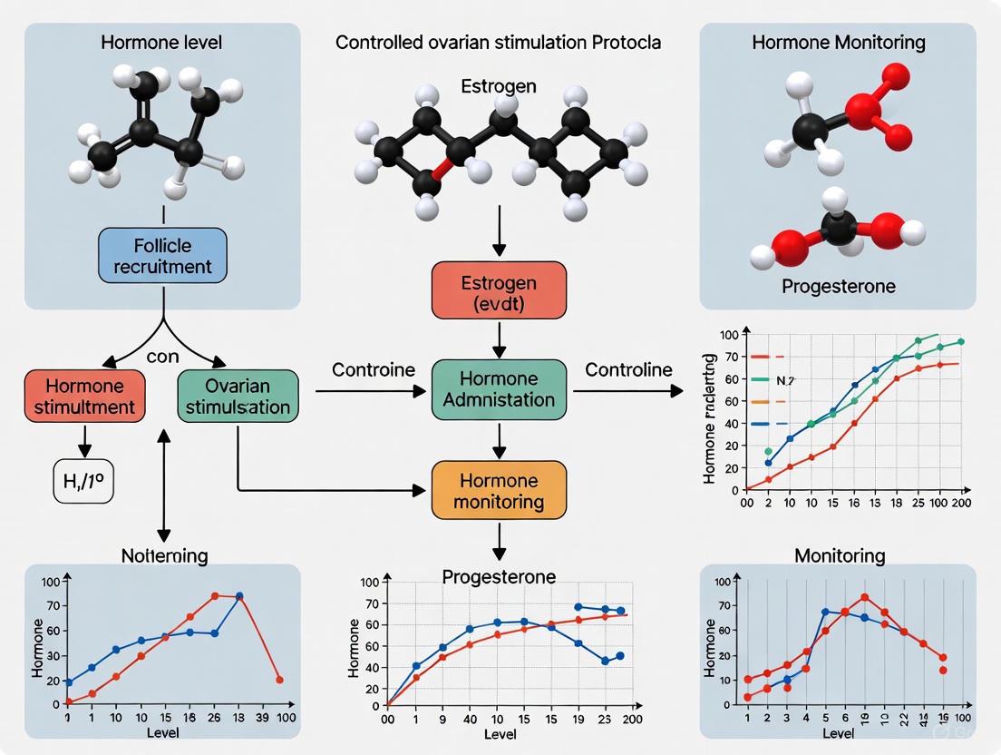
Hormone Monitoring in Controlled Ovarian Stimulation: Protocols, Precision, and Future Directions
This article provides a comprehensive analysis of hormone monitoring practices within controlled ovarian stimulation (COS) protocols for assisted reproductive technology (ART).
Hormone Monitoring in Controlled Ovarian Stimulation: Protocols, Precision, and Future Directions
Abstract
This article provides a comprehensive analysis of hormone monitoring practices within controlled ovarian stimulation (COS) protocols for assisted reproductive technology (ART). It examines the foundational principles and global utilization patterns of hormonal assays, explores established and emerging methodological applications, and discusses strategies for troubleshooting and optimizing cycle outcomes. A critical evaluation of the evidence validating various monitoring approaches is presented, including a comparative analysis of their impact on key performance indicators such as pregnancy rates and the prevention of ovarian hyperstimulation syndrome (OHSS). Tailored for researchers, scientists, and drug development professionals, this review synthesizes current clinical practices, highlights technological innovations, and identifies pivotal areas for future biomedical research to enhance treatment personalization and efficacy.
The Role of Hormone Monitoring in COS: Global Practices and Core Principles
Hormonal monitoring is a cornerstone of controlled ovarian stimulation (COS) in assisted reproductive technology (ART). It provides the critical data required to individualize treatment, maximize the efficacy and safety of ovarian stimulation, and generate high-quality data for clinical research and drug development. The primary objectives of this monitoring are twofold: to optimize oocyte yield and quality, and to prevent iatrogenic complications, most notably ovarian hyperstimulation syndrome (OHSS). This document details the application notes and experimental protocols for comprehensive hormonal monitoring within the context of advanced COS research.
Core Objectives of Hormonal Monitoring
The hormonal monitoring framework in COS is designed to achieve several synergistic objectives that align with both clinical and research goals.
Optimizing Oocyte Yield and Quality: Precise tracking of estradiol (E₂), luteinizing hormone (LH), and progesterone levels allows for the determination of the optimal timing for oocyte maturation trigger, ensuring the retrieval of a maximum number of metaphase II (MII) oocytes [1]. Furthermore, biomarkers such as growth differentiation factor-9 (GDF-9) and bone morphogenetic protein-15 (BMP-15) in cumulus cells have been identified as reliable indicators of oocyte developmental potential, linking hormonal environments to embryological outcomes [1].
Preventing Ovarian Hyperstimulation Syndrome (OHSS): OHSS is a serious, iatrogenic complication of COS. Monitoring identifies patients at high risk, characterized by rapidly rising E₂ levels and a high follicular count [2]. This risk stratification enables the implementation of preventive strategies, such as the use of a gonadotropin-releasing hormone (GnRH) agonist trigger instead of human chorionic gonadotropin (hCG) and the adoption of a "freeze-all" embryo strategy [2] [3].
Individualizing Stimulation Protocols: Hormonal profiles, combined with ovarian reserve markers like Anti-Müllerian Hormone (AMH), guide the selection and dosing of gonadotropins. Research indicates that dosing based on individualized ovarian reserve testing is recommended to decrease the risk of OHSS [2]. This personalized approach is essential for managing patients with diverse profiles, including those with LH/FSH deficiency [4].
Generating Robust Research Data: Standardized hormonal monitoring protocols are indispensable for comparing the efficacy and safety of novel stimulation protocols, gonadotropin formulations, and adjuvant medications in clinical trials [5] [3].
Quantitative Hormonal Parameters and Clinical Correlations
Effective monitoring relies on the interpretation of key hormonal parameters against established benchmarks. The tables below summarize critical values and their implications for cycle management.
Table 1: Interpretation of Key Hormonal Levels During COS
| Hormone | Phase | Typical Range | Research & Clinical Significance |
|---|---|---|---|
| FSH | Baseline (Day 2-3) | 1.37 - 9.9 IU/L [6] | High baseline may indicate diminished ovarian reserve; used for initial dosing calculations. |
| Estradiol (E₂) | Mid-late Stimulation | Variable; rate of rise is key | A steep rise is associated with high oocyte yield but also increased OHSS risk [2]. Low E₂ relative to follicle count may indicate LH deficiency [4]. |
| LH | Baseline (Day 2-3) | 1.37 - 9.9 IU/L [6] | Low baseline may suggest need for LH supplementation [4]. |
| During Stimulation | <5 IU/L (in antagonist cycles) | A surge >10-15 IU/L indicates a premature LH surge, requiring cycle management. | |
| Progesterone | Late Stimulation | <1.5 ng/mL | Premature elevation can indicate premature luteinization, potentially impacting endometrial receptivity. |
Table 2: OHSS Classification and Associated Features [2]
| OHSS Stage | Clinical Features | Laboratory Features |
|---|---|---|
| Mild | Abdominal distension, discomfort, nausea | No significant alterations |
| Moderate | Mild features + ultrasonographic ascites | Hemoconcentration (Hct >41%), Elevated WBC |
| Severe | Clinical ascites, hydrothorax, oliguria | Severe hemoconcentration (Hct >45%), electrolyte imbalances |
| Critical | ARDS, thromboembolism, anuria | Worsening of severe findings |
Detailed Experimental Protocols for Hormonal Assessment
Protocol: Serial Hormonal Monitoring in a GnRH Antagonist Cycle
This protocol is a standard model for assessing ovarian response and is widely used as a control in comparative studies [5].
Objective: To track hormonal dynamics for determining the gonadotropin dose, antagonist start day, and trigger timing while collecting data for research on follicular development.
Materials:
- Serum collection tubes (SST)
- Automated immunoassay systems (e.g., chemiluminescence) for E₂, LH, P4
- Ultrasound machine with a high-frequency transvaginal probe
Workflow:
- Baseline Assessment (Cycle Day 2-3): Perform transvaginal ultrasound for antral follicle count (AFC) and collect blood for FSH, LH, and E₂.
- Stimulation Phase (Day 5 onward): Administer recombinant FSH (rFSH) or highly purified human menopausal gonadotropin (HP-hMG). Monitor E₂ and LH every 1-2 days via blood draw alongside follicular tracking via ultrasound.
- GnRH Antagonist Administration: Introduce a GnRH antagonist (e.g., Cetrorelix 0.25 mg/day) when the leading follicle reaches 12-14 mm or E₂ >1,468 pmol/L [5]. Continue daily monitoring of E₂, LH, and P4.
- Final Oocyte Maturation Trigger: Administer trigger when ≥3 follicles reach >17mm. For high-risk OHSS patients, use a GnRH agonist trigger (e.g., Triptorelin 0.2 mg) instead of hCG [2].
- Oocyte Retrieval: Perform 34-36 hours post-trigger.
Protocol: Quantification of Oocyte Quality Biomarkers (GDF-9 & BMP-15) in Cumulus Cells
This laboratory protocol supports translational research linking stimulation protocols to oocyte competence [1].
Objective: To isolate cumulus cells (CCs) and quantify the expression levels of GDF-9 and BMP-15 mRNA to assess oocyte developmental potential across different COS protocols.
Materials:
- Hyaluronidase
- PBS buffer (pH 7.4)
- Microcentrifuge tubes
- RNA extraction kit (e.g., Qiagen RNeasy Micro Kit)
- cDNA synthesis kit
- Real-time PCR system and TaqMan probes for GDF-9 and BMP-15
Workflow:
- CC Collection: 40-42 hours post-trigger, treat cumulus-oocyte complexes with hyaluronidase. Mechanically denude oocytes and transfer the separated CCs to a microtube [1].
- Washing and Storage: Wash CCs repeatedly with PBS, centrifuge, remove supernatant, and store the cell pellet at -80°C.
- RNA Extraction & cDNA Synthesis: Extract total RNA from CC pellets following the manufacturer's protocol. Quantify RNA and perform reverse transcription to synthesize cDNA.
- qPCR Amplification: Perform real-time qPCR using GAPDH or similar as a housekeeping gene. Use the 2^(-ΔΔCt) method to calculate the relative expression levels of GDF-9 and BMP-15.
- Data Correlation: Correlate expression levels with embryological outcomes (fertilization rate, high-quality blastocyst formation rate) and the COS protocol used (e.g., agonist vs. antagonist) [1].
Signaling Pathways and Workflow Visualization
The following diagrams illustrate the key physiological pathways and experimental workflows involved in hormonal monitoring.
Hypothalamic-Pituitary-Ovarian (HPO) Axis
VEGF-Mediated Pathway in OHSS Pathogenesis
Experimental Workflow for Hormonal Monitoring & Biomarker Analysis
The Scientist's Toolkit: Key Research Reagents & Materials
Table 3: Essential Reagents and Materials for Hormonal Monitoring Research
| Item | Function/Application | Example References |
|---|---|---|
| Recombinant FSH (rFSH) | Standardized FSH source for ovarian stimulation; control arm in trials comparing gonadotropin formulations. | Gonal-f (Merck Serono) [5] [3] |
| Highly Purified hMG (HP-hMG) | Contains both FSH and hCG-driven LH activity; studied for its potential to yield better-quality embryos and lower OHSS risk in high responders. | Menopur (Ferring) [3] |
| GnRH Antagonist | Prevents premature LH surge in stimulation cycles; the basis for flexible and shorter protocols. | Cetrorelix (Cetrotide) [1] [5] |
| GnRH Agonist | Used for pituitary downregulation in long protocols or as a trigger for final oocyte maturation to reduce OHSS risk. | Triptorelin (Gonapeptyl) [1] [5] |
| Medroxyprogesterone Acetate (MPA) | Progestin used in Progestin-Primed Ovarian Stimulation (PPOS) to prevent LH surges. | Tarlusal (Deva) [5] |
| Chemiluminescence Immunoassay Kits | Gold standard for sensitive and specific quantitative measurement of serum FSH, LH, E₂, and P4. | Various commercial suppliers [6] |
| TaqMan Probes for qPCR | For precise quantification of gene expression biomarkers (e.g., GDF-9, BMP-15) in cumulus cells. | Applied Biosystems [1] |
Within the realm of Assisted Reproductive Technologies (ART), controlled ovarian stimulation (COS) is a fundamental component aimed at maximizing the yield of mature oocytes [7]. The meticulous monitoring of hormonal dynamics during COS cycles is critical for optimizing follicular development, determining the timing for oocyte maturation trigger, and ultimately improving cumulative live birth rates (CLBRs) [7] [8]. This document provides detailed Application Notes and Protocols for the monitoring of three key hormones—estradiol (E2), progesterone, and luteinizing hormone (LH). Framed within a broader thesis on COS protocol research, this content is designed to support researchers, scientists, and drug development professionals in standardizing methodologies and interpreting complex hormonal data. The following sections synthesize current market data with clinical experimental protocols to offer a comprehensive toolkit for advanced reproductive research.
The adoption of hormone assays in clinical practice is driven by diagnostic needs, technological advancements, and the growing prevalence of hormonal disorders. The following tables summarize the quantitative market data for each hormone, reflecting their commercial and, by extension, their clinical application footprint.
Table 1: Global Market Overview for Key Hormones in Clinical Practice
| Hormone | Market Size (2024/2025) | Projected Market Size (2033/2034) | Projected CAGR | Primary Application Segments |
|---|---|---|---|---|
| Estradiol | USD 11.12 billion (2024) [9] | USD 19.46 billion (2034) [9] | 5.77% (2025-2034) [9] | Menopause Symptom Management (55% share), Osteoporosis Prevention & Treatment [9] |
| Progesterone | USD 1.52 billion (2024) [10] [11] | USD 5.05 billion (2034) [10] [11] | 12.74% (2025-2034) [10] [11] | Menopause (Dominant), Hormone Replacement Therapy, In-Vitro Fertilization (IVF) [10] [11] |
| LH (Test Kits) | USD 1.2 billion (2024) [12] | USD 2.1 billion (2033) [12] | 7.5% (2026-2033) [12] | At-Home Fertility Planning, Clinical & Hospital Use [12] |
Table 2: Analysis of Adoption Trends by Formulation and Route of Administration
| Hormone / Aspect | Dominant Segment | Fastest-Growing Segment | Key Regional Trends |
|---|---|---|---|
| Estradiol | Oral Tablets/Capsules (40% share) [9] | Transdermal Patches & Vaginal Products [9] | North America dominated the market in 2024; Asia-Pacific is the fastest-growing region [9]. |
| Progesterone | Injectables (Route of Administration) [10] [11] | Oral (Route of Administration) [10] [11] | North America held the largest revenue share in 2024; Asia Pacific is expected to grow rapidly [10]. |
| LH Assays | Strip Tests (Product Type) [12] | Digital Ovulation Tests (Product Type) [12] | North America dominates; growth is fueled by digital/in-app integration and at-home testing [12]. |
Application Notes: Clinical Significance & Experimental Evidence
Estradiol (E2) in COS Monitoring
Estradiol, secreted by granulosa cells of developing follicles, is a traditional cornerstone for monitoring follicular growth and maturation during COS [7] [8]. Its serum levels are expected to rise steadily with follicular growth. However, recent large-scale retrospective evidence has nuanced its clinical significance.
- Association with Cumulative Live Birth Rate (CLBR): A study of 27,487 conventional COS cycles found that an unexpected E2 decline during monitoring occurred in 10.3% of patients and was associated with a statistically significant decrease in CLBR (55% vs. 66.3% in controls) [7].
- Mediating Factors: This decrease in CLBR was primarily mediated (72.5%-76.5%) through a reduction in viable embryo yield, as fewer oocytes were retrieved and fewer embryos were formed from cycles exhibiting an E2 decline [7].
- Clinical Implications: These findings affirm that E2 remains a critical biomarker for predicting COS outcomes. The data suggest that a single decline may have a modest effect, but consecutive E2 declines are associated with worse outcomes (Adjusted OR 0.72) [7]. Interestingly, increasing the gonadotropin (Gn) dose after an E2 decline did not improve CLBR, indicating the need for alternative intervention strategies [7].
Progesterone in Luteal Phase Support and Beyond
Progesterone is essential for endometrial receptivity and embryo implantation. Its use is well-established in luteal phase support in ART and in hormone replacement therapy (HRT) for menopausal women.
- Menopause Management: In postmenopausal HRT, progestogen is primarily used to protect the endometrium from the proliferative effects of estrogen [10] [11].
- Expanding Applications: Research is exploring progesterone's role beyond reproduction, including its potential use as progesterone receptor modulators (PRMs) in managing hormone receptor-positive breast and endometrial cancers [11].
- Formulation Trends: While injectable progesterone has been dominant, particularly in fertility treatments, the oral segment is experiencing the fastest growth. This is driven by the development of sustained-release micronized progesterone formulations that improve bioavailability and patient compliance [10] [11].
Luteinizing Hormone (LH) in Ovulation Trigger and Monitoring
LH assays are pivotal for detecting the endogenous LH surge in natural cycles and for planning the timing of the oocyte retrieval after hCG trigger in COS cycles.
- At-Home Fertility Planning: The demand for at-home LH detection kits is a major market driver. These strip, midstream, and digital tests empower individuals to identify the fertile window for conception planning [12].
- Digital Integration: A key trend is the innovation in digital connectivity. Smart ovulation tests integrated with mobile apps provide real-time tracking, data analytics, and personalized insights, enhancing user experience and reliability [12].
- Clinical Use: In fertility clinics, LH levels are part of the standard hormonal panel monitored during COS to assess response and prevent premature ovulation, especially in antagonist cycles [7].
Emerging Biomarkers: Inhibin A
Research continues to seek biomarkers with superior predictive value. Serum Inhibin A, primarily secreted by mature antral follicles, has emerged as a promising candidate.
- Comparative Accuracy: A retrospective study of 84 IVF cycles found that both Inhibin A and E2 on the day of hCG trigger were significantly correlated with the number of mature oocytes retrieved [8].
- Potential Clinical Utility: The correlation of Inhibin A with oocyte yield strengthened with increasing follicular size, suggesting it may offer a more accurate assessment of follicular maturity compared to E2 alone. The study concluded that Inhibin A, combined with transvaginal ultrasound, could potentially improve the timing of the hCG trigger [8].
Experimental Protocols
Protocol 1: Monitoring Serum Estradiol and LH During Controlled Ovarian Stimulation
Objective: To serially monitor serum E2 and LH levels for tracking follicular development and determining the timing for final oocyte maturation trigger in a COS cycle.
Materials
- Research Reagent Solutions:
- GnRH Agonist/Antagonist: For pituitary down-regulation or prevention of premature LH surge.
- Gonadotropins (FSH/hMG): For ovarian stimulation.
- Recombinant hCG: For triggering final oocyte maturation.
- Serum Separation Tubes: For blood sample collection.
- Automated Immunoassay Analyzer: For quantitative measurement of E2 and LH.
- Chemiluminescence or ELISA Kits: Validated for E2 and LH detection.
Workflow Diagram:
Methodology:
- Baseline Assessment: On cycle day 2-3, perform a transvaginal ultrasound (TVUS) to assess antral follicle count and rule of ovarian cysts. Collect a baseline blood sample for E2, FSH, and LH [7].
- Ovarian Stimulation: Initiate gonadotropin (Gn) stimulation with FSH or hMG. The starting dose (75-300 IU) is determined by patient age, BMI, and ovarian reserve [7].
- Serial Monitoring:
- Trigger Decision: Once TVUS confirms that the average diameter of at least one follicle has reached 18mm (or two follicles reach 17mm), administer recombinant hCG (200-250 μg) to trigger final oocyte maturation [7].
- Oocyte Retrieval: Perform transvaginal ultrasound-guided oocyte retrieval 35-37 hours post-hCG trigger [7].
Data Analysis:
- Plot E2 levels against days of stimulation. An unexpected decline (>10-15% from previous measurement) should be noted and its association with oocyte yield and embryo quality investigated [7].
- Monitor LH levels to ensure suppression in agonist cycles or to detect a premature surge in antagonist cycles.
Protocol 2: Protocol for Comparative Analysis of Inhibin A and Estradiol
Objective: To compare the correlation of serum Inhibin A and Estradiol levels with the number of mature oocytes retrieved in IVF cycles.
Materials
- Research Reagent Solutions:
- Specific Inhibin A ELISA Kit: Validated for quantitative serum analysis.
- Estradiol Immunoassay Kit: As used in Protocol 1.
- Serum Collection Tubes.
- Microplate Reader and Washer: For ELISA execution.
- Standard Laboratory Centrifuge.
Workflow Diagram:
Methodology:
- Study Population: Include women undergoing COS for IVF using an antagonist protocol. Obtain ethical approval and informed consent [8].
- Sample Collection: On the day of hCG trigger, collect a blood sample from each participant prior to the trigger injection [8].
- Sample Analysis:
- Process the blood sample to obtain serum.
- Aliquot the serum and run, in parallel, the specific Inhibin A ELISA and the Estradiol immunoassay according to the manufacturers' instructions.
- Outcome Measurement: Record the total number of oocytes retrieved and the number of mature (Metaphase II) oocytes for each patient following the oocyte retrieval procedure [8].
- Statistical Analysis:
- Perform linear regression analysis to determine the correlation (R² value) between serum Inhibin A levels and the number of mature oocytes.
- Perform linear regression analysis to determine the correlation (R² value) between serum E2 levels and the number of mature oocytes.
- Compare the correlation coefficients (R²) of the two hormones to assess which biomarker shows a stronger association with mature oocyte yield [8].
The Scientist's Toolkit: Essential Research Reagents
Table 3: Key Research Reagent Solutions for Hormone Monitoring Studies
| Item | Function/Application | Specific Examples/Notes |
|---|---|---|
| Gonadotropins (FSH/hMG) | To stimulate the development of multiple ovarian follicles during COS [7]. | Starting dose determined by patient profile; subject to dose adjustment during cycle [7]. |
| GnRH Agonist/Antagonist | To control the pituitary gland, preventing a premature LH surge that could lead to early ovulation [7]. | Agonist protocols involve down-regulation; antagonist protocols involve co-treatment during stimulation [7]. |
| Recombinant hCG | To trigger final oocyte maturation, mimicking the natural LH surge [7]. | Administered subcutaneously when lead follicles reach optimal size [7]. |
| Serum Blood Collection Tubes | For the collection and processing of patient blood samples for serum-based hormone assays. | Essential for ensuring sample integrity for accurate E2, LH, and Inhibin A measurement. |
| Automated Immunoassay Systems | For the high-throughput, quantitative measurement of hormone levels (E2, LH, Progesterone) in serum [13]. | Systems using chemiluminescence technology are widely adopted in clinical laboratories. |
| Inhibin A ELISA Kit | For the specific quantitative measurement of Inhibin A in serum for research purposes [8]. | Used to investigate its potential as a superior biomarker of follicular maturity compared to E2 [8]. |
| Micronized Progesterone Formulations | Used for luteal phase support in ART and in HRT; object of research for improved bioavailability [10] [11]. | Available in oral, vaginal, and injectable forms; sustained-release oral formulations are a key research area [11]. |
This application note provides a detailed framework for monitoring key hormonal dynamics—Estradiol (E2), Progesterone (P4), Luteinizing Hormone (LH), and Follicle-Stimulating Hormone (FSH)—during Controlled Ovarian Stimulation (COS) for in vitro fertilization (IVF). Within the broader thesis of optimizing COS protocols, we present standardized protocols and quantitative benchmarks to guide researchers and drug development professionals in assessing ovarian response, triggering final oocyte maturation, and supporting the luteal phase. The data and methodologies herein are synthesized from current literature and clinical evidence to support robust experimental design and diagnostic development.
Controlled Ovarian Stimulation (COS) is a cornerstone of assisted reproductive technology (ART), aimed at inducing multi-follicular development to obtain multiple competent oocytes [14] [15]. The hormonal interplay of E2, P4, LH, and FSH during this process is critical for follicular growth, endometrial receptivity, and ultimately, treatment success. Monitoring these hormones across stimulation visits allows for the individualization of therapy, helping to optimize oocyte yield while mitigating risks such as Ovarian Hyperstimulation Syndrome (OHSS) [14] [16]. The evolving landscape of ART, including a shift towards "freeze-all" cycles and GnRH agonist triggering, necessitates a re-evaluation of traditional monitoring practices [16]. This document establishes precise application notes and protocols for tracking hormonal dynamics, providing a foundation for clinical research and the development of novel therapeutic and diagnostic agents.
Hormonal Reference Ranges and Clinical Significance
The following table summarizes the quantitative benchmarks and clinical significance of key hormones at critical time points during a COS cycle.
Table 1: Hormonal Reference Ranges and Clinical Significance During COS
| Hormone | Phase / Time Point | Typical Range / Threshold | Clinical & Research Significance |
|---|---|---|---|
| FSH | Baseline (Cycle Day 2-3) | Patient-specific (e.g., 5-15 IU/L) | Used to determine starting gonadotropin dose; high baseline may indicate diminished ovarian reserve [15]. |
| During Stimulation (e.g., Day 5) | N/A (Dose-dependent) | Serum level correlates with weight-adjusted starting dose (r² = 0.352); insufficient dose requires increase >5%, leading to heterogeneous follicle size and fewer mature oocytes [15]. | |
| LH | Baseline (Cycle Day 2-3) | Patient-specific | Establish baseline prior to GnRH analog administration [17] [18]. |
| After GnRH Agonist/Antagonist | <1.2 IU/L (Severe Deficiency) [17]>50% decrease post-antagonist (Oversuppression) [18] | Iatrogenic deficiency can occur, potentially impacting oocyte quality and pregnancy rates. Supplementation with r-hLH may be beneficial in specific patient subgroups (e.g., Poseidon low prognosis groups) [17]. | |
| Estradiol (E2) | Baseline (Cycle Day 2-3) | <60 pg/ml [19] | Assess cycle baseline before stimulation initiation. |
| During Stimulation | Rises significantly with follicular growth [19] | Traditionally used with TVUS to monitor response; however, evidence suggests TVUS-only monitoring may be non-inferior to combined (TVUS + E2) monitoring for clinical pregnancy and OHSS rates [14]. | |
| At Trigger (Peak) | 1,000 - 4,000 pg/ml [19] | Correlates with follicular development. Low E2 in relation to follicular response may indicate insufficient LH activity [17]. | |
| Progesterone (P4) | Baseline (Cycle Day 2-3) | <1.5 ng/ml (NEP) [20] | Elevated Progesterone (EP) >1.5 ng/ml at baseline shows no significant impact on Live Birth Rate (LBR) [20]. |
| At Ovulation Trigger | >1.5 ng/ml (EP) [20] | EP at trigger is associated with lower LBR and Clinical Pregnancy Rate (CPR) for Day 3 embryo transfers, but not for Day 5 (blastocyst) transfers [20]. | |
| Pregnancy Test Day (After Fresh ET) | ≥16.5 ng/ml [21] | A threshold of ≥16.5 ng/ml is associated with higher ongoing pregnancy and live birth rates. Levels below this may indicate benefit from prolonged luteal support [21]. |
Detailed Experimental Protocols for Hormone Monitoring
Protocol 1: Serum Hormone Measurement and Assay Methodology
This protocol outlines the standard procedure for quantifying E2, P4, LH, and FSH in serum during COS cycles.
1. Sample Collection:
- Timing: Collect venous blood samples at defined monitoring visits: baseline (cycle day 2-3), during stimulation (e.g., day 5, day 8), on the day of ovulation trigger, and on the day of pregnancy test (day 15 post-oocyte retrieval) [21] [18].
- Processing: Centrifuge samples to separate serum. Aliquot and freeze at -20°C or lower until analysis.
2. Hormone Quantification:
- Recommended Assay: Electrochemiluminescence Immunoassay (ECLIA), e.g., Cobas systems [21] [18].
- Quality Control: Implement standard operating procedures adhering to Good Clinical Practice (GCP) and Good Laboratory Practice (GLP). Include internal quality controls and participate in external quality assurance schemes.
- Key Performance Parameters:
3. Data Interpretation:
- Compare individual patient results against the reference ranges and thresholds provided in Table 1.
- Track hormone kinetics (e.g., the rate of E2 rise, the degree of LH suppression) to guide clinical decisions.
Protocol 2: Clinical Monitoring Workflow in a GnRH Antagonist Cycle
This protocol describes the integration of hormonal and ultrasound monitoring within a standard GnRH antagonist COS cycle for research and clinical application.
1. Baseline Assessment (Cycle Day 2-3):
- Procedure: Perform transvaginal ultrasound (TVUS) for antral follicle count (AFC) and to rule out significant cysts. Collect baseline blood sample for E2, P4, and LH.
- Research Consideration: The utility of a baseline scan is being re-evaluated, as "random start" protocols yield equivalent oocyte numbers, and AFC can vary throughout the cycle [16].
2. Ovarian Stimulation Initiation:
- Procedure: Administer recombinant FSH (rFSH) starting on cycle day 2-3. The starting dose is determined by ovarian reserve markers (AMH, AFC, age) [15].
- Fixed-Dose Period: Maintain the initial rFSH dose for the first 5-7 days, as early follicle size on day 5 predicts subsequent growth and time to trigger (r² = 0.58–0.62) [16] [15].
3. Mid-Stimulation Monitoring (Stimulation Day 5-7):
- Procedure: Initiate GnRH antagonist. Perform TVUS to measure follicle size and count. Collect blood for E2 and LH measurement.
- Decision Points:
4. Final Monitoring & Trigger Timing (Typically Day 9-12):
- Procedure: Perform TVUS and measure serum E2 and P4.
- Trigger Criteria: Administer ovulation trigger (hCG or GnRH agonist) when at least 3 follicles reach ≥18 mm in diameter [15] [19].
- Critical Research Data:
- Record P4 level at trigger. For Day 3 embryo studies, P4 >1.5 ng/mL is a key negative outcome predictor [20].
- Record total and mature oocyte yield post-retrieval.
5. Luteal Phase Support (LPS) and Outcome Assessment:
- Procedure: Initiate luteal phase support (e.g., vaginal progesterone) from the day of oocyte retrieval [21].
- Research Endpoint Measurement:
Visualization of Hormonal Dynamics and Workflows
Hormonal Signaling Pathways in COS
The following diagram illustrates the interplay of exogenous drugs and endogenous hormonal pathways during COS.
COS Monitoring Clinical Workflow
This flowchart details the sequential steps and decision points in a typical COS monitoring protocol.
The Scientist's Toolkit: Research Reagent Solutions
Table 2: Essential Reagents and Materials for Hormonal Dynamics Research
| Item | Function / Application | Examples / Specifications |
|---|---|---|
| Recombinant FSH (rFSH) | Stimulates multi-follicular growth; primary interventional drug in COS. | Gonal-F (Merck Serono), Follistim (Merck) [15]. |
| Recombinant LH (r-hLH) | Supplements endogenous LH deficiency; used as an adjuvant to rFSH in specific patient populations. | Luveris (Merck) [17]. |
| GnRH Antagonists | Prevents premature LH surge by blocking pituitary GnRH receptors. | Cetrorelix (Cetrotide, Merck Serono), Ganirelix (Orgalutran, MSD) [17] [15]. |
| GnRH Agonists | Used in "long" or "short" protocols for pituitary downregulation; can also be used for final oocyte maturation trigger. | Leuprolide (Lupron) [19], Triptorelin (Decapeptyl) [21]. |
| hCG | Triggers final oocyte maturation; mimics the natural LH surge. | Recombinant hCG alfa (Ovitrelle, Serono) [21]. |
| Micronized Progesterone | Luteal phase support to prepare and maintain the endometrium for implantation. | Vaginal progesterone (Utrogestan, Besins International) [21]. |
| Immunoassay Systems | Quantitative measurement of serum E2, P4, LH, and FSH levels. | Electrochemiluminescence Immunoassay (ECLIA) on platforms like Cobas 8000 (Roche Diagnostics) [21]. |
| Ultrasound System | Transvaginal ultrasound (TVUS) for tracking follicular growth and endometrial lining. | 2D/3D systems with high-resolution probes (e.g., GE Voluson E8) for precise follicle tracking [19]. |
Comparative Utility of Hormonal Assays versus Ultrasound-Only Monitoring
This application note provides a comparative analysis of monitoring protocols in controlled ovarian stimulation (COS), focusing on the relative utility of combined hormonal assay and ultrasound monitoring versus ultrasound-only approaches. Within the context of developing optimized COS protocols, this analysis underscores that the selection of a monitoring strategy involves balancing clinical outcomes, cost-effectiveness, and patient burden. Evidence indicates that while hormonal monitoring provides critical biochemical data for individualizing treatment, ultrasound-only monitoring can be a cost-effective and efficient alternative without compromising primary success rates in selected patient populations and protocols [22] [23].
Key comparative data from a clinical study is summarized in Table 1 below.
Table 1: Comparative Outcomes of Ultrasound-Only vs. Combination Monitoring in IVF
| Outcome Measure | Ultrasound-Only Monitoring (Group I, n=110) | Combination Monitoring (Group II, n=96) | P-value |
|---|---|---|---|
| Clinical Pregnancy Rate | 23.4% | 22.9% | Not Significant |
| Take-Home Baby Rate | 14.8% | 14.3% | Not Significant |
| OHSS Rate | 1 patient | 1 patient | Not Significant |
| Average Monitoring Cost (Jordanian Dinar) | 78 JD | 222 JD | < 0.0001 |
The findings reveal no statistically significant differences in clinical pregnancy rates, take-home baby rates, or the incidence of ovarian hyperstimulation syndrome (OHSS) between the two monitoring strategies [22]. However, the cost of monitoring was significantly lower in the ultrasound-only group, and this protocol was also found to be more convenient and less time-consuming for both patients and the clinical team [22].
Controlled ovarian stimulation (COS) is a fundamental pharmacological intervention in Assisted Reproductive Technology (ART) aimed at inducing the development of multiple ovarian follicles to yield a sufficient number of mature oocytes for retrieval [24]. The success of in vitro fertilization (IVF) is partly dependent on obtaining an optimal number of oocytes while avoiding complications such as OHSS [25]. Cycle monitoring, the process of tracking follicular development and endocrine response, is therefore a standard of care in medically assisted reproduction (MAR) to evaluate and individualize treatment [23].
The two primary modalities for cycle monitoring are:
- Transvaginal Ultrasound: Provides real-time, direct visualization of the ovaries to assess the number and size of developing follicles and evaluate endometrial thickness [26] [27].
- Serum Hormonal Assays: Quantify circulating levels of key reproductive hormones, most commonly estradiol (E2), progesterone (P4), and luteinizing hormone (LH), to infer follicular activity and endocrine status [23] [28].
The combination of ultrasound and hormonal monitoring is widely practiced globally [23]. However, the added clinical value of routine hormonal monitoring alongside ultrasound has been a subject of debate, particularly given the increased costs, patient inconvenience, and logistical burden associated with frequent blood draws [22] [23]. This note examines the evidence for and against the utility of hormonal assays in this setting.
Experimental Protocols and Methodologies
Protocol for Combination Hormonal and Ultrasound Monitoring
This protocol details the methodology for monitoring a COS cycle using both serial transvaginal ultrasounds and serum hormonal level assessments, as commonly employed in clinical practice and research [23] [27].
Objective: To closely track follicular growth and endocrine response to gonadotropin stimulation for precise timing of ovulation trigger and dose adjustment, while mitigating the risk of OHSS.
Materials:
- Equipment: High-resolution transvaginal ultrasound machine with a high-frequency probe (e.g., 5-9 MHz); Phlebotomy supplies; Centrifuge.
- Analyzers: Automated immunoassay analyzer for hormone quantification (e.g., Roche Elecsys).
- Key Reagents: Assay-specific kits for E2, P4, and LH.
Procedure:
- Baseline Assessment (Cycle Day 2-4):
- Perform a transvaginal ultrasound to assess the antral follicle count (AFC) in both ovaries and exclude the presence of functional cysts [27].
- Collect a venous blood sample for baseline measurement of E2, P4, and LH [27].
- The results from this assessment are used to confirm a quiescent ovarian state and finalize the stimulation protocol.
Stimulation Phase Monitoring (Approximately Day 6-7 onwards, then every 1-2 days):
- Conduct serial transvaginal ultrasounds to track the number and diameter of growing follicles. Follicles are typically measured in three dimensions, and the mean diameter is calculated [24].
- Concurrently, collect blood samples for serum E2 and P4 analysis. The frequency of monitoring increases as follicles approach maturity [23].
- Data Integration and Decision Points:
- Gonadotropin Dose Adjustment: E2 levels are used by many specialists to guide dose adjustments. A slow rise in E2 may prompt a dose increase, while a very rapid rise may indicate a risk of hyper-response and lead to a dose decrease [23].
- OHSS Risk Assessment: A steep rise in E2 levels (e.g., >3000 pg/mL) in the presence of a high number of intermediate-sized follicles is a key risk indicator for OHSS [23].
- Trigger Timing: The combination of lead follicle sizes reaching 17-20 mm and corresponding E2 levels informs the final decision for administering the ovulation trigger (hCG or GnRH agonist) [24].
Luteal Phase Support:
- Post-retrieval, a mid-luteal phase P4 measurement may be taken to assess the adequacy of luteal phase support, though this practice is not universal [23].
Protocol for Ultrasound-Only Monitoring
This protocol outlines a monitoring strategy that relies exclusively on transvaginal ultrasound, omitting routine serum hormone testing.
Objective: To achieve successful COS outcomes with a simplified, more cost-effective, and less burdensome monitoring regimen.
Materials:
- Equipment: High-resolution transvaginal ultrasound machine with a high-frequency probe (e.g., 5-9 MHz).
Procedure:
- Baseline Assessment (Cycle Day 2-4):
- Stimulation Phase Monitoring (Approximately Day 10-12, frequency as needed):
- Conduct serial transvaginal ultrasounds to monitor follicular growth and endometrial lining thickness [27].
- Decision Points:
- Gonadotropin Dose Adjustment: Decisions are based solely on follicular growth patterns observed via ultrasound. A lack of adequate follicular development would prompt a dose increase, while excessive multifollicular development would lead to a dose reduction or cycle cancellation to prevent OHSS [22].
- Trigger Timing: The ovulation trigger is administered based primarily on the size of the leading follicles, typically when at least three follicles reach 17-20 mm in diameter [22] [24]. The absence of a premature LH surge is inferred clinically rather than biochemically.
Signaling Pathway and Workflow Visualizations
The following diagrams illustrate the physiological pathways involved in COS and the logical workflow for selecting a monitoring protocol.
COS Endocrine Feedback Pathways
Monitoring Protocol Selection Workflow
Research Reagent Solutions and Essential Materials
The following table details key reagents and materials essential for implementing the experimental protocols described in this note.
Table 2: Key Research Reagents and Materials for COS Monitoring
| Item | Function / Application | Examples / Specifications |
|---|---|---|
| Recombinant FSH (rFSH) | Stimulates multi-follicular development; used in various COS protocols. | Gonal-f, Puregon [25] [24] |
| Human Menopausal Gonadotropin (hMG) | Urinary-derived gonadotropin with FSH and LH activity for ovarian stimulation. | Menopur [22] [25] |
| GnRH Agonists | Suppresses pituitary function to prevent premature LH surge in "long" or "short" protocols. | Leuprolide, Buserelin [25] [24] |
| GnRH Antagonants | Provides immediate suppression of pituitary LH release; used in antagonist protocols. | Cetrorelix, Ganirelix [25] [24] |
| hCG / Recombinant hCG | Triggers final oocyte maturation; mimics the natural LH surge. | Ovidrel, Pregnyl [22] [24] |
| Serum Hormone Assays | Quantitative measurement of E2, P4, and LH levels in serum for monitoring. | Roche Elecsys E170 module, ECLIA method [29] [23] |
| Anti-Müllerian Hormone (AMH) Assay | Assess ovarian reserve prior to treatment; predicts ovarian response. | Beckman Coulter Access, DSL AMH ELISA [28] |
Discussion and Future Perspectives
The comparative analysis underscores a nuanced clinical landscape. The primary argument for combination monitoring is its ability to provide a more complete picture of the ovarian response. Serum E2 levels offer an indirect measure of follicular health and number, which can be particularly useful when ultrasound findings are ambiguous or in predicting hyper-response and OHSS risk before it is fully apparent on ultrasound [23]. Furthermore, progesterone monitoring is critical for detecting a premature luteinization, which can compromise oocyte quality and is not detectable by ultrasound alone.
Conversely, the ultrasound-only protocol presents a compelling case based on efficiency and resource allocation. The significant reduction in cost (approximately 65% cheaper in one study) and patient burden, without a statistically significant drop in live birth rates, makes it an attractive option for specific patient cohorts and healthcare systems [22]. This approach reduces the physical and emotional strain on patients associated with frequent venipuncture and long wait times at clinics [23].
Future directions in COS monitoring are leaning towards technological innovations that further reduce patient burden. The development of remote urine-based hormonal assays is being investigated as a potential alternative to serum testing. These assays have shown good correlation with serum levels for E2, P4, and LH and could form part of a digital health solution integrating home-based testing and telemedicine [23]. This aligns with broader FemTech trends focusing on non-invasive techniques and empowering patients through self-monitoring [30]. The integration of artificial intelligence to interpret complex hormonal and ultrasound data also holds promise for optimizing individualized treatment plans in the future [30].
In the realm of assisted reproductive technology (ART), the individualized selection of controlled ovarian stimulation (COS) protocols is paramount for optimizing treatment outcomes. Baseline ovarian reserve assessment provides the foundational data informing this selection, with antral follicle count (AFC) and anti-Müllerian hormone (AMH) emerging as the most significant biomarkers of ovarian response [31]. These markers allow clinicians to predict ovarian response to stimulation and tailor protocols accordingly, balancing the risks of poor response and ovarian hyperstimulation syndrome (OHSS).
The clinical utility of AMH and AFC extends beyond mere prediction of oocyte yield; when interpreted through validated classification systems like the POSEIDON criteria, they facilitate sophisticated protocol stratification that aligns stimulation strategies with individual patient profiles [32]. This application note delineates the quantitative thresholds, predictive values, and protocol selection frameworks supported by contemporary evidence, providing researchers and clinicians with structured methodologies for implementing ovarian reserve-guided treatment pathways.
Quantitative Thresholds and Predictive Values
AMH and AFC serve as reliable predictors of ovarian response, with established thresholds correlating with hyporesponse and hyperresponse. The POSEIDON classification utilizes specific boundaries (AFC < 5 and AMH < 1.2 ng/mL) to identify patients with diminished ovarian reserve [32]. Conversely, recent research has established precise thresholds for predicting hyperresponse, defined as the retrieval of ≥15 oocytes, with variations observed across age groups [33].
Table 1: AMH and AFC Thresholds for Predicting Ovarian Response
| Response Category | Biomarker | Overall Population | Women <35 years | Women ≥35 years |
|---|---|---|---|---|
| Hyperresponse [33] | AMH (ng/mL) | ≥4.38 | ≥4.95 | ≥4.33 |
| AFC | ≥16 | ≥18 | ≥15 | |
| Poor Response [32] | AMH (ng/mL) | <1.2 | <1.2 | <1.2 |
| AFC | <5 | <5 | <5 |
The predictive performance of these biomarkers differs, with AFC demonstrating superior discriminatory capacity for hyperresponse (AUC 0.80) compared to AMH (AUC 0.71) in the overall population [33]. This highlights the complementary value of both assessments in clinical practice.
Protocol Selection Based on Ovarian Reserve Profile
Stratification for Normal and Discordant Reserve Markers
Patients presenting with concordant AMH and AFC values within the normal range (AMH ≥1.2 ng/mL, AFC ≥5) typically respond well to conventional GnRH antagonist protocols [32] [33]. However, specific thresholds warrant caution; women with AMH ≥4.38 ng/mL or AFC ≥16 require careful gonadotropin dosing and trigger strategies to mitigate OHSS risk [33].
For patients with discordant AMH and AFC values, protocol selection requires more nuanced consideration:
- Women with low AMH and normal AFC (Group 2: AFC ≥5, AMH <1.2 ng/mL) demonstrate significantly improved outcomes with GnRH agonist ultra-long protocols, showing higher numbers of total oocytes, clinical pregnancy rates (CPR), and live birth rates (LBR) compared to GnRH antagonist protocols [32].
- Women with normal AMH and low AFC (Group 3: AFC <5, AMH ≥1.2 ng/mL) show a trend toward better outcomes with GnRH agonist long protocols, though statistical significance was not reached in all studies [32].
Special Considerations for Diminished Ovarian Reserve
For women with unequivocally diminished ovarian reserve (AFC <5 and AMH <1.2 ng/mL), strategic supplementation with human menopausal gonadotropin (HMG) during GnRH antagonist cycles may improve outcomes. Research indicates that adding HMG (75-150 U/d) during the mid-late follicular phase (when the lead follicle reaches 10-14 mm) significantly increases the number of retrieved oocytes, mature oocytes, and usable embryos compared to both no supplementation and early supplementation approaches [34]. This mid-late HMG supplementation strategy also resulted in a higher fresh cycle clinical pregnancy rate [34].
Figure 1: Protocol Selection Based on Ovarian Reserve Profile
Experimental Protocols and Assessment Methodologies
AMH Laboratory Assessment Protocol
Principle: Serum AMH levels are quantified using enzyme-linked immunosorbent assay (ELISA) techniques, which provide reliable measurements of this glycoprotein hormone produced by granulosa cells of primary, preantral, and small antral follicles [31] [32].
Specimen Collection:
- Collect 3-5 mL venous blood in serum separation tubes during days 2-3 of the spontaneous menstrual cycle
- Allow samples to clot at room temperature for 30 minutes
- Centrifuge at 1000 × g for 15 minutes
- Aliquot serum and store at -20°C if not assayed immediately
Assay Procedure (ELISA):
- Bring all reagents and samples to room temperature
- Add 50 μL of standard, control, or patient sample to appropriate wells
- Add 100 μL of enzyme conjugate to each well
- Incubate for 60 minutes at room temperature
- Aspirate and wash wells 4 times with wash buffer
- Add 100 μL of substrate solution to each well
- Incubate for 15 minutes at room temperature protected from light
- Add 100 μL of stop solution to each well
- Read absorbance at 450 nm within 30 minutes
Interpretation: Calculate AMH concentration from standard curve. Values <1.2 ng/mL indicate diminished ovarian reserve, while values ≥4.38 ng/mL indicate increased hyperresponse risk [32] [33].
Technical Notes: AMH levels demonstrate relatively consistent within-cycle and between-cycle variability in ovulating women [31]. Levels may be decreased in women using hormonal contraceptives and should be interpreted with caution in these patients [31].
Antral Follicle Count (AFC) Ultrasonography Protocol
Principle: Transvaginal ultrasonography performed during the early follicular phase quantifies antral follicles (2-10 mm in diameter) in both ovaries, providing a direct anatomical assessment of the recruitable follicular cohort [31] [32].
Equipment:
- High-frequency transvaginal transducer (≥5 MHz)
- Ultrasound system with caliper measurement capability
- Standard ultrasonography gel
Procedure:
- Perform examination during menstrual days 2-4
- Systematically scan each ovary in longitudinal and transverse planes
- Identify and measure all follicles 2-10 mm in mean diameter
- Record the count for each ovary separately
- Calculate total AFC as the sum of both ovaries
Quality Control:
- Experienced operators should perform assessments to ensure reliability
- Maintain consistent measurement criteria across patients
- Participate in regular reliability training to minimize inter-observer variability [32]
Interpretation: AFC <5 indicates diminished ovarian reserve, while AFC ≥16 indicates increased hyperresponse risk [32] [33].
Research Reagent Solutions
Table 2: Essential Research Reagents for Ovarian Reserve Assessment
| Reagent/Material | Manufacturer Examples | Research Application |
|---|---|---|
| AMH ELISA Kit | Kangrun Biotech (China) | Quantitative serum AMH measurement [32] |
| Recombinant FSH | Gonal-f (Merck Serono), Puregon | Ovarian stimulation in COS protocols [34] [1] |
| Human Menopausal Gonadotropin (HMG) | Guangzhou Lizhu Group, Livzon | LH-containing preparation for supplementation [34] |
| GnRH Agonist | Decapeptyl (Ferring), Leuprolide acetate | Pituitary suppression in long protocols [32] [1] |
| GnRH Antagonist | Cetrotide (Merck Serono), Ganirelix | Prevention of premature LH surges [34] [32] |
| hCG Trigger | Ovidrel (Merck Serono), Pregnyl | Final oocyte maturation induction [32] [1] |
Baseline ovarian reserve assessment using AMH and AFC provides an evidence-based foundation for individualized COS protocol selection. The established thresholds and stratification strategies presented herein enable researchers and clinicians to optimize ovarian response while mitigating treatment risks. The precise methodological protocols ensure reproducible assessment techniques, while the reagent solutions facilitate standardized implementation across research and clinical settings. Future research directions should focus on refining predictive models through multi-marker integration and exploring molecular mechanisms underlying variable ovarian response to further personalize stimulation strategies.
Methodologies in Hormone Monitoring: From Serum Assays to At-Home Technologies
Within the context of controlled ovarian stimulation (COS) for assisted reproductive technology (ART), precise monitoring of serum hormone levels is a critical determinant of successful outcomes. The follicular phase of the cycle demands particular attention, as the dynamic interplay of estradiol (E2), progesterone (P4), and luteinizing hormone (LH) dictates follicular growth, endometrial receptivity, and the optimal timing for ovulation trigger or embryo transfer [35] [36]. This protocol document outlines standardized methodologies for serum hormone monitoring during the follicular phase, framed within broader research on optimizing COS protocols. The guidelines and data presented herein are designed for researchers, scientists, and drug development professionals engaged in the refinement of ovarian stimulation strategies. A global survey of ART specialists confirms that hormonal monitoring is widely utilized by approximately 80% of clinicians, with E2 being the most frequently tracked hormone to adjust gonadotropin dosing and predict ovarian hyperstimulation syndrome (OHSS) [35].
Hormone-Specific Clinical Significance & Monitoring Protocols
Estradiol (E2)
- Clinical Significance: E2 is the primary biomarker for follicular growth and maturation. It is secreted by granulosa cells in response to follicle-stimulating hormone (FSH) and reflects the quantity and quality of the developing follicular cohort. Monitoring E2 trajectories allows for the assessment of ovarian response and the prevention of both excessive stimulation (OHSS) and poor response [35].
- Monitoring Protocol: Serum E2 levels are typically monitored at the initiation of gonadotropins (baseline) and subsequently every 2-3 days during stimulation. The most intense monitoring occurs as follicles approach maturity. A global practice survey indicates that E2 is the most commonly monitored hormone across all clinic visits during stimulation, with its measurement used by 74% of respondents for OHSS prediction [35]. In natural cycles, a peak in E2 followed by a sharp decline of over 50% is a strong predictor of imminent ovulation, occurring within the same or the following day [37].
Luteinizing Hormone (LH)
- Clinical Significance: LH supports follicular development by stimulating androgen production in theca cells, which are then aromatized to E2 in granulosa cells. In GnRH agonist or antagonist protocols, the primary goal is to suppress the premature LH surge that can lead to oocyte maturation defects and cycle cancellation [36]. Research indicates that even in suppressed cycles, a specific "LH window" is necessary for optimal steroidogenesis and oocyte competence [36].
- Monitoring Protocol: In GnRH agonist cycles (e.g., the follicular-phase long protocol), pituitary suppression leads to low endogenous LH levels. Monitoring LH on the day of trigger (LHHCG) is recommended. A large retrospective cohort study (n=4502 cycles) demonstrated that while live birth rates were not significantly affected, the number of retrieved oocytes, fertilized oocytes, and embryos showed a trend of gradual decrease as LHHCG levels increased [36]. The study stratified LH levels into groups, with an LH ≤ 0.5 IU/L associated with the highest oocyte yield [36]. In natural cycles, the LH surge is the primary signal for impending ovulation, though its kinetics can be variable [37].
Progesterone (P4)
- Clinical Significance: During the follicular phase, P4 levels are expected to remain low. A premature rise in P4 (PPR) can indicate premature luteinization, which may negatively impact endometrial synchrony and implantation potential. Recent evidence has also highlighted the role of a small, autonomous preovulatory P4 rise in triggering the ovulatory process [38].
- Monitoring Protocol: Serum P4 is typically measured alongside E2 and LH during monitoring visits. The proportion of clinicians measuring P4 increases significantly on or just before the day of ovulation triggering, from 34.3% in mid-stimulation visits to 67.7% [35]. In natural cycles, a P4 level ≥ 2 nmol/L (∼0.63 ng/mL) has a high sensitivity (91.5%) for predicting ovulation the next day, though specificity is lower (62.7%) [37]. Machine learning models have identified a preovulatory P4 level of ≥ 0.65 ng/ml as a top predictor of ovulation within 24 hours, with an accuracy exceeding 92% [38].
Table 1: Key Hormone Thresholds and Their Clinical Implications during the Follicular Phase
| Hormone | Timing | Threshold Level | Clinical Implication | Citation |
|---|---|---|---|---|
| LH | On HCG day (GnRH agonist protocol) | ≤ 0.5 IU/L | Associated with higher numbers of retrieved oocytes, fertilized oocytes, and embryos. | [36] |
| Progesterone (P4) | Preovulatory (Natural Cycle) | ≥ 0.65 ng/mL | Predicts ovulation within 24 hours with >92% accuracy. | [38] |
| Progesterone (P4) | Preovulatory (Natural Cycle) | > 2 nmol/L (∼0.63 ng/mL) | 91.5% sensitivity for predicting ovulation the next day. | [37] |
| Estradiol (E2) | Late Follicular Phase (Natural Cycle) | Drop from peak level | 100% associated with ovulation emergence the same or next day. | [37] |
Quantitative Hormone Dynamics and Data Analysis
Understanding the expected hormonal trajectories is fundamental for interpreting patient-specific data. The following table synthesizes quantitative hormone values from research on natural and stimulated cycles.
Table 2: Quantitative Hormone Values Across the Periovulatory Period in Natural Cycles
| Day Relative to Ovulation (D0) | Estradiol (E2) pmol/L (Mean ± SEM) | Luteinizing Hormone (LH) IU/L (Mean ± SEM) | Progesterone (P4) nmol/L (Mean ± SEM) |
|---|---|---|---|
| D(-2) | 1378 ± 66.0 (Peak) | - | - |
| D(-1) | - | 51.9 ± 1.9 (Peak) | 3.2 ± 0.9 |
| D(0) (Ovulation) | 393 ± (58% decrease from D-1) | - | 5.1 ± 0.1 |
| D(+1) | - | - | > 5 nmol/L (94.3% PPV for D0) |
Data adapted from a prospective cohort study with daily hormonal and ultrasound monitoring [37].
Experimental Workflow for Hormone Monitoring
The following diagram illustrates the integrated clinical decision-making pathway for follicular phase hormone monitoring, combining ultrasound and hormonal data.
The Scientist's Toolkit: Research Reagent Solutions
This section details essential materials and assays used in the featured research for reliable hormone monitoring.
Table 3: Essential Research Reagents and Assays for Serum Hormone Monitoring
| Reagent / Assay | Function / Application | Research Context |
|---|---|---|
| Electrochemiluminescence Immunoassay (ECLIA) | Quantitative measurement of serum E2, P4, and LH levels. High precision and automation suitable for high-throughput clinical labs. | Used for daily hormone level assessments in natural cycle studies with reported precision metrics (e.g., CV% ≤ 10% for P4 samples ≤ 1 ng/ml) [38]. |
| Recombinant Human FSH (e.g., Gonal-f) | For controlled ovarian stimulation to induce multi-follicular growth. | Standard gonadotropin used in GnRH antagonist and PPOS protocols [1] [5]. |
| GnRH Agonist (e.g., Triptorelin) | For pituitary downregulation in long protocols to prevent premature LH surge. | Administered as a single 3.75 mg dose in the follicular-phase long protocol [36]. |
| GnRH Antagonist (e.g., Cetrorelix) | For immediate suppression of the LH surge in flexible or fixed antagonist protocols. | Initiated when leading follicle reaches 12-14 mm in diameter [1] [5]. |
| Recombinant hCG (e.g., Ovidrel) | Used to trigger final oocyte maturation, mimicking the natural LH surge. | Standard trigger medication administered when follicular and hormonal criteria are met [1] [36]. |
The administration of a trigger shot for final oocyte maturation is a critical determinant of success in assisted reproductive technology (ART) cycles. This protocol outlines the essential hormonal monitoring and threshold assessments required on the day of trigger to optimize the yield of mature metaphase II oocytes. We detail the endocrine profiles and kinetics associated with different trigger types—human chorionic gonadotropin (hCG), gonadotropin-releasing hormone agonist (GnRHa), and kisspeptin—and provide evidence-based guidelines for timing oocyte retrieval. The application of these precise monitoring strategies is fundamental to improving laboratory outcomes and advancing drug development in reproductive medicine.
In controlled ovarian stimulation (COS) protocols, the final maturation of oocytes is induced by an exogenous trigger that replicates the natural mid-cycle luteinizing hormone (LH) surge. The efficacy of this process is contingent upon precise hormonal monitoring on the day of trigger to ensure optimal follicular maturation and coordinate the retrieval of oocytes with maximal developmental competence. This document, framed within broader research on hormone monitoring during COS, provides detailed application notes and protocols for researchers and scientists on the critical hormonal thresholds and monitoring practices essential for successful final oocyte maturation.
Quantitative Hormonal Profiles of Different Triggers
The endocrine response following the trigger shot varies significantly based on the mechanism of action of the agent used. The table below summarizes the peak levels and kinetics of LH-like activity following administration of hCG, GnRHa, and kisspeptin triggers, based on a cohort study of 499 IVF cycles [39].
Table 1: Hormonal Kinetics and Peak Levels Following Different Triggers
| Trigger Type | Mechanism of Action | Peak Hormone Level | Time to Peak (hours) | Key Associations |
|---|---|---|---|---|
| hCG | Direct LH receptor agonist | hCG: 121 IU/L | 24 | Negative association with patient body weight [39]. |
| GnRHa | Pituitary gonadotropin release | LH: 140 IU/L | ~4 | LH rise positively predicted by pre-trigger LH levels [39]. |
| Kisspeptin | Hypothalamic GnRH release | LH: 41 IU/L | ~4 | LH rise positively predicted by pre-trigger LH levels [39]. |
Progesterone rise during oocyte maturation occurs precipitously following each trigger and is a strong predictor of the number of mature oocytes retrieved. Counter-intuitively, this progesterone rise is negatively associated with the magnitude of the LH rise following all three triggers [39].
Critical Hormonal Thresholds and Monitoring on Trigger Day
Hormonal monitoring on the day of trigger is a multi-factorial decision. While ultrasound assessment of follicular size is primary, hormonal data provides critical supplementary information for timing and personalizing the trigger.
Common Practice and Hormone Utilization
A global survey of ART specialists revealed that on or just before the day of ovulation triggering [40]:
- 71% of respondents measure estradiol (E2)
- 67.7% measure progesterone (P4)
- 31.5% measure luteinizing hormone (LH)
This represents a significant increase in the measurement of P4 and LH compared to earlier monitoring visits during stimulation, underscoring their specific importance for the final trigger decision [40].
Luteinizing Hormone (LH) Thresholds
The required LH-like exposure for successful oocyte maturation differs by trigger type due to their distinct pharmacokinetics.
- The odds of achieving a satisfactory mature oocyte yield are increased by the level of hCG/LH achieved post-trigger [39].
- Following a GnRHa trigger, the induced LH surge is characterized by a shorter duration compared to the sustained action of hCG [39].
- In postpartum women, a higher LH threshold may be required to trigger ovulation, suggesting a decreased ovarian responsiveness to LH [41].
Progesterone (P4) Rise
The rise in progesterone is a critical event following the trigger.
- The progesterone rise is a strong positive predictor of the number of mature oocytes retrieved [39].
- The magnitude of the progesterone rise per mature oocyte at 12 hours post-trigger is greater following a GnRHa trigger than following hCG or kisspeptin triggers [39].
- A premature rise in progesterone on the day of trigger has been associated with altered endometrial receptivity, often leading to a "freeze-all" cycle strategy.
Optimal Timing from Trigger to Retrieval
The interval between trigger administration and oocyte retrieval (often referred to as the "trigger-to-retrieval interval") is critical for maximizing the yield of mature Metaphase II (MII) oocytes. Evidence indicates that the optimal interval differs based on the trigger type used.
Table 2: Optimal Trigger-to-Retrieval Intervals for Oocyte Maturity
| Trigger Type | Shorter Interval (Hours) | MII Oocytes Retrieved (Mean ± SD) | Longer Interval (Hours) | MII Oocytes Retrieved (Mean ± SD) | Key Findings |
|---|---|---|---|---|---|
| GnRHa | Shorter (~35h) | 4.3 ± 5.3 [42] | Longer (~36.5+h) | 7.2 ± 6.5 [42] | Longer intervals yield significantly more MII oocytes and higher blastocyst formation [42]. |
| hCG | Shorter (~35h) | 6.9 ± 5.8 [42] | Longer (~36.5+h) | 4.0 ± 4.6 [42] | Shorter intervals are associated with a higher MII oocyte yield [42]. |
The differences in optimal timing are likely due to the distinct signaling pathways and durations of action of the triggers. The prolonged steroidogenic action of hCG may allow for a broader window for maturation, whereas the shorter, sharper LH surge induced by GnRHa may require a longer period to complete nuclear and cytoplasmic maturation effectively.
Experimental Protocols for Trigger Monitoring
The following protocol provides a detailed methodology for monitoring and executing the trigger shot in a GnRH antagonist co-treated cycle, as used in foundational studies [39].
Patient Selection and Stimulation
- Inclusion Criteria: Women aged 18-42, BMI 18-30 kg/m², undergoing IVF/ICSI.
- Ovarian Stimulation: Initiate with recombinant FSH (150-300 IU) starting on cycle day 2-3.
- GnRH Antagonist Co-treatment: Administer a GnRH antagonist (e.g., Cetrotide 0.25 mg) daily once the leading follicle reaches 12-14 mm in diameter to prevent a premature LH surge [5].
Monitoring and Trigger Criteria
- Ultrasound Monitoring: Perform serial transvaginal ultrasounds every 2-3 days to track follicular growth.
- Trigger Timing: Administer the trigger when at least three follicles reach a diameter of ≥17-18 mm [39] [5].
- Hormonal Blood Draws:
- Pre-Trigger Baseline: Measure serum LH, E2, and P4 on the morning of the trigger decision.
- Post-Trigger Kinetics: For research purposes, collect blood at defined intervals post-trigger (e.g., 0, 4, 12, 24, 36 hours) to profile LH/hCG, E2, and P4 levels [39].
Trigger Administration
Choose one of the following based on the patient's risk profile and treatment plan:
- hCG Trigger: Administer 250 µg recombinant hCG subcutaneously [39].
- GnRHa Trigger: Administer 0.2-0.4 mg Triptorelin subcutaneously [39].
- Kisspeptin Trigger: Administer Kisspeptin-54 at 6.4-12.8 nmol/kg as a single bolus or split dose [39].
Oocyte Retrieval
- Schedule oocyte retrieval based on the evidence-based intervals detailed in Table 2:
- Immediately after retrieval, the embryology laboratory will identify and denude the cumulus-oocyte complexes to assess nuclear maturity, defined by the presence of a polar body (Metaphase II).
Signaling Pathways and Experimental Workflow
Signaling Pathways of Ovulation Triggers
The following diagram illustrates the distinct biological pathways through which hCG, GnRHa, and kisspeptin stimulate final oocyte maturation.
Experimental Workflow for Trigger Day Monitoring
This workflow outlines the key procedural steps from monitoring to oocyte retrieval.
The Scientist's Toolkit: Research Reagent Solutions
The following table catalogs essential reagents and materials required for implementing the experimental protocols described in this document.
Table 3: Essential Research Reagents for Trigger Monitoring Studies
| Item | Function & Application in Research | Example Product/Catalog |
|---|---|---|
| Recombinant hCG | Direct LH receptor agonist; used for final oocyte maturation in stimulation protocols. | Ovitrelle (Choriogonadotropin alfa) [5] |
| GnRHa (Triptorelin) | Agonist analog to induce a pituitary LH/FSH surge for trigger. | Gonapeptyl (Triptorelin acetate) [5] |
| Kisspeptin-54 | Research compound to stimulate endogenous GnRH release for a tempered LH surge. | Bachem Holding AG [39] |
| Recombinant FSH | For controlled ovarian hyperstimulation to develop multiple follicles. | Gonal-f (Follitropin alfa) [5] |
| GnRH Antagonist | To prevent premature LH surges during stimulation in antagonist protocols. | Cetrotide (Ganirelix) [5] |
| Medroxyprogesterone Acetate | Progestin for PPOS protocols to prevent LH surges during follicular phase. | Tarlusal [5] |
| ELISA/CLIA Kits | For quantitative measurement of serum LH, hCG, E2, and P4 levels in monitoring. | Multiple commercial vendors |
| Mira Analyzer | Quantitative urine hormone monitor for E3G, LH, and PDG; useful for longitudinal tracking studies. | Mira Fertility Tracker [41] |
Rigorous hormonal monitoring on the day of trigger is a cornerstone of successful COS. Understanding the distinct pharmacokinetics and optimal retrieval windows for hCG, GnRHa, and kisspeptin triggers allows researchers and clinicians to personalize protocols. Adherence to evidence-based thresholds for follicular size, coupled with an understanding of the associated hormonal changes, maximizes the yield of mature oocytes. This protocol provides a framework for standardized application in both clinical research and the development of novel therapeutic agents in reproductive medicine.
The precision of controlled ovarian stimulation (COS) protocols in assisted reproductive technology is paramount for successful outcomes. A critical component of this precision is the accurate monitoring of key reproductive hormones. Recent innovations in quantitative at-home urinary hormone monitors represent a significant advancement, offering researchers and clinicians non-invasive tools to track luteinizing hormone (LH), estrone-3-glucuronide (E3G), and pregnanediol glucuronide (PdG) with laboratory-grade accuracy [43] [44]. These devices enable detailed profiling of the luteal phase, revealing dynamics such as luteinization, progestation, and luteolysis, which are essential for evaluating cycle normality and optimizing fertility interventions [44]. This document provides application notes and experimental protocols for validating these tools within a research context focused on COS protocol development.
Validated At-Home Hormone Monitoring Systems
Multiple at-home monitoring systems have undergone recent validation studies. The table below summarizes key quantitative devices and their performance characteristics.
Table 1: Validated Quantitative At-Home Hormone Monitors
| Device Name | Hormones Measured | Technology | Key Validation Findings | References |
|---|---|---|---|---|
| Inito Fertility Monitor (IFM) | E3G, PdG, LH | Smartphone-connected reader, lateral flow assay (competitive ELISA for E3G/PdG, sandwich for LH) | Average CV: 4.95% (E3G), 5.05% (PdG), 5.57% (LH). High correlation with laboratory ELISA. 100% specificity for novel ovulation confirmation criterion. | [43] [45] |
| Mira Monitor | E3G, LH, PdG (on separate sticks) | Fluorescence-based assay | LH surge identification highly correlated with ClearBlue Monitor (R=0.94 postpartum, R=0.83 perimenopause). E3G levels significantly higher on "High" vs. "Low" CBFM days. | [46] [44] |
| Oova | LH, E3G, PdG | Smartphone-based quantitative lateral flow reader | Claims 99% correlation with blood testing in independent lab assessments. Trusted by 400+ clinics for quantitative data. | [47] |
Figure 1: Experimental workflow for at-home urinary hormone monitoring, showcasing the primary technological pathways.
Core Experimental Validation Protocols
Protocol for Assay Precision and Accuracy Validation
This protocol is adapted from the validation study of the Inito Fertility Monitor [43].
Objective: To determine the coefficient of variation (CV) and recovery percentage of the monitor compared to laboratory-based ELISA.
Materials:
- At-home hormone monitor (e.g., Inito, Mira) and corresponding test strips.
- Control urine samples (e.g., male urine with negligible target hormone levels).
- Purified metabolites (LH, E3G, PdG) for spiking (e.g., from Sigma-Aldrich).
- Laboratory ELISA kits for LH (e.g., DRG LH ELISA EIA-1290), E3G (e.g., Arbor Estrone-3-Glucuronide EIA K036-H5), and PdG (e.g., Arbor Pregnanediol-3-Glucuronide EIA K037-H5).
Method:
- Sample Preparation: Prepare a series of standard solutions by spiking control urine with known concentrations of LH, E3G, and PdG.
- Testing: Analyze each standard solution in multiple replicates (n≥10 recommended) using the at-home monitor.
- Reference Method: Test the same samples in triplicate using the laboratory ELISA kits according to the manufacturer's instructions.
- Data Analysis:
- Precision: Calculate the intra-assay Coefficient of Variation (CV) for each hormone concentration using the formula: CV (%) = (Standard Deviation / Mean) × 100.
- Accuracy (Recovery): Calculate the recovery percentage as (Measured Concentration by Monitor / Expected Spiked Concentration) × 100.
- Correlation: Perform linear regression analysis to establish the correlation (R²) between the values obtained from the monitor and the ELISA.
Protocol for Clinical Correlation with Serum and Ultrasound
This protocol is based on a study comparing the Mira monitor with serum hormones and transvaginal ultrasound (TVUS) [48].
Objective: To correlate urinary hormone levels measured by at-home monitors with serum hormone levels and the day of ovulation confirmed by ultrasound.
Materials:
- At-home hormone monitor (e.g., Mira).
- Phlebotomy supplies for daily serum sampling.
- Refrigerated centrifuge and -80°C freezer for serum storage.
- Access to a transvaginal ultrasound machine.
Method:
- Participant Recruitment: Recruit participants with regular menstrual cycles, no known endocrine disorders, and not on hormonal contraception.
- Sample Collection:
- Participants provide daily first morning urine samples for the at-home monitor.
- Concurrent daily blood samples are collected for serum analysis of Estradiol (E2), Progesterone (P), and LH.
- Ultrasound Monitoring: Begin transvaginal sonography ~7 days before the estimated day of ovulation. Continue daily until two days after dominant follicle (DF) collapse. Define:
- Day -1: Last day of maximum DF diameter.
- Day 0: First day of DF collapse (ovulation occurs between Day -1 and Day 0).
- Data Analysis:
- Index all hormone values (serum and urinary) to Day 0.
- Compare the day of the urinary LH peak with the serum LH peak and the Day 0 determined by ultrasound.
- Analyze the correlation between the rise in urinary PdG and the rise in serum progesterone in the luteal phase.
Table 2: Key Reagent Solutions for Hormone Monitoring Research
| Research Reagent / Material | Function / Application | Example Source / Catalog |
|---|---|---|
| Purified LH, E3G, PdG Metabolites | For spiking experiments to create standard curves and assess assay accuracy and linearity. | Sigma-Aldrich (e.g., E3G #E2127, PdG #903620) [43] |
| Laboratory ELISA Kits | Gold-standard reference method for validating the quantitative results from at-home monitors. | Arbor Assays (E3G, PdG), DRG (LH) [43] |
| Control Urine | A matrix with negligible hormone levels for preparing standard solutions in spike-and-recovery studies. | Commercially sourced or pooled male urine [43] |
| Potential Interferents | To test assay specificity against common substances that may cause cross-reactivity. | e.g., hCG, acetaminophen, ascorbic acid, caffeine [43] |
Figure 2: Logical relationship between urinary hormone metabolites, at-home monitoring devices, and gold-standard validation methods.
Data Analysis and Application in COS Research
The validation of these tools enables sophisticated analysis for COS research. Quantitative tracking of PdG allows for the detailed characterization of the luteal phase into distinct processes: luteinization (initial PdG rise post-LH), progestation (PdG plateau), and luteolysis (PdG decline) [44]. Case studies using Mira and Inito monitors have identified abnormal luteal phases, such as cycles with prolonged luteinization and anovulatory cycles, which are characterized by the absence of an LH surge and no subsequent PdG rise [44]. Integrating this urinary hormone data provides a quantitative method to assess the impact of different COS protocols on endometrial receptivity and luteal phase sufficiency, moving beyond the imprecision of a single "day 21" progesterone test [44].
Premature Ovarian Insufficiency (POI) and oncofertility represent two distinct yet interconnected clinical challenges in reproductive medicine where precise hormonal monitoring is critical. POI is a clinical condition characterized by the loss of ovarian function before age 40, indicated by irregular menstrual cycles alongside biochemical confirmation of ovarian insufficiency [49]. Recent data indicate a higher prevalence of POI than previously thought, approximately 3.5% [49]. This condition carries significant implications for bone health, cardiovascular function, neurological health, sexual function, and overall quality of life [49]. The diagnostic landscape for POI has evolved, with current guidelines recommending that only one elevated FSH level >25 IU/L is sufficient for diagnosis, replacing previous requirements for repeated measurements [49].
In oncofertility, the primary concern is preserving reproductive potential before gonadotoxic cancer treatments, requiring accurate assessment of ovarian reserve and function despite the urgent timeline of cancer therapy. Both populations require specialized hormonal monitoring approaches that differ fundamentally from standard protocols used in normal ovarian aging or routine fertility assessments. The unique hormonal milieu and pathophysiology in these conditions necessitate tailored monitoring strategies that inform both clinical management and drug development research.
Quantitative Hormonal Parameters and Diagnostic Criteria
Table 1: Diagnostic and Monitoring Parameters for POI and Oncofertility Applications
| Parameter | POI Diagnostic Criteria | Oncofertility Application | Monitoring Considerations | Technological Platforms |
|---|---|---|---|---|
| FSH | >25 IU/L (single measurement sufficient) [49] | Baseline assessment pre-treatment; trend monitoring post-treatment | Significant fluctuations may occur; combine with clinical symptoms | Serum immunoassays; automated platforms |
| AMH | Not recommended as standalone diagnostic; useful where diagnostic uncertainty exists [49] | Primary marker for ovarian reserve assessment pre- and post-chemotherapy | Less cyclic variation than FSH; better stability | ELISA, automated immunoassays |
| LH | Typically elevated alongside FSH | Limited utility for reserve assessment; useful for complete ovarian function evaluation | Requires interpretation with estradiol levels | Serum immunoassays; urinary dipstick (Mira) [50] |
| Estradiol | Often <30 pg/mL | Baseline assessment; monitoring during stimulation cycles | Low levels confirm hypoestrogenic state | LC-MS/MS (gold standard); immunoassays |
| Progesterone/PdG | Not diagnostic but relevant for HRT management | Assessment of ovulatory function recovery | Urinary PdG confirms ovulation (≥5 μg/mL threshold) [48] | Serum immunoassays; urinary PdG (Mira, Oova) [50] [48] |
Table 2: Comparison of Hormone Monitoring Technologies for Research Applications
| Technology Platform | Sample Type | Analytes | Research Utility | Limitations |
|---|---|---|---|---|
| Serum Immunoassays | Blood | FSH, LH, E2, P, AMH | Gold standard for quantitative precision | Invasive; not suitable for frequent home monitoring |
| Urinary Hormone Monitoring (Mira) | First-morning urine | LH, E3G, PDG | At-home frequent sampling; real-cycle dynamics | Fluctuations greater than serum levels [48] |
| Oova Platform | Urine | LH, PdG | AI-powered quantitative tracking; personalized baselines [50] | Requires validation against serum measures |
| Transvaginal Sonography | Imaging | Follicle development, endometrial thickness | Direct structural correlation with hormonal data | Operator-dependent; not for home use |
Experimental Protocols for Hormonal Assessment
Protocol 1: Comprehensive POI Diagnostic Workup
Objective: To establish a definitive diagnosis of POI and assess associated health implications.
Materials:
- Serum separation tubes
- Centrifuge
- Automated immunoassay system (FSH, LH, estradiol, AMH)
- DXA scanner (bone density assessment)
- Fasting lipid profile materials
Procedure:
- Patient Evaluation:
- Document menstrual history (oligo/amenorrhea for ≥4 months)
- Record vasomotor symptoms, psychological assessment
- Obtain family history of POI or autoimmune disorders
Blood Collection and Analysis:
- Collect venous blood (non-fasting)
- Process serum within 2 hours of collection
- Analyze FSH, LH, estradiol, AMH in single batch to minimize variability
- Interpret results: FSH >25 IU/L with low estradiol confirms POI [49]
Additional Assessments:
- Perform DXA scan for bone mineral density
- Assess cardiovascular risk factors (fasting lipid profile, blood pressure)
- Consider karyotyping and FMMR1 premutation testing based on clinical indication
Follow-up:
- Schedule multidisciplinary consultation (reproductive endocrinology, psychology, cardiology)
- Initiate hormone therapy discussion unless contraindicated
Protocol 2: Fertility Preservation Hormonal Monitoring in Oncofertility
Objective: To assess ovarian reserve and function before and after gonadotoxic cancer treatment to inform fertility preservation decisions.
Materials:
- Serum separation tubes
- Centrifuge
- AMH and FSH immunoassay kits
- Transvaginal ultrasound with high-frequency transducer
- Cryopreservation equipment
Procedure:
- Pre-treatment Baseline Assessment (Preferably early follicular phase):
- Collect serum for AMH, FSH, estradiol
- Perform antral follicle count (AFC) via transvaginal ultrasound
- Document ovarian volume and morphology
Stimulation Cycle Monitoring (If time permits):
- Baseline ultrasound and hormonal assessment (cycle day 2-3)
- Initiate controlled ovarian stimulation protocol
- Monitor every 2-3 days with estradiol levels and follicle tracking
- Trigger ovulation when ≥3 follicles reach 17-18mm diameter
- Schedule oocyte retrieval 36 hours post-trigger
Post-treatment Monitoring (3, 6, and 12 months post-chemotherapy):
- Assess AMH recovery trajectory
- Monitor menstrual cycle resumption
- Document FSH and estradiol levels
- Repeat AFC if ovarian function appears to be recovering
Protocol 3: Longitudinal Urinary Hormone Tracking for Cycle Mapping
Objective: To characterize menstrual cycle dynamics and ovulatory function using at-home urinary hormone monitoring.
Materials:
- Mira monitor or Oova kit [50]
- Test wands/cartridges for LH, E3G, and PDG
- Smartphone with dedicated application
- Data export capabilities
Procedure:
- Platform Setup:
- Initialize fertility monitoring platform according to manufacturer specifications
- Input user characteristics (age, cycle length history)
- Establish baseline hormone levels through initial testing
Daily Testing Protocol:
- Collect first-morning urine sample
- Apply sample to test cartridge according to manufacturer instructions
- Scan cartridge using smartphone application
- Record results in application database
- Continue testing throughout complete menstrual cycle
Data Interpretation:
Analysis:
- Compare self-reported cycle length to calculated length from hormone data
- Note age-related patterns (follicular phase shortens with age while luteal phase lengthens) [50]
- Correlate hormone patterns with symptoms
Visual Workflows for Hormonal Monitoring
Hormonal Monitoring Clinical Decision Pathway
Biomarker Analysis Method Comparison
Research Reagent Solutions and Essential Materials
Table 3: Essential Research Reagents and Materials for Hormonal Monitoring Studies
| Reagent/Material | Specifications | Research Application | Key Considerations |
|---|---|---|---|
| FSH Immunoassay Kit | Automated platform-compatible; sensitivity <0.5 IU/L | POI diagnosis; ovarian function assessment | Calibrate to WHO standards; recognize pulsatile secretion |
| AMH ELISA Kit | Second-generation assay; minimal protease interference | Ovarian reserve quantification in oncofertility | More stable than FSH throughout cycle; best predictor |
| LH Urinary Strips | Quantitative; AI-powered reading (Oova platform) [50] | At-home cycle mapping; ovulation prediction | Identify surge relative to individual baseline |
| PdG/P Urine Assay | Pregnanediol-3-glucuronide detection; ≥5 μg/mL threshold [48] | Ovulation confirmation; luteal phase assessment | Correlate with serum progesterone (3-5 ng/mL post-ovulation) |
| Estrone-3-glucuronide (E3G) Test | Urinary estrogen metabolite assay | Fertile window initiation tracking | More variable than serum estradiol [48] |
| Transvaginal Ultrasound | High-frequency transducer (≥7MHz) | Antral follicle count; follicle tracking | Standardized measurement technique required |
| Data Analysis Software | R, Python with specialized packages | Hormone pattern analysis; cycle variability assessment | Account for age-related trends [50] |
Discussion and Research Implications
The evolving landscape of hormonal monitoring for POI and oncofertility presents significant opportunities for drug development and clinical protocol refinement. Current evidence indicates that serum estradiol and progesterone pairs may serve as superior biomarkers for signaling the start of the 6-day fertile window compared to urinary E3G measurements [48]. However, both serum and urinary hormone monitoring successfully identify the ovulation/luteal transition interval, supporting the integration of home-based monitoring technologies into comprehensive research protocols [48].
For POI management, recent guideline updates reflect improved understanding of diagnostic criteria and therapeutic needs. The simplification of FSH testing requirements (single measurement >25 IU/L) may facilitate earlier diagnosis and intervention [49]. Hormone therapy remains cornerstone for mitigating long-term sequelae, but optimal dosing regimens and specific considerations for iatrogenic POI require further investigation through well-designed clinical trials [49].
In oncofertility, the urgent timeline for fertility preservation decisions underscores the need for rapid, accurate ovarian reserve assessment. AMH has emerged as the most valuable biomarker in this context due to its cycle stability and strong predictive value. Future research directions should focus on validating integrated monitoring systems that combine serum biomarkers with urinary hormone patterns and imaging parameters to create personalized fertility preservation protocols.
The success of assisted reproductive technology (ART) relies on the precise synchronization of a developmentally competent embryo with a receptive endometrium, a period often termed the "window of implantation" [51]. The luteal phase, the time after ovulation or progesterone administration, is critical for establishing and maintaining this receptivity. During this phase, the endometrium undergoes a complex series of molecular and cellular changes, driven primarily by progesterone, to create a hospitable environment for the implanting blastocyst [51]. Disruptions in the hormonal milieu or the endometrial response are significant contributors to implantation failure. Therefore, meticulous monitoring of endometrial preparation and robust luteal phase support (LPS) are fundamental to optimizing pregnancy outcomes in ART, particularly in frozen embryo transfer (FET) cycles, the number of which has more than doubled in the past decade [51]. This document provides detailed application notes and experimental protocols for researchers and drug development professionals focused on refining these critical processes within the broader context of controlled ovarian stimulation (COS) protocols.
Quantitative Outcomes of Endometrial Preparation Protocols
The choice of endometrial preparation protocol is a key determinant of FET success. Current strategies range from purely natural cycles to fully programmed artificial cycles, each with distinct endocrine profiles and clinical outcomes.
Table 1: Comparative Pregnancy Outcomes of Natural/Modified Natural vs. Programmed FET Cycles
| Outcome Measure | Natural/mNC-FET | Programmed (HRT)-FET | Statistical Significance |
|---|---|---|---|
| Live Birth Rate (LBR) | 34.7% [52], 51.2% [53] | 34.8% [52], 50.1% [53] | Comparable (aRR 1.02, 95% CI 0.80–1.29) [52] |
| Clinical Pregnancy Rate | 42.9% [52], 54.3% [52] | 42.0% [52], 51.3% [52] | Comparable [52] |
| Miscarriage Rate | 7.8% [52] | 7.1% [52] | Comparable [52] |
| Clinical Pregnancy Loss | 14.0% [53] | 17.0% [53] | Significantly lower in natural cycles [53] |
| Hypertensive Disorders | 6.1% [53] | 8.8% [53] | Significantly lower in natural cycles [53] |
| Postpartum Haemorrhage | 2.0% [53] | 6.1% [53] | Significantly lower in natural cycles [53] |
A large retrospective cohort study of 2365 FET cycles demonstrated that with intensive LPS (vaginal progesterone and oral dydrogesterone), both modified natural cycles (mNC) and hormone replacement therapy (HRT) cycles yield equivalent live birth and pregnancy rates in ovulatory women [52]. However, a pivotal multicenter RCT presented at ESHRE 2025, which included 4,376 women, revealed crucial differences in maternal safety. While live birth rates were nearly identical, the natural ovulation regimen was associated with a significantly lower risk of adverse obstetric outcomes, including hypertensive disorders and postpartum haemorrhage [53]. This highlights that protocol selection should consider not only efficacy but also maternal health implications.
The success of any protocol is also contingent on adequate luteal phase support. Serum progesterone (P4) monitoring and supplementation in programmed cycles remain areas of active investigation.
Table 2: Impact of Serum Progesterone Monitoring and Rescue Strategies in Artificial FET Cycles
| Study Design | Intervention for Low P4 (<10 ng/mL) | Key Finding | Effect on Ongoing Pregnancy Rate |
|---|---|---|---|
| RCT (n=824) [53] | Standard MVP (800 mg/day) vs. MVP + IM P4 (50 mg) | Significant benefit of IM rescue | Increased from 28.6% to 35.2% (RR 1.22) [53] |
| RCT (n=270) [53] | Standard MVP (400 mg bid) vs. Increased MVP (400 mg tid) | No significant benefit of increased vaginal dose | No significant difference [53] |
Conflicting evidence from recent prospective studies indicates that the route of rescue progesterone administration may be a critical factor. While intramuscular (IM) supplementation improved outcomes, merely increasing the dose of vaginal micronized progesterone (MVP) did not, suggesting that suboptimal absorption may not be fully overcome with higher vaginal doses [53].
Experimental Protocols for Endometrial Preparation and Evaluation
Protocol 1: Modified Natural Cycle (mNC) for Frozen Embryo Transfer
This protocol is suitable for ovulatory women with regular menstrual cycles and leverages endogenous hormone production.
Materials:
- Recombinant or urinary hCG (e.g., 5000 IU) [52].
- Vaginal micronized progesterone (e.g., 800 mg daily) [52].
- Oral dydrogesterone (e.g., 30 mg daily) [52].
- Ultrasound machine with transvaginal probe.
- Chemiluminescent immunoassay system for serum hormone measurement.
Procedure:
- Baseline Assessment: Perform a transvaginal ultrasound on cycle days 2–4 to confirm absence of ovarian cysts and a thin endometrium [52].
- Follicular Monitoring: Begin serial ultrasound monitoring on approximately cycle day 7. Track the growth of the dominant follicle and measure endometrial thickness and pattern [52].
- Ovulation Trigger: Administer 5000 IU of hCG intramuscularly when the leading follicle reaches a mean diameter of ≥16 mm, endometrial thickness is ≥7 mm, and serum progesterone is <1.5 ng/mL [52].
- Luteal Phase Support (LPS): Initiate intensive LPS the day after hCG trigger. The regimen consists of:
- Embryo Transfer:
- LPS Continuation: Continue combined LPS until 12 weeks of gestation in ongoing pregnancies [52].
Protocol 2: Artificial Cycle (HRT) for Frozen Embryo Transfer
This protocol uses exogenous hormones to control endometrial development and is suitable for women with ovulatory dysfunction or for scheduling flexibility.
Materials:
- Oral estradiol valerate (e.g., 6 mg daily) [52].
- Vaginal micronized progesterone (e.g., 800 mg daily) [52].
- Oral dydrogesterone (e.g., 30 mg daily) [52].
- Ultrasound machine with transvaginal probe.
Procedure:
- Estrogen Priming: Initiate oral estradiol valerate (6 mg daily) on cycle day 2–4 [52].
- Endometrial Monitoring: Perform a follow-up ultrasound on approximately day 10 of estrogen administration to assess endometrial development. The cycle may be cancelled if the endometrium remains below 7 mm after 21 days of estrogen [52].
- Progesterone Initiation: Commence progesterone supplementation once the endometrial thickness reaches a minimum of 7 mm. This typically occurs on day 14 of estrogen administration [52].
- Luteal Phase Support (LPS): Initiate intensive LPS upon progesterone start. The regimen is identical to the mNC protocol:
- Embryo Transfer:
- Hormone Continuation: Continue estrogen and combined LPS until 7 and 12 weeks of gestation, respectively, in pregnant patients [52].
Protocol 3: Assessment of Endometrial Receptivity Biomarker (NF-κB)
This protocol describes the evaluation of NF-κB, an inflammatory biomarker associated with thin endometrium and impaired receptivity [54].
Materials:
- Pipelle endometrial biopsy cannula.
- Tissue storage: Phosphate-buffered saline (PBS) and 10% formalin.
- NF-κB/p65 primary antibody.
- ELISA kit for quantitative NF-κB protein analysis.
- Immunohistochemistry (IHC) reagents: citrate buffer for antigen retrieval, peroxidase block, chromogenic substrate (e.g., AEC), hematoxylin counterstain.
Procedure:
- Patient Stratification: Stratify patients based on reproductive history and endometrial thickness (EMT). Key groups include RIF with thin endometrium (EMT ≤7 mm), RIF with normal endometrium (>7 mm), and fertile controls [54].
- Sample Collection: Perform an endometrial biopsy during the mid-secretory phase (LH+7 or P+7), corresponding to the window of implantation. Use a Pipelle cannula to obtain tissue [54].
- Sample Processing: Split the biopsy specimen for different analyses:
- NF-κB Quantification (ELISA):
- Homogenize the frozen tissue sample.
- Perform the protein extraction and quantify total protein concentration.
- Run the ELISA according to the manufacturer's instructions to determine NF-κB concentration, expressed as ng/mg of total protein [54].
- NF-κB Localization (IHC):
- Section paraffin-embedded tissue to 5 µm thickness.
- Deparaffinize and rehydrate the sections.
- Perform heat-induced antigen retrieval using citrate buffer in a microwave.
- Block endogenous peroxidase activity.
- Incubate with NF-κB/p65 primary antibody.
- Apply appropriate secondary antibody and chromogenic substrate (AEC).
- Counterstain with Mayer's hematoxylin [54].
- Histoscore Calculation: Score the IHC slides using a semi-quantitative method (Histoscore = Extent × Intensity). Extent is the percentage of positive cells (0.1 for <25%, 0.4 for 26-50%, etc.), and Intensity is scored from 0 (none) to 3 (strong) [54].
Signaling Pathways and Experimental Workflows
Progesterone Signaling in Endometrial Receptivity
The following diagram illustrates key signaling pathways involved in progesterone-mediated endometrial preparation, highlighting potential disruption points and therapeutic targets.
Diagram Title: Progesterone Signaling and NF-κB Disruption in Implantation.
Endometrial Biomarker Analysis Workflow
This workflow outlines the experimental procedure for evaluating endometrial receptivity using the NF-κB biomarker.
Diagram Title: NF-κB Biomarker Analysis Workflow.
The Scientist's Toolkit: Research Reagent Solutions
Table 3: Essential Reagents and Materials for Endometrial Receptivity Research
| Item | Function/Application | Example Specifications |
|---|---|---|
| Vaginal Micronized Progesterone | Luteal phase support; induces secretory endometrial transformation [52]. | Cyclogest, 800 mg daily dose [52]. |
| Oral Dydrogesterone | Synthetic progesterone; part of intensive LPS regimens [52]. | Duphaston, 30 mg daily dose [52]. |
| Oral Estradiol Valerate | Estrogen priming in HRT cycles; promotes endometrial proliferation [52]. | Progynova, 6 mg daily starting dose [52]. |
| Recombinant hCG | Final oocyte maturation trigger; timing of ovulation in mNC cycles [52]. | 5000 IU dose for trigger [52]. |
| NF-κB/p65 Antibody | Primary antibody for IHC and/or WB; detects NF-κB expression in endometrial tissue [54]. | Used for immunohistochemical staining [54]. |
| Pipelle Endometrial Biopsy Cannula | Minimally invasive device for obtaining endometrial tissue samples for research [54]. | CooperSurgical Pipelle [54]. |
| CD138 Antibody | IHC marker for plasma cells; used to diagnose and exclude chronic endometritis [54]. | Ensures sample quality by excluding inflammation [54]. |
| GnRH Antagonist (Cetrorelix) | Prevents premature LH surge in controlled ovarian stimulation protocols [5]. | Cetrotide, 0.125–0.25 mg/day [5]. |
| Recombinant FSH | Gonadotropin for controlled ovarian stimulation; promotes multi-follicular growth [5]. | Gonal-f, dose 150-300 IU/day [5]. |
Optimizing COS Outcomes: Data-Driven Protocols and AI-Enhanced Decision Making
Within the broader research on controlled ovarian stimulation (COS) protocols, the precision of gonadotropin dosing represents a critical determinant of success in assisted reproductive technology (ART). The fundamental challenge lies in the significant inter-patient variability in ovarian sensitivity, where a "one-size-fits-all" approach to gonadotropin dosing can lead to either suboptimal ovarian response or the serious complication of ovarian hyperstimulation syndrome (OHSS) [55] [56]. This application note details the scientific and methodological frameworks for utilizing hormonal biomarkers, particularly anti-Müllerian hormone (AMH), to achieve individualized gonadotropin dosing. The shift from empirical dosing to algorithm-driven protocols is essential for standardizing treatment outcomes, optimizing resource utilization in drug development, and improving the safety profile of ovarian stimulation therapies.
Predictive Biomarkers and Quantitative Dosing Algorithms
The foundation of personalized dosing rests on the correlation between baseline hormonal levels and ovarian response. Key biomarkers have been established that allow for the prediction of ovarian sensitivity before stimulation initiation.
Table 1: Key Biomarkers for Predicting Ovarian Response to Gonadotropin Stimulation
| Biomarker | Biological Function | Predictive Value for Ovarian Response | Clinical Utility in Dosing |
|---|---|---|---|
| Anti-Müllerian Hormone (AMH) | Glycoprotein produced by granulosa cells of preantral and small antral follicles | Strong positive correlation with oocyte yield; primary predictor of excessive response [57] [55] | Most significant variable in dosing nomograms; identifies patients requiring lower doses (<150 IU) [55] [56] |
| Antral Follicle Count (AFC) | Sonographic count of follicles 2–10 mm in diameter | Direct quantitative assessment of recruitable follicle cohort; correlates with oocyte yield [55] | Combined with AMH in multivariate models; identifies low (AFC 1-3) and high responders [55] [58] |
| Basal Follicle-Stimulating Hormone (FSH) | Pituitary hormone stimulating follicular growth | Negative correlation with ovarian reserve; elevated levels indicate diminished response [57] | Used in earlier nomograms; largely superseded by AMH in modern algorithms [55] |
| Age | Chronological age of patient | Independent negative predictor of oocyte quality and yield [57] [56] | Key moderating factor in dosing models; older patients may require higher doses only in specific subgroups [58] |
Machine learning analyses have further refined our understanding of the relative importance of these predictors. For predicting metaphase II (MII) oocyte counts, a gradient-boosting model identified AMH and AFC as the two most important features, followed by the outcome of previous stimulation and patient age [56]. Body mass index (BMI) and other accompanying symptoms were found to be less impactful but still contributed to the cumulative predictive effect [56].
Table 2: Impact of Gonadotropin Dose on MII Oocyte Yield Relative to Predicted Response (Based on Machine Learning Analysis of 9,598 Stimulation Cycles) [56]
| Predicted MII Oocyte Group | Optimal Daily Dose (IU) | Observed Effect of Higher Dosing (>225 IU) |
|---|---|---|
| Low (1-3 oocytes) | 225 IU | Lower and higher doses were less effective |
| Suboptimal (4-8 oocytes) | 150-225 IU | Decline in oocyte count observed with increasing dosage |
| High (9-12 oocytes) | 225 IU | Lower and higher doses were less effective |
Experimental Protocols for Individualized Dosing
Development of a Nomogram for GnRH Antagonist Protocols
Protocol Title: Individualized Gonadotropin Starting Dose Calculation Using a Novel Nomogram for GnRH Antagonist Protocols
Background: While previous nomograms were developed for GnRH agonist protocols, the GnRH antagonist protocol requires a distinct algorithm due to differences in follicle synchronization and stimulation duration [55].
Methodology:
- Patient Population: Women aged 20-45 years undergoing their first IVF/ICSI cycle with a GnRH antagonist protocol. Exclude patients with endocrine disorders, PCOS, endometriosis, or history of ovarian surgery [55].
- Baseline Assessment:
- Draw blood samples for AMH measurement on day 2-3 of the menstrual cycle.
- Perform transvaginal ultrasonography for Antral Follicle Count (AFC) on day 2-3 of the cycle.
- Record patient's Body Mass Index (BMI).
- Multivariate Model Application: The gonadotropin starting dose is determined by a validated model based on serum AMH, AFC, and BMI, which accounts for 59% of the variability in ovarian sensitivity (OS), defined as the number of oocytes retrieved per starting gonadotropin dose (IU) [55].
- Stimulation Protocol:
- Initiate COS with recombinant or urinary FSH on day 2-3 of the menstrual cycle.
- Add GnRH antagonist (e.g., Cetrorelix 0.25 mg) on stimulation day 5 or 6.
- Trigger final oocyte maturation with hCG or GnRH agonist when ≥3 follicles reach >17 mm diameter.
- Perform oocyte retrieval 34-36 hours post-trigger [55].
Validation: The nomogram demonstrated a concordance index (C-index) of 0.833 (95% CI, 0.829-0.837) with good performance upon internal validation via bootstrap resampling [55].
Protocol for a Gonadotropin Starting Dose Calculator for Japanese Patients
Protocol Title: Development and Use of a Gonadotropin Starting Dose Calculator Incorporating Target Oocytes and Stimulation Duration
Background: This protocol describes the creation of a calculator that incorporates the target number of oocytes and stimulation duration, enabling national standardization of COS [57].
Methodology:
- Patient Population: Patients undergoing COS using a GnRH antagonist protocol. Exclude those with hypothalamic dysfunction, endometriosis, or history of ovarian surgery/chemotherapy [57].
- Predictor Variables:
- Use Age and AMH as predictive variables, selected via backward stepwise multiple regression.
- Initial serum FSH was not selected in the final model for this population.
- Calculator Development:
- The Oocyte Sensitivity Index (OSI) is defined as: OSI = Number of oocytes retrieved / (Starting dose of gonadotropin × Duration days of COS).
- The multiple regression equation predicting OSI is decomposed to solve for the starting dose based on the target number of oocytes and planned stimulation duration [57].
- Clinical Application:
Figure 1: Workflow for Individualized Gonadotropin Dosing. This diagram outlines the sequential process from initial patient assessment to oocyte retrieval, highlighting the central role of biomarker-driven dose calculation.
Advanced Monitoring and Protocol Selection Frameworks
Remote Monitoring of Ovarian Response
Recent technological advances enable the decentralization of COS monitoring, reducing patient and clinic burden.
Protocol Title: Reliability of Self-Scans Using a Smartphone-Based Vaginal Ultrasound Device for Ovarian Stimulation Monitoring
Methodology:
- Device & Training: Provide patients with a smartphone-based vaginal ultrasound probe (e.g., Pulsenmore). Train participants in a clinic on proper self-scanning techniques for visualizing the uterus and ovaries [59].
- Scanning Protocol: Patients perform self-scans from home at each monitoring timepoint within 3 hours before or after standard in-clinic (IC) sonography. A sonographer provides remote guidance in real-time [59].
- Data Comparison: Compare self-scan (FC) and IC measurements for:
- Endometrial thickness (ET)
- Number of follicles ≥10 mm
- Identification of leading follicle >14 mm
- Number of stimulated follicles pre-trigger [59].
- Outcome Correlation: Correlate FC and IC findings with the number of total and mature (MII) oocytes retrieved post-trigger [59].
Results: FC measurements closely matched IC findings for AFC (ρ=0.86, P<.001), number of stimulated follicles ≥10 mm (ρ=0.84, P<.001), and pre-trigger ET (ρ=0.54, P=.002), with an 87.1% concordance in identifying endometrial adequacy (≥7 mm) [59].
Protocol Selection Based on Molecular Markers
Different COS protocols differentially affect oocyte quality markers, providing a biological basis for protocol selection.
Experimental Protocol: Comparing the Impact of COS Protocols on GDF-9 and BMP-15 Expression in Cumulus Cells [1]
- Patient Grouping: Divide ICSI patients into four COS protocol groups:
- Group A: Short-acting GnRH agonist (luteal phase)
- Group B: Long-acting GnRH agonist (follicular phase)
- Group C: Micro-stimulation protocol
- Group D: Antagonist protocol [1].
- Cumulus Cell Collection: Collect cumulus cells (CCs) after oocyte retrieval 36 hours post-trigger. Denude CCs using hyaluronidase, wash with PBS, and store at -80°C [1].
- RNA Extraction and qPCR: Extract mRNA from CCs and measure the relative expression levels of GDF-9 and BMP-15 using real-time quantitative PCR (Q-PCR) [1].
- Correlation with Outcomes: Correlate GDF-9 and BMP-15 expression levels with oocyte maturation (GV, MI, MII), fertilization status, and embryo quality (high-quality cleavage embryos and blastocysts) [1].
Key Findings: GDF-9 and BMP-15 levels were significantly higher in MII oocytes and in normally fertilized oocytes and high-quality embryos. The short-acting luteal phase and long-acting follicular phase protocols resulted in higher expression of these oocyte quality markers compared to the antagonist and micro-stimulation protocols [1].
Figure 2: Stimulation Protocol Selection Framework. This decision-pathway diagram links patient biomarker profiles to recommended stimulation protocols and gonadotropin dosing strategies.
The Scientist's Toolkit: Research Reagent Solutions
Table 3: Essential Reagents and Materials for Gonadotropin Dosing Research
| Research Tool | Specific Examples | Research Application & Function |
|---|---|---|
| Recombinant Gonadotropins | Gonal-F (follitropin alfa), Puregon | Standardized FSH source for dose-response studies; enables precise IU dosing [55] [56] |
| Urinary-derived Gonadotropins | Menopur (menotropin), HMG Ferring | Contains both FSH and LH activity; used for comparative efficacy studies [57] [56] |
| GnRH Antagonists | Cetrorelix (Cetrotide), Ganirelix | For pituitary suppression in antagonist protocols; prevents premature LH surge [57] [55] |
| GnRH Agonists | Triptorelin, Leuprolide acetate | For pituitary downregulation in long protocols; used for final oocyte maturation trigger [1] [5] |
| Progestins for PPOS | Medroxyprogesterone acetate (Tarlusal) | Orally administered alternative to prevent LH surges in progestin-primed ovarian stimulation [5] |
| AMH Detection Assays | Elecsys AMH assay (Roche) | Automated, quantitative measurement of serum AMH levels for ovarian reserve assessment [57] [55] |
| Home Ultrasound Device | Pulsenmore follicle count vaginal self-scan device | Enables remote monitoring of follicular development and endometrial thickness in decentralized trials [59] |
| RNA Extraction & qPCR Kits | Various commercial systems | For quantifying expression of oocyte quality markers (GDF-9, BMP-15) in cumulus cells [1] |
Ovarian Hyperstimulation Syndrome (OHSS) is a serious iatrogenic complication of Controlled Ovarian Stimulation (COS) in Assisted Reproductive Technology (ART). With moderate-to-severe OHSS occurring in approximately 1-5% of in vitro fertilization (IVF) cycles, its prevention remains a paramount concern for clinicians and researchers [2]. The syndrome's pathophysiology involves increased capillary permeability mediated by vascular endothelial growth factor (VEGF), triggered by human chorionic gonadotropin (hCG), leading to fluid shifts from intravascular to extravascular spaces [2]. This application note examines the critical role of hormonal monitoring, particularly estradiol (E2) levels, in predicting and preventing OHSS, while providing evidence-based protocols for researchers and clinicians working in reproductive medicine and drug development.
Quantitative Data on Hormonal Predictors
Key Hormonal and Biochemical Predictors of OHSS
| Predictor | Risk Threshold | Associated OHSS Risk | Evidence Grade |
|---|---|---|---|
| Peak Estradiol (E2) | >3500 pg/mL | Significantly increased | Strong [60] |
| Antimüllerian Hormone (AMH) | >3.4 ng/mL | High risk | Strong [2] [60] |
| Number of Follicles | >20 on hCG day | High risk | Moderate [60] |
| Oocytes Retrieved | >24 | Significantly increased | Strong [60] |
| Estradiol Decline During COS | Any decline from previous measurement | 10.3% incidence in cycles; associated with reduced cumulative live birth rates | Moderate [7] |
Efficacy of Pharmacological Interventions for OHSS Prevention
| Intervention | Moderate/Severe OHSS Reduction (RR) | SUCRA Ranking | Evidence Quality |
|---|---|---|---|
| Calcium | RR 0.14 (95% CI: 0.04, 0.46) | 92.4% | High [60] |
| Hydroxyethyl Starch (HES) | RR 0.25 (95% CI: 0.07, 0.73) | N/A | High [60] |
| Cabergoline | RR 0.43 (95% CI: 0.24, 0.71) | N/A | Moderate [60] |
| GnRH Agonist Trigger | Significant reduction | N/A | Strong [2] |
| Freeze-All Strategy | Significant reduction | N/A | Strong [2] |
Estradiol Dynamics in OHSS Prediction
Estradiol Decline as a Predictive Biomarker
Recent evidence from a large retrospective cohort study (n=27,487 COS cycles) indicates that unexpected E2 decline during monitoring occurs in approximately 10.3% of cycles and is associated with significantly decreased cumulative live birth rates (CLBRs) [7]. In both unmatched and matched cohorts, CLBRs were significantly decreased (unmatched: 66.3% versus 55%, P<0.001; matched: 59% versus 55%, P=0.003). This E2 decline also correlates with decreased oocyte yield and embryo yield, with mediation analyses showing that 76.5% of the decrease in CLBR was attributable to reduced embryo yield [7].
Monitoring Protocols for Estradiol Assessment
Standardized E2 Monitoring Protocol:
- Frequency: Serial measurements every 1-2 days once leading follicles reach >12mm diameter
- Critical Threshold: E2 >3500 pg/mL indicates high OHSS risk
- Decline Assessment: Compare each measurement with previous value; note that even single declines may impact outcomes
- Consecutive Declines: Two consecutive declines associated with worse outcomes (adjusted OR 0.72, 95% CI: 0.56,0.94) [7]
Interpretation Framework:
- Rising E2 patterns typically indicate normal follicular development
- Any decline should trigger increased monitoring frequency
- Consecutive declines may necessitate protocol modification
- E2 trends should be correlated with follicular growth patterns
Experimental Protocols for Hormonal Assessment
Protocol 1: Comprehensive OHSS Risk Assessment
Objective: To systematically identify patients at high risk for OHSS using multimodal assessment.
Materials:
- Serum collection tubes
- Automated immunoassay systems
- Transvaginal ultrasound equipment
- Standardized data collection forms
Methodology:
- Baseline Assessment (Cycle Day 2-3):
- Measure serum AMH, FSH, E2
- Perform antral follicle count (AFC) via transvaginal ultrasound
- Document patient age, BMI, and PCOS status
Stimulation Monitoring (Day 6 until trigger):
- Serial E2 measurements every 1-2 days
- Follicular tracking every 1-2 days
- Document gonadotropin doses and any adjustments
Trigger Day Assessment:
- Record peak E2 level
- Document total follicles ≥10mm, ≥14mm, ≥17mm
- Calculate follicle-to-oocyte index (FOI)
Risk Stratification:
- Apply POSEIDON criteria for patient classification [61]
- Identify patients meeting ≥1 high-risk criterion
- Implement appropriate preventive strategies
Validation Parameters:
- Correlation between predicted and actual oocyte yield
- OHSS incidence rates across risk categories
- Predictive value of individual parameters
Protocol 2: Intervention Efficacy Assessment
Objective: To evaluate the effectiveness of OHSS prevention strategies in high-risk patients.
Study Design: Randomized controlled trial or prospective cohort study
Inclusion Criteria:
- AMH >3.4 ng/mL or AFC >24
- Previous OHSS history
- E2 >3500 pg/mL during stimulation
- ≥20 follicles ≥10mm on trigger day
Intervention Arms:
- GnRH agonist trigger + luteal phase support
- Freeze-all strategy with subsequent frozen embryo transfer
- Cabergoline prophylaxis (0.5mg/day for 8 days starting day of trigger)
- Calcium supplementation (elemental calcium 500mg bid)
- Control group (standard hCG trigger with fresh transfer)
Primary Outcome: Incidence of moderate-to-severe OHSS Secondary Outcomes: Oocyte yield, maturation rate, fertilization rate, clinical pregnancy rate, live birth rate
Statistical Analysis:
- Sample size calculation based on assumed OHSS rate reduction
- Intention-to-treat analysis
- Logistic regression for confounding factors
Visualization of Monitoring and Intervention Pathways
OHSS Risk Assessment and Intervention Pathway
OHSS Prevention Intervention Strategies
The Scientist's Toolkit: Research Reagent Solutions
Essential Research Materials for OHSS Prediction Studies
| Reagent/Assay | Function | Application in OHSS Research |
|---|---|---|
| Electrochemiluminescence Immunoassays | Quantitative measurement of serum E2 | Serial monitoring during COS cycles; detection of E2 decline patterns [7] |
| AMH Automated Assays (Elecsys AMH) | Assessment of ovarian reserve | Baseline OHSS risk stratification [62] |
| Recombinant Gonadotropins (Follitropin alfa/delta) | Controlled ovarian stimulation | Personalized stimulation protocols; dose-response studies [62] |
| GnRH Agonists/Antagonists | Pituitary suppression and trigger | OHSS prevention protocols; agonist triggering studies [2] |
| VEGF ELISA Kits | Measurement of vascular permeability factor | Pathophysiological studies of OHSS mechanisms [2] |
| Cell Culture Media | In vitro follicle and granulosa cell culture | Mechanistic studies of E2 production and regulation |
| RNA Sequencing Kits | Transcriptomic analysis | Molecular studies of follicular response to stimulation |
Discussion and Future Directions
The integration of hormonal monitoring, particularly E2 dynamics, with other predictive factors provides a robust framework for OHSS prevention. The recent evidence demonstrating the clinical significance of E2 decline patterns during COS represents an important advancement in individualized risk assessment [7]. Future research directions should focus on:
- Personalized Medicine Approaches: Integration of genetic markers (FSHR, AMH polymorphisms) with hormonal parameters for refined risk prediction [61]
- Novel Biomarkers: Exploration of Inhibin A as a potential complement to E2 monitoring for assessing follicular maturity [8]
- Advanced Predictive Modeling: Development of machine learning algorithms incorporating multiple parameters for dynamic risk assessment
- Drug Development: Targeted therapies addressing the VEGF-mediated capillary permeability underlying OHSS pathophysiology
The combination of GnRH antagonist protocols, agonist triggering, and freeze-all strategies represents the current gold standard for high-risk patients, with pharmacological adjuvants (cabergoline, calcium) providing additional protection [2] [60]. As research continues to refine our understanding of hormonal predictors, the development of increasingly precise prevention protocols will enhance both safety and efficacy in ART.
In the field of assisted reproductive technology (ART), controlled ovarian stimulation (COS) is a critical step that directly influences the quantity and quality of oocytes retrieved. The endocrine profile on the day of human chorionic gonadotropin (hCG) administration serves as a crucial biomarker for predicting cycle outcomes. Artificial intelligence (AI) driven clinical decision support systems (CDSS) are revolutionizing this domain by transforming complex hormonal data and patient parameters into actionable, personalized clinical recommendations [63] [64]. These systems leverage machine learning to analyze extensive datasets, uncovering subtle relationships between patient characteristics, stimulation protocols, and pregnancy outcomes that may elude conventional statistical methods. This document outlines the application, experimental protocols, and key methodologies for AI-based prediction of hCG-day hormones and subsequent pregnancy grading, providing a framework for researchers and clinicians engaged in hormone monitoring research within COS protocols.
AI-CDDSS Architecture and Predictive Targets
The AI-driven CDSS functions as an integrated analytical engine designed to personalize ovarian stimulation. It utilizes patient-specific baseline data to simulate and predict key endocrine and follicular response markers on the day of hCG trigger [63].
- Input Parameters: The system integrates multifactorial patient data, including:
- Baseline Demographics: Age, body mass index (BMI).
- Infertility Etiology: Cause and duration of infertility.
- Ovarian Reserve Markers: Basal anti-Müllerian hormone (AMH), antral follicle count (AFC), and day-3 levels of follicle-stimulating hormone (FSH), luteinizing hormone (LH), and estradiol (E2).
- Ultrasound Metrics: Ovarian volume and endometrial thickness at baseline.
- AI Engine: An adaptive ensemble model, often incorporating feature importance algorithms (e.g., ACA-FI) and predictive classifiers (e.g., IRF), processes these inputs [63].
- Primary Predictive Outputs: The model forecasts four critical endpoints on the hCG day:
- Progesterone (P) level
- Number of oocytes retrieved (NOR)
- Estradiol (E2) level
- Endometrial thickness (EMT)
Pregnancy Outcome Grading System
The predicted values for P, NOR, E2, and EMT are synthesized into a composite score that stratifies patients into pregnancy probability tiers [63]. The following table summarizes this grading system.
Table 1: Pregnancy Grading System Based on Predicted hCG-Day Parameters
| Grade Level | Total Score Range | Predicted Pregnancy Rate |
|---|---|---|
| Level IV | 15 - 16 | 0.55 |
| Level III | 13 - 14 | 0.44 |
| Level II | 11 - 12 | 0.24 |
| Level I | 4 - 10 | 0.07 |
This grading system enables clinicians to objectively assess the anticipated success of a cycle based on the stimulation response and allows for protocol adjustments before embryo transfer.
Experimental Validation & Performance
Validation Dataset and Workflow
The development and validation of such an AI-CDDSS require a robust, retrospective dataset. A representative study analyzed anonymized data from 17,791 patients undergoing OS and IVF/ICSI [63]. The model is trained to establish the complex relationships between baseline inputs and the four hCG-day outcomes. Its performance is then prospectively validated on new patient cohorts.
The workflow for developing and deploying the AI-CDDSS is illustrated below.
Key Performance Metrics
Implementation of the AI-CDDSS has demonstrated significant improvements in both clinical and economic outcomes [63].
Table 2: Performance Metrics of AI-CDDSS in Clinical Validation
| Metric | Pre-Implementation | Post-Implementation | Statistical Significance |
|---|---|---|---|
| Clinical Pregnancy Rate | 0.452 | 0.512 | p < 0.001 |
| Mean Cost Per Cycle | ¥7,385 | ¥7,242 | p = 0.018 |
| Incremental Cost-Effectiveness Ratio (ICER) | - | Saving ¥2,383 per additional clinical pregnancy | - |
| Patient Grade Improvement | Number of Patients | Patients Improved to Higher Grade | Improvement Rate |
| Level I to Better | 1,355 | 1,355 | 100% |
| Level II to Better | 2,341 | 2,290 | 97.8% |
| Level III to Better | 3,839 | 1,448 | 37.7% |
The system prioritized a GnRH antagonist protocol for 54.64% of patients, resulting in per-patient time savings of 15.39–33.48 days and cost reductions of ¥989–¥2,623 compared to non-optimal protocols [63].
Detailed Experimental Protocols
Protocol for AI Model Development and Training
This protocol details the steps for creating the predictive AI model core.
Objective: To develop an ensemble machine learning model capable of accurately predicting hCG-day hormone levels (P, E2), oocyte yield (NOR), and endometrial thickness (EMT) from patient baseline characteristics.
Materials:
- Data Source: Anonymized electronic health records (EHR) from patients who underwent COS.
- Software: Python 3.x with scikit-learn, XGBoost, or R with caret and randomForest packages.
Methodology:
- Data Curation:
- Extract structured data for a minimum of 10,000 completed ART cycles.
- Inclusion Criteria: Cycles with complete data for baseline inputs and the four target hCG-day outcomes.
- Data Cleaning: Handle missing values using advanced imputation techniques (e.g., k-nearest neighbors). Remove outliers beyond 3 standard deviations for continuous variables.
- Feature Selection:
- Perform univariate analysis to identify significant predictors.
- Use multivariate algorithms (e.g., Random Forest feature importance, LASSO regression) to select the most potent set of non-redundant features for the model.
- Model Training:
- Divide the dataset randomly into a training set (80%) and a hold-out test set (20%).
- Train multiple algorithms (e.g., Random Forest, XGBoost, Support Vector Machines) on the training set.
- Use k-fold cross-validation (e.g., k=5 or 10) on the training set to tune hyperparameters and prevent overfitting.
- Model Validation:
- Evaluate the final chosen model on the untouched hold-out test set.
- Primary Metrics: Report R² (coefficient of determination) and Mean Absolute Error (MAE) for the regression tasks (P, E2, NOR, EMT).
Protocol for Clinical Validation of the Pregnancy Grading System
This protocol validates the entire AI-CDDSS workflow in a clinical setting.
Objective: To prospectively assess the impact of the AI-CDDSS on clinical pregnancy rates and treatment efficiency.
Materials:
- The trained AI model integrated into a user-friendly software interface.
- A prospective cohort of patients scheduled for their first or second IVF cycle.
Methodology:
- Patient Enrollment:
- Recruit a consecutive series of eligible patients (e.g., n > 4,000) providing informed consent.
- Record all baseline characteristics as defined in the model's input requirements.
- Intervention:
- Input patient data into the AI-CDDSS.
- The system recommends an OS protocol (e.g., GnRH antagonist, long agonist, ultra-long agonist) based on simulated outcomes.
- The clinician makes the final protocol choice, considering the AI recommendation.
- Outcome Measurement:
- Primary Endpoint: Clinical pregnancy rate, confirmed by ultrasound observation of a gestational sac at 7 weeks.
- Secondary Endpoints:
- Live birth rate.
- Total gonadotropin dose used.
- Incidence of ovarian hyperstimulation syndrome (OHSS).
- Total treatment cost per cycle.
- Statistical Analysis:
- Compare outcomes between patients where the AI recommendation was followed vs. not followed, or against a historical control group.
- Use Chi-squared tests for categorical data (pregnancy rates) and t-tests for continuous data (cost, medication dose). A p-value < 0.05 is considered statistically significant.
The Scientist's Toolkit: Key Reagents & Materials
The following table lists essential reagents and materials critical for conducting research and clinical protocols in AI-driven prediction of COS outcomes.
Table 3: Essential Research Reagents and Materials for Hormone Monitoring and AI Model Development
| Item Name | Function/Application | Specification Notes |
|---|---|---|
| Immunoassay Kits | Quantitative measurement of serum hormones (AMH, FSH, LH, E2, P) on specific cycle days. | Chemiluminescence (CLIA) or Electrochemiluminescence (ECLIA) platforms are standard. Ensure lot-to-lot consistency. |
| Recombinant Gonadotropins | For controlled ovarian stimulation (e.g., Gonal-f). | Used in the clinical protocols that the AI model is designed to optimize. |
| GnRH Agonists/Antagonists | For pituitary suppression during COS (e.g., Leuprolide, Cetrorelix). | Key variables in the AI model's protocol selection. |
| Recombinant hCG | For final oocyte maturation trigger (e.g., Ovitrelle). | Standardizes the endpoint for "hCG-day" measurements. |
| High-Resolution Ultrasound | For transvaginal monitoring of follicular growth and endometrial thickness. | Critical for collecting AFC and EMT data, which are key model inputs and outputs. |
| Time-Lapse Incubators (TLS) | For continuous, non-invasive embryo culture and imaging. | Can provide additional morphokinetic data for future AI model integration [65]. |
| Data Analysis Software | (e.g., Python with Pandas/Scikit-learn, R). | For data cleaning, feature engineering, and machine learning model development. |
Visualizing the Clinical Decision Pathway
The logical flow of how the AI system transforms patient data into a clinical recommendation is encapsulated in the following decision pathway diagram.
Controlled ovarian stimulation (COS) is a fundamental step in assisted reproductive technology, yet patient response varies significantly. Poor responders and hyper-responders represent two distinct populations requiring tailored protocols to optimize outcomes and minimize risks such as ovarian hyperstimulation syndrome (OHSS) or cycle cancellation. This document provides application notes and experimental protocols for identifying these patients using hormonal criteria and implementing protocol switching, framed within advanced hormone monitoring research.
Diagnostic Hormonal Criteria for Patient Stratification
Prospective identification of poor and hyper-responders enables preemptive protocol customization. Key biomarkers include anti-Müllerian hormone (AMH), antral follicle count (AFC), and basal follicle-stimulating hormone (FSH), which correlate strongly with ovarian reserve and response patterns [66] [67].
Table 1: Hormonal and Ultrasonographic Criteria for Stratifying Ovarian Response
| Parameter | Poor Responder | Normal Responder | Hyper-Responder |
|---|---|---|---|
| AMH | <1.1 ng/mL [68] | 1.1–3.4 ng/mL | >3.4 ng/mL [67] |
| AFC | <7 [68] | 7–24 | >24 [67] |
| Basal FSH | >10 IU/mL [66] | <10 IU/mL | Normal or low |
| Age | ≥37 years [66] or ≥40 [68] | <37 years | Any age, often younger |
| Previous Oocyte Yield | ≤3 oocytes [68] | 10–15 oocytes | >18–20 oocytes [67] |
Protocol Switching: Rationale and Triggers
Protocol switching involves altering the stimulation strategy mid-cycle based on early hormonal or follicular growth dynamics. This approach individualizes treatment in real-time, improving safety and efficacy.
Switching Triggers for Hyper-Responders
Hyper-responders risk OHSS, characterized by excessive follicular growth and elevated estradiol (E₂). Switching to a GnRH antagonist-based protocol or a "freeze-all" strategy mitigates this risk [67] [69].
- Indicator: Rapid follicular recruitment (>15 follicles ≥11 mm by day 6) or E₂ >1,468 pmol/L [67] [1].
- Action: Transition from a long agonist protocol to an antagonist, or use a GnRH agonist trigger instead of hCG to reduce OHSS risk [25] [67].
- Evidence: GnRH antagonists provide immediate LH suppression, shortening stimulation and lowering OHSS incidence without compromising live birth rates [25].
Switching Triggers for Poor Responders
Poor responders exhibit inadequate follicular growth and low oocyte yield. Switching to mild stimulation or agonist-flare protocols may enhance response [66] [68].
- Indicator: ≤3 follicles ≥10 mm after 5 days of conventional dosing [66].
- Action: Shift from long agonist protocols to micro-stimulation or antagonist protocols with androgen priming [68].
- Evidence: Mild stimulation (≤150 IU gonadotropins) yields comparable pregnancy rates to high-dose regimens while reducing medication burden [68].
Diagram: Protocol Switching Decision Pathway
Experimental Protocols for Hormonal Monitoring
Validating protocol switches requires rigorous lab methodologies. Below is a protocol for assessing oocyte quality markers under different COS regimens.
Protocol: Quantifying GDF-9 and BMP-15 in Cumulus Cells
Objective: Analyze expression of oocyte-secreted factors (GDF-9, BMP-15) under different COS protocols to assess oocyte developmental potential [1].
Materials:
- Patients: ≤35 years, AMH ≥1.1 ng/mL, AFC ≥7, excluding PCOS/endometriosis [1].
- Groups:
- A: Short-acting luteal phase protocol (GnRH-a 0.1 mg/d + gonadotropins)
- B: Long-acting follicular phase protocol (GnRH-a 3.75 mg + gonadotropins)
- C: Micro-stimulation (letrozole 5 mg/d + hMG 75–150 IU)
- D: Antagonist (rFSH 150–300 IU + cetrorelix 0.25 mg/d) [1].
Methodology:
- COS & Oocyte Retrieval: Trigger with hCG 250 µg when ≥2 follicles reach ≥18 mm. Retrieve cumulus-oocyte complexes (COCs) transvaginally 36 hours post-trigger [1].
- Cumulus Cell (CC) Collection: Denude COCs with hyaluronidase. Isolate CCs, wash with PBS, and store at −80°C [1].
- RNA Extraction & qPCR: Extract mRNA from CCs. Perform reverse transcription and quantify GDF-9/BMP-15 via real-time qPCR using GAPDH as reference [1].
- Outcome Measures: Relate GDF-9/BMP-15 levels to oocyte maturity (GV/MI/MII), fertilization, and blastocyst formation [1].
Table 2: Key Research Reagent Solutions
| Reagent | Function | Example Product & Manufacturer |
|---|---|---|
| Recombinant FSH | Stimulates multi-follicular growth | Gonal-f (Merck Serono) [1] |
| GnRH Agonist | Suppresses pituitary via down-regulation | Leuprolide Acetate (Shanghai Livzon) [1] |
| GnRH Antagonist | Prevents premature LH surge via receptor blockade | Cetrorelix (Cetrotide, Merck Serono) [1] |
| Hyaluronidase | Dissociates cumulus cells for oocyte denudation | Not Specified (Enzyme Solution) [1] |
| qPCR Kits | Quantifies mRNA expression of GDF-9/BMP-15 | Not Specified (SYBR Green/Probe-Based) [1] |
Diagram: Experimental Workflow for Cumulus Cell Analysis
Data Analysis and Application
Table 3: Impact of COS Protocols on GDF-9/BMP-15 Expression and Outcomes
| COS Protocol | GDF-9 Expression | BMP-15 Expression | High-Quality Blastocyst Rate |
|---|---|---|---|
| Short-Acting Luteal | High [1] | High [1] | Increased [1] |
| Long-Acting Follicular | Moderate | High [1] | Increased [1] |
| Micro-Stimulation | Low | Low [1] | Reduced [1] |
| Antagonist | Low [1] | Low [1] | Reduced [1] |
- Key Finding: Long-acting and short-acting luteal protocols yield higher GDF-9/BMP-15 levels, correlating with improved oocyte maturity and blastocyst formation versus antagonist/micro-stimulation protocols [1].
- Application: Reserve micro-stimulation for poor responders prioritizing reduced medication burden over optimal oocyte yield [68].
Protocol switching guided by real-time hormonal criteria represents a paradigm shift in COS personalization. Integrating AMH, AFC, and on-treatment follicular tracking allows dynamic adaptation to optimize outcomes for poor and hyper-responders. Future research should leverage AI-driven hormone monitoring and molecular biomarkers like GDF-9/BMP-15 to refine switching algorithms further [30] [70] [1].
Application Notes: Efficacy and Economic Value of Streamlined Protocols
The adoption of streamlined and minimal-monitoring protocols in controlled ovarian stimulation (COS) represents a significant shift towards increasing the efficiency and accessibility of assisted reproductive technologies (ART). The following application notes summarize the core research findings supporting this paradigm shift.
Optimizing Follicular Tracking Visits
Traditional COS involves frequent monitoring visits, which are resource-intensive for clinics and burdensome for patients. Evidence from a large, retrospective database analysis of 9,294 ultrasound scans across 2,322 IVF cycles suggests that monitoring can be safely streamlined. The study employed machine learning models to identify the most predictive time points for clinical decisions.
- Key Finding: The earliest cycle day with high predictive accuracy for both the timing of the oocyte maturation trigger and the risk of over-response (a key risk factor for Ovarian Hyperstimulation Syndrome, OHSS) is Stimulation Day 5 [71].
- Implication: Prioritizing a monitoring scan on or around Day 5 of stimulation can form the backbone of a safe, effective, and less burdensome monitoring strategy. This approach minimizes face-to-face interactions without compromising the ability to make critical clinical decisions [71].
Economic and Clinical Outcomes of Minimal Monitoring
The economic impact of reducing monitoring intensity is a critical consideration for clinics and healthcare systems.
Table 1: Cost and Outcome Comparison of Minimal vs. Conventional Ovarian Stimulation in Poor Responders
| Parameter | Minimal Stimulation IVF (MS-IVF) | Conventional IVF (C-IVF) | Difference (95% CI) |
|---|---|---|---|
| Pregnancy Rate per Cycle | Based on 35 cycles | Based on 57 cycles | -5.1% (-13.2 to 5.2) |
| Medication Cost per Cycle | €-1260 (95% CI, -1401 to -1118) | ||
| Probability of being Cost-effective | Ranged from 1 to 0.76 for willingness-to-pay of €0 to €15,000 per pregnancy [72] |
A prospective observational study focusing on poor responders (women >35 years meeting Bologna criteria) demonstrated that Minimal Ovarian Stimulation IVF (MS-IVF) is a cost-effective alternative to Conventional IVF (C-IVF). The primary driver of this conclusion is the dramatically lower medication cost associated with MS-IVF, while the difference in pregnancy rates was not statistically significant [72]. This principle of cost-saving through minimal monitoring is reinforced by health economic outcomes from other medical fields, such as a "minimal monitoring" (MINMON) strategy for Hepatitis C treatment, which was also found to be cost-saving while maintaining high efficacy [73].
Clinical Significance of Hormone Level Fluctuations
While reducing monitoring frequency is feasible, the data obtained from remaining hormone tests remain critically informative.
- Estradiol (E2) Decline: A large retrospective study of 27,487 COS cycles found that an unexpected decline in serum E2 levels during monitoring occurred in 10.3% of patients and was associated with a statistically significant decrease in cumulative live birth rates (CLBRs) (66.3% vs 55%, P<0.001) [7].
- Mediating Factor: This decrease in CLBR was primarily mediated by a reduction in viable embryo yield (72.5-76.5% mediated effect), suggesting that E2 decline may reflect compromised oocyte developmental potential [7].
- Clinical Takeaway: E2 levels remain a crucial biomarker. A single decline warrants attention, and consecutive declines are associated with worse outcomes, indicating that hormone monitoring retains its value within a streamlined schedule [7].
Experimental Protocols
This section provides detailed methodologies for key experiments cited in the application notes, enabling researchers to replicate or adapt these approaches.
Protocol 1: Machine Learning-Driven Follicular Tracking
This protocol is derived from the retrospective analysis that identified optimal monitoring time points [71].
2.1.1 Study Design and Data Collection
- Design: Retrospective database analysis.
- Participants: 1,875 women undergoing 2,322 IVF/ICSI cycles at a single tertiary referral centre.
- Data Extraction: Collect anonymized data including:
- Patient demographics (age, diagnosis, AMH, AFC).
- Cycle characteristics (medication type and dosage).
- Detailed follicular tracking data from all ultrasound scans (follicle counts by size).
- Cycle outcomes (trigger date, number of oocytes collected, OHSS occurrence).
2.1.2 Machine Learning and Statistical Analysis
- Outcome Definitions:
- Trigger Timing: The actual day of oocyte maturation trigger administration.
- Over-response: Defined as >18 follicles ≥11 mm on trigger day and/or 18 oocytes collected [71].
- Model Training:
- Use a Random Forest Regressor (for predicting trigger day) and a classifier for over-response, implemented in Python (e.g., scikit-learn).
- Input features for a given scan day: patient age, baseline follicle count, and current follicle count by size.
- Employ 5-fold cross-validation at the treatment cycle level to evaluate model generalizability.
- Performance Evaluation:
- For trigger day prediction, report Mean Squared Error (MSE).
- For over-response prediction, report the Area Under the Receiver Operating Characteristic Curve (AUC).
- Train and evaluate models for each stimulation day (e.g., Day 5, 6, 7) to identify the earliest day with optimal predictive power (e.g., High AUC, low MSE) [71].
Protocol 2: Assessing Impact of Estradiol Decline on Cumulative Live Birth
This protocol is based on the large-scale retrospective study investigating E2 decline [7].
2.2.1 Study Population and Definitions
- Cohort: Patients undergoing conventional COS cycles.
- Inclusion/Exclusion: Include patients with a complete cycle (either achieved a live birth or had all embryos transferred). Exclude non-conventional protocols (e.g., natural, mild stimulation).
- Exposure Definition: E2 decline is defined as a serum E2 value during monitoring that is lower than the value from the immediately preceding visit.
2.2.2 Statistical Analysis Plan
- Primary Outcome: Cumulative Live Birth Rate (CLBR) per initiated cycle.
- Confounder Adjustment:
- Use Propensity Score Matching to create a balanced cohort of patients with and without E2 decline based on baseline characteristics (e.g., age, AMH, AFC, stimulation protocol).
- Alternatively, use multivariate Generalized Linear Models (GLM) with pre-specified confounders selected via a Direct Acyclic Graph (DAG).
- Mediation Analysis: Perform formal mediation analysis to determine the proportion of the effect of E2 decline on CLBR that is mediated by a reduction in the number of embryos available for transfer [7].
Protocol 3: Economic Evaluation of Minimal Ovarian Stimulation
This protocol outlines the economic comparison of MS-IVF versus C-IVF [72].
2.3.1 Study Design and Treatment Protocols
- Design: Prospective observational cohort study.
- Population: Poor responders (Bologna criteria) aged >35 years.
- Groups:
- Intervention Group (MS-IVF):
- Cycle Day 3-7: Letrozole 5mg/day orally.
- Cycle Day 7 onward: Subcutaneous hMG 150 IU/day (dose adjustable) + continued letrozole until trigger.
- GnRH antagonist added when leading follicle reaches 13-14 mm.
- Control Group (C-IVF):
- Cycle Day 3 onward: Subcutaneous hMG 300 IU/day (dose adjustable).
- GnRH antagonist added when leading follicle reaches 13-14 mm.
- Trigger: hCG trigger administered when at least one follicle reaches ≥17mm.
- Intervention Group (MS-IVF):
2.3.2 Cost-Effectiveness Analysis
- Perspective: Payer's perspective (patient out-of-pocket costs in this setting).
- Time Horizon: 10 months (no discounting needed).
- Costing:
- Collect data on medication consumption from medical records.
- Assign unit costs to calculate total medication cost per cycle for each protocol.
- Effectiveness Measure: Pregnancy rate per initiated cycle.
- Analysis:
- Use Seemingly Unrelated Regressions (SUR) adjusted for propensity scores to estimate differences in costs and effects between groups.
- Calculate the Incremental Cost-Effectiveness Ratio (ICER).
- Plot cost-effectiveness acceptability curves to show the probability of MS-IVF being cost-effective across a range of willingness-to-pay thresholds [72].
Workflow and Pathway Visualizations
Streamlined Follicular Monitoring Workflow
The diagram below outlines the patient pathway in a streamlined monitoring protocol, informed by machine learning insights [71].
Impact Pathway of Estradiol Decline
This diagram illustrates the proposed mechanistic pathway by which an unexpected decline in estradiol (E2) impacts cumulative live birth rates, as identified in clinical research [7].
The Scientist's Toolkit: Research Reagent Solutions
Table 2: Essential Reagents and Materials for COS Monitoring Research
| Item | Function/Application in Research | Example/Note |
|---|---|---|
| Recombinant FSH | Standardized gonadotropin for controlled ovarian stimulation in protocol studies. | Gonal-F (Merck Serono) [5] |
| Human Menopausal Gonadotropin (hMG) | Contains both FSH and LH activity; used in conventional and minimal stimulation protocols. | Used in C-IVF and MS-IVF protocols [72] |
| GnRH Antagonist | Prevents premature LH surge in antagonist protocols. | Cetrotide (Merck Serono) [5] [72] |
| Medroxyprogesterone Acetate | Oral progestin for preventing LH surge in the PPOS protocol. | Tarlusal (Deva) [5] |
| Letrozole | Aromatase inhibitor used in minimal stimulation protocols to reduce estrogen production and modulate follicular response. | Used in MS-IVF protocol [72] |
| Recombinant hCG | Triggers final oocyte maturation. | Ovitrelle (Merck Serono) [7] [5] |
| GnRH Agonist | Alternative trigger for final oocyte maturation, especially in high-responder patients. | Triptorelin (Gonapeptyl) [5] |
| Anti-Müllerian Hormone (AMH) Assay | Key biomarker for assessing ovarian reserve in patient stratification. | Used to define poor responders [72] |
| Machine Learning Library | For developing predictive models of cycle outcomes (e.g., trigger day, over-response). | Scikit-learn in Python [71] |
Evidence-Based Validation: Comparing Monitoring Strategies and Their Clinical Impact
Application Notes and Protocol
Controlled ovarian hyperstimulation (COH) is a fundamental step in assisted reproductive technology (ART), designed to stimulate the development of multiple follicles to maximize oocyte yield [74]. Traditional monitoring of ovarian stimulation during in vitro fertilization (IVF) and intracytoplasmic sperm injection (ICSI) cycles has combined transvaginal ultrasonography (TVUS) with serum estradiol (E2) level measurements [14]. TVUS assesses follicular development and endometrial status, while serum E2 provides indirect evidence of follicular maturity and function. However, the necessity of combined monitoring remains controversial, with debates centering on whether it is time-consuming, expensive, and inconvenient compared to a simplified TVUS-only approach [14].
This document provides application notes and detailed protocols based on a systematic assessment of the current evidence regarding these two monitoring strategies, with a specific focus on their impact on pregnancy rates and ovarian hyperstimulation syndrome (OHSS) incidence. This analysis is situated within a broader thesis investigating optimization strategies for controlled ovarian stimulation protocols, particularly through refined monitoring techniques.
The most comprehensive evidence comes from a Cochrane systematic review, last updated in March 2020, which analyzed six randomized controlled trials involving 781 women [14]. The key outcomes are summarized in the table below.
Table 1: Key Outcomes from Cochrane Review: TVUS-only vs. Combined Monitoring (TVUS + E2) [14]
| Outcome | Number of Studies (Participants) | Pooled Effect Estimate (95% CI) | Certainty of Evidence | Interpretation |
|---|---|---|---|---|
| Live Birth per Woman | No studies reported | Not estimable | No evidence | Primary outcome not reported by included studies. |
| Clinical Pregnancy per Woman | 4 (N=617) | OR 1.10 (0.79 to 1.54) | ⊕⊕⊝⊝ Low | Little to no difference between strategies. |
| Mean Number of Oocytes Retrieved | 5 (N=596) | MD 0.32 (-0.60 to 1.24) | ⊕⊕⊝⊝ Low | Little to no difference between strategies. |
| OHSS Rate (per woman) | 6 (N=781) | OR 1.03 (0.48 to 2.20) | ⊕⊕⊝⊝ Low | Little to no difference in the incidence of OHSS. |
| Cycle Cancellation Rate | 2 (N=115) | OR 0.57 (0.07 to 4.39) | ⊕⊕⊝⊝ Low | Little to no difference between strategies. |
Evidence Quality and Interpretation: The Cochrane review concluded that the evidence did not suggest combined monitoring is more efficacious than TVUS-only monitoring regarding clinical pregnancy rates and OHSS incidence [14]. However, the overall quality of the evidence was low, limited by imprecision and poor reporting of study methodology. Therefore, the results should be interpreted with caution, and the choice of method may depend on factors such as convenience, cost, and patient preferences.
Global Clinical Practice Patterns
Despite the guideline suggestions and systematic review conclusions, real-world practice indicates a strong preference for combined monitoring. A 2023 global survey of 528 ART specialists from 88 countries revealed that 98.9% use TVUS, and 79.5% also employ hormonal monitoring (HM) during cycle monitoring [40].
Table 2: Hormonal Monitoring Practices in Real-World Clinical Settings [40]
| Monitoring Aspect | Practice Detail | Percentage of Respondents |
|---|---|---|
| Overall HM Use | Use HM during any monitoring visit | 79.5% |
| Gonadotropin Dose Adjustment | Adjust dose based on US findings | 81.0% |
| Adjust dose based on hormonal levels | 61.7% | |
| Adjust dose specifically based on E2 levels | 50.0% | |
| OHSS Prediction | Use E2 monitoring for OHSS prediction | 74.0% |
| Ovulation Trigger Timing | Use HM for timing the ovulation trigger | 45.0% |
This survey highlights a significant disconnect between evidence and practice, with most specialists believing hormones play an important role in monitoring and decision-making, particularly for OHSS prevention [40].
Detailed Experimental Protocols for Monitoring
Protocol A: Combined Monitoring (TVUS + Serum Hormones)
Principle: This protocol integrates follicular morphology with endocrine function to guide stimulation and trigger decisions [14] [40].
Materials:
- High-resolution transvaginal ultrasound system with a 5-9 MHz probe.
- Phlebotomy supplies for serum collection.
- Access to immunoassay platforms for measuring E2, progesterone (P4), and luteinizing hormone (LH).
Procedure:
- Baseline Assessment (Cycle Day 2/3): Perform TVUS to exclude functional cysts and count antral follicles. Measure serum E2, FSH, and LH to confirm ovarian quiescence.
- Stimulation Monitoring (Starting ~Day 5-6 and every 1-3 days thereafter):
- TVUS: Measure the diameter of all growing follicles in two dimensions. Track endometrial thickness and pattern.
- Hormonal Assay: Measure serum E2 levels. The rate of E2 rise informs follicular maturity and may influence gonadotropin dosing [40].
- Pre-Trigger Assessment:
- TVUS: Confirm that at least 1-3 follicles have reached a lead diameter of 17-20 mm.
- Hormonal Assay: Measure E2, P4, and LH.
- E2: Correlates with follicular cohort maturity. Used for OHSS risk assessment; a very high level (>3000 pg/mL) may warrant cycle cancellation or "freeze-all" strategy [40] [75].
- P4: A premature rise (>1.5 ng/mL) may indicate premature luteinization and affect endometrial receptivity.
- LH: To detect a premature LH surge.
- Ovulation Trigger: Administer hCG or GnRH agonist when criteria for follicle size and hormonal levels are met.
Protocol B: Ultrasonography-Only Monitoring
Principle: This simplified protocol relies solely on follicular and endometrial morphology assessed via TVUS to guide treatment, reducing patient burden and costs [14].
Materials:
- High-resolution transvaginal ultrasound system with a 5-9 MHz probe.
Procedure:
- Baseline Assessment (Cycle Day 2/3): Perform TVUS to exclude functional cysts and confirm ovarian quiescence.
- Stimulation Monitoring (Starting ~Day 5-6 and every 1-3 days thereafter):
- TVUS: Measure the diameter of all growing follicles. Track endometrial thickness and pattern.
- Gonadotropin dose adjustments are made based on follicular growth rate and number.
- Pre-Trigger Assessment:
- TVUS: Confirm that at least 1-3 follicles have reached a lead diameter of 17-20 mm. The decision to trigger is based solely on follicular size.
- Ovulation Trigger: Administer hCG or GnRH agonist when follicular size criteria are met.
Diagram 1: COH monitoring protocol decision flow. The combined path uses follicle size and hormone levels for trigger decision, while the ultrasound-only path relies on follicle size.
The Scientist's Toolkit: Research Reagent Solutions
Table 3: Essential Materials for Investigating Ovarian Stimulation Monitoring
| Item | Function/Application in Research |
|---|---|
| Recombinant & Urinary Gonadotropins (e.g., FSH, hMG) | Used in controlled ovarian stimulation protocols to induce multi-follicular development [74] [5]. |
| GnRH Analogues (Agonists e.g., Triptorelin; Antagonists e.g., Cetrorelix) | To prevent premature luteinizing hormone (LH) surge. A key variable in different stimulation protocols (Long, Antagonist) [74] [1]. |
| Progestins (e.g., Medroxyprogesterone Acetate) | For progestin-primed ovarian stimulation (PPOS) protocols, an alternative for preventing LH surges [5]. |
| Aromatase Inhibitors (e.g., Letrozole) & SERMs (e.g., Clomiphene Citrate) | Used in minimal/mild stimulation protocols to induce ovulation with less gonadotropin [74] [68]. |
| Immunoassay Kits (for E2, P4, LH, AMH, FSH) | Quantifying serum hormone levels for endocrine monitoring and ovarian reserve assessment [14] [40]. |
| hCG / GnRH Agonist Trigger | To induce final oocyte maturation prior to retrieval. The choice impacts OHSS risk [74] [75]. |
| Hyaluronidase | Enzyme used to denude oocytes of surrounding cumulus cells for ICSI and subsequent cumulus cell research [1]. |
| Real-time Quantitative PCR (qPCR) Reagents | For analyzing gene expression in cumulus cells or embryos (e.g., GDF-9, BMP-15) to assess oocyte quality under different protocols [1]. |
Advanced Research Pathways and Biomarker Investigation
Beyond routine monitoring parameters, research is exploring molecular markers to better assess oocyte developmental potential. Key oocyte-secreted factors (OSFs) like Growth Differentiation Factor-9 (GDF-9) and Bone Morphogenetic Protein-15 (BMP-15) are under investigation.
Protocol: Analyzing OSF Expression in Cumulus Cells [1]
Objective: To compare the expression levels of GDF-9 and BMP-15 in cumulus cells (CCs) from patients undergoing different COS protocols and correlate them with oocyte maturity and embryo development.
Workflow:
- Patient Grouping & COS: Assign patients to different COS protocols (e.g., Long Agonist, Antagonist, Micro-stimulation).
- Oocyte Retrieval & Denudation: Collect cumulus-oocyte complexes (COCs) 36 hours post-trigger. Treat with hyaluronidase to separate CCs.
- Sample Collection & Storage: Wash CCs in PBS, centrifuge, and store pellets at -80°C.
- RNA Extraction & qPCR: Extract total RNA from CCs. Synthesize cDNA. Perform real-time qPCR using specific primers for GDF-9 and BMP-15. Normalize to a housekeeping gene (e.g., GAPDH).
- Correlation with Outcomes: Correlate relative expression levels with oocyte maturity (GV, MI, MII), fertilization status, and embryo quality (high-quality cleavage embryo, blastocyst).
Diagram 2: Experimental workflow for cumulus cell biomarker analysis. This research protocol connects stimulation protocols to molecular outcomes.
Application Note: Studies have shown that GDF-9 and BMP-15 levels are significantly higher in MII oocytes and are associated with normal fertilization and high-quality embryo development. Furthermore, different stimulation protocols (e.g., long-acting follicular phase protocol) can yield different expression levels of these biomarkers, suggesting a molecular basis for protocol efficacy [1]. This type of analysis provides a deeper, mechanistic understanding of how monitoring and stimulation decisions ultimately affect oocyte quality.
The precision of controlled ovarian stimulation (COS) hinges on accurate, real-time monitoring of physiological responses. Validating novel biomarkers and emerging at-home monitoring devices against established clinical standards—serum hormone levels and ultrasound imaging—is therefore a critical frontier in reproductive medicine research. This process ensures that new, often less invasive, tools are reliable for clinical decision-making in drug development and personalized COS protocol design. This document provides detailed application notes and experimental protocols to standardize this validation process for researchers and scientists.
The following tables consolidate key quantitative findings from recent studies on biomarkers relevant to ovarian stimulation and other endocrine monitoring contexts.
Table 1: Performance Metrics of Novel Serum Biomarkers
This table summarizes the diagnostic and prognostic performance of newly identified serum biomarkers from recent cancer studies, illustrating the model validation approaches applicable to COS research [76] [77].
| Biomarker / Model | Cancer Type | Area Under Curve (AUC) | Sensitivity / Specificity | Key Finding |
|---|---|---|---|---|
| 4-Biomarker Model (CHI3L1, FCGBP, VSIG2, TFF2) | Gastric Cancer | Superior modeling accuracy [76] | Not Specified | Validated via RT-PCR and ELISA; correlated with immune cell infiltration [76]. |
| Interleukin-10 (IL-10) | Ovarian Hyperstimulation Syndrome (OHSS) | 0.633 [78] | 80.0% / 71.8% [78] | Baseline level ≥33.5 ng/L associated with increased OHSS risk [78]. |
| SVM Metabolite Model (90 metabolites) | Cholangiocarcinoma (Recurrence) | Predictive accuracy comparable to clinical standards [77] | Not Specified | LysoPCs, LysoPEs, kynurenine were among top metabolites for recurrence prediction [77]. |
Table 2: Performance of Wearable Monitoring Devices
This table outlines the clinical validation results for a wearable ultrasound patch, demonstrating the rigorous testing required for at-home monitoring devices [79].
| Device / Biomarker | Validation Method | Key Performance Metric | Clinical Context |
|---|---|---|---|
| Wearable Ultrasound Patch (Blood Pressure) | Arterial Line (Gold Standard) | Closely agreed with measurements [79] | Cardiac catheterization lab & ICU; non-invasive continuous monitoring [79]. |
| Wearable Ultrasound Patch (Blood Pressure) | Blood Pressure Cuff | Closely matched results in all tests [79] | Daily activities (cycling, eating, mental arithmetic, postural changes) [79]. |
| At-Home Hormone Tests (Various) | Lab-collected tests | Variable; potential for user error [80] | FDA states many are Laboratory Developed Tests (LDTs) without pre-market review [80]. |
Detailed Experimental Protocols for Biomarker Validation
Protocol for Identification and Validation of Serum Protein Biomarkers
This protocol is adapted from an integrated bioinformatics and clinical validation workflow for discovering novel protein biomarkers [76].
A. Sample Collection and Cohort Design
- Cohort Definition: Recruit patients according to strict inclusion/exclusion criteria (e.g., age ≤ 35, regular menstrual cycle, specific AMH range) [1]. Divide into relevant phenotypic groups (e.g., high vs. low responders, OHSS vs. non-OHSS).
- Sample Collection: Collect venous blood (e.g., ~10 mL [78]) at predetermined time points during a COS cycle. Process serum immediately and store at -80°C.
B. Biomarker Discovery & Selection
- Data Mining: Utilize public databases (e.g., TCGA, GEO) to identify differentially expressed genes (DEGs) in target tissues [76].
- Computational Analysis: Employ machine learning algorithms (LASSO regression, Random Forest) combined with multivariate Cox (multiCox) analysis to narrow down candidate hub genes from DEGs [76].
- Selection Criteria: Prioritize genes that encode secreted proteins to ensure they are detectable in serum [76].
C. Experimental Validation
- RNA Extraction & RT-qPCR: Extract mRNA from collected granulosa cells or other relevant tissues. Use reverse transcription-quantitative PCR (RT-qPCR) to measure the relative expression levels of candidate biomarkers (e.g., GDF-9, BMP-15) [1]. Calculate relative expression using the 2^–ΔΔCt method.
- Protein Quantification (ELISA): Affirm serum protein levels using Enzyme-Linked Immunosorbent Assay (ELISA) kits specific to the target biomarkers. Use standard curves for absolute quantification [76].
- Statistical Correlation: Correlate biomarker levels with COS outcomes (oocyte maturity, fertilization rate, high-quality blastocyst rate) [1] and OHSS incidence [78].
D. Clinical Utility Assessment
- ROC Analysis: Perform Receiver Operating Characteristic (ROC) curve analysis to determine the diagnostic power (AUC) of the biomarker and establish optimal clinical cut-off values [76] [78].
- Survival/Outcome Analysis: Use Kaplan-Meier curves and log-rank tests to assess the prognostic value of the biomarker for long-term outcomes like cumulative live birth rate [76].
Protocol for Validating At-Home Devices Against Clinical Gold Standards
This protocol outlines the key steps for validating the accuracy of wearable or at-home devices, using a wearable ultrasound patch as a paradigm [79].
A. Study Design and Participant Recruitment
- Design: Conduct a prospective cohort validation study involving a sufficient number of subjects (e.g., n > 100) to ensure statistical power [79].
- Cohort: Include participants across a range of activities (rest, exercise, postural changes) and clinical settings (e.g., cardiac catheterization lab) to test device robustness [79].
B. Simultaneous Data Acquisition
- Test Device: Deploy the at-home/wearable device according to manufacturer instructions. For a wearable ultrasound patch, adhere it to the skin (e.g., forearm) for continuous data acquisition [79].
- Gold Standard Reference: Simultaneously, collect measurements using the clinical gold standard.
C. Data Analysis and Correlation
- Statistical Comparison: Use Bland-Altman plots to assess the agreement between the test device and the gold standard. Calculate correlation coefficients (e.g., Pearson's r).
- Accuracy Metrics: Report key metrics such as mean absolute error, sensitivity, and specificity against the reference standard [79].
D. Clinical Validation
- Outcome Correlation: The ultimate validation is correlating the device's readouts with clinically relevant endpoints. For example, a wearable ultrasound patch's blood pressure data should predict cardiovascular events, and an at-home hormone test's progesterone metabolite (PdG) level should correlate with ultrasound-confirmed ovulation [80].
The Scientist's Toolkit: Key Research Reagent Solutions
Table 3: Essential Materials and Reagents for Biomarker and Device Validation
| Item / Reagent | Function / Application | Specific Example / Note |
|---|---|---|
| RT-qPCR Reagents | Quantifies mRNA expression levels of target genes in tissue samples (e.g., cumulus cells) [1]. | Used to measure GDF-9 and BMP-15 expression; requires specific primers, reverse transcriptase, and fluorescent dyes [1]. |
| ELISA Kits | Validates and quantifies the concentration of specific protein biomarkers in serum or plasma [76]. | Critical for confirming the presence of novel biomarker proteins like CHI3L1 or TFF2 identified via bioinformatics [76]. |
| UPLC-MS Systems | Conducts untargeted metabolomics and lipidomics profiling of serum to discover novel metabolite biomarkers [77]. | Identified ~4,241 metabolites in serum; essential for discovering recurrence-associated metabolites like LysoPCs and kynurenine [77]. |
| Piezoelectric Transducers | The core component of wearable ultrasound devices, converting electrical signals into ultrasound waves and back [81]. | Integrated into flexible, stretchable patches for continuous, non-invasive physiological monitoring (e.g., blood flow) [81] [79]. |
| GnRH Agonists/Antagonists | Forms the basis of various COS protocols, enabling controlled manipulation of the hormonal milieu for research [1]. | Examples: short/long-acting luteal phase protocols, antagonist protocols. Impacts expression of oocyte quality factors (GDF-9, BMP-15) [1]. |
| Certified Laboratory Services | Provides the gold-standard analysis for hormone levels (e.g., FSH, E2, AMH) against which at-home tests are validated [80]. | At-home test kits must be processed in CLIA-certified labs to ensure result reliability and aid in validation [80]. |
The rigorous validation of novel biomarkers and at-home devices through correlation with serum levels and ultrasound is paramount for advancing personalized COS protocols. The integrated use of bioinformatics, machine learning, precise wet-lab techniques, and comprehensive clinical testing provides a robust framework for translating research discoveries into reliable clinical tools. The protocols and data summaries presented here offer a foundational roadmap for researchers in drug development and reproductive science to standardize validation processes, ultimately contributing to improved patient outcomes in assisted reproduction.
Controlled ovarian stimulation (COS) is a foundational component of assisted reproductive technology (ART), with gonadotropin-releasing hormone (GnRH) analogues playing a critical role in preventing premature luteinizing hormone (LH) surges. The two predominant classes of GnRH analogues—agonists and antagonists—differ fundamentally in their mechanisms of action and clinical application. These differences extend to their distinct impacts on hormonal profiles throughout the stimulation cycle, necessitating protocol-specific monitoring strategies. Within the context of a broader thesis on COS protocol optimization, this review systematically examines the comparative efficacy of hormonal monitoring between GnRH antagonist and agonist protocols. We synthesize current evidence on endocrine dynamics, provide structured experimental protocols, and analyze implications for clinical outcomes and drug development.
Comparative Analysis of Protocol Efficacy and Hormonal Outcomes
Table 1: Key Hormonal and Outcome Differences Between GnRH Agonist and Antagonist Protocols
| Parameter | GnRH Agonist Long Protocol | GnRH Antagonist Protocol | Evidence Quality |
|---|---|---|---|
| LH Suppression Mechanism | Gradual desensitization after initial "flare-up" [74] | Immediate, competitive receptor blockade [74] [82] | Established |
| Treatment Duration | Longer [74] [82] | Shorter (by ~1 day) [82] | High [82] |
| Gonadotropin Consumption | Higher (by >200 IU) [82] | Lower [74] [82] | High [82] |
| Oocyte Yield | Generally higher number of oocytes retrieved [74] [83] | Slightly lower number of oocytes [82] | Moderate |
| LH & E2 on hCG Day | Higher E2 levels [74] [82] | Lower E2 levels, more stable LH [74] [82] | Moderate |
| OHSS Risk | Higher risk, especially in PCOS [74] [82] | Significantly lower risk [84] [82] | High [82] |
| Live Birth Rate (General POP) | No significant difference [84] [85] [82] | No significant difference [84] [85] [82] | High (for general POP) |
| Cumulative Pregnancy Rate (POSEIDON Groups) | Superior in poor responders [83] | Inferior in poor responders [83] | Moderate [83] |
The GnRH agonist long protocol, historically the gold standard, induces an initial "flare-up" stimulation of gonadotropin secretion followed by pituitary desensitization [74]. This process leads to a longer treatment duration and higher gonadotropin consumption but is associated with a higher number of oocytes retrieved and better folliculogenesis in some patient populations [74] [82]. In contrast, the GnRH antagonist protocol provides immediate suppression of the pituitary gland through competitive receptor blockade, resulting in a significantly shorter stimulation duration and reduced gonadotropin requirement [74] [82].
A critical distinction lies in the risk of ovarian hyperstimulation syndrome (OHSS). Evidence consistently demonstrates that the antagonist protocol is associated with a markedly lower risk of OHSS, particularly in high-risk groups like women with polycystic ovary syndrome (PCOS) [74] [82]. This safety profile is a major advantage. Regarding the paramount outcome of live birth, large-scale analyses and a recent 2025 retrospective cohort study show no significant difference between the two protocols in the general population [84] [85] [82]. However, protocol efficacy is highly patient-stratified. For example, in patients classified under the POSEIDON criteria (poor ovarian responders), a modified long GnRH agonist protocol yielded a significantly higher cumulative pregnancy rate (51.7%) compared to a non-down-regulation protocol (34.5%) [83].
Hormonal Monitoring Strategies and Experimental Protocols
Standard Monitoring Workflow
The following diagram illustrates the generalized workflow for hormonal monitoring in a GnRH antagonist protocol, highlighting key decision points.
Detailed Experimental Protocol for an LH-Based Modified Antagonist Regimen
A 2022 multi-center randomized controlled trial detailed an innovative LH-based modified GnRH antagonist protocol, which provides a robust template for precise hormonal monitoring [86].
Objective: To evaluate the clinical efficacy and cost-effectiveness of an LH-tiered antagonist protocol compared to a conventional flexible antagonist protocol in normal responders.
Population: Infertile patients aged 23-38, with regular menstrual cycles, an antral follicle count (AFC) of 8-20, and a body mass index (BMI) of 18-28 kg/m². Key exclusion criteria include PCOS and uterine abnormalities [86].
Stimulation and Intervention:
- Ovarian Stimulation: Recombinant FSH (150-300 IU) is initiated on cycle day 2/3. The dose is fixed for the first 4-5 days [86].
- Monitoring & Randomization: Hormonal (LH, E2) and ultrasonographic monitoring is performed on stimulation day 1, day 4-5, and every 2 days thereafter. Participants are randomized to the control or study group at the initiation of stimulation [86].
- Intervention Arms:
- Control Group (Conventional Flexible Protocol): GnRH antagonist (0.25 mg/day) is initiated when at least one follicle reaches ≥14 mm in diameter. Administration continues daily until the day of trigger [86].
- Study Group (LH-Based Modified Protocol): From day 6 of stimulation, the GnRH antagonist dose is tailored based on the serum LH level [86]:
- LH ≤4 IU/L: No antagonist.
- 4 < LH ≤6 IU/L: 0.125 mg/day for two days.
- 6 < LH ≤10 IU/L: 0.25 mg/day for two days.
- 10 < LH ≤15 IU/L: 0.375 mg/day for one day.
- LH >15 IU/L: 0.5 mg/day for one day. The decision to continue antagonist administration is based on subsequent LH measurements being >4 IU/L.
Triggering and Outcome Measures:
- Final Oocyte Maturation: Trigger is administered with 0.2 mg triptorelin and 2000-3000 IU hCG when ≥2 follicles reach ≥18 mm [86].
- Primary Outcome: Cumulative ongoing pregnancy rate per oocyte retrieval [86].
- Key Hormonal Data Points: Serum levels of LH, E2, and progesterone are measured at baseline, during stimulation (aligning with monitoring visits), and on the day of trigger [86].
Signaling Pathways and Molecular Mechanisms of GnRH Analogues
The fundamentally different mechanisms of action of agonists and antagonists at the pituitary gonadotrope level are a primary driver for the distinct hormonal monitoring needs of each protocol.
GnRH agonists are decapeptides with modified amino acids that increase their half-life and binding affinity relative to the native hormone [74]. Upon administration, they initially provide sustained stimulation of gonadotropin secretion (the "flare" effect), which is followed by receptor downregulation and desensitization of the pituitary gland, leading to profound suppression of FSH and LH release [74]. This characteristic flare effect is absent in antagonist protocols.
Conversely, GnRH antagonists are also modified decapeptides but act as competitive antagonists [74] [82]. They immediately and reversibly occupy the GnRH receptors on pituitary gonadotropes, blocking the binding of endogenous GnRH. This action induces a rapid, dose-dependent suppression of gonadotropin secretion without the initial flare, facilitating more flexible and shorter treatment cycles [74] [82]. The direct ovarian and endometrial effects of these analogues are an area of ongoing research, with some evidence suggesting impacts on endometrial receptivity and oocyte quality that are independent of their pituitary actions [82].
The Scientist's Toolkit: Research Reagent Solutions
Table 2: Essential Reagents for Hormonal Monitoring Research in COS Protocols
| Reagent / Assay | Primary Function in Research | Key Considerations |
|---|---|---|
| Recombinant FSH (e.g., Gonal-F) | Standardized ovarian stimulation; enables comparison of follicular response between protocols [87] [86]. | Purity and batch-to-batch consistency are critical for experimental reproducibility. |
| GnRH Agonists (Triptorelin, Leuprorelin) | Induce pituitary downregulation; study "flare-up" endocrinology and desensitization kinetics [74] [83]. | Different half-lives (short vs. long-acting) can be selected based on protocol design. |
| GnRH Antagonants (Cetrorelix, Ganirelix) | Investigate immediate pituitary suppression without flare; explore flexible dosing strategies [74] [86]. | Cetrorelix and Ganirelix are considered comparable for pregnancy outcomes [84]. |
| Immunoassays for LH, FSH, E2, P | Quantify hormone levels for protocol monitoring and endpoint analysis (e.g., LH surge prevention) [87] [88] [86]. | Automation (e.g., Immulite) improves precision; harmonization of assays across sites is key for multi-center trials. |
| Anti-Müllerian Hormone (AMH) Assay | Stratify patients by ovarian reserve (e.g., POSEIDON criteria); use as a covariate in analysis [83] [89]. | A key predictive biomarker for ovarian response, often used in patient selection models. |
| Recombinant hCG / GnRH Agonist | Trigger final oocyte maturation; study different triggering pharmacodynamics and LH activity profiles [82] [86]. | GnRH agonist triggering is only feasible in antagonist cycles and mitigates OHSS risk. |
Discussion and Future Research Directions
The evidence confirms that GnRH antagonist and agonist protocols are not interchangeable but represent distinct therapeutic strategies with unique hormonal monitoring landscapes. The choice between them should be guided by patient-specific factors, including ovarian reserve, PCOS status, and previous response to stimulation, underscoring the necessity of a personalized medicine approach in ART.
Future research should focus on refining patient stratification biomarkers. The GnRH agonist challenge test (GAST), which measures the E2 response to a single GnRHa dose in the early follicular phase, has shown promise as a dynamic predictor of ovarian response in antagonist cycles, potentially outperforming basal AMH or AFC [88]. Furthermore, optimizing luteal phase support in antagonist cycles remains a critical area of investigation. Emerging evidence suggests that adjunctive use of GnRH agonists during the luteal phase can significantly improve clinical pregnancy and live birth rates, particularly in blastocyst transfer cycles, possibly by enhancing endometrial receptivity [90].
From a drug development perspective, these findings highlight opportunities for novel antagonist formulations with optimized pharmacokinetics and for compounds that leverage the extra-pituitary GnRH receptors found in ovarian and endometrial tissues. The integration of sophisticated hormonal monitoring with individualized protocol selection represents the forefront of innovation in the pursuit of improved ART outcomes.
Within the realm of Assisted Reproductive Technology (ART), Controlled Ovarian Stimulation (COS) is a critical phase aimed at obtaining an optimal number of mature oocytes. The personalization of these protocols is paramount for maximizing success rates while minimizing risks such as Ovarian Hyperstimulation Syndrome (OHSS) [91]. Current research is increasingly focused on leveraging biomarkers and advanced technologies to refine these protocols. This document applies an economic and workflow validation framework to these advancements, quantitatively assessing their impact on time-savings and cost-reduction for research and clinical development laboratories. The objective is to provide a validated methodology for evaluating the operational and financial efficiency of novel COS monitoring strategies.
Current Practices and Quantitative Benchmarks
Understanding the baseline of current clinical practice is essential for evaluating the impact of optimized protocols. A recent global survey of ART specialists provides critical benchmark data on the standard of care [40].
Table 1: Global Practices in Ovarian Stimulation Monitoring (n=528 Respondents)
| Monitoring Aspect | Percentage of Practitioners | Key Rationale |
|---|---|---|
| Use Ultrasound (US) for monitoring | 98.9% | Standard for tracking follicular development |
| Use Hormonal Monitoring (HM) during any cycle visit | 79.5% | Complement to US for a comprehensive response assessment |
| Adjust gonadotropin dose during OS | 87.0% | To optimize follicle growth and prevent poor response or OHSS |
| Base dose adjustment on Hormonal Levels | 61.7% | Objective data for dose titration |
| Use Oestradiol (E2) for OHSS prediction | 74.0% | Key hormone for assessing hyper-response risk |
Despite these established practices, professional society guidelines, such as those from ESHRE, have noted that the evidence for the superior efficacy of combined US and serum E2 monitoring over US alone is low to moderate quality, highlighting a need for more robust validation of personalized approaches [40]. Furthermore, the market for advanced monitoring is evolving rapidly. The continuous hormone monitoring market, valued at USD 325.7 million in 2025, is projected to grow to USD 716.2 million by 2035, indicating strong industry investment and belief in technological advancement [92].
Validated Biomarkers for Protocol Personalization
The shift towards personalized COS relies on biomarkers that accurately predict ovarian response. The following table summarizes the key biomarkers and their validation status.
Table 2: Validated Biomarkers for Protocol Personalization in COS
| Biomarker | Category | Primary Clinical Utility | Validation Level |
|---|---|---|---|
| Anti-Müllerian Hormone (AMH) | Hormonal | Accurate predictor of ovarian reserve and response to COS; guides stimulation dosing [91]. | High |
| Antral Follicle Count (AFC) | Functional / Ultrasonographic | Determines the dose of FSH required; predicts treatment success [91]. | High |
| Follicle-Stimulating Hormone (FSH) | Hormonal | Basic prognosis for success; gross patient categorization [91]. | Established / Baseline |
| Oestradiol (E2) | Hormonal | Used by 74% of practitioners for OHSS prediction; monitored on trigger day [40]. | Established / Clinical Benchmark |
| Progesterone (P4) | Hormonal | Prevents premature luteinization; measured by 67.7% on/prior to trigger day [40]. | Established / Clinical Benchmark |
| Genetic Profile | Genetic | Future potential to predict individual patient's response based on genotype [91]. | Experimental |
As concluded in the research, "no single biomarker can stand alone as a guide to determine the best treatment option" [91]. The future of personalized COS lies in the integrated use of hormonal, functional, and genetic biomarkers.
Economic Analysis of Optimized Protocols
The implementation of optimized, biomarker-driven protocols has significant economic implications for research and clinical operations. The core economic benefit stems from avoiding costly and inefficient one-size-fits-all approaches.
The following diagram illustrates the economic decision-making pathway for validating and implementing a new optimized COS protocol, linking R&D investment directly to operational and financial outcomes.
Adopting a cost-optimization mindset, as opposed to simplistic cost-cutting, is crucial. Traditional cost-cutting can undermine strategic initiatives and lead to the loss of skilled talent, with only 11% of organizations able to sustain such cuts over three years [93]. In contrast, continuous cost optimization rebalances the cost structure with an eye on revenue-generation and profitability objectives [93]. In the context of COS research, this means investing in predictive biomarkers and efficient workflows to reduce the long-term cost per successful outcome rather than merely reducing the cost of a single cycle.
The integration of Artificial Intelligence (AI) is a powerful force multiplier in this economic model. In procurement and supply chain management, AI can streamline manual work by up to 30% and reduce overall costs by 15% to 45% [94]. Applied to COS, AI can analyze vast datasets to predict patient-specific responses to gonadotropins, optimize drug procurement based on predictive algorithms, and automate data analysis from monitoring devices. This translates to more efficient use of research budgets and operational resources.
Experimental Protocols for Workflow Validation
To empirically validate the efficiency gains of a new monitoring protocol or biomarker, researchers must employ structured experimental designs. The following section provides a detailed methodology for such validation.
Protocol: Time-and-Motion Study for a Novel Hormonal Monitoring Workflow
Objective: To quantitatively compare the hands-on technician time and total process duration between a standard hormonal monitoring workflow and a proposed optimized workflow.
Materials:
- Research Reagent Solutions: See Section 6 for a detailed list.
- Equipment: Centrifuge, ELISA/multiplex analyzer, laboratory information management system (LIMS).
- Software: Data analysis software (e.g., R, Python, GraphPad Prism).
Methodology:
- Workflow Mapping: Visually map both the standard and optimized protocols using a methodical approach. Document every step, from sample accessioning to final data reporting, noting peripheral systems and any existing workarounds [95].
- Pilot Testing: Before full implementation, test the redesigned workflows. Begin with tests independent of new technology, then scale up to technology-dependent tests, using vendor assistance if applicable [95]. Solicit volunteer staff for testing to minimize resistance.
- Data Collection: For a set number of simulated patient cycles (e.g., N=20 per protocol), record:
- Total Hands-on Time: Total time laboratory personnel are actively processing samples.
- Total Turnaround Time (TAT): Time from sample receipt to result availability.
- Error Rate: Number of procedural errors or data entry mistakes requiring correction.
Validation & Analysis:
- Use a statistical test (e.g., unpaired t-test) to compare the mean hands-on time and TAT between the two groups.
- Calculate the percentage reduction in time and error rate for the optimized protocol.
- The results provide quantitative data on workflow efficiency gains, which can be used for subsequent cost analysis.
Protocol: Cost-Benefit Analysis of a Personalized Dosing Algorithm
Objective: To determine the economic impact of a biomarker-driven dosing algorithm compared to a standard fixed-dose protocol.
Materials:
- Historical data on gonadotropin consumption and cycle outcomes.
- Current drug and testing costings.
- Cost-benefit analysis model (e.g., in Microsoft Excel).
Methodology:
- Define Cost Drivers: Identify all direct costs associated with a COS cycle (e.g., gonadotropins, hormone assays, ultrasound scans, clinician time, cycle cancellation costs).
- Model Scenarios:
- Standard Protocol: Apply average drug consumption and standard monitoring frequency to a cohort.
- Optimized Protocol: Apply a personalized algorithm (e.g., based on AMH/AFC) that adjusts starting dose and subsequent gonadotropin consumption. Include the cost of the additional biomarker test(s).
- Outcome Valuation: Model outcomes such as reduced OHSS (saving the cost of management), reduced cycle cancellations, and improved efficiency freeing up clinic capacity.
Validation & Analysis:
- Calculate the total cost per initiated cycle for both scenarios.
- Perform a sensitivity analysis on key variables (e.g., drug price, cost of biomarker test) to determine the robustness of the model.
- The output is the net cost saving (or incremental cost) per cycle and the return on investment (ROI) for implementing the personalized algorithm.
The Scientist's Toolkit: Research Reagent Solutions
The successful validation of optimized COS protocols relies on a suite of reliable research reagents and tools.
Table 3: Essential Research Materials for COS Protocol Validation
| Research Tool | Function in Validation | Example Application |
|---|---|---|
| AMH ELISA/IEMA Kits | Quantifies serum AMH levels to stratify patients for personalized dosing protocols [91]. | Correlating AMH levels with ovarian response (oocytes retrieved) in a clinical trial. |
| Multiplex Immunoassay Panels | Simultaneously measures multiple hormones (E2, P4, LH, FSH) from a single sample, saving time and sample volume. | High-frequency hormonal monitoring during a stimulation cycle to model hormone dynamics. |
| Genetic SNP Panels | Genotypes polymorphisms in genes related to folliculogenesis and drug metabolism (e.g., FSH receptor) [91]. | Investigating genetic predictors of poor or hyper-response to standard gonadotropin doses. |
| Cell Culture Assays (e.g., Granulosa Cell Lines) | In vitro models for studying the molecular mechanisms of gonadotropin action and new drug candidates. | Testing the potency and efficacy of novel recombinant gonadotropins. |
| AI-Powered Data Analysis Platform | Integrates and analyzes complex, multi-parametric data (hormonal, ultrasound, genetic, outcome) to identify predictive patterns. | Developing a machine learning model to predict the optimal starting dose of FSH. |
The economic and workflow validation of optimized COS protocols is not an ancillary activity but a core component of modern reproductive science and clinic management. By adopting a rigorous framework that incorporates detailed time-and-motion studies and comprehensive cost-benefit analyses, researchers and drug developers can move beyond simple clinical efficacy. The data generated provides a compelling business case for the adoption of personalized medicine in ART, demonstrating that investments in advanced biomarkers, AI-driven data analysis, and streamlined workflows yield significant returns through enhanced operational efficiency, reduced drug waste, and ultimately, more successful and sustainable patient outcomes.
Application Notes: Integrating AI and Hormonal Thresholds in Clinical Trial Design
The integration of Artificial Intelligence (AI) and the validation of personalized hormonal thresholds are pivotal for advancing controlled ovarian stimulation (COS) protocols. These innovations address the core challenge of inter-patient variability, moving beyond one-size-fits-all regimens towards truly personalized, predictive, and preemptive treatment strategies. This document outlines their application in modern clinical trials, providing a framework for researchers and drug development professionals.
1. The Paradigm of Personalized Hormonal Thresholds The identification of precise hormonal thresholds is transforming the timing of final oocyte maturation. A 2025 retrospective dual-center cohort study (n=1118 NC-IVF/ICSI cycles) established a serum LH threshold of 19.055 mIU/mL on the day of trigger for patients with diminished ovarian reserve (DOR). This threshold, identified via ROC analysis (AUC=0.945, specificity=93.3%), enables precise clinical path stratification [96]. Utilizing this threshold, patients with LH levels at or below this value are candidates for exogenous trigger administration, which has demonstrated superior outcomes in oocyte yield, high-quality embryo rate, and live birth rates compared to reliance on an endogenous LH surge, particularly benefitting the 35-39 age subgroup (Live Birth Rate, OR=6.25) [96].
2. AI as a Predictive Engine for Individualized Protocols AI and machine learning (ML) models are being deployed to optimize complex clinical decisions and mitigate risks throughout the COS process. A key application is the prediction and prevention of Ovarian Hyperstimulation Syndrome (OHSS). Advanced predictive systems now operate across four critical stages: pre-stimulation, trigger day, post-retrieval, and post-transfer. By incorporating multi-stage patient data and using techniques like K-fold cross-validation, these models provide dynamic, individualized risk assessments, allowing for proactive protocol adjustments [97]. Furthermore, AI is enhancing data analysis from rapid diagnostics; for instance, ML-based systems using U-Net semantic segmentation networks can automatically analyze immunochromatographic test strips (e.g., for hCG), improving detection accuracy for weak positive samples and classifying concentration ranges, which streamlines hormonal monitoring [98].
3. Synergistic Integration for Future Trials The convergence of precise hormonal thresholds and AI-driven analytics creates a closed-loop ecosystem for clinical research. Thresholds provide the validated, actionable endpoints that AI models use to generate recommendations. Simultaneously, AI continuously refines these thresholds by analyzing new multimodal data from clinical trials, electronic health records, and wearable devices. This synergy is central to developing adaptive trial designs, where interim data triggers protocol modifications, thereby enhancing trial efficiency and the success rates of novel therapeutic agents [99].
Table 1: Key Hormonal Thresholds and AI Model Performance from Recent Studies
| Metric / Parameter | Reported Value | Clinical / Research Context | Source & Performance |
|---|---|---|---|
| LH Threshold (Trigger Day) | 19.055 mIU/mL | Decision point for exogenous trigger in DOR patients undergoing NC-IVF/ICSI [96]. | AUC: 0.945; Specificity: 93.3% [96]. |
| hCG Dose (Animal Model) | 1000 IU | Threshold dose for inducing accessory corpus luteum formation and increasing progesterone levels in cattle [100]. | Accessory CL formation: 61.5% (vs. 0% control); P4 levels significantly increased from Day 6 (P=0.04) [100]. |
| AI OHSS Prediction | 4-Stage Model | Predictive model framework (pre-stimulation, trigger day, post-retrieval, post-transfer) for OHSS risk [97]. | Utilizes K-fold cross-validation for model stability and accuracy [97]. |
Table 2: Clinical Outcomes Associated with Exogenous vs. Endogenous Trigger Strategies
| Outcome Measure | Exogenous Trigger Group | Endogenous LH Group | Statistical Significance & Notes |
|---|---|---|---|
| Live Birth Rate (LBR) | Significantly Higher | Lower | For patients aged 35-39, OR = 6.25 (95% CI: 1.34-29.23) [96]. |
| Clinical Pregnancy Rate (CPR) | Significantly Higher | Lower | P < 0.05 after PSM and logistic regression [96]. |
| High-Quality Embryo Rate | Significantly Higher | Lower | P < 0.05; benefit observed across all age groups [96]. |
| Oocyte Yield Rate | Significantly Higher | Lower | P < 0.05 [96]. |
Experimental Protocols for Validation Studies
Protocol 1: Validating a Hormonal Threshold for Trigger Decision
1. Objective: To prospectively validate the efficacy of a specific LH threshold (e.g., 19.055 mIU/mL) in optimizing reproductive outcomes in a defined patient population.
2. Patient Population:
- Inclusion: Women <38 years old with a diagnosis of DOR (meeting at least two criteria: AMH <1.1 ng/mL, AFC ≤5, basal FSH >10 IU/L) undergoing NC-IVF/ICSI [96].
- Exclusion: Uterine anomalies, uncontrolled systemic disease, or presence of endocrine disorders not related to DOR.
3. Study Design:
- A prospective, randomized, controlled trial is recommended for highest-quality evidence.
- Intervention Arm: Patients with trigger-day LH ≤19.055 mIU/mL receive a standardized exogenous trigger (e.g., hCG 250μg or GnRHa 0.2mg).
- Control Arm: Patients with trigger-day LH ≤19.055 mIU/mL do not receive an exogenous trigger and are monitored for an endogenous LH surge.
- Note: For ethical considerations, patients with LH >19.055 mIU/mL can be managed according to standard clinic protocols and analyzed as an observational cohort.
4. Procedures and Monitoring:
- Ovarian Monitoring: Transvaginal ultrasound and serum hormone assessments (E2, LH, P4) are performed following standardized NC-IVF/ICSI monitoring schedules [96].
- Trigger Administration: The exogenous trigger is administered when the leading follicle is ≥14 mm, serum E2 ≥150 pg/mL, and the patient's LH level is at or below the defined threshold.
- Oocyte Retrieval: Scheduled for 34 hours post-trigger or 24-36 hours after a detected endogenous LH surge [96].
5. Outcome Measures:
- Primary Endpoint: Live Birth Rate per initiated cycle.
- Secondary Endpoints: Clinical Pregnancy Rate, high-quality embryo rate, oocyte yield rate, and cycle cancellation rate [96].
Protocol 2: Developing and Validating a Multi-Stage AI Predictive Model
1. Objective: To develop and validate an AI model that predicts the risk of OHSS at four sequential stages of a COS cycle.
2. Data Collection and Feature Engineering:
- Data Source: Use a large, curated dataset from one or multiple reproductive centers.
- Staged Feature Selection: Collect distinct feature sets for each of the four stages [97]:
- Pre-stimulation: Age, BMI, AFC, AMH, basal FSH.
- Trigger Day: Number of follicles >12mm, peak E2 level, total gonadotropin dose, LH and P4 levels.
- Post-retrieval: Number of oocytes retrieved, fertilization rate.
- Post-transfer: Endometrial thickness, pregnancy status.
- Outcome Labeling: Cases are labeled based on the diagnosis of moderate/severe OHSS.
3. Model Training and Validation:
- Algorithm Selection: Employ machine learning classifiers such as Random Forest, Support Vector Machines, or Neural Networks.
- Training Technique: Use K-fold cross-validation on the training set to ensure model stability and prevent overfitting [97].
- Performance Assessment: Validate the final model on a held-out test set. Report AUC, sensitivity, specificity, and positive predictive value for each predictive stage.
4. Clinical Workflow Integration:
- The model's risk score is integrated into the electronic health record system.
- Pre-stimulation: Identifies high-risk patients for consideration of a GnRH antagonist protocol with GnRH agonist trigger.
- Trigger Day: A high-risk score can guide the decision to withhold hCG trigger, coast, or use a low-dose hCG regimen.
- Post-retrieval/Transfer: Guides symptom monitoring and counseling for patients identified as at-risk for late-onset OHSS [97].
Workflow and Pathway Visualizations
AI-Driven Hormonal Threshold Clinical Workflow
LH/hCG Triggering and Luteal Phase Support Pathway
The Scientist's Toolkit: Research Reagent Solutions
Table 3: Essential Reagents and Materials for Hormonal Threshold and AI Validation Studies
| Item/Category | Function in Research | Specific Examples / Notes |
|---|---|---|
| Immunoassay Kits | Quantitative measurement of serum hormone levels (LH, hCG, E2, P4) for threshold determination and monitoring. | Automated electrochemiluminescence (ECLIA) or ELISA kits. Critical for ensuring the precision of the LH threshold value (e.g., 19.055 mIU/mL) [96]. |
| Recombinant Gonadotropins | Used in controlled ovarian stimulation protocols to standardize the follicular growth phase. | Recombinant FSH (e.g., Gonal-f); Long-acting FSH-CTP (e.g., Elonva, Corifollitropin alfa) to reduce injection frequency [101]. |
| Trigger Agents | To induce final oocyte maturation in a controlled manner after validating the need via LH threshold. | hCG-based (e.g., urinary hCG, recombinant hCG (Ovidrel)); GnRH Agonist (e.g., Triptorelin (Decapeptyl)) for GnRH antagonist cycles [96] [102]. |
| Cell Culture Media | For in-vitro embryo culture and development post-retrieval. Assessment of embryo quality is a key outcome. | Sequential media systems (e.g., G-1 v5 PLUS, G2 from Vitrolife) for culturing embryos to the cleavage and blastocyst stages [96] [102]. |
| AI/ML Software & Compute | To build, train, and validate predictive models for outcomes like OHSS risk or optimal trigger timing. | Python with libraries (scikit-learn, TensorFlow/PyTorch); Access to GPU-accelerated computing resources for handling large multimodal datasets [98] [99] [97]. |
| Immunochromatographic Strip Readers | Automated, quantitative reading of rapid tests (e.g., for urinary LH). Can be integrated with ML for improved accuracy. | Systems utilizing U-Net semantic segmentation and classification networks to analyze test strip images and output concentration categories, reducing weak positive sample false negatives [98]. |
Conclusion
Hormone monitoring remains a cornerstone of personalized controlled ovarian stimulation, with global practices deeply integrated into clinical decision-making for dose adjustment and OHSS prevention, despite ongoing debate about its necessity in all scenarios. The future of COS monitoring is poised for a significant transformation, driven by technological innovation. The integration of artificial intelligence for outcome prediction and protocol selection, alongside the validation of quantitative at-home urinary hormone monitors, promises a new era of highly personalized, cost-effective, and patient-centric treatment. Key research imperatives include establishing robust, AI-derived hormonal thresholds for diverse patient populations, conducting large-scale prospective trials to definitively validate the efficacy of novel monitoring technologies, and further exploring the molecular basis of hormonal responses to develop next-generation therapeutics. For researchers and drug developers, these advancements highlight critical pathways for innovation in diagnostic tools, predictive algorithms, and targeted therapies to ultimately improve ART success rates worldwide.
References
- 1. The impact of different ovarian stimulation protocols on ... [https://www.frontiersin.org/journals/endocrinology/articles/10.3389/fendo.2025.1572388/full]
- 2. Prevention of moderate and severe ovarian ... [https://www.asrm.org/practice-guidance/practice-committee-documents/prevention-and-treatment-of-moderate-and-severe-ovarian-hyperstimulation-syndrome-a-guideline/]
- 3. Prevention of ovarian hyperstimulation syndrome [https://pmc.ncbi.nlm.nih.gov/articles/PMC12496434/]
- 4. Management of LH/FSH deficiency among assisted ... [https://pmc.ncbi.nlm.nih.gov/articles/PMC12361164/]
- 5. Progestin primed ovarian stimulation yields comparable ... [https://www.nature.com/articles/s41598-025-24192-4]
- 6. Follicle-Stimulating Hormone (FSH) [https://emedicine.medscape.com/article/2089048-overview]
- 7. Unexpected estradiol decline during ovarian stimulation ... [https://pmc.ncbi.nlm.nih.gov/articles/PMC12580126/]
- 8. Comparison of serum inhibin A and estradiol levels for ... [https://www.sciencedirect.com/science/article/pii/S2468784725000856]
- 9. Estrogen Replacement Therapy Market Size 2025 to 2034 [https://www.precedenceresearch.com/estrogen-replacement-therapy-market]
- 10. Progesterone Market Size Drives at 12.74% CAGR to Hit ... [https://finance.yahoo.com/news/progesterone-market-size-drives-12-150000912.html]
- 11. Progesterone Market Soars USD 5.05 Bn at 12.74% CAGR ... [https://www.towardshealthcare.com/insights/progesterone-market-sizing]
- 12. Global Ovulation Test Market Driven by Rising Fertility ... [https://finance.yahoo.com/news/global-ovulation-test-market-driven-144500010.html]
- 13. United States Luteinizing Hormone Elisa Test Kit Market ... [https://www.linkedin.com/pulse/united-states-luteinizing-hormone-elisa-test-kit-kimuc/]
- 14. Monitoring of stimulated cycles in assisted reproduction (IVF ... [https://pmc.ncbi.nlm.nih.gov/articles/PMC8094870/]
- 15. FSH Requirements for Follicle Growth During Controlled ... [https://www.frontiersin.org/journals/endocrinology/articles/10.3389/fendo.2019.00579/full]
- 16. Monitoring of controlled ovarian stimulation in IVF - PMC [https://pmc.ncbi.nlm.nih.gov/articles/PMC11263399/]
- 17. Luteinizing hormone supplementation in controlled ovarian ... [https://pmc.ncbi.nlm.nih.gov/articles/PMC11111922/]
- 18. Luteinizing hormone profiles during ovarian stimulation in ... [https://pmc.ncbi.nlm.nih.gov/articles/PMC11652183/]
- 19. Ovarian Stimulation IVF Protocols Medications and Drugs ... [https://www.advancedfertility.com/ivf-in-detail/ovarian-stimulation]
- 20. Progesterone level in assisted reproductive technology [https://www.nature.com/articles/s41598-024-81539-z]
- 21. Serum progesterone concentration on pregnancy test day ... [https://pmc.ncbi.nlm.nih.gov/articles/PMC10338216/]
- 22. Ultrasound or ultrasound and hormonal determinations for ... [https://pubmed.ncbi.nlm.nih.gov/9989897/]
- 23. Self-Monitoring of Urinary Hormones in Combination with ... [https://pmc.ncbi.nlm.nih.gov/articles/PMC8592080/]
- 24. Principles of controlled ovarian stimulation for assisted ... [https://www.sciencedirect.com/science/article/abs/pii/S1751721423000301]
- 25. Controlled ovarian stimulation protocols for assisted ... [https://pmc.ncbi.nlm.nih.gov/articles/PMC6473393/]
- 26. Understanding Hormonal Health: How Ultrasounds Can ... [https://atlantaultrasound.com/blog/b/understanding-hormonal-health-how-ultrasounds-can-detect-irregularities]
- 27. Cycle Monitoring: What to Expect, Benefits, and Limitations [https://www.onefertilitykitchenerwaterloo.com/cycle-monitoring-what-expect-benefits-limitations/]
- 28. Female Hormone Tests: A Complete Guide to Assess ... - Synlab [https://www.synlab-sd.com/en/blog/womens-health-en/female-hormone-tests-a-complete-guide-to-assess-your-health-and-fertility/]
- 29. testing pitfalls and summary of guidance on sex hormone ... [https://pmc.ncbi.nlm.nih.gov/articles/PMC1767292/]
- 30. Hormonal Wellness Initiative Trends for 2025 [https://globalwellnessinstitute.org/global-wellness-institute-blog/2025/03/28/hormonal-wellness-initiative-trends-for-2025/]
- 31. Testing and interpreting measures of ovarian reserve: a ... [https://www.asrm.org/practice-guidance/practice-committee-documents/testing-and-interpreting-measures-of-ovarian-reserve-a-committee-opinion-2020/]
- 32. Efficacy of three COS protocols and predictability of AMH ... [https://pmc.ncbi.nlm.nih.gov/articles/PMC8403432/]
- 33. Antimüllerian hormone and antral follicle count thresholds ... [https://www.sciencedirect.com/science/article/pii/S0015028224024014]
- 34. Timing of HMG supplementation and clinical outcomes ... [https://pmc.ncbi.nlm.nih.gov/articles/PMC8075798/]
- 35. Real-world practices of hormone monitoring during ovarian ... [https://pmc.ncbi.nlm.nih.gov/articles/PMC10714002/]
- 36. Is it necessary to monitor the serum luteinizing hormone (LH ... [https://rbej.biomedcentral.com/articles/10.1186/s12958-022-00888-4]
- 37. Prediction of ovulation: new insight into an old challenge [https://www.nature.com/articles/s41598-023-47241-2]
- 38. Preovulatory progesterone levels are the top indicator for ... [https://pmc.ncbi.nlm.nih.gov/articles/PMC11337897/]
- 39. Endocrine Requirements for Oocyte Maturation Following ... [https://pmc.ncbi.nlm.nih.gov/articles/PMC7573298/]
- 40. Real-world practices of hormone monitoring during ovarian ... [https://www.frontiersin.org/journals/endocrinology/articles/10.3389/fendo.2023.1260783/full]
- 41. Using Quantitative Hormone Monitoring to Identify the ... [https://pmc.ncbi.nlm.nih.gov/articles/PMC10672941/]
- 42. Optimal timing for triggering oocyte maturation during in ... [https://www.sciencedirect.com/science/article/pii/S266633412500114X]
- 43. Validation of urinary reproductive hormone measurements ... [https://www.nature.com/articles/s41598-023-36539-w]
- 44. Using Quantitative Hormonal Fertility Monitors to Evaluate the ... [https://pmc.ncbi.nlm.nih.gov/articles/PMC9866173/]
- 45. Validation of urinary reproductive hormone measurements ... [https://pubmed.ncbi.nlm.nih.gov/37286704/]
- 46. Validating At-Home Urinary Hormone Measurements in ... [https://pmc.ncbi.nlm.nih.gov/articles/PMC12040536/]
- 47. Oova Hormone Monitoring Kits - Fertility & Perimenopause ... [https://www.oova.life/?srsltid=AfmBOopfpPLNbFq1stiiIivEQSblEvwupIIYhCV56i63rtsgRbFJCXtl]
- 48. Comparison of Day-Specific Serum LH, Estradiol, and ... [https://www.mdpi.com/1648-9144/60/8/1207]
- 49. Evidence-based guideline: Premature Ovarian Insufficiency [https://www.asrm.org/practice-guidance/practice-committee-documents/evidence-based-guideline-premature-ovarian-insufficiency--2024/]
- 50. Using Hormone Data and Age to Pinpoint Cycle Day within ... [https://pmc.ncbi.nlm.nih.gov/articles/PMC10384168/]
- 51. Optimising endometrial preparation in frozen embryo transfer [https://www.sciencedirect.com/science/article/abs/pii/S1751721425001642]
- 52. Intensive luteal phase support in hormone replacement ... [https://pmc.ncbi.nlm.nih.gov/articles/PMC12270165/]
- 53. Highlights from ESHRE 2025: Advances in Endometrial ... [https://www.emjreviews.com/reproductive-health/congress-review/highlights-from-eshre-2025-advances-in-endometrial-preparation-strategies-for-frozen-embryo-transfer-j150125/]
- 54. NF-κB as an Inflammatory Biomarker in Thin Endometrium [https://www.mdpi.com/2075-4418/15/14/1762]
- 55. A Novel Nomogram for Individualized Gonadotropin ... [https://www.frontiersin.org/journals/endocrinology/articles/10.3389/fendo.2021.688654/full]
- 56. Exploring gonadotropin dosing effects on MII oocyte retrieval in ovarian stimulation | Journal of Assisted Reproduction and Genetics [https://link.springer.com/article/10.1007/s10815-024-03102-z]
- 57. The gonadotropins starting dose calculator, which can be ... [https://pmc.ncbi.nlm.nih.gov/articles/PMC9853467/]
- 58. Who may benefit from an increased gonadotropin dosing in ... [https://www.sciencedirect.com/science/article/abs/pii/S0301211523007637]
- 59. Monitoring Ovarian Stimulation for Assisted Reproduction ... [https://www.jmir.org/2025/1/e72607]
- 60. A Network Meta-Analysis of Randomized Controlled Trials [https://www.frontiersin.org/journals/endocrinology/articles/10.3389/fendo.2022.808517/full]
- 61. The most appropriate indicators of successful ovarian ... [https://rbej.biomedcentral.com/articles/10.1186/s12958-024-01331-6]
- 62. Predicting the optimal timing for triggering in controlled ... [https://pmc.ncbi.nlm.nih.gov/articles/PMC12581242/]
- 63. the optimal protocol choice for IVF-ET [https://www.sciencedirect.com/science/article/pii/S2090123225008355]
- 64. Artificial intelligence and assisted reproductive technology [https://www.sciencedirect.com/science/article/pii/S1028455924002742]
- 65. MAIA platform for routine clinical testing: an artificial ... [https://www.nature.com/articles/s41598-025-17755-y]
- 66. Poor responders in in vitro fertilization (IVF) therapy [https://pmc.ncbi.nlm.nih.gov/articles/PMC3987493/]
- 67. Treatment algorithms for high responders: What we can ... [https://www.sciencedirect.com/science/article/pii/S1521693422001821]
- 68. Comparison of pregnancy rates for poor responders using ... [https://www.asrm.org/practice-guidance/practice-committee-documents/comparison-of-pregnancy-rates-for-poor-responders-using-ivf-with-mild-ovarian-stimulation-versus-conventional-ivf-a-guideline-2018/]
- 69. A novel GnRH-antagonist protocol by switching to ... [https://www.sciencedirect.com/science/article/pii/S0929664619311490]
- 70. AI-Driven Hormone Monitoring: Precision Medicine in 2025 [https://heclinics.com/ai-driven-hormone-monitoring-precision-medicine/]
- 71. Streamlining follicular monitoring during controlled ovarian ... [https://pmc.ncbi.nlm.nih.gov/articles/PMC7665450/]
- 72. Effectiveness and Cost-effectiveness of Minimal Ovarian ... [https://pmc.ncbi.nlm.nih.gov/articles/PMC10279446/]
- 73. Health economic outcomes of a minimal monitoring ... [https://pmc.ncbi.nlm.nih.gov/articles/PMC12014070/]
- 74. Comparison of different stimulation protocols used in in ... [https://pmc.ncbi.nlm.nih.gov/articles/PMC4486909/]
- 75. Ovarian Hyperstimulation Syndrome (OHSS): A Narrative ... [https://www.mdpi.com/2075-4426/14/9/915]
- 76. Identification and Validation of Four Serum Biomarkers ... [https://pmc.ncbi.nlm.nih.gov/articles/PMC11907202/]
- 77. Identification of serum metabolite biomarkers and ... [https://www.nature.com/articles/s41598-025-97641-9]
- 78. Fibrinogen and cytokines as biomarkers of ovarian ... [https://pubmed.ncbi.nlm.nih.gov/41224701/]
- 79. Researchers develop clinically validated, wearable ... [https://www.sciencedaily.com/releases/2024/11/241120122153.htm]
- 80. 5 Best At-Home Hormone Tests of 2025 [https://www.healthline.com/health/hormone-test-at-home]
- 81. Wearable Ultrasound Devices for Therapeutic Applications [https://pmc.ncbi.nlm.nih.gov/articles/PMC12381330/]
- 82. Conventional GnRH antagonist protocols versus long GnRH agonist protocol in IVF/ICSI cycles of polycystic ovary syndrome women: a systematic review and meta-analysis | Scientific Reports [https://www.nature.com/articles/s41598-022-08400-z]
- 83. Comparison between the modified long gonadotropin ... [https://www.frontiersin.org/journals/endocrinology/articles/10.3389/fendo.2023.1189357/full]
- 84. What is the optimal GnRH antagonist protocol for ovarian ... [https://pmc.ncbi.nlm.nih.gov/articles/PMC10152179/]
- 85. GnRH antagonist protocol versus long-acting ... [https://pubmed.ncbi.nlm.nih.gov/41025217/]
- 86. Luteinizing hormone-based modified GnRH antagonist ... [https://www.frontiersin.org/journals/endocrinology/articles/10.3389/fendo.2022.922950/full]
- 87. Comparison of the GnRH agonist and antagonist protocol on the same patients in assisted reproduction during controlled ovarian stimulation cycles - PMC [https://pmc.ncbi.nlm.nih.gov/articles/PMC3759499/]
- 88. GnRH agonist early follicular challenge test as a predictor ... [https://www.nature.com/articles/s41598-024-65059-4]
- 89. Gonadotropin-releasing hormone antagonist protocol is ... [https://www.sciencedirect.com/science/article/pii/S0015028225005916]
- 90. Effect of adding gonadotropin-releasing hormone agonists ... [https://www.nature.com/articles/s41598-025-25355-z]
- 91. , functional and genetic biomarkers in Hormonal ... controlled ovarian [https://pubmed.ncbi.nlm.nih.gov/22309877/]
- 92. Continuous Hormone Monitoring Market Growth 2025-2035 [https://www.futuremarketinsights.com/reports/continuous-hormone-monitoring-market]
- 93. Prioritize Cost Optimization Over Cost Cutting To Achieve ... [https://www.forbes.com/sites/jimdeloach/2025/06/02/prioritize-cost-optimization-over-cost-cutting-to-achieve-meaningful-results/]
- 94. From Buzz to Bottom Line - Cost Savings Using GenAI [https://www.bcg.com/publications/2025/from-buzz-to-bottom-line-cost-reductions-using-genai]
- 95. Making CPOE Work: Redesign Workflows to Optimize ... [https://www.psqh.com/marapr06/cpoe.html]
- 96. a propensity score matching and logistic regression analysis [https://pmc.ncbi.nlm.nih.gov/articles/PMC12268907/]
- 97. CN114842937B - 用于ohss预测的智能监测方法及系统 [https://patents.google.com/patent/CN114842937B/zh]
- 98. 一种基于机器学习的免疫层析浓度检测方法及系统 [https://patents.google.com/patent/CN112071423A/zh]
- 99. AI赋能临床试验,开创全链条深度协同的新范式 [https://www.phirda.com/artilce_39064.html?module=trackingCodeGenerator]
- 100. 马与人绒毛膜促性腺激素(eCG/hCG)调控黄体发育提升牛 ... [https://www.ebiotrade.com/newsf/2025-4/20250422095243699.htm]
- 101. 国内首款「长效重组人卵泡刺激素」获批,或重塑50 亿市场 [https://bydrug.pharmcube.com/news/detail/88c769b91d63ce9e6a63fb767fceaff9]
- 102. 父母染色体状态对年轻复发性流产患者整倍体囊胚发育阶段与形态 ... [https://www.ebiotrade.com/newsf/2025-9/20250920010943888.htm]