Estradiol and Progesterone Dynamics: Decoding Hormonal Fluctuations in the Healthy Menstrual Cycle for Clinical and Research Applications
This article provides a comprehensive analysis of the rhythmic fluctuations of estradiol and progesterone throughout the healthy human menstrual cycle, tailored for researchers, scientists, and drug development professionals.
Estradiol and Progesterone Dynamics: Decoding Hormonal Fluctuations in the Healthy Menstrual Cycle for Clinical and Research Applications
Abstract
This article provides a comprehensive analysis of the rhythmic fluctuations of estradiol and progesterone throughout the healthy human menstrual cycle, tailored for researchers, scientists, and drug development professionals. It covers the foundational endocrinology of the follicular and luteal phases, explores advanced methodological approaches for hormone quantification, discusses clinical implications and optimization strategies in reproductive medicine, and reviews validation techniques and comparative analyses of hormonal thresholds. The synthesis of this information aims to inform the development of targeted therapies, refine diagnostic criteria, and guide future biomedical research.
The Endocrine Architecture of the Menstrual Cycle: Phases, Hormonal Patterns, and Systemic Effects
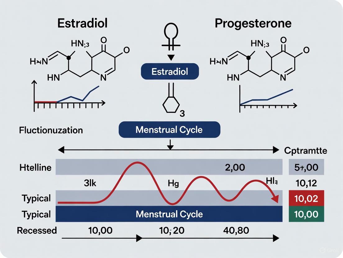
The human menstrual cycle represents a quintessential example of systemic endocrine regulation, coordinating parallel ovarian and uterine cycles through precisely timed hormonal fluctuations. For researchers and drug development professionals, understanding the intricate interplay between estradiol and progesterone is paramount, as these hormones not only govern reproductive physiology but also influence a wide array of peripheral systems. This whitepaper provides a technical analysis of follicular, ovulatory, and luteal transitions, framing these processes within contemporary research on hormonal patterning in eumenorrheic cycles. We present quantitative hormonal data, experimental methodologies for cycle phase verification, and visualization tools to elucidate the complex signaling pathways that regulate female reproductive function.
Hormonal Regulation and Phase Definitions
The menstrual cycle is orchestrated by the hypothalamic-pituitary-ovarian (HPO) axis, which synchronizes two distinct but parallel cycles: the ovarian cycle (changes within the ovaries) and the uterine cycle (changes within the endometrium) [1] [2]. These cycles are divided into phases based on dominant physiological events, with hormonal signaling serving as the primary communication mechanism between brain, ovaries, and uterus [1].
The Ovarian Cycle Phases
The ovarian cycle encompasses the follicular phase, ovulation, and the luteal phase, characterized by specific hormonal milieus and structural changes within the ovary [1] [2].
Follicular Phase: This phase begins with the onset of menses and ends at ovulation [1]. Follicle-stimulating hormone (FSH) stimulates the development of approximately 15-20 ovarian follicles, each containing an immature egg [3]. One follicle becomes dominant, secreting increasing amounts of estradiol as it matures to a size of approximately 18-29mm [2]. For most individuals, this phase lasts approximately 10-22 days, though duration shows significant interindividual variability [1].
Ovulation: Triggered by a surge in luteinizing hormone (LH) from the anterior pituitary, ovulation represents the release of a mature oocyte from the dominant follicle approximately 14 days before the next menstrual period [1] [4]. The LH surge is itself initiated when estradiol reaches a critical threshold and switches from negative to positive feedback on the pituitary [2] [4].
Luteal Phase: Following ovulation, the ruptured follicle transforms into the corpus luteum, a transient endocrine structure that secretes progesterone and some estrogen to support potential pregnancy [1]. This phase typically lasts 14 days with minimal variation (range: 9-16 days) [1]. If pregnancy does not occur, the corpus luteum regresses after 9-11 days, triggering menstrual shedding [1].
The Uterine Cycle Phases
The uterine cycle runs in parallel to the ovarian cycle, consisting of the menstruation, proliferative, and secretory phases, each characterized by distinct endometrial changes [1] [2].
Menstruation: The shedding of the uterine lining marks day 1 of the cycle, occurring when estrogen and progesterone levels are at their lowest due to corpus luteum regression [1] [2]. This phase typically lasts 3-8 days [2].
Proliferative Phase: Occurring alongside the follicular phase, this stage involves rebuilding of the endometrial lining under the influence of rising estradiol levels from the developing follicles [1] [2]. The endometrium thickens from approximately 2-3mm to 8-12mm [2].
Secretory Phase: Corresponding with the luteal phase, progesterone from the corpus luteum transforms the endometrium into a receptive state for implantation, promoting secretory activity and vascular development [1] [2]. Without pregnancy, hormonal support withdraws, leading to tissue breakdown and the initiation of menses.
Table 1: Quantitative Hormonal Fluctuations Across Menstrual Cycle Phases
| Cycle Phase | Estradiol (pg/mL) | Progesterone (ng/mL) | FSH (mIU/mL) | LH (mIU/mL) |
|---|---|---|---|---|
| Early Follicular | Low (20-50) [5] | Low (<0.5) [2] | Elevated (3-10) [2] | Low (2-8) [2] |
| Late Follicular | High (150-400) [5] | Low (<0.5) [2] | Decreasing (1-5) [2] | Rising (10-25) [2] |
| Mid-Luteal | Moderate (50-150) [5] | High (5-20) [2] | Low (1-5) [2] | Low (2-8) [2] |
| Late Luteal | Decreasing | Decreasing | Low | Low |
Table 2: Structural and Functional Changes in Ovarian and Uterine Cycles
| Cycle Phase | Ovarian Events | Endometrial Changes | Dominant Hormonal Regulators |
|---|---|---|---|
| Follicular | Recruitment of follicular cohort; selection and growth of dominant follicle [1] [2] | Proliferation and thickening (up to 8-12mm) [2] | FSH, rising estradiol [1] |
| Ovulatory | Rupture of dominant follicle and oocyte release [1] [4] | Cervical mucus changes to facilitate sperm entry [2] | LH surge, declining estradiol [1] |
| Luteal | Formation and function of corpus luteum [1] [2] | Secretory transformation; preparation for implantation [1] | Progesterone, moderate estradiol [1] |
Experimental Protocols for Menstrual Cycle Research
Robust experimental methodologies are essential for investigating hormonal fluctuations and their physiological correlates. The following protocols represent current best practices for menstrual cycle research.
Protocol for Cycle Phase Verification and Hormonal Assessment
Objective: To accurately determine menstrual cycle phase and correlate with hormonal status [5] [6].
Participant Selection Criteria:
- Naturally menstruating females (no hormonal contraception) [5] [6]
- Regular cycles (21-35 days) for at least 3 prior months [5] [6]
- No pregnancy/breastfeeding in previous 6 months [6]
- Age range: 18-40 years [5] [6]
Cycle Phase Determination Method:
- Cycle Day Calculation: Count from day 1 of menstrual bleeding (heavy flow) [2]
- Hormonal Verification:
- Urinary LH Kits: Participant self-testing to detect LH surge; ovulation confirmed by positive test [5] [6]
- Basal Body Temperature (BBT) Tracking: Post-ovulatory progesterone rise causes sustained BBT increase of approximately 0.3-0.5°C [7]
- Serial Hormonal Assays: For precise confirmation, collect blood samples for estradiol and progesterone quantification via ELISA or LC-MS/MS [5]
Phase Classification:
- Menstruation/Early Follicular: Days 1-7 from menses onset, confirmed by low estradiol and progesterone [5] [6]
- Late Follicular: 2 days after bleeding cessation until ovulation, characterized by high estradiol [6]
- Ovulation: Day of positive LH test [5] [6]
- Mid-Luteal: 7 days post-ovulation, confirmed by elevated progesterone [6]
Protocol for Assessing Cognitive Performance Across Cycle Phases
Objective: To evaluate potential cognitive fluctuations across menstrual phases using the Mnemonic Similarity Task (MST) [5].
Experimental Design:
- Task: Mnemonic Similarity Task (MST) - behavioral index of hippocampal pattern separation [5]
- Procedure: 128 trials presenting objects categorized as "target" (previously seen), "lure" (similar to previous), or "foil" (new) [5]
- Measures: Correct identification of lures as "similar" indicates successful pattern separation [5]
- Testing Timepoints: Four sessions across one complete cycle [5]
- Counterbalancing: Participants randomly assigned to different testing order sequences to control for practice effects [6]
Data Analysis:
- Primary outcome: Accuracy for lure trials across cycle phases [5]
- Statistical approach: Repeated measures ANOVA with within-subject factor (phase) and between-subject factors (athletic status) [6]
Visualization of Menstrual Cycle Signaling Pathways
Hypothalamic-Pituitary-Ovarian Axis Regulation
Parallel Ovarian and Uterine Cycle Changes
The Scientist's Toolkit: Research Reagent Solutions
Table 3: Essential Research Reagents for Menstrual Cycle Studies
| Reagent/Kit | Manufacturer Examples | Research Application | Technical Notes |
|---|---|---|---|
| LH Urinary Detection Kits | Clearblue, First Response | At-home ovulation detection for phase timing [5] [6] | Qualitative results; cost-effective for longitudinal studies |
| Enzyme-Linked Immunosorbent Assay (ELISA) | R&D Systems, Abcam, Sigma-Aldrich | Quantitative measurement of estradiol, progesterone, FSH, LH in serum/plasma [5] | High sensitivity required for low hormonal levels in early follicular phase |
| Mnemonic Similarity Task (MST) | Custom implementation | Behavioral assessment of hippocampal pattern separation [5] | 128-trial format; standardized lure similarity indices |
| Basal Body Thermometers | Femometer, iSnowMed | Tracking ovulatory temperature shift for phase confirmation [7] | Digital precision to 0.01°C; Bluetooth connectivity for data logging |
| Cognitive Test Batteries | Gorilla Experiment Builder, Inquisit | Assessing attention, inhibition, spatial anticipation [6] | Web-based administration enables remote testing |
| RNA Extraction Kits | Qiagen, Thermo Fisher | Gene expression analysis in endometrial biopsies across cycle phases | Stabilize samples in RNAlater for cyclic transcriptome studies |
Research Implications and Future Directions
Current evidence demonstrates that estradiol and progesterone fluctuations throughout the menstrual cycle exert measurable effects on physiological and cognitive function, though with significant individual variability [5] [6]. Recent research indicates cognitive performance, particularly tasks involving hippocampal pattern separation, peaks during the high-estradiol late follicular phase and reaches its lowest point during the mid-luteal phase when progesterone is dominant [5]. Interestingly, these objective measures often contradict subjective perceptions, with many individuals reporting worse performance during menstruation despite no measurable cognitive deficits [6].
From a therapeutic development perspective, these findings highlight several critical considerations. First, the timing of drug interventions targeting hormonal systems must account for cyclical endocrine patterns. Second, clinical trial designs should standardize phase assessment methodologies to reduce confounding variables. Third, individual variability in hormonal responsiveness necessitates personalized approaches rather than one-size-fits-all solutions.
Future research should prioritize elucidating the molecular mechanisms through which cyclic hormonal fluctuations influence peripheral systems, developing more precise phase verification protocols, and exploring how modifiable factors like physical activity level might modulate cycle-related symptoms and performance changes [6]. Such investigations will advance both fundamental understanding of female physiology and the development of targeted interventions for cycle-related disorders.
Estradiol (E2) serves as the primary regulator of the menstrual cycle, orchestrating a complex sequence of events from follicular recruitment to the preovulatory surge that triggers ovulation. This whitepaper examines the quantitative dynamics of estradiol production throughout the follicular phase, detailing the endocrine parameters that govern follicle development, selection, and dominance. Within the context of broader research on estradiol-progesterone fluctuation patterns in the healthy menstrual cycle, we present methodological frameworks for investigating these processes, including advanced analytical techniques for hormone quantification and experimental models for elucidating receptor-mediated mechanisms. The precise coordination of estradiol signaling with other reproductive hormones represents a critical area for therapeutic development in reproductive medicine and drug discovery.
The menstrual cycle is characterized by precisely coordinated fluctuations in reproductive hormones, with estradiol-17β (E2) serving as the primary estrogen regulating follicular development and ovulation. Within the framework of research on estradiol-progesterone fluctuation patterns in the healthy menstrual cycle, understanding E2 dynamics provides crucial insights into female reproductive physiology and pathology. The follicular phase of the cycle, spanning from menses onset until ovulation, is marked by progressively increasing E2 production from developing ovarian follicles, culminating in a preovulatory surge that triggers the luteinizing hormone (LH) release essential for ovulation [8].
Estradiol operates through genomic and non-genomic signaling pathways mediated by two nuclear estrogen receptors (ERα and ERβ). These receptors exhibit distinct expression patterns and functional roles throughout folliculogenesis [9]. The rhythmicity of E2 production during the menstrual cycle generates metabolic patterns beyond the reproductive axis, influencing neurotransmitter precursors, glutathione metabolism, urea cycle function, and nutrient utilization [10]. This technical review examines E2 dynamics from follicular recruitment through the preovulatory surge, with emphasis on quantitative hormone profiles, receptor interactions, and methodological approaches for experimental investigation.
Molecular Mechanisms of Estradiol Production and Signaling
The Two-Cell, Two-Gonadotropin Theory of Estrogen Synthesis
Estradiol biosynthesis in ovarian follicles occurs through the coordinated actions of theca and granulosa cells in a process known as the two-cell, two-gonadotropin mechanism. LH receptors located on theca cells stimulate conversion of cholesterol to androstenedione and testosterone. These androgen precursors then diffuse across the basement membrane to granulosa cells, which possess abundant follicle-stimulating hormone (FSH) receptors. FSH activates the aromatase enzyme (CYP19) in granulosa cells, converting the androgens to estrone and ultimately to estradiol via 17-β-hydroxysteroid dehydrogenase type I [8].
Table 1: Daily Production Rates of Sex Steroids During the Menstrual Cycle
| Sex Steroids | Early Follicular | Preovulatory | Mid-Luteal |
|---|---|---|---|
| Progesterone (mg) | 1 | 4 | 25 |
| 17α-Hydroxyprogesterone (mg) | 0.5 | 4 | 4 |
| Androstenedione (mg) | 2.6 | 4.7 | 3.4 |
| Testosterone (μg) | 144 | 171 | 126 |
| Estrone (μg) | 50 | 350 | 250 |
| Estradiol (μg) | 36 | 380 | 250 |
Data from Baird DT. Fraser IS. Blood production and ovarian secretion rates of estradiol-17β and estrone in women throughout the menstrual cycle. J Clin Endocrinol Metab 38: 1009-1017. 1974. @ The Endocrine Society. Values expressed in milligrams or micrograms per 24 hours [8].
Estrogen Receptor Dynamics and Signaling Pathways
Estradiol exerts its effects through two primary receptors: ERα (encoded by ESR1) and ERβ (encoded by ESR2). Both are nuclear receptors that regulate gene transcription upon E2 binding [9]. Recent research demonstrates that these receptors exhibit distinct expression patterns and functional roles throughout the menstrual cycle and across reproductive aging. During the pubertal transition in Duolang sheep, ovarian ERβ expression gradually increases from prepuberty to postpuberty, while ERα expression in the hypothalamus and pituitary decreases during puberty with a postpuberty rebound, suggesting differentiated regulatory functions [9].
The dynamics of ER protein interactions change significantly with age and hormonal status. Research in ventral hippocampus tissue demonstrates quantitative changes in ERβ protein-protein interactions with E2 replacement that are age-dependent, providing a potential mechanism for age-related changes in E2 responsiveness after menopause [11]. These receptor dynamics have implications for neuroprotection and cognitive function across the lifespan.
Figure 1: Two-Cell, Two-Gonadotropin Theory of Estradiol Synthesis. This signaling pathway illustrates the coordinated mechanism of estradiol production in ovarian follicles, requiring both theca and granulosa cells with their respective gonadotropin receptors.
Quantitative Dynamics of Estradiol During the Follicular Phase
Follicular Recruitment, Selection, and Dominance
Follicular development progresses through three distinct stages: recruitment, selection, and dominance. The recruitment stage occurs during menstrual cycle days 1-4, when rising FSH levels recruit a cohort of follicles from the non-proliferating pool. Between cycle days 5-7, selection occurs wherein one follicle is chosen from the recruited cohort to ovulate, while the remaining follicles undergo atresia. Anti-Müllerian hormone (AMH), produced by granulosa cells, plays a significant role in follicle selection. By cycle day 8, the dominant follicle establishes its precedence by promoting its own growth while suppressing maturation of other ovarian follicles [8].
The follicular phase encompasses the period from the first day of menses until ovulation. Serum E2 levels rise in parallel with growing follicle size and increasing granulosa cell numbers. Each granulosa cell possesses approximately 1500 FSH receptors by the secondary stage of follicular development, with receptor numbers remaining relatively constant throughout subsequent development [8]. The rise in E2 secretion increases the total number of estradiol receptors on granulosa cells, creating a positive feedback loop that amplifies E2 production as the follicular phase progresses.
Hormonal Fluctuations and the Preovulatory Surge
The late follicular phase is characterized by a dramatic rise in E2 production, primarily from the dominant follicle. For the positive feedback effect on LH release to occur, estradiol levels must exceed 200 pg/mL for approximately 50 hours [8]. This sustained elevation triggers the hypothalamic-pituitary axis to generate the preovulatory LH surge that induces ovulation.
Table 2: Estradiol Reference Ranges Across the Menstrual Cycle
| Cycle Phase | Estradiol (pg/mL) | Key Physiological Events |
|---|---|---|
| Early Follicular | 20-80 [12] | FSH rise recruits follicle cohort |
| Mid-Follicular | 60-150 [8] | Dominant follicle selection |
| Late Follicular | 200-500 [12] | Endometrial proliferation, positive feedback on pituitary |
| Preovulatory Peak | 200-500 [12] | Triggers LH surge (>200 pg/mL for ~50h) [8] |
| Mid-Luteal | 60-200 [12] | Support of endometrial secretion |
The luteal phase of the cycle remains relatively constant at 14 days in all women, while variability in total cycle length derives primarily from differences in the follicular phase duration, which can range from 10 to 16 days [8]. Following ovulation, the ruptured follicle transforms into the corpus luteum, which produces both progesterone and estradiol to support the endometrial lining in preparation for potential implantation.
Methodological Approaches for Investigating Estradiol Dynamics
Analytical Techniques for Estradiol Quantification
Mass spectrometry-based approaches have emerged as the gold standard for quantifying estrogens in clinical and research settings. Conventional immunoassay techniques have come under scrutiny due to concerns about selectivity, accuracy, and precision, particularly at the lower concentration ranges relevant to menstrual cycle physiology [13].
Liquid Chromatography-Tandem Mass Spectrometry (LC-MS/MS) Protocols:
- Sample Preparation: Liquid-liquid extraction (LLE) or solid-phase extraction (SPE) approaches are preferred, with derivatization remaining necessary for detection in smaller sample volumes
- Sensitivity: Limits of quantification typically range from 0.5-5 pg/mL for estrone and estradiol, with higher limits for bioactive metabolites
- Matrices: Validated for biological fluids including urine, serum, plasma, and saliva
- Specificity: Capable of distinguishing between estradiol isomers and metabolites with high structural similarity [13]
Experimental Considerations for Menstrual Cycle Studies:
- Sampling Frequency: Dense sampling protocols (e.g., every 2 hours) around the preovulatory period to capture the LH surge dynamics [9]
- Phase Verification: Combination of serum hormones, urinary luteinizing hormone, and self-reported menstrual cycle timing for precise 5-phase cycle classification [10]
- Population Homogeneity: Strict inclusion criteria for normally cycling women, with exclusion of hormonal contraceptive users and individuals with endocrine disorders
In Vitro Models for Estradiol Signaling Research
Primary granulosa cell cultures provide a valuable model system for investigating E2 effects on follicular development. The following protocol outlines standard methodology for granulosa cell isolation and stimulation:
Granulosa Cell Isolation and Culture Protocol:
- Ovarian Source: Obtain fresh ovaries from appropriate model systems
- Follicle Aspiration: Aspirate early antral follicles (2-5 mm) under sterile conditions
- Cell Collection: Filter follicular fluid through 70 μm mesh to remove oocytes and debris
- Centrifugation: Pellet cells at 1500× g for 5 minutes and resuspend in DMEM with 10% FBS and 1% penicillin-streptomycin
- Cell Identification: Confirm granulosa cell identity via immunofluorescence staining for follicle-stimulating hormone receptor (FSHR)
- Experimental Stimulation: Culture cells at 37°C with 5% CO2 and stimulate with physiological E2 concentrations (0-1000 ng/mL) for dose-response studies [9]
This model system has demonstrated that ERα exhibits a biphasic expression pattern in granulosa cells, peaking at 250 ng/mL E2 and decreasing at higher concentrations, while ERβ and GnRH expression increase in a dose-dependent manner [9].
The Researcher's Toolkit: Essential Reagents and Materials
Table 3: Essential Research Reagents for Estradiol Dynamics Investigation
| Reagent/Category | Specific Examples | Research Application |
|---|---|---|
| Antibodies | Anti-FSHR antibody [9], Anti-LHR monoclonal antibody 3B5 [14] | Cell identification, receptor localization |
| Cell Culture Media | DMEM with 10% FBS, 1% penicillin-streptomycin [9] | Granulosa cell maintenance and experimentation |
| Hormone Standards | 17β-estradiol (E2), Estrone (E1), Estriol (E3) [13] | Mass spectrometry calibration, treatment studies |
| ELISA Kits | Commercial sheep-specific E2 ELISA [9] | Serum/plasma hormone quantification |
| qPCR Reagents | PerfectStart Green qPCR SuperMix, PrimeScript RT reagent Kit [9] | Gene expression analysis of ERα, ERβ, steroidogenic enzymes |
| Extraction Materials | Solid-phase extraction cartridges, derivatization reagents [13] | Sample preparation for mass spectrometry |
Follicular Wave Theory and Estradiol Dynamics
The traditional model of single follicle recruitment per cycle has been supplemented by the follicular wave theory, which proposes that follicles grow in synchronous waves throughout the menstrual cycle. Approximately 68% of reproductive-aged women exhibit two waves of follicle development during one interovulatory interval, while 32% experience three waves [15].
In women with two follicular waves, the first wave begins during the early luteal phase and is typically anovulatory, while the second wave emerges in the late luteal or early follicular phase and contains the ovulatory follicle. Women with three follicular waves experience an additional wave during the mid-luteal phase. The hormonal regulation of wave emergence involves subtle interactions between FSH, inhibin, estradiol, and progesterone [15].
This paradigm has important clinical implications, particularly for controlled ovarian stimulation protocols. Luteal-phase stimulation has been developed for fertility preservation in cancer patients who cannot delay oncological treatment to await the traditional follicular phase start, demonstrating that viable oocytes can be obtained regardless of menstrual cycle phase [15].
Figure 2: Experimental Workflow for Menstrual Cycle Research. This methodology diagram outlines a comprehensive approach for investigating estradiol dynamics across the menstrual cycle, incorporating both clinical and laboratory components.
The dynamics of estradiol from follicular recruitment through the preovulatory surge represent a sophisticated endocrine coordination system essential for human reproduction. Understanding these precise fluctuation patterns within the broader context of menstrual cycle physiology provides critical insights for developing targeted therapeutic interventions. The quantitative data and methodological frameworks presented herein offer researchers standardized approaches for investigating estradiol dynamics in both basic science and clinical applications.
Future research directions should focus on elucidating the molecular mechanisms underlying estrogen receptor signaling dynamics across different reproductive stages, developing more refined experimental models that recapitulate the human menstrual cycle, and translating these findings into clinical applications for disorders of menstrual cycle rhythmicity. The integration of advanced analytical techniques with sophisticated experimental designs will continue to enhance our understanding of estradiol dynamics in health and disease.
The corpus luteum, a transient endocrine structure formed from the ovulated follicle, serves as the primary source of progesterone during the luteal phase of the menstrual cycle. Its precise regulatory function is fundamental to endometrial receptivity, embryo implantation, and the maintenance of early pregnancy. This whitepaper synthesizes current research on progesterone patterns, detailing the quantitative dynamics of progesterone secretion, the pathophysiology of luteal phase deficiency (LPD), and advanced methodologies for its assessment. Within the broader context of estradiol-progesterone fluctuation patterns in healthy menstrual cycle research, we present structured data, experimental protocols, and analytical tools to guide drug development and clinical investigation. The intricate interplay between progesterone pulsatility, endometrial transformation, and metabolic rhythmicity underscores the necessity for precise measurement techniques and targeted therapeutic interventions to address luteal phase defects.
The luteal phase constitutes the second half of the ovarian cycle, initiated by ovulation and characterized by the formation and function of the corpus luteum [16]. This temporary structure develops from the remnants of the dominant follicle after it releases its oocyte. The primary endocrine function of the corpus luteum is the secretion of progesterone and, to a lesser extent, estradiol, which collectively prepare the endometrium for implantation [16] [17].
The molecular and cellular basis of progesterone production involves the transformation of follicular theca and granulosa cells into luteal cells, which synthesize and secrete progesterone in response to pulsatile luteinizing hormone (LH) stimulation [16] [18]. Progesterone's cardinal function is to transform the estrogen-primed proliferative endometrium into a secretory state. This involves inducing stromal edema, stimulating glandular secretion of glycogen and glycoproteins, and promoting vascular development, thereby creating a receptive environment for a blastocyst [17] [19]. The hormone also exerts a negative feedback effect on the hypothalamic-pituitary axis, suppressing gonadotropin-releasing hormone (GnRH) pulse frequency and preventing further follicular development during the luteal phase [19].
If pregnancy does not occur, the corpus luteum undergoes luteolysis approximately 10-14 days after ovulation, leading to a precipitous decline in progesterone and estradiol levels. This hormone withdrawal causes vasoconstriction in the endometrial spiral arteries, tissue ischemia, and the eventual shedding of the functional endometrial layer, manifesting as menstruation [16] [17]. In the event of conception, the developing blastocyst begins secreting human chorionic gonadotropin (hCG), which rescues the corpus luteum (a process known as the "maternal recognition of pregnancy"). The corpus luteum continues to produce progesterone until around the 7-9th week of gestation, after which the placenta assumes the primary role of progesterone production in a process called the luteal-placental shift [16] [19].
Quantitative Progesterone Dynamics in the Luteal Phase
Understanding the normal rhythmicity and expected concentrations of progesterone is critical for identifying pathological states such as luteal phase deficiency (LPD).
Table 1: Reference Ranges for Serum Progesterone Across the Menstrual Cycle and Pregnancy
| Reproductive Status | Phase | Progesterone Reference Range | Notes |
|---|---|---|---|
| Premenopausal Adult Female | Follicular Phase | < 50 ng/dL (< 0.5 ng/mL) [19] | Baseline, low level |
| Luteal Phase | 300 - 2500 ng/dL (3 - 25 ng/mL) [19] | Peak occurs 6-8 days post-ovulation [18] | |
| Pregnancy | First Trimester | 725 - 4400 ng/dL (7.25 - 44 ng/mL) [19] | Corpus luteum is primary source |
| Second Trimester | 1950 - 8250 ng/dL (19.5 - 82.5 ng/mL) [19] | Placental takeover complete | |
| Third Trimester | 6500 - 22900 ng/dL (65 - 229 ng/mL) [19] | Produced entirely by placenta |
Progesterone secretion is inherently pulsatile, correlating with LH pulses, and concentrations can fluctuate up to eight-fold within 90 minutes [18]. This variability complicates single-point measurements for diagnostic purposes. A single mid-luteal phase progesterone level >3 ng/mL is typically considered evidence that ovulation has occurred, but this does not confirm an adequate luteal phase [17] [18]. The luteal phase length is relatively fixed, typically lasting 12 to 14 days (range 11-17 days). A clinically defined LPD is often associated with a luteal phase of ≤10 days [18].
Research within the context of estradiol-progesterone fluctuation patterns reveals that these hormonal shifts have systemic metabolic consequences. A comprehensive metabolomics study found that the luteal phase is characterized by significant decreases in plasma amino acids, derivatives, and specific lipid species, suggesting a state of increased nutrient utilization potentially driven by the progesterone peak [10].
Table 2: Metabolic Patterns Across the Menstrual Cycle
| Metabolite Class | Observed Change in Luteal Phase | Statistical Significance | Proposed Interpretation |
|---|---|---|---|
| Amino Acids & Biogenic Amines (e.g., Ornithine, Arinine, Alanine) | Significant decrease | 37 metabolites met FDR threshold (q<0.20) for Luteal-Menstrual contrast [10] | Indicative of an anabolic state and increased nitrogen utilization [10] |
| Phospholipids (e.g., LPCs, PCs) | Significant decrease | 17 lipid species met FDR threshold (q<0.20) for Luteal-Follicular contrast [10] | Cyclic rhythmicity in energy substrates |
| Vitamin D (25-OH Vitamin D) | Lowest in Luteal, peaks in Menstrual phase | Significant for Luteal-Menstrual & Ovine-Menstrual contrasts (q<0.20) [10] | Potential interaction with sex hormone rhythms |
Pathophysiology and Diagnosis of Luteal Phase Deficiency
Luteal Phase Deficiency (LPD) is a clinical condition characterized by inadequate progesterone secretion or action, resulting in an impaired endometrial response that is unreceptive to implantation [18]. The pathophysiological basis for LPD can be traced to disturbances in the hypothalamic-pituitary-ovarian axis.
Etiology and Pathophysiology
The primary mechanisms underlying LPD include:
- Inadequate Progesterone Production: This can stem from impaired folliculogenesis, leading to a dysfunctional corpus luteum. Contributing factors include low follicular-phase FSH levels, altered FSH/LH ratios, or abnormal GnRH pulsatility [18].
- Endometrial Progesterone Resistance: In some cases, serum progesterone levels may be adequate, but the endometrium exhibits a defective response to the hormone, preventing normal secretory transformation and decidualization [18].
Several clinical conditions are associated with an increased risk of LPD, such as hypothalamic amenorrhea, eating disorders, excessive exercise, hyperprolactinemia, thyroid dysfunction, obesity, polycystic ovary syndrome (PCOS), endometriosis, and advanced reproductive age [18].
Diagnostic Methodologies and Protocols
Diagnosing LPD remains challenging due to the pulsatile nature of progesterone secretion and the invasiveness of the most definitive test.
Protocol: Endometrial Biopsy
The historical gold standard for diagnosing LPD has been the endometrial biopsy.
- Principle: A tissue sample of the endometrium is obtained in the late luteal phase and histologically dated according to established criteria (e.g., Noyes' criteria). A discrepancy of more than two days between the histological date and the expected date (based on the ovulation day) in two separate cycles was considered diagnostic for LPD [18].
- Limitations: The procedure is invasive, subjective, and has poor inter- and intra-observer reliability. Furthermore, studies have shown it cannot reliably differentiate between fertile and infertile women, limiting its clinical utility [18].
Protocol: Serum Progesterone Measurement
- Principle: Single or multiple serum progesterone measurements are taken during the mid-luteal phase (typically around day 21 of a 28-day cycle) to assess corpus luteum function.
- Procedure: A single mid-luteal level >3 ng/mL confirms ovulation but is insufficient to rule out LPD due to progesterone pulsatility. Serial measurements (e.g., every 2-3 days) provide a more integrated assessment of progesterone exposure [18].
- Limitations: The wide, rapid fluctuations in progesterone levels make it difficult to define a reliable diagnostic threshold for LPD [18].
Protocol: Urinary Hormone Metabolite Monitoring with Quantitative Fertility Monitors
Advanced quantitative fertility monitors represent a non-invasive method for detailed luteal phase profiling.
- Principle: These devices use lateral flow assays to measure the concentration of urinary hormone metabolites, including pregnanediol glucuronide (PDG, a major progesterone metabolite), luteinizing hormone (LH), and estrone-3-glucuronide (E3G, an estrogen metabolite) [20].
- Procedure: First-morning urine samples are collected daily. Test sticks are analyzed by a monitor (e.g., Mira, Inito) that syncs with a smartphone app to graphically display quantitative hormone levels. This allows for the identification of the LH surge (to pinpoint ovulation) and the subsequent dynamic rise and fall of PDG throughout the luteal phase [20].
- Data Analysis: The profile allows researchers to assess key luteal phase processes: luteinization (the initial formation of the corpus luteum, reflected by the interaction of the LH surge and the initial PDG rise), progestation (the PDG plateau that supports the endometrium), and luteolysis (the decline of PDG at the end of the cycle) [20]. Abnormal patterns, such as a slow rise, low plateau, or shortened duration of elevated PDG, can indicate LPD.
Diagram 1: LPD Diagnostic Workflow
Advanced Research Tools and Experimental Reagents
The following toolkit compiles essential reagents and methodologies for investigating corpus luteum function and progesterone dynamics in a research setting.
Table 3: Research Reagent Solutions for Luteal Phase Investigation
| Research Tool / Reagent | Function / Application | Experimental Notes |
|---|---|---|
| Quantitative Urinary PDG/LH/E3G Assays (e.g., Mira, Inito monitors) | Non-invasive, high-frequency longitudinal tracking of luteal phase hormone dynamics. Identifies luteinization, progestation, and luteolysis processes [20]. | Provides a quantitative profile superior to single serum measurements for assessing luteal phase adequacy and identifying subtle defects [20]. |
| Enzyme-Linked Immunosorbent Assay (ELISA) | Quantifies serum levels of progesterone, estradiol, LH, FSH. The standard for hormone level validation in clinical studies. | Must account for pulsatile secretion of progesterone; serial measurements are often necessary for accurate assessment [18]. |
| Recombinant Gonadotropins (FSH, LH, hCG) | Used in ovarian stimulation protocols to study corpus luteum formation and function under controlled conditions, and to rescue the corpus luteum in early pregnancy models [21] [22]. | hCG is used to trigger ovulation and support the corpus luteum in assisted reproductive technology (ART) cycles [22]. |
| Progesterone Receptor Antagonists (e.g., Mifepristone) | Investigational tools to block progesterone action at the receptor level, enabling the study of progesterone withdrawal and its role in endometrial breakdown [19]. | Useful for modeling the end of the luteal phase and studying the mechanisms of menstruation. |
| Vaginal Progesterone (Micronized Progesterone) | The cornerstone for luteal phase support in ART and for treating LPD. Serves as the active comparator in therapeutic trials [22] [18]. | Allows for study of endometrial response to exogenous progesterone and assessment of treatment efficacy. |
Diagram 2: Hormonal Regulation Pathway
The corpus luteum is a pivotal determinant of reproductive success through its regulated secretion of progesterone during the luteal phase. Advanced research techniques, particularly quantitative urinary hormone monitoring, are refining our understanding of progesterone patterns and the definition of LPD. Future investigations should leverage mathematical modeling and in silico clinical trials to simulate luteal phase dynamics and optimize stimulation and support protocols [21]. Furthermore, research must continue to elucidate the complex dialogue between the corpus luteum, the endometrium, and the developing embryo, with a focus on the molecular basis of endometrial progesterone resistance. Integrating metabolic data with hormonal profiles offers a novel systems-biology approach to understanding the luteal phase as a key rhythmic process in female physiology, paving the way for personalized therapeutic strategies in infertility and recurrent pregnancy loss.
Interplay of Gonadotropins (FSH, LH) with Ovarian Steroids
The menstrual cycle represents a meticulously orchestrated biological rhythm, central to human reproduction. This whitepaper delineates the complex interplay between pituitary gonadotropins—follicle-stimulating hormone (FSH) and luteinizing hormone (LH)—and the ovarian steroids, estradiol and progesterone. Framed within estradiol-progesterone fluctuation patterns in the healthy menstrual cycle, this review synthesizes the endocrine regulation of folliculogenesis, ovulation, and luteal formation. We explore the hypothalamic-pituitary-ovarian (HPO) axis, quantitative hormone dynamics, underlying molecular mechanisms, and critical experimental methodologies. This resource is designed to inform researchers, scientists, and drug development professionals in the field of reproductive endocrinology, providing structured data, signaling pathways, and essential research tools.
The human menstrual cycle is a quintessential example of a tightly regulated endocrine process, essential for the perpetuation of the species. It is governed by the dynamic, reciprocal interactions between the hypothalamus, pituitary gland, and ovaries—collectively known as the HPO axis [23] [24]. Central to this dialogue are the gonadotropins (FSH and LH) and the ovarian steroids (estradiol and progesterone). Their fluctuating concentrations direct a sequence of events encompassing ovarian follicle development, ovulation, and preparation of the uterine endometrium for implantation [8]. Disruptions in this intricate interplay can lead to a spectrum of gynecological conditions, including infertility, polycystic ovarian syndrome (PCOS), and endometriosis [25] [23]. A precise understanding of these hormonal fluctuations and their mechanisms of action is therefore paramount for both basic reproductive biology and the development of novel therapeutic interventions.
The Hypothalamic-Pituitary-Ovarian (HPO) Axis: A Regulatory Framework
The HPO axis functions as a closed-loop feedback system, initiating and modulating the menstrual cycle.
2.1. Hypothalamic and Pituitary Roles The rhythmic release of gonadotropin-releasing hormone (GnRH) from the hypothalamus is the fundamental pulse generator for the cycle. GnRH is secreted in a pulsatile manner into the hypophyseal portal circulation, stimulating the anterior pituitary to synthesize and secrete FSH and LH [26] [27]. The frequency and amplitude of GnRH pulses are critical; low-frequency pulses preferentially favor FSH secretion, while high-frequency pulses favor LH secretion [25] [27]. The sensitivity of the pituitary gonadotropes to GnRH also changes throughout the cycle, increasing significantly during the midcycle surge due to a self-priming effect of GnRH [27].
2.2. Ovarian Feedback and Cycle Phases The ovaries respond to FSH and LH by driving follicular development and steroidogenesis. In return, the ovarian steroids, estradiol and progesterone, provide feedback at the level of the hypothalamus and pituitary. The cycle can be divided into two main phases based on ovarian activity:
- Follicular/Proliferative Phase: This phase begins with menses and ends at ovulation. It is characterized by the recruitment and selection of a dominant ovarian follicle, which secretes increasing amounts of estradiol. Initially, low and then rising levels of estradiol exert negative feedback on FSH secretion [8] [24].
- Luteal/Secretory Phase: This phase begins after ovulation and lasts until the onset of the next menses. The ruptured follicle transforms into the corpus luteum, which secretes large quantities of progesterone and estradiol. Progesterone dominates this phase and, in conjunction with estradiol, inhibits GnRH pulse frequency, maintaining negative feedback [8] [27].
A unique event occurs at the end of the follicular phase: when estradiol levels rise above a threshold (~200 pg/mL) for approximately 50 hours, the feedback switches from negative to positive. This triggers the surge of GnRH and the subsequent LH surge that is essential for ovulation [8] [25].
Molecular Mechanisms of Hormone Action
3.1. Gonadotropin Receptors and Signaling FSH and LH mediate their effects via specific G-protein coupled receptors (GPCRs).
- FSH Receptor (FSHR): The FSHR is located exclusively on the granulosa cells of the ovary [28]. FSH binding activates a Gs protein-coupled pathway, leading to the activation of adenylate cyclase and an increase in intracellular cyclic AMP (cAMP). This cascade activates protein kinase A (PKA), which in turn phosphorylates downstream targets, including the transcription factor CREB. This signaling pathway is responsible for FSH's key actions: stimulating aromatase expression, promoting follicular growth, and inducing LH receptor expression on granulosa cells of the preovulatory follicle [25] [28].
- LH Receptor (LHCGR): The LHCGR is expressed on theca cells throughout follicular development and is induced on granulosa cells of large antral follicles by FSH [28]. LH binding also primarily signals through the Gq/11 pathway, activating phospholipase C (PLC). PLC hydrolyzes phosphatidylinositol 4,5-bisphosphate (PIP2) to generate inositol trisphosphate (IP3) and diacylglycerol (DAG). IP3 triggers the release of calcium from intracellular stores, while DAG activates protein kinase C (PKC). This signaling is crucial for androgen production in theca cells and for the final stages of oocyte maturation and ovulation [27] [28].
Table 1: Gonadotropin Receptor Characteristics
| Characteristic | FSH Receptor (FSHR) | LH/hCG Receptor (LHCGR) |
|---|---|---|
| Primary Cell Location | Granulosa Cells | Theca Cells, Granulosa Cells (preovulatory) |
| Protein Family | G-protein Coupled Receptor (GPCR) | G-protein Coupled Receptor (GPCR) |
| Key Signaling Pathways | Gs / cAMP / PKA | Gq / PLC / IP3 / Ca²⁺ / PKC |
| Main Biological Actions | Follicle growth, Aromatase induction, LH receptor induction | Androgen synthesis, Ovulation trigger, Luteal maintenance |
3.2. Steroidogenesis: The Two-Cell Theory The synthesis of ovarian steroids requires the cooperative interaction between theca and granulosa cells, as described by the two-cell, two-gonadotropin model [8].
- Theca Cells: Under LH stimulation, theca cells convert cholesterol into androstenedione and testosterone (C19 steroids).
- Granulosa Cells: These androgens are then diffused to the granulosa cells. Under FSH stimulation, granulosa cells express high levels of the enzyme aromatase (CYP19), which converts the androgens into estrogens—specifically, androstenedione to estrone and testosterone to estradiol [8] [25]. This compartmentalization ensures efficient production of estradiol, the hallmark steroid of the developing follicle.
3.3. The Role of Neuroprogesterone in Positive Feedback Emerging evidence highlights that the brain is not merely a passive recipient of peripheral steroid signals but an active site of steroidogenesis. A key discovery is the role of neuroprogesterone in the positive feedback mechanism. The rising estradiol levels during the late follicular phase stimulate the synthesis of progesterone de novo within the hypothalamus [29] [30]. This "neuroprogesterone" is synthesized in astrocytes and acts locally on estrogen-induced progesterone receptors in the hypothalamus. Blocking this hypothalamic progesterone synthesis prevents the LH surge and ovulation, even in the presence of adequate peripheral estradiol levels, establishing neuroprogesterone as a necessary component of the ovulatory trigger [29] [30].
Quantitative Hormonal Dynamics Across the Menstrual Cycle
The median duration of a menstrual cycle is 28 days, though cycles between 25 and 30 days are common. The luteal phase is relatively fixed at approximately 14 days, while the variability in cycle length is primarily due to differences in the duration of the follicular phase [8].
Table 2: Daily Production Rates of Sex Steroids During the Menstrual Cycle Data from Baird DT. Fraser IS. J Clin Endocrinol Metab 38: 1009-1017, 1974. Adapted from Endotext [8].
| Sex Steroid | Early Follicular | Preovulatory | Mid-Luteal |
|---|---|---|---|
| Progesterone (mg/24h) | 1 | 4 | 25 |
| 17α-Hydroxyprogesterone (mg/24h) | 0.5 | 4 | 4 |
| Androstenedione (mg/24h) | 2.6 | 4.7 | 3.4 |
| Testosterone (μg/24h) | 144 | 171 | 126 |
| Estrone (μg/24h) | 50 | 350 | 250 |
| Estradiol (μg/24h) | 36 | 380 | 250 |
Table 3: Pulsatile Secretion of Luteinizing Hormone (LH) Data adapted from Endotext [8].
| Cycle Phase | Pulse Frequency (minutes) | Pulse Amplitude (mIU/ml/min) |
|---|---|---|
| Early Follicular | 80 ± 3 | 0.43 ± 0.02 |
| Late Follicular | 53 ± 1 | 0.70 ± 0.03 |
| Mid-Luteal | 177 ± 15 | 0.26 ± 0.02 |
Experimental Protocols for Investigating the HPO Axis
5.1. GnRH Stimulation Test Purpose: To assess the functional integrity of the pituitary gonadotropes and, indirectly, the hypothalamus. It is used in the diagnosis of disorders like hypogonadotropic hypogonadism and central precocious puberty [26]. Detailed Methodology:
- Baseline Sample: An intravenous (IV) line is established, and a baseline blood sample is drawn to measure FSH, LH, and sex steroids.
- GnRH Administration: A bolus of synthetic GnRH (e.g., 100 µg) is administered intravenously or subcutaneously.
- Serial Sampling: Subsequent blood samples are collected at timed intervals, typically at 15, 30, 45, 60, and 120 minutes post-injection.
- Analysis: LH and FSH levels are measured in all samples. A normal response is a significant rise in LH (and to a lesser extent, FSH), peaking at 30-60 minutes.
- Interpretation: An exaggerated response suggests a lack of prior gonadal steroid negative feedback (as in primary gonadal failure). A blunted or absent response indicates a pituitary defect, while a delayed response may suggest a hypothalamic origin [26].
5.2. Blocking Neuroprogesterone Synthesis In Vivo Purpose: To investigate the necessity of hypothalamic progesterone synthesis for the LH surge and ovulation [29] [30]. Detailed Methodology:
- Animal Model: Use gonadally intact, cycling female rats.
- Surgery and Cannulation: Implant a cannula into the lateral cerebral ventricle for direct intracerebroventricular (ICV) infusion.
- Inhibitor Infusion: On the morning of proestrus, infuse a steroidogenic enzyme inhibitor such as aminoglutethimide (AGT), a P450 side-chain cleavage (P450scc) enzyme inhibitor, or a specific 3β-hydroxysteroid dehydrogenase (3β-HSD) inhibitor into the lateral ventricle. Control animals receive vehicle infusion.
- Monitoring: Monitor the estrous cycle via vaginal cytology. In inhibitor-treated animals, the cycle will arrest in proestrus.
- Sample Collection: On the afternoon of proestrus, collect blood via decapitation or cardiac puncture to measure peripheral LH, FSH, estradiol, and progesterone. Simultaneously, microdissect hypothalamic tissue for measurement of local progesterone levels via radioimmunoassay (RIA) or mass spectrometry.
- Ovarian Analysis: Examine ovaries for the presence of ovulated oocytes or forming corpora lutea. AGT-treated rats will show an absence of ovulation despite normal peripheral estradiol levels [30].
Signaling Pathway and System Workflow Visualizations
Diagram 1: HPO Axis & Feedback Loops. This diagram illustrates the primary structures, hormones, and feedback loops within the Hypothalamic-Pituitary-Ovarian axis and their effects on the uterus.
Diagram 2: Two-Cell Gonadotropin Theory. This diagram details the cooperative steroidogenesis between theca and granulosa cells for estradiol production, highlighting key signaling pathways and enzymatic conversions.
The Scientist's Toolkit: Essential Research Reagents
Table 4: Key Reagents for Investigating Gonadotropin-Ovarian Interactions
| Reagent / Material | Function / Application | Example Use Case |
|---|---|---|
| Synthetic GnRH | Stimulates pituitary release of FSH and LH in vivo or in vitro. | GnRH stimulation test; studying pulsatile hormone secretion [26] [27]. |
| Recombinant FSH & LH | Provides pure gonadotropin preparations for cell culture or animal studies. | Ovarian follicle culture in vitro; superovulation protocols in animal models [25]. |
| GnRH Agonists (e.g., Leuprolide) | Initially stimulate, then chronically suppress the HPG axis by downregulating pituitary GnRH receptors. | Treating hormone-dependent cancers (prostate, breast); managing endometriosis; in IVF protocols to prevent premature LH surge [26] [25]. |
| GnRH Antagonists (e.g., Cetrorelix) | Competitively block the GnRH receptor, providing immediate suppression of FSH and LH release. | In IVF protocols for rapid suppression of the LH surge [26] [25]. |
| Enzyme Inhibitors (e.g., Aminoglutethimide) | Inhibits key steroidogenic enzymes (e.g., P450scc). | Investigating the role of neurosteroids in the LH surge; studying adrenal and gonadal steroidogenesis [29] [30]. |
| Specific siRNA/shRNA | Knocks down expression of target genes (e.g., FSHR, LHCGR, StAR, Aromatase). | Elucidating the specific function of a protein in steroidogenic signaling pathways in cell lines [28]. |
| Antibodies (for IHC, WB) | Detect and localize specific proteins (e.g., FSHR, LHCGR, ER, PR, steroidogenic enzymes). | Determining cellular expression patterns of receptors in ovarian tissue sections [28]. |
| Primary Cell Cultures (Granulosa, Theca) | Provide an ex vivo model to study cell-specific responses to hormones. | Investigating the mechanisms of FSH-induced aromatase activity or LH-induced androgen production [28] [30]. |
The menstrual cycle is characterized by dynamic fluctuations in estradiol and progesterone, which extend beyond reproductive function to modulate systemic metabolism. This whitepaper synthesizes evidence from clinical and experimental studies to elucidate how cyclic hormonal variations influence glucose homeostasis, lipid metabolism, cognitive performance, and body composition. Targeting researchers and drug development professionals, we provide structured quantitative data, experimental protocols, and visualizations of key signaling pathways to guide future investigations into metabolic health in menstruating individuals.
The menstrual cycle comprises two primary phases—the follicular phase (dominated by estradiol) and the luteal phase (dominated by progesterone)—driven by the hypothalamic-pituitary-ovarian (HPO) axis. Emerging research highlights that these hormonal oscillations regulate metabolic pathways, including insulin sensitivity, lipogenesis, and energy expenditure [7] [31]. Understanding these fluctuations is critical for developing therapies for metabolic disorders and optimizing drug efficacy in premenopausal women.
Metabolic Fluctuations Across Menstrual Cycle Phases
Hormonal variations during the menstrual cycle induce systemic metabolic changes, summarized in Table 1.
Table 1: Metabolic Parameters Across Menstrual Cycle Phases
| Phase | Hormonal Profile | Glucose Metabolism | Lipid Metabolism | Cognitive Performance |
|---|---|---|---|---|
| Early Follicular | Low estradiol, low progesterone | Insulin resistance potential | Stable LDL-C, HDL-C | Lower reaction times [6] |
| Late Follicular | High estradiol, low progesterone | Enhanced insulin sensitivity | Reduced LDL-C, increased HDL-C | Improved spatial anticipation [6] |
| Ovulation | Peak estradiol, LH surge | Optimal glucose utilization | Favorable lipid profile | Fastest reaction times [6] |
| Mid-Luteal | High progesterone, moderate estradiol | Reduced insulin sensitivity [31] | Elevated LDL-C, triglycerides [31] | Slower reaction times [6] |
- Glucose Homeostasis: Estradiol enhances insulin sensitivity by activating estrogen receptor alpha (ERα) in skeletal muscle and liver, while progesterone may antagonize this effect [31].
- Lipid Metabolism: Estradiol decline during the luteal phase increases LDL-C and triglycerides, elevating cardiovascular risk [31].
- Cognitive Function: Executive function peaks during ovulation (high estradiol) and declines in the luteal phase (high progesterone) [6].
Experimental Protocols for Metabolic and Cognitive Assessment
Protocol 1: Assessing Cognitive Performance Across Cycles
Objective: Measure attention, inhibition, and spatial anticipation across menstrual phases [6]. Methodology:
- Participants: Recruit eumenorrheic women (ages 18–40) not using hormonal contraception.
- Phase Verification: Track phases via urinary luteinizing hormone (LH) kits or basal body temperature [7] [6].
- Cognitive Tasks:
- Go/No-Go Task: Assess inhibitory control.
- Spatial Anticipation Task: Evaluate timing and coordination.
- Data Collection: Test at four phases: menstruation (early follicular), late follicular, ovulation, and mid-luteal.
- Analysis: Compare reaction times and errors using repeated-measures ANOVA.
Protocol 2: Evaluating Glucose and Lipid Metabolism
Objective: Quantify insulin resistance and lipid dynamics [31] [32]. Methodology:
- Participants: Include women with regular cycles and exclude those with metabolic diseases.
- Blood Sampling: Measure fasting glucose, insulin, LDL-C, HDL-C, and triglycerides at each phase.
- Hormonal Assays: Quantify estradiol, progesterone, LH, and FSH via ELISA.
- Statistical Analysis: Use multivariate regression to correlate hormonal levels with metabolic parameters.
Signaling Pathways and Metabolic Regulation
HPO Axis and Metabolic Integration
The HPO axis coordinates hormonal fluctuations that indirectly influence metabolism via central and peripheral pathways.
Estrogen-Mediated Glucose Regulation
Estradiol activates ERα in metabolic tissues to enhance insulin sensitivity and suppress lipogenesis.
The Scientist's Toolkit: Research Reagent Solutions
Table 2: Essential Reagents for Menstrual Cycle Metabolic Studies
| Reagent/Tool | Function | Example Application |
|---|---|---|
| Urinary LH Kits | Detects ovulation via luteinizing hormone surge | Phase verification in cognitive studies [6] |
| ELISA Kits | Quantifies serum estradiol, progesterone, insulin | Hormonal and metabolic profiling [31] |
| Basal Body Thermometers | Tracks temperature shifts for ovulation detection | Cycle phase categorization [7] |
| Cognitive Batteries | Assesses executive function (e.g., Go/No-Go tasks) | Evaluating phase-dependent cognitive changes [6] |
| Lipoprotein Assays | Measures LDL-C, HDL-C, and triglycerides | Lipid metabolism analysis [31] |
Discussion and Future Directions
Cyclic hormonal fluctuations significantly impact systemic metabolism, with estradiol enhancing insulin sensitivity and lipid homeostasis, while progesterone may counter these effects. The integration of cognitive data further highlights the broad influence of menstrual cycles on physiological and neurological function. Future research should prioritize:
- Personalized Metabolic Modeling: Developing phase-specific interventions for women with obesity or PCOS [33] [32].
- Drug Development: Considering hormonal phases in clinical trials to optimize dosing and efficacy.
- Molecular Mechanisms: Elucidating tissue-specific ERα and ERβ signaling using single-cell transcriptomics.
The menstrual cycle is a key determinant of systemic metabolic health, influencing processes from glucose regulation to cognitive performance. By adopting standardized experimental protocols and leveraging advanced reagents, researchers can decode the complex interplay between hormonal rhythms and metabolism, paving the way for precision medicine in women’s health.
Analytical Techniques for Quantifying Hormonal Rhythms: From HPLC to LC-MS/MS
Accurate measurement of steroid hormones, particularly estradiol and progesterone, is fundamental to research on fluctuation patterns across the healthy menstrual cycle. The choice of analytical platform directly impacts data reliability, sensitivity, and biological validity. This technical guide provides an in-depth comparison of three core platforms—immunoassays, high-performance liquid chromatography with fluorescence detection (HPLC-FLD), and liquid chromatography-tandem mass spectrometry (LC-MS/MS)—framed within the context of female reproductive endocrinology research. Understanding the capabilities and limitations of each method is crucial for researchers and drug development professionals designing studies to unravel the complex interplay between hormones, brain, and behavior.
The following table summarizes the core characteristics of each analytical platform relevant to sex hormone research.
Table 1: Comparison of Key Analytical Platforms for Sex Hormone Quantification
| Feature | Immunoassays (e.g., ELISA, RIA) | HPLC-FLD | LC-MS/MS (including UPLC-MS/MS) |
|---|---|---|---|
| Fundamental Principle | Antibody-antigen binding for detection | Chromatographic separation followed by fluorescence detection | Chromatographic separation followed by mass-based detection |
| Typical Sensitivity (LoQ) | Variable; often insufficient at low concentrations [34] | ~10 ng/mL for estradiol [35] | 1-5 pg/mL for estradiol in serum [36] [37] |
| Specificity | Low to moderate; prone to cross-reactivity with similar molecules [38] [34] | High (with good separation) | Very High; separates and identifies analytes by mass |
| Multiplexing Capability | Low; typically single analyte or few analytes | Moderate | High; can simultaneously quantify dozens of steroids [39] |
| Sample Throughput | High | Moderate | Moderate to High (with modern systems) |
| Sample Volume Required | Low | Moderate | Low to Moderate |
| Capital & Operational Cost | Low | Moderate | High |
| Best Suited For | High-throughput screening where ultimate accuracy is not critical | Environments with budget constraints where hormones are at high concentrations | Research requiring high sensitivity, specificity, and multiplexing, especially for low-concentration hormones [38] [40] |
Performance in Hormone Research Context
Limitations of Immunoassays
While widely used due to their low cost and high throughput, immunoassays show significant limitations in the context of menstrual cycle research. A 2025 comparative study of salivary hormones concluded that ELISA showed "poor performance" for measuring estradiol and progesterone, with these hormones being "much less valid than testosterone" [38] [40]. The study highlighted that only LC-MS/MS showed the expected physiological differences in estradiol and testosterone levels between men and women, and machine-learning models classified hormone data more successfully when using LC-MS/MS-derived values [40]. These findings underscore that immunoassays may lack the necessary precision and accuracy to reliably detect the subtle yet biologically critical fluctuations of estradiol and progesterone across the menstrual cycle.
The Superiority of LC-MS/MS
Liquid chromatography-tandem mass spectrometry has emerged as the gold-standard technique for steroid hormone profiling. Its superior specificity eliminates the cross-reactivity issues that plague immunoassays. Furthermore, its exceptional sensitivity is crucial for accurately quantifying the low picogram-per-milliliter concentrations of estradiol present in postmenopausal women, men, and certain phases of the menstrual cycle [36] [34].
UPLC-MS/MS methods have been validated for the simultaneous quantitation of both endogenous and synthetic sex steroids in human serum, achieving lower limits of quantitation (LLOQ) as low as 1 pg/mL for estradiol and 25 pg/mL for various progestogens [36] [37]. This sensitivity and multiplexing capability makes it ideal for comprehensive hormone profiling. A key advantage for long-term studies is its demonstrated long-term robustness, with both accuracy and precision remaining within ±15% over a two-year period, a critical feature for longitudinal clinical trials [36].
Niche for HPLC-FLD
HPLC with fluorescence detection serves as a viable alternative when mass spectrometry is inaccessible due to its high cost and operational complexity. The efficiency and quick separation of HPLC combined with the sensitivity and selectivity of fluorescence detection make HPLC-FLD a potent tool for routine measurement [41]. However, most steroids lack intrinsic fluorescence, necessitating a derivatization step before analysis [35] [41]. For instance, a 2025 study developed an HPLC-FLD method for estradiol and its metabolites in saliva and serum, using derivatization with dansyl chloride to enable detection. This method achieved a linear range of 10–300 ng/mL and a LOQ of 10 ng/mL [35]. While this sensitivity is insufficient for measuring low physiological levels, it can be adequate for pharmaceutical applications or high-concentration samples.
Detailed Experimental Protocols
Protocol: UPLC-MS/MS for Serum Estradiol and Progestogens
This robust protocol is adapted from a 2019 study for the simultaneous quantitation of estrogens and progestogens in human serum [36] [37].
Sample Preparation (Liquid-Liquid Extraction):
- Pipette 0.5 mL of serum sample into a glass tube.
- Spike with 10 µL of 20 ng/mL testosterone-d3 (internal standard for progestogens) and/or 25 µL of 0.5 ng/mL estradiol-d5 (internal standard for estrogens).
- Add 3 mL of n-butylchloride. Vortex vigorously for 2 minutes.
- Centrifuge for 10 minutes at high speed (e.g., >3000 RCF) to separate phases.
- Transfer approximately 2.5 mL of the organic (upper) layer to a new tube.
- Evaporate the organic layer to dryness under a gentle stream of nitrogen at 40°C.
Derivatization of Estrogens (to enhance ionization):
- Reconstitute the dried extract for estrogens analysis in 50 µL of 50 mM bicarbonate buffer (pH 10.5).
- Add 50 µL of 1 mg/mL dansyl chloride in acetonitrile.
- Heat the mixture at 60°C for 3 minutes to complete the derivatization reaction.
UPLC-MS/MS Analysis:
- Chromatography:
- Column: Waters UPLC BEH C18 (1.7 µm, 2.1 mm × 150 mm).
- Temperature: 55°C.
- Mobile Phase: Gradient of water and methanol, both with modifiers like formic acid or ammonium acetate.
- Flow Rate: 0.3 mL/min.
- Injection Volume: 7.5 µL.
- Mass Spectrometry:
- Ionization: Heated Electrospray Ionization (HESI) in positive mode.
- Detection: Selected Reaction Monitoring (SRM).
- Data Analysis: Quantification is achieved by comparing the peak area ratio of the analyte to its corresponding deuterated internal standard against a calibration curve prepared in stripped human serum.
- Chromatography:
Protocol: HPLC-FLD for Estrogens in Saliva/Serum
This protocol is based on a 2025 method developed for quantifying estradiol and its metabolites [35].
Sample Preparation (Solid-Phase Microextraction - SPME):
- Prepare the sample matrix (saliva or serum), likely involving protein precipitation or dilution.
- Perform solid-phase microextraction using a divinylbenzene sorbent.
- Elute the analytes using methanol as the desorption agent.
Derivatization (to introduce a fluorophore):
- Derivatize the extracted analytes with dansyl chloride.
- The reaction is performed to attach the fluorescent dansyl group to the estrogen molecules.
HPLC-FLD Analysis:
- Chromatography:
- Column: Poroshell 120 EC-C18 (2.7 µm, 2.1 × 100 mm).
- Temperature: 50°C.
- Mobile Phase: Gradient of water with 0.1% formic acid and methanol.
- Flow Rate: 0.5 mL/min.
- Gradient: Increases from 76% to 100% methanol over 8 minutes.
- Detection:
- Fluorescence Detection: Excitation (λEX) at 350 nm and Emission (λEM) at 530 nm.
- Chromatography:
Workflow and Decision Pathway
Diagram 1: Platform selection workflow.
The Scientist's Toolkit: Key Research Reagent Solutions
The following table details essential reagents and materials required for setting up the described analytical methods.
Table 2: Key Research Reagent Solutions for Hormone Analysis
| Reagent / Material | Function / Application | Example from Literature |
|---|---|---|
| Dansyl Chloride | A derivatizing agent that introduces a fluorescent moiety to estrogens to enable sensitive detection by FLD or enhance ionization for MS. | Used in both HPLC-FLD [35] and UPLC-MS/MS [36] protocols for estrogen analysis. |
| Deuterated Internal Standards (e.g., Testosterone-d3, Estradiol-d5) | Added to samples at known concentrations to correct for procedural losses, matrix effects, and instrument variability during MS analysis, improving accuracy and precision. | Critical component in the UPLC-MS/MS method for quantitation in serum [36] [37]. |
| Solid-Phase Microextraction (SPME) Sorbent | Used for efficient extraction and clean-up of analytes from complex biological matrices like saliva or serum prior to analysis. | A divinylbenzene sorbent was used for SPME in the HPLC-FLD method [35]. |
| Liquid-Liquid Extraction Solvent (n-butylchloride) | An organic solvent used to efficiently extract steroid hormones from serum or plasma samples during sample preparation for MS. | Used in the UPLC-MS/MS protocol for serum [36]. |
| Charcoal-Stripped Human Serum | A matrix depleted of endogenous hormones, used for preparing calibration standards and quality controls to match the sample matrix and ensure accurate quantification. | Served as the blank matrix for preparing calibration curves in the UPLC-MS/MS method [36] [37]. |
{#abstract}
The accurate quantification of estradiol (E2) and progesterone (P4) is paramount for research on the healthy menstrual cycle, a dynamic process characterized by predictable fluctuations in these hormones [42]. However, their low circulating concentrations, particularly of estradiol, and complex biological matrices present significant analytical challenges. This technical guide details the advanced sample preparation strategies—specifically, extraction and derivatization—required to achieve the sensitivity and specificity needed for robust hormone analysis. Within the context of menstrual cycle research, where patterns reveal critical metabolic and behavioral shifts [10], these methods enable the precise mapping of hormonal rhythms. We provide a comprehensive overview of modern techniques, including evaluations of solid-phase extraction, liquid-liquid extraction, and the critical role of chemical derivatization for mass spectrometry, complete with structured protocols and reagent toolkits for the practicing scientist.
{#introduction}
The menstrual cycle is a fundamental biological rhythm driven by the coordinated fluctuation of ovarian hormones, primarily estradiol (E2) and progesterone (P4) [42]. Research into the patterns of these hormones in healthy women provides a crucial baseline for understanding a wide range of physiological and psychological phenomena, from metabolic changes and immune function to social behavior and pain perception [43] [44] [10]. The typical cycle is divided into follicular and luteal phases, characterized by a late-follicular E2 peak, an ovulatory luteinizing hormone (LH) surge, and a mid-luteal P4 peak alongside a secondary E2 rise [42] [45].
Accurately quantifying these hormonal variations is methodologically challenging. Estradiol circulates at very low concentrations (often in the pg/mL range in serum), and progesterone, while more abundant, also requires highly specific detection methods [46] [47]. Immunoassays, once the standard, are increasingly recognized for their limitations, including cross-reactivity with other steroids and poor agreement between different kits [46] [47]. Consequently, liquid chromatography-tandem mass spectrometry (LC-MS/MS) has emerged as the gold standard due to its superior specificity and sensitivity [47]. However, the success of LC-MS/MS hinges on effective sample preparation to isolate steroids from their matrix and enhance their ionization efficiency. This guide delves into the core challenges and solutions in extracting and derivatizing E2 and P4 for reliable analysis within menstrual cycle research.
{#extraction}
Sample Extraction and Clean-up Techniques
The first critical step in analyzing E2 and P4 is their extraction and purification from biological samples like serum, plasma, or saliva. This process removes interfering compounds and pre-concentrates the analytes.
{#extraction-table} Table 1: Common Extraction Techniques for Steroid Hormones
| Technique | Principle | Advantages | Limitations | Common Applications |
|---|---|---|---|---|
| Solid-Phase Extraction (SPE) | Analytes are partitioned between a solid sorbent and a liquid sample. Impurities are washed away, and target analytes are eluted with a strong solvent [48]. | Excellent clean-up efficiency; high reproducibility; ability to automate [48]. | Can be more costly than LLE; requires method optimization for sorbent type [48]. | Widely used for serum/plasma samples prior to LC-MS/MS [46]. |
| Liquid-Liquid Extraction (LLE) | Relies on the partitioning of analytes between two immiscible liquids (e.g., organic solvent and aqueous sample) [49]. | Simple, low-cost, and does not require specialized equipment [49]. | Often less selective than SPE; emulsion formation; requires large solvent volumes [48]. | A conventional method, sometimes used for urinary steroids [49]. |
| Hybrid: QuEChERS | "Quick, Easy, Cheap, Effective, Rugged, and Safe"; involves solvent extraction followed by dispersive SPE clean-up [48]. | Rapid; effective for complex and fatty matrices; minimal solvent use [48]. | May require extensive optimization for specific analytes and matrices [48]. | Gaining traction for various biological and environmental matrices [48]. |
Advanced techniques like Solid-Phase Analytical Derivatization (SPAD) hybridize extraction and derivatization into a single step. In one demonstrated approach for steroid hormones, a C18 cartridge was used to simultaneously extract the analytes and facilitate their trimethylsilyl derivatization directly on the solid support, reducing preparation time and potentially increasing derivative yield [49].
Figure 1: A generalized workflow for the sample preparation and analysis of estradiol and progesterone, highlighting the key steps of extraction and derivatization.
{#derivatization}
Chemical Derivatization for Enhanced MS Sensitivity
For mass spectrometric detection, particularly with electrospray ionization (ESI), E2 and P4 are not ideal analytes due to poor ionization efficiency. Chemical derivatization introduces functional groups that dramatically enhance sensitivity and can improve chromatographic behavior.
{#derivatization-table} Table 2: Comparison of Derivatization Reagents for Estradiol
| Reagent | Derivatization Conditions | Key MS/MS Fragmentation | Advantages | Drawbacks |
|---|---|---|---|---|
| Dansyl Chloride (DNS-Cl) | Reaction with NaHCO₃/Na₂CO₃ buffer, ~90 min [46]. | Reagent-derived ion (m/z 171) [46]. | Currently the most-used method; significant sensitivity gain vs. underivatized E2 [46]. | High background noise; poor specificity due to common fragment from all isobars [46]. |
| MPDNP-F | One-step reaction with DMAP catalyst, 15 min at 60°C [46]. | Skeleton-containing product ion ([M–NO₂–H]⁺) [46]. | Rapid, one-step; high specificity; uses volatile catalyst (DMAP); cleaner baselines [46]. | Reagent requires synthesis; not yet commercially widespread [46]. |
| Pyridine-3-sulfonyl Chloride (PyS-Cl) | Initial derivatization followed by methylation (two-step) [46]. | Skeleton-containing product ion ([M–SO₂]⁺) from molecular cation [46]. | Provides specific product ion containing E2-skeleton [46]. | Lengthy, two-step derivatization process [46]. |
The core challenge derivatization addresses is the low proton affinity of E2. While dansyl chloride is popular, it generates a predominant fragment ion derived from the reagent itself (m/z 171), which can also be produced by other interfering compounds in the sample, leading to reduced specificity [46]. A more advanced strategy involves reagents like MPDNP-F (1-(2,4-dinitro-5-fluorophenyl)-4,4-dimethylpiperazinium iodide), which is designed to produce a skeleton-containing product ion during MS/MS. This means the detected fragment ion retains the core structure of E2, thereby guaranteeing that the signal is specific to the target analyte and not an interferent [46]. Furthermore, the MPDNP-F method replaces non-volatile inorganic salt catalysts with a volatile organic catalyst (DMAP), which is more compatible with LC-MS/MS instrumentation [46].
Figure 2: A conceptual diagram contrasting the fragmentation pathways of dansyl and MPDNP-F derivatives, highlighting the critical advantage of skeleton-containing product ions for assay specificity.
{#protocols}
Detailed Experimental Protocols
This section provides a detailed methodology for a state-of-the-art derivatization and analysis protocol for estradiol.
Protocol: Sensitive LC/ESI-MS/MS Quantification of Serum/Plasma Estradiol using MPDNP-F Derivatization [46]
1. Sample Preparation and Extraction
- Sample Volume: Use 100 µL of serum or plasma.
- Internal Standard: Add a stable isotope-labeled internal standard (e.g., 2H4-E2) to account for procedural losses and matrix effects.
- Extraction: Pass the sample through a preconditioned solid-phase extraction cartridge (e.g., Oasis HLB or Strata-X). Wash with water and elute the analytes with an organic solvent like ethyl acetate or acetonitrile. Evaporate the eluent to dryness under a gentle stream of nitrogen.
2. Derivatization Reaction
- Reagent: Reconstitute the dried extract in 50 µL of acetonitrile containing MPDNP-F reagent.
- Catalyst: Add 4-dimethylaminopyridine (DMAP) as a catalyst.
- Reaction Conditions: Incubate the mixture at 60°C for 15 minutes.
- Reaction Stop & Analysis: After incubation, the reaction mixture can be diluted and directly injected into the LC-MS/MS system.
3. LC/ESI-MS/MS Analysis
- Chromatography: Employ reverse-phase liquid chromatography to separate the derivatized E2 from other components.
- Ionization: Positive electrospray ionization (ESI+) mode.
- MS Detection: Operate the mass spectrometer in selected reaction monitoring (SRM) mode.
- SRM Transition: Monitor the transition from the molecular cation ([M]⁺) of the MPDNP-E2 derivative to the skeleton-containing product ion ([M–NO₂–H]⁺). This specific transition is the key to the method's high specificity.
This method has been validated to precisely and accurately quantify E2 at concentrations as low as 5.0 pg/mL, making it suitable for monitoring the low levels present in post-menopausal women, men, and during the early follicular phase of the menstrual cycle [46].
{toolkit}
The Scientist's Toolkit: Key Research Reagent Solutions
Table 3: Essential Reagents and Materials for Estradiol/Progesterone Analysis
| Item | Function & Rationale |
|---|---|
| MPDNP-F | A quaternized derivatization reagent that introduces a permanent positive charge and a dinitrophenyl group, enabling high-sensitivity detection in positive ESI-MS and yielding a specific E2-skeleton product ion upon fragmentation [46]. |
| Dansyl Chloride | A widely used derivatization reagent that introduces a tertiary amine group, which is readily protonated, thereby significantly improving the ionization efficiency of estradiol in ESI-MS [46]. |
| Oasis HLB / Strata-X Cartridges | Solid-phase extraction cartridges containing a hydrophilic-lipophilic balanced polymer sorbent. Effective for the extraction of a wide range of analytes, including steroid hormones, from biological fluids [46] [49]. |
| 4-Dimethylaminopyridine (DMAP) | A volatile organic catalyst used in the MPDNP-F derivatization reaction. It is superior to non-volatile inorganic salts (e.g., NaHCO₃) as it is less detrimental to the LC-MS/MS instrument while efficiently catalyzing the reaction [46]. |
| Stable Isotope-Labeled Internal Standards (e.g., ²H₄-E2) | Chemically identical to the analyte but with a different mass. They are added at the beginning of sample preparation to correct for variable extraction recovery, matrix effects, and instrument performance, ensuring quantitative accuracy [46]. |
{#conclusion}
The rigorous investigation of estradiol and progesterone fluctuation patterns in the healthy menstrual cycle demands analytical methods of the highest caliber. Overcoming the challenges of low hormone concentration and complex matrix effects requires a sophisticated sample preparation workflow. As detailed in this guide, this involves a two-pronged strategy: first, efficient extraction and clean-up using techniques like SPE, and second, chemical derivatization with advanced reagents like MPDNP-F to confer optimal MS detectability and specificity. Moving away from traditional, less specific immunoassays and even conventional derivatization methods towards these robust LC-MS/MS workflows is critical. The protocols and tools outlined herein provide researchers and drug development professionals with a clear pathway to generate reliable, high-quality data, thereby deepening our understanding of the fundamental endocrine rhythms that govern female physiology.
This technical guide provides an in-depth examination of core method validation parameters—sensitivity, specificity, and limit of quantitation (LOQ)—within the context of analyzing estradiol and progesterone fluctuations across healthy menstrual cycles. For researchers and drug development professionals, rigorous method validation is paramount to generating reliable data from complex biological matrices. This whitepaper outlines standardized definitions, experimental protocols, and performance criteria aligned with regulatory guidance, providing a framework for validating assays that can accurately capture the dynamic hormonal landscape of the menstrual cycle.
In endocrine research, particularly the study of fluctuating reproductive hormones like estradiol and progesterone throughout the menstrual cycle, the quality of analytical data is foundational to valid scientific conclusions. The analysis of these hormones in biological matrices (e.g., serum, plasma) presents significant challenges due to their low endogenous concentrations, structural similarities to other steroids, and the complexity of the matrix itself [50]. Method validation establishes that an analytical procedure is suitable for its intended use by demonstrating that key parameters meet predefined acceptance criteria. This document focuses on three critical validation parameters: sensitivity, specificity, and the Limit of Quantitation (LOQ), framing them within the practical needs of menstrual cycle research.
Core Definitions and Regulatory Context
A clear understanding of terminology is essential. The following definitions are consistent with guidelines from bodies such as the Clinical and Laboratory Standards Institute (CLSI) [51].
- Limit of Quantitation (LOQ): The lowest concentration of an analyte that can be quantitatively determined with acceptable precision and accuracy [52] [51]. At the LOQ, the analyte response must be discrete, identifiable, and reproducible. The precision, expressed as coefficient of variation (%CV), should be ≤20%, and accuracy (relative error, %RE) should be within ±20% of the nominal concentration [52]. The LOQ is distinct from the Limit of Detection (LOD), which is the lowest concentration that can be detected but not necessarily quantified [53].
- Sensitivity: In the context of method validation, sensitivity often refers to the lowest concentrations reliably measured (the LOD and LOQ). However, more precisely, it is the ability of a method to discriminate small differences in analyte concentration, which is mathematically represented by the slope of the calibration curve [52].
- Specificity: The ability of the analytical method to measure the analyte unequivocally in the presence of other components, including metabolites, impurities, or matrix constituents that may be expected to be present [54] [55]. For steroid hormones, this is crucial due to the presence of structurally similar compounds.
Table 1: Key Performance Criteria for LLOQ in Bioanalytical Method Validation [52]
| Parameter | Acceptance Criterion | Typical Validation Requirement |
|---|---|---|
| Precision (%CV) | ≤ 20% | Minimum of 5 replicates |
| Accuracy (%RE) | ± 20% | Minimum of 5 replicates |
| Signal | At least 5 times the blank signal | Analyte response should be discrete and identifiable |
Methodologies for Parameter Determination
Determining the Limit of Quantitation (LOQ)
Several established approaches can be used to determine the LOQ, each with specific protocols [52].
- Signal-to-Noise Ratio: A practical and common approach, particularly in chromatographic methods. The LOQ is defined as the concentration that yields a signal-to-noise ratio of 10:1 [52]. This requires analyzing a blank sample (the matrix without the analyte) to measure the baseline noise and a low-concentration sample to measure the analyte signal.
- Standard Deviation of the Blank and Slope of the Calibration Curve: The LOQ can be calculated using the formula: LOQ = 10(SD/S), where SD is the standard deviation of the blank response, and S is the slope of the calibration curve [52]. This approach requires a minimum of 10 replicate measurements of a blank sample to establish a robust SD.
- Precision Profile (Eurachem Approach): This empirical method involves analyzing at least six replicates of samples at decreasing concentration levels near the expected LOQ. The calculated %CV is plotted against the concentration, and the LOQ is determined by interpolation at the 20% CV level [52].
- Accuracy Profile (Total Error Approach): This is considered an advanced and comprehensive method, as it integrates both precision (random error) and accuracy (systematic error/bias). The LOQ is the lowest concentration that fulfills the pre-defined acceptability limits for total error [52].
Establishing Sensitivity
The fundamental measure of a method's sensitivity is the slope of the calibration curve. A steeper slope indicates a greater change in instrument response per unit change in analyte concentration, enabling better discrimination between closely spaced concentrations. The working sensitivity, however, is defined by the LOD and LOQ, which must be low enough to be physiologically relevant. For estradiol, which can fluctuate from 36 μg/24h in the early follicular phase to 380 μg/24h pre-ovulation [8], the method's LOQ must be adapted to the expected concentrations and the aim of the study [52].
Demonstrating Specificity
Specificity is validated by analyzing a minimum of six independent sources of the blank biological matrix [52]. The absence of interfering peaks should be confirmed at the retention times of both the analyte and the internal standard. For LC-MS/MS methods, this is achieved by using multiple reaction monitoring (MRM) transitions. The specificity for estradiol and progesterone assays must be challenged against other steroid hormones (e.g., testosterone, androstenedione, 17-hydroxyprogesterone) to ensure no cross-reactivity or signal contribution.
Application in Menstrual Cycle Hormone Research
The menstrual cycle is characterized by precise, dynamic fluctuations in estradiol and progesterone, driven by the hypothalamic-pituitary-ovarian axis [8]. The luteal phase is relatively constant at 14 days, while the follicular phase varies from 10 to 16 days, accounting for most cycle length variability [8]. Capturing these profiles analytically demands methods with high sensitivity and specificity.
Table 2: Daily Production Rates of Estradiol and Progesterone Across the Menstrual Cycle [8]
| Phase of Cycle | Estradiol (μg/24h) | Progesterone (mg/24h) |
|---|---|---|
| Early Follicular | 36 | 1 |
| Preovulatory | 380 | 4 |
| Mid-Luteal | 250 | 25 |
A validated LC-MS/MS method for contraceptive steroids, including ethinyl estradiol and various progestins, achieved an LLOQ of 0.009 ng/mL for most analytes, demonstrating the high sensitivity required for monitoring low serum concentrations [56]. The sample preparation involved solid-supported liquid extraction, and the method was validated for specificity against a panel of 10 compounds, ensuring accurate measurement in the complex serum matrix [56].
The Scientist's Toolkit: Research Reagent Solutions
The following table details essential reagents and materials critical for developing and validating methods for hormone analysis in biological matrices.
Table 3: Essential Research Reagents for Hormone Analysis Validation
| Reagent / Material | Function in Analysis | Example from Literature |
|---|---|---|
| Charcoal-Stripped Serum | Used to prepare calibration standards and quality control samples; provides a analyte-free matrix that mimics the biological sample. | Used for preparing calibration curves in validated LC-MS/MS methods [56]. |
| Isotopic Labeled Internal Standards | Corrects for analyte loss during sample preparation and matrix effects during ionization; essential for precision and accuracy. | E2-d5, P4-C3, and other synthetic isotopic standards are used in LC-MS/MS [56]. |
| Solid-Supported Liquid Extraction (SLE) Plates | A sample preparation technique for efficient cleanup and extraction of analytes from complex biological matrices like serum or plasma. | Employed for extracting a panel of contraceptive steroids from serum [56]. |
| Chromatography Column (e.g., Biphenyl) | Separates the target analytes from each other and from matrix interferences prior to detection. | A Raptor Biphenyl column was used for the separation of 10 steroids [56]. |
| Mass Spectrometry Reference Material | Certified standards used for instrument calibration and method development to ensure identity and quantity. | Sourced from specialist manufacturers (e.g., Toronto Research Chemicals, Cerilliant) [56]. |
Experimental Workflow for a Validation Study
A comprehensive validation study for an LC-MS/MS method targeting estradiol and progesterone should follow a structured workflow. The process begins with method development and optimization of sample preparation (e.g., extraction) and instrument parameters (e.g., MRM transitions). The core validation phase involves conducting experiments to establish specificity, sensitivity, linearity, and the LOQ, followed by assessments of accuracy, precision, and matrix effects. The method is applied to real samples only after all validation parameters meet acceptance criteria.
Robust method validation is non-negotiable for generating credible data in endocrinology research. Accurately tracing the intricate patterns of estradiol and progesterone throughout the menstrual cycle hinges on a method's sensitivity to measure low concentrations, its specificity to distinguish between similar steroids, and a well-characterized LOQ that defines the lower boundary of reliable quantification. By adhering to the structured protocols and criteria outlined in this guide—covering everything from foundational definitions and calculation methods to practical reagent tools—researchers can ensure their analytical methods are fit-for-purpose, thereby strengthening the foundation of reproductive health and drug development science.
In the pharmaceutical industry, demonstrating the stability of a drug substance (DS) or drug product (DP) over its shelf life is a fundamental regulatory requirement [57]. Stability-indicating methods (SIMs) are chromatographic or analytical procedures developed and validated to separate and quantify both the active pharmaceutical ingredient (API) and its related compounds, including process impurities and degradation products [57]. The core purpose of a SIM is to accurately measure the API and any potential degradation products that may form under various storage conditions, without analytical interference [58]. This provides assurance that the drug product will maintain its identity, strength, quality, and purity throughout its intended shelf life.
For hormonal formulations, which include contraceptives and hormone replacement therapies, the development of robust SIMs is particularly crucial. These products, often composed of steroidal APIs like ethinylestradiol and levonorgestrel, are used to prevent pregnancy, manage menopausal symptoms, or treat conditions like endometriosis [58]. The chemical complexity of these molecules, coupled with their typically low dosage, presents distinct analytical challenges that SIMs must overcome.
The Menstrual Cycle: A Context of Dynamic Hormonal Equilibrium
The development of hormonal formulations is intrinsically linked to an understanding of the physiological system they are designed to modulate or replace—the female menstrual cycle. A regular menstrual cycle, typically ranging from 21 to 35 days, is characterized by predictable fluctuations in the primary ovarian hormones, estradiol (E2) and progesterone (P4) [59]. These hormonal shifts define three main phases:
- Early Follicular Phase: Begins with the onset of menses and is characterized by low levels of both E2 and P4 [5] [59].
- Pre-Ovulatory Phase (Late Follicular): E2 levels rise significantly, peaking just before ovulation [5] [59].
- Mid-Luteal Phase: Following ovulation, P4 levels rise to their highest concentration, while E2 settles to moderate levels [5] [59].
These rhythmic hormonal changes exert widespread effects on the body. Table 1 summarizes key physiological and neurological findings linked to these fluctuations in healthy women, illustrating the dynamic environment in which hormonal pharmaceuticals operate.
Table 1: Physiological and Neurological Correlates of Menstrual Cycle Phases
| Cycle Phase | Estradiol (E2) | Progesterone (P4) | Observed Physiological/Neurological Correlates |
|---|---|---|---|
| Early Follicular | Low | Low | Lowest whole-brain dynamical complexity (node-metastability) [59]. |
| Pre-Ovulatory | High (Peak) | Low | Highest whole-brain dynamical complexity; peak pattern separation performance on Mnemonic Similarity Task [5] [59]. |
| Mid-Luteal | Moderate | High (Peak) | Intermediate whole-brain dynamical complexity; reduced pattern separation performance [5] [59]. |
| Perimenstrual | Low | Low | Increased prevalence of physical symptoms (e.g., headaches, pain) potentially impacting exercise motivation and performance [7]. |
This cyclical pattern represents a tightly regulated system. Hormonal formulations are designed to interact with this system, making it imperative that the drugs themselves remain stable and potent. Any degradation of the API could lead to unintended physiological consequences, ineffective therapy, or potential safety concerns.
Core Principles of Stability-Indicating Method Development
Defining Method Objectives and Understanding Drug Substance Chemistry
The development of a SIM begins with a clear definition of the method's objectives [57]. This involves identifying all analytes that require separation and quantification, which typically includes the API and any known or potential degradation products and impurities. A critical first step is gaining a comprehensive understanding of the drug substance's chemistry, including its physicochemical properties and potential degradation pathways [57]. For instance, if an API has a nucleophilic center, it may be susceptible to reaction with electrophiles during base degradation [57]. This knowledge guides the initial selection of chromatographic conditions and informs the design of forced degradation studies.
The Role of Forced Degradation Studies
Forced degradation studies, also known as stress testing, are the cornerstone of demonstrating that a method is "stability-indicating" [57]. These studies involve intentionally exposing the drug substance and product to harsh conditions to accelerate degradation and generate potential degradation products. The goal is to create samples that contain all likely degradants, which are then used to verify that the analytical method can separate these compounds from the main API and from each other.
Recommended stress conditions include [57] [60]:
- Acid and Base Hydrolysis: Typically using 0.1N to 1N HCl or NaOH at room temperature or elevated temperatures for 1-7 days.
- Oxidation: Exposure to 0.3% to 3% hydrogen peroxide (H₂O₂) at room temperature for several hours to days.
- Thermal Degradation: Solid and solution states may be exposed to elevated temperatures (e.g., 70°C) for up to several weeks.
- Photodegradation: Exposure to fluorescent and UV light.
A key consideration during forced degradation is the extent of degradation. It is generally recommended that the API should be degraded no more than 5-10%, as degradation beyond 10-20% can lead to the formation of secondary degradation products that may not be relevant under normal storage conditions [57].
Methodologies and Workflows for SIM Development
Chromatographic Separation and Optimization
Reversed-phase high-performance liquid chromatography (HPLC) is the most widely used separation technique for SIMs in the pharmaceutical industry [57]. The development process involves optimizing multiple parameters to achieve the required separation:
- Sample Solvent and Preparation: The solvent must effectively dissolve the drug substance without dissolving interfering excipients [61].
- Mobile Phase Composition and pH: Adjusting the pH of the mobile phase is a powerful tool for separating ionizable compounds [57].
- Column Type: The stationary phase (e.g., C18, C8, pentafluorophenyl) is selected based on the chemical structure of the analytes [57] [61].
- Temperature: Column temperature can be varied to improve resolution and efficiency.
- Detection: While UV detection, particularly with a diode array detector (DAD) for peak purity assessment, is most common, mass spectrometry (MS) is invaluable for identifying unknown degradation products [57].
The following workflow diagram outlines the key stages in developing and validating a stability-indicating method.
Advanced Approaches: Quality by Design (QbD) vs. One Factor at a Time (OFAT)
The traditional OFAT approach to method development, where one parameter is varied at a time while others are held constant, is increasingly being superseded by the Analytical Quality by Design (AQbD) approach [58]. AQbD, as outlined in ICH Q8(R2), is a systematic, knowledge-driven development process based on sound science and risk analysis [58]. It employs statistical tools like Design of Experiments (DoE) to model the relationship between analytical method parameters (e.g., mobile phase pH, column temperature) and performance outcomes (e.g., resolution, peak tailing).
The advantages of AQbD over OFAT are significant [58]:
- It allows for the optimization of multiple parameters simultaneously through a reduced number of experiments.
- It defines a "method operable design region" (MODR), within which method adjustments can be made without the need for revalidation.
- It provides a more robust and economical method by systematically exploring variability.
For example, a stability-indicating method for ethinylestradiol and levonorgestrel was successfully developed using the DoE approach, enabling efficient optimization of chromatographic conditions to simultaneously quantify the APIs and their main impurities [58].
Validation and Application of Stability-Indicating Methods
Method Validation Parameters
Once developed, a SIM must be validated to demonstrate that it is suitable for its intended purpose. Validation is performed according to international guidelines, such as those from the International Council for Harmonisation (ICH) [60]. The table below outlines the key validation parameters and their typical acceptance criteria.
Table 2: Key Validation Parameters for Stability-Indicating Methods
| Validation Parameter | Description | Typical Acceptance Criteria |
|---|---|---|
| Specificity/Selectivity | Ability to measure analyte accurately in the presence of impurities, degradants, or excipients. | No interference observed; peak purity > 990 [57]. |
| Linearity & Range | The method produces results directly proportional to analyte concentration. | R² > 0.999 [62] [60]. |
| Accuracy | Closeness of measured value to the true value. | Recovery of 98%–102% [60]. |
| Precision | Degree of agreement among individual test results (Repeatability & Intermediate Precision). | %RSD < 2.0 [62] [60]. |
| Limit of Detection (LOD) | Lowest amount of analyte that can be detected. | Signal-to-Noise ratio ~3:1 [60]. |
| Limit of Quantification (LOQ) | Lowest amount of analyte that can be quantified with acceptable precision and accuracy. | Signal-to-Noise ratio ~10:1 [60]. |
| Robustness | Capacity to remain unaffected by small, deliberate variations in method parameters. | Method complies with system suitability criteria [60]. |
Analytical Challenges and Solutions for Hormonal Formulations
The analysis of hormonal formulations presents specific challenges that SIMs must address:
- Low Dosage and High Potency: Hormonal drugs like ethinylestradiol are often active in very low doses (e.g., 0.03 mg) [58]. This limits sample dilution and demands high method sensitivity to detect and quantify low-level impurities [61].
- Excipient Interference: The presence of formulation excipients can interfere with the analysis of the low-concentration API. Strategies to overcome this include using selective detectors (e.g., MS, fluorescence), careful selection of detection wavelengths, and employing sample preparation techniques like solid-phase extraction (SPE) to isolate the API [57] [61].
- Containment: For highly potent hormones, analyst safety requires proper handling controls, such as containment hoods or isolators, which can introduce complexity and variability into the testing process [61].
The Scientist's Toolkit: Essential Reagents and Materials
The following table details key reagents, materials, and instrumentation commonly employed in the development and execution of stability-indicating methods for hormonal formulations.
Table 3: Research Reagent Solutions for SIM Development
| Item | Function/Application | Example from Literature |
|---|---|---|
| HPLC/UHPLC System | Core instrumentation for chromatographic separation. | Agilent 1200/1260 series; Dionex UltiMate 3000 [62] [60]. |
| Diode Array Detector (DAD) | UV detection with peak purity assessment capability. | Standard in modern HPLC for identifying co-eluting peaks [57]. |
| Reverse-Phase C18 Column | Common stationary phase for separating a wide range of APIs and degradants. | LiChrospher RP-Select B; ACE C18 [62] [60]. |
| Mass Spectrometer (MS) | Definitive identification of unknown degradation products. | Used for structural elucidation of impurities [57]. |
| Forced Degradation Reagents | To intentionally degrade the API and generate degradation products. | 0.1-1N HCl/NaOH; 0.3-3% H₂O₂ [57] [60]. |
| Solid-Phase Extraction (SPE) | Sample preparation technique to clean up samples and reduce excipient interference. | Used for thyroid hormone analysis to improve accuracy [61]. |
Stability-indicating methods are indispensable tools in the development and quality control of hormonal drug products. Their rigorous development, leveraging approaches like AQbD and comprehensive forced degradation studies, ensures that these sensitive pharmaceuticals remain safe, effective, and of high quality throughout their shelf life. By framing this analytical science within the context of the dynamic hormonal fluctuations of the healthy menstrual cycle, researchers and drug development professionals gain a more holistic understanding of the critical intersection between analytical chemistry, pharmaceutical regulation, and human physiology. The continued evolution of analytical technologies and methodologies will be vital in meeting the challenges posed by next-generation, highly potent hormonal therapies.
The menstrual cycle is a complex, orchestrated sequence of hormonal fluctuations that govern reproductive function and exert widespread effects on physiological systems. Estradiol (E2) and progesterone (P4) represent the primary ovarian steroid hormones, with concentrations varying significantly across cycle phases [5] [7]. Research indicates that the late follicular phase is characterized by a pronounced peak in estradiol, which subsequently falls after ovulation, while progesterone rises to dominate the mid-luteal phase [5] [63]. Understanding the precise patterns of these fluctuations is crucial not only for fundamental physiology but also for investigating their role in cognition, mental health, physical performance, and various hormone-sensitive disease states [5] [7] [63].
A significant challenge in this field is the accurate quantification of these hormones in biological samples. Estrogens circulate at extremely low concentrations (often in the picogram-per-milliliter range), necessitating highly sensitive and selective analytical methods [35] [64]. Furthermore, the complex biological matrices of serum, saliva, or urine can interfere with analysis, making efficient sample preparation a critical step. This technical guide details the integration of two powerful techniques—Solid-Phase Microextraction (SPME) and High-Sensitivity Fluorescence Detection (FLD)—to address these challenges, providing a robust framework for advanced research on the hormonal dynamics of the healthy menstrual cycle.
Core Principle: Coupling SPME with Fluorescence Detection
The synergy between SPME and fluorescence detection creates a powerful analytical pipeline. SPME serves as a highly efficient sample preparation and pre-concentration step, isolating and enriching target analytes from complex biological samples. This is followed by a derivatization process that renders the naturally non-fluorescent estrogens detectable by FLD. The combined approach offers significant advantages:
- Enhanced Sensitivity and Selectivity: SPME efficiently cleans up the sample matrix, reducing background interference, while derivatization for FLD provides a highly sensitive and specific signal [35].
- Micro-Sampling and Minimal Sample Consumption: SPME is amenable to small sample volumes, which is particularly valuable for serial sampling in longitudinal menstrual cycle studies [35].
- Cost-Effectiveness and Accessibility: While LC-MS/MS is often considered the gold standard for sensitivity, the HPLC-FLD platform presents a more accessible and cost-effective alternative for many laboratories, without compromising performance for a wide range of concentrations [35].
Solid-Phase Microextraction (SPME) Methodology
SPME is a non-exhaustive, solvent-free extraction technique that integrates sampling, extraction, concentration, and sample introduction into a single step.
SPME Procedure for Estrogen Analysis
A typical SPME protocol for extracting estrogens from blood serum or saliva, as demonstrated in recent research, involves the following steps [35]:
- Sorbent Selection: A divinylbenzene sorbent is commonly used for the extraction of relatively low-polarity steroid hormones like estradiol and its metabolites.
- Sample Preparation: Biological samples (e.g., serum or saliva) are typically diluted with a buffer to adjust ionic strength and pH, optimizing binding efficiency.
- Extraction: The SPME fiber is immersed in the prepared sample and incubated with agitation for a defined period. During this time, the target analytes partition from the sample matrix to the sorbent coating.
- Washing: After extraction, the fiber is briefly rinsed with a gentle solvent (e.g., water or a water-methanol mixture) to remove loosely adsorbed matrix components, thereby reducing non-specific interference.
- Desorption: The captured analytes are released from the fiber into a suitable solvent for subsequent analysis. Methanol has been successfully used as a desorption agent for estrogens, providing high recovery rates [35].
Advantages Over Traditional Techniques
SPME offers distinct advantages over traditional liquid-liquid extraction (LLE):
- It eliminates the need for large volumes of hazardous organic solvents.
- It reduces the number of processing steps, minimizing potential sample loss.
- It lowers the risk of emulsion formation, leading to more robust and reproducible results [35].
Fluorescence Detection and Derivatization Strategies
Most steroid hormones, including estradiol, lack intrinsic fluorescence, preventing their direct analysis by FLD. To overcome this limitation, a derivatization step is employed, where the analyte is chemically tagged with a fluorophore.
Derivatization with Dansyl Chloride
Dansyl chloride (DNS-Cl) is a prevalent derivatization agent for estrogens [35]. The procedure generally involves:
- Reaction: The extracted and dried analytes are reacted with a dansyl chloride solution in a suitable buffer (e.g., carbonate buffer) at an elevated temperature (e.g., 60°C) for a specific duration.
- Mechanism: Dansyl chloride reacts with the phenolic hydroxyl group present in estrogens, forming a highly fluorescent dansyl derivative.
- Detection: The resulting derivatives are excited at a wavelength of 350 nm (λEX), and the fluorescence emission is measured at 530 nm (λEM) [35].
Alternative Sensing Platforms
Beyond traditional HPLC-FLD, novel biosensing platforms exploit fluorescence for ultra-high sensitivity detection:
- Aptamer-Based Sensors: These use nucleic acid aptamers as recognition elements. A fluorescent aptasensor coupled with hybridization chain reaction (HCR) and horseradish peroxidase (HRP) amplification has demonstrated a limit of detection (LOD) for E2 as low as 0.2 pg/mL [65].
- Nanoparticle-Enhanced Sensors: Gold nanoparticles (AuNPs) are used as nanoplatforms for signal amplification. Their large surface-to-volume ratio and versatile surface biochemistry allow them to carry numerous fluorescent probes, drastically increasing sensitivity. Such methods have achieved LODs in the fg/mL range [64].
- Fluorescence Complementation Biosensors: These are engineered proteins where the ligand-binding event triggers the reassembly of a split fluorescent protein, generating a detectable signal [66].
Integrated Experimental Workflow
The following diagram and table summarize the complete integrated workflow from sample to result.
SPME-FLD Workflow
Example Chromatographic Conditions for Estradiol Analysis
Table 1: Exemplar HPLC-FLD conditions for the separation of estradiol and its metabolites after SPME and derivatization, adapted from [35].
| Parameter | Specification | Purpose/Rationale |
|---|---|---|
| Column | Poroshell 120 EC-C18 (2.1 × 100 mm, 2.7 µm) | High-efficiency core-shell particles for fast, high-resolution separation. |
| Temperature | 50°C | Optimized for peak shape and retention time consistency. |
| Mobile Phase | Water with 0.1% Formic Acid (A) and Methanol (B) | Common reverse-phase solvents; acid improves chromatography. |
| Flow Rate | 0.5 mL/min | Suitable for column dimensions and MS compatibility if needed. |
| Gradient | 76% B to 100% B (0-8 min), back to 76% B (8.1-11 min) | Elutes analytes of varying polarities; rapid re-equilibration. |
| Detection (FLD) | λEX = 350 nm / λEM = 530 nm | Optimal wavelengths for dansyl chloride derivatives. |
Performance Metrics and Data Comparison
Analytical Figures of Merit
The performance of the SPME-HPLC-FLD method can be evaluated using the following standard metrics, which are benchmarked against other common techniques.
Table 2: Comparison of analytical techniques for estradiol (E2) detection. LLOQ = Lower Limit of Quantification. Data synthesized from [35] [64] [65].
| Detection Technique | Sample Preparation | Linear Range (E2) | LOD/LLOQ (E2) | Key Advantages / Applications |
|---|---|---|---|---|
| HPLC-FLD with SPME | SPME | 10–300 ng/mL | LLOQ: 10 ng/mL | Cost-effective; excellent for higher concentrations; suitable for method development. |
| Fluorescent Aptasensor | Liquid-liquid extraction or dilution | 1–100 pg/mL | LOD: 0.2 pg/mL | Ultra-high sensitivity; potential for portable devices; complex sample matrix may interfere. |
| LC-MS/MS | Liquid-liquid extraction (e.g., MTBE) | Varies | LLOQ: 0.5 pg/mL | Gold standard for sensitivity & specificity; requires expensive instrumentation and expertise. |
| Electrochemical Sensor | Often direct analysis or simple dilution | 1–100,000 nM | LOD: ~0.23-43 nM | Portable, rapid results; useful for field testing; can suffer from interference. |
The Scientist's Toolkit: Essential Research Reagents
Table 3: Key reagents and materials for implementing SPME and fluorescence detection for estrogen analysis.
| Reagent/Material | Function/Description | Example Use Case |
|---|---|---|
| Divinylbenzene SPME Fiber | Extraction phase for SPME; selectively absorbs estrogens from aqueous samples. | Solid-phase microextraction of estradiol from diluted serum [35]. |
| Dansyl Chloride (DNS-Cl) | Derivatizing agent; reacts with phenolic -OH group of estrogens to form a fluorescent compound. | Fluorescent derivatization for HPLC-FLD detection [35]. |
| Poroshell 120 EC-C18 Column | HPLC column with core-shell technology; provides high-efficiency separations. | Chromatographic separation of estradiol, 2-OHE2, and 2-MeOE2 [35]. |
| Recombinant ERα Protein | Bioreceptor; specifically binds estradiol with high affinity. | Functionalizing biosensors for selective E2 detection [66] [67]. |
| Gold Nanoparticles (AuNPs) | Signal amplification nanoplatform; large surface area for attaching fluorescent probes or antibodies. | Ultra-high-sensitivity fluorescence immunoassays [64]. |
| Aptamer specific for E2 | Synthetic single-stranded DNA/RNA oligonucleotide that binds E2 with high specificity. | Recognition element in fluorescent aptasensors for E2 detection [65] [68]. |
The integration of Solid-Phase Microextraction with High-Sensitivity Fluorescence Detection represents a refined and powerful methodology for probing the intricate dynamics of estradiol and progesterone in menstrual cycle research. This combined approach effectively balances performance, practicality, and cost, making it an invaluable tool for researchers and drug development professionals. By providing detailed protocols, performance benchmarks, and a comprehensive toolkit, this guide serves as a foundation for advancing our understanding of endocrine physiology and its profound impact on health and disease. As biosensor technology continues to evolve, the potential for even more sensitive, rapid, and point-of-care diagnostic applications in this field is immense.
Clinical Translation and Optimization: Hormonal Thresholds for Assisted Reproduction and Therapeutic Intervention
Frozen embryo transfer (FET) cycles represent a cornerstone of assisted reproductive technology (ART), accounting for a substantial proportion of ART cycles and births [69]. Success in these cycles is critically dependent on achieving endometrial receptivity through precise hormonal preparation. This whitepaper synthesizes current research to analyze the roles and predictive thresholds of estradiol (E2) and progesterone (P4) in hormonally prepared FET cycles. The findings underscore that serum progesterone levels on the day of embryo transfer exhibit the strongest association with pregnancy success, while a specific estradiol threshold on transfer day has also been identified as a significant independent predictor. These insights are framed within the context of physiological hormonal fluctuation patterns observed in the healthy menstrual cycle, providing a critical framework for optimizing luteal phase support protocols and improving clinical outcomes in FET.
The rise of frozen embryo transfer (FET) cycles marks a significant evolution in assisted reproductive technology, driven by advances in cryopreservation techniques. According to the Society for Assisted Reproductive Technology, embryo cryopreservation was involved in more than 75% of ART cycles and accounted for 25% of births in 2022 [69]. In programmed FET cycles, where ovulation is suppressed and no corpus luteum forms, exogenous estradiol and progesterone are administered sequentially to induce endometrial development and maturation for implantation [69]. Embryo implantation relies fundamentally on a receptive endometrium, with progesterone orchestring essential biochemical and molecular changes in the endometrium that transition it from a proliferative to a secretory state [69]. The synchronization between endometrial maturation and embryo developmental stage is crucial for successful outcomes, making the timing, dosage, and serum levels of hormonal support critical variables in FET protocols [69].
This technical guide examines the hormonal predictors of FET success within the context of natural menstrual cycle physiology. By understanding the rhythmic fluctuations of estradiol and progesterone in healthy cycles, researchers and clinicians can better define optimal thresholds for hormonally prepared FET cycles. The following sections provide a systematic analysis of hormonal thresholds, detailed experimental methodologies, and clinical implications for drug development and protocol refinement.
Hormonal Dynamics in the Physiologic Menstrual Cycle
The natural menstrual cycle provides the fundamental blueprint for hormonal replacement in FET cycles. Understanding these physiologic patterns is essential for creating artificial cycles that mimic endogenous hormonal activity.
The menstrual cycle is characterized by two interconnected cycles: the ovarian cycle (follicular, ovulatory, and luteal phases) and the uterine cycle (menstruation, proliferative, and secretory phases) [1]. The median duration is 28 days, with most cycles ranging between 25 to 30 days [8]. The luteal phase is relatively constant at 14 days in all women, while variability in cycle length primarily derives from the follicular phase, which can range from 10 to 16 days [8].
Table: Phases of the Normal Menstrual Cycle
| Phase | Timing (Days) | Dominant Hormones | Key Ovarian Events | Key Endometrial Events |
|---|---|---|---|---|
| Menstrual | 1-5 | Low E2, Low P4 | Follicle recruitment begins | Shedding of endometrial lining |
| Follicular/Proliferative | 1-14 | Rising E2, Low P4 | Follicular development, dominant follicle selection | Endometrial proliferation and thickening |
| Ovulatory | ~14 | LH surge, E2 peak | Release of mature oocyte | Window for fertilization |
| Luteal/Secretory | 15-28 | Rising P4, Moderate E2 | Corpus luteum formation | Endometrial secretion, preparation for implantation |
Estradiol and Progesterone Patterns
In natural cycles, estradiol levels progressively increase during the follicular phase, typically exceeding 200 pg/mL in the late follicular stage and maintaining that level for at least 50 hours prior to ovulation [70]. The corpus luteum produces approximately 25 mg of progesterone daily during the mid-luteal phase [8]. This progesterone peak is essential for the transition of the endometrium to a receptive state capable of supporting implantation.
For successful implantation, an appropriately developed endometrium synchronized with the embryo's developmental stage is required [69]. After adequate estradiol priming, the duration of progesterone exposure primarily drives endometrial receptivity [69]. The concept of the "window of implantation" emphasizes that exact timing between endometrial maturation and embryo development is crucial [69].
Hormonal Thresholds for FET Success
Progesterone Thresholds
Progesterone levels during FET cycles have been extensively studied, with mounting evidence supporting specific thresholds for optimal outcomes. A systematic review and meta-analysis published in 2024 encompassing 64 studies (N = 57,988 women) provided comprehensive insights into progesterone's role across ART cycles [71].
Table: Progesterone Thresholds and FET Outcomes
| Progesterone Timing | Threshold | Outcome Measure | Impact | Study Details |
|---|---|---|---|---|
| Baseline (Follicular Phase) | >1.5 ng/mL | Live Birth Rate (LBR) | No significant impact | 2 studies, N=309 [71] |
| At Ovulation Trigger (Fresh Cycles) | >1.5 ng/mL | LBR with Day 3 Embryos | Significant reduction (OR 0.37) | 6 studies, N=13,870 [71] |
| At Ovulation Trigger (Fresh Cycles) | >1.5 ng/mL | LBR with Day 5 Embryos | No significant impact | 3 studies, N=5,174 [71] |
| On Embryo Transfer Day (FET) | Strongest association | Clinical Pregnancy & Live Birth | Strongest predictor | Prospective cohort [69] |
The most significant findings indicate that progesterone level on the day of embryo transfer (T3) demonstrates the strongest association with pregnancy success (clinical pregnancy and live birth) [69]. This highlights that adequate luteal phase progesterone is more critical than follicular phase levels in FET outcomes.
Estradiol Thresholds
While progesterone has traditionally received more attention in luteal phase support, emerging evidence indicates that estradiol levels also play a significant role in FET success. A 2025 retrospective study of 175 HRT-FET cycles identified a specific estradiol threshold on embryo transfer day that significantly impacts pregnancy outcomes [70].
The decision tree analysis revealed a serum estradiol threshold of 201 pg/mL on embryo transfer day [70]. Patients with E2 > 201 pg/mL had significantly higher clinical pregnancy rates (62.6% vs. 2.6%, p < 0.001) and significantly lower early pregnancy loss rates (7.25% vs. 87.5%, p < 0.001) [70]. Logistic regression confirmed a significant positive association between higher serum E2 and clinical pregnancy rates, establishing E2 > 201 pg/mL as an independent predictor of successful outcomes [70].
This threshold aligns remarkably with physiologic patterns observed in natural cycles, where estradiol levels typically exceed 200 pg/mL in the late follicular stage [70]. The parallel suggests that mimicking this natural estrogenic environment in artificial cycles supports improved endometrial receptivity and pregnancy maintenance.
Experimental Protocols and Methodologies
Study Design and Patient Selection
Recent studies investigating hormonal predictors in FET cycles have employed rigorous methodological approaches. A representative prospective cohort study by Yuceturk et al. provides an exemplary model for this research [69]:
Inclusion Criteria:
- Women aged <40 years
- At least one good-quality blastocyst
- Single blastocyst transfer cycles
- Regular menstrual cycles (21-35 days)
Exclusion Criteria:
- Endometriosis (AFS stage 3-4)
- Uterine pathologies (fibroids, polyps, Müllerian anomalies, adenomyosis)
- Hydrosalpinx
- History of recurrent implantation failure (≥3 failed embryo transfers)
- Medical disorders affecting endometrial receptivity (diabetes, thyroid disorders, thrombophilia)
Ethical Considerations: The study protocol received approval from the institutional review board and ethics committee (ATADEK-2019/14-67), with all participants providing written informed consent [69].
Hormonal Preparation and Monitoring Protocol
A standardized hormone replacement therapy protocol was implemented across studies [69] [70]:
- Pituitary Down-Regulation: GnRH agonist administration in the mid-luteal phase of the previous cycle.
- Estradiol Priming: Oral estradiol valerate (2 mg every 8 hours) beginning on day 2-3 of the menstrual cycle.
- Endometrial Assessment: Transvaginal ultrasound after 10-12 days of estradiol administration to confirm endometrial thickness ≥7 mm with triple-line pattern.
- Progesterone Initiation: Intramuscular progesterone (50 mg/day) commenced while continuing estradiol valerate.
- Blastocyst Transfer: Performed on the 4th day of progesterone administration.
- Luteal Phase Support: Continued progesterone supplementation with estradiol (6 mg orally) until gestational week 12 in pregnant patients.
Hormonal Assessment Time Points
Studies consistently measured serum hormone levels at key time points [69]:
- T1: Day of progesterone initiation
- T2: Day before embryo transfer
- T3: Day of embryo transfer
Blood samples were collected, and serum estrogen and progesterone were analyzed using chemiluminescence methods (e.g., ADVIA Centaur XP Automated Chemiluminescence System) with intra- and inter-assay coefficients of variation <5% and <10%, respectively [70].
Outcome Measures
Primary and secondary outcomes were systematically defined across studies:
Primary Outcome:
- Clinical pregnancy: Presence of an intrauterine gestational sac with fetal heartbeat detected at 7 weeks of gestation [70].
Secondary Outcomes:
- Biochemical pregnancy: Positive β-hCG without subsequent visualization of a gestational sac [69].
- Implantation rate: Number of gestational sacs per number of embryos transferred [69].
- Miscarriage rate: Loss of clinical pregnancy before 12 weeks of gestation [70].
- Live birth rate: Delivery of a live infant after 24 weeks of gestation [71].
Statistical Analysis
Comprehensive statistical approaches were employed:
- Normality assessment using Kolmogorov-Smirnov test [70]
- Decision tree analysis with CART growing algorithm and Gini index splitting criterion to determine optimal hormone thresholds [70]
- Receiver Operating Characteristic (ROC) curve analysis to evaluate sensitivity, specificity, and area under the curve (AUC) [70]
- Binary logistic regression to assess effects of various independent variables on pregnancy outcomes [70]
- Random-effects meta-analysis for systematic reviews (DerSimonian and Laird method) [71]
Research Reagent Solutions
Table: Essential Research Materials for Hormonal FET Studies
| Reagent/Equipment | Function/Application | Specifications |
|---|---|---|
| Chemiluminescence Immunoassay System | Quantitative measurement of serum E2 and P4 levels | ADVIA Centaur XP or equivalent; CV <5% intra-assay, <10% inter-assay [70] |
| Oral Estradiol Valerate | Endometrial proliferation and preparation | Typically administered at 2 mg every 8 hours [70] |
| Intramuscular Progesterone | Luteal phase support and endometrial transformation | Typically 50 mg/day administered intramuscularly [70] |
| GnRH Agonist | Pituitary down-regulation | Leuprolide acetate or equivalent in mid-luteal phase [70] |
| Transvaginal Ultrasound System | Endometrial thickness and pattern assessment | High-frequency transducer (≥7 MHz) for precise measurement [69] |
| β-hCG Assay | Detection of biochemical pregnancy | Quantitative chemiluminescent microparticle immunoassay [69] |
Signaling Pathways and Experimental Workflows
Hormonal Signaling Pathway in FET Cycle
Diagram 1: Hormonal signaling pathway in FET cycle. This workflow illustrates the sequential administration of GnRH agonist, estradiol, and progesterone, culminating in endometrial preparation for embryo transfer and ongoing luteal support.
Hormonal Assessment Experimental Workflow
Diagram 2: Hormonal assessment experimental workflow. This methodology outlines the systematic approach to patient selection, serial blood collection at key time points, hormone quantification, and statistical analysis correlating hormone levels with clinical outcomes.
Discussion and Clinical Implications
Interpretation of Hormonal Thresholds
The identification of specific hormonal thresholds has profound implications for FET protocol optimization. The progesterone level on the day of embryo transfer emerging as the strongest predictor of success underscores the critical importance of adequate luteal phase support [69]. This finding aligns with the physiological role of progesterone in natural cycles, where it orchestrates the essential biochemical and molecular changes in the endometrium necessary for implantation [69].
The estradiol threshold of 201 pg/mL on transfer day provides a complementary biomarker for cycle optimization [70]. This threshold remarkably mirrors the natural cycle physiology where estradiol levels exceed 200 pg/mL in the late follicular phase [70]. The parallel suggests that maintaining sufficient estrogenic activity throughout the preparation phase, even after progesterone initiation, supports endometrial receptivity and pregnancy maintenance. The dramatic difference in clinical pregnancy rates (62.6% vs. 2.6%) based on this threshold highlights its clinical significance [70].
Methodological Considerations for Future Research
The heterogeneity in study protocols and progesterone thresholds identified in the 2024 systematic review indicates the need for standardized methodologies in future research [71]. Key considerations include:
- Timing of Assessments: Consistent timing of hormonal measurements relative to progesterone initiation and embryo transfer
- Threshold Definitions: Development of universally accepted critical values for both estradiol and progesterone
- Patient Stratification: Accounting for variables such as age, embryo quality, and endometrial thickness
- Assay Standardization: Implementation of uniform laboratory techniques across research centers
The differential impact of elevated progesterone at ovulation trigger on day 3 versus day 5 embryo transfers highlights the importance of considering embryo developmental stage in protocol design [71]. This finding suggests that blastocyst-stage embryos may be more resilient to certain hormonal perturbations, possibly due to their inherent biological robustness.
Implications for Drug Development
For pharmaceutical researchers and developers, these findings highlight several promising directions:
- Formulation Optimization: Development of improved progesterone delivery systems to maintain stable serum levels
- Combination Therapies: Exploration of optimal estradiol-progesterone ratios throughout the FET cycle
- Personalized Dosing: Creation of algorithms for individualized hormone supplementation based on patient characteristics
- Monitoring Technologies: Innovation in point-of-care testing for rapid hormone level assessment
The recognition that both estradiol and progesterone levels on transfer day significantly impact outcomes suggests that luteal phase support could be enhanced by monitoring both hormones and adjusting supplementation accordingly [69] [70].
This analysis establishes clear hormonal predictors for success in frozen embryo transfer cycles, framed within the context of physiological menstrual cycle patterns. The evidence demonstrates that serum progesterone level on the day of embryo transfer has the strongest association with clinical pregnancy and live birth rates, while an estradiol threshold of 201 pg/mL on transfer day serves as an independent predictor of successful outcomes. These findings provide researchers and drug development professionals with specific targets for protocol optimization and therapeutic innovation.
The alignment between optimal hormonal thresholds in artificial FET cycles and natural cycle physiology reinforces the importance of mimicking endogenous hormonal patterns when designing ART protocols. Future research should focus on validating these thresholds across diverse patient populations, standardizing assessment methodologies, and developing personalized approaches to hormonal supplementation in FET cycles.
The Impact of Low Progesterone on Implantation Failure and Live Birth Rates
Progesterone plays an indispensable role in establishing and maintaining pregnancy, with its rhythmic fluctuations throughout the menstrual cycle critically influencing endometrial receptivity and embryonic implantation. This in-depth technical review synthesizes current evidence demonstrating that low luteal progesterone levels are significantly associated with decreased ongoing pregnancy rates and increased miscarriage rates, particularly in artificial cycles without corpus luteum formation. We present comprehensive quantitative data establishing specific progesterone thresholds predictive of reproductive success, with levels below 9.8-10.6 ng/ml on the day of embryo transfer substantially reducing live birth rates in frozen embryo transfer cycles. The molecular mechanisms through which estrogen and progesterone receptor signaling mediates endometrial transformation are detailed, alongside experimental protocols for assessing endometrial receptivity and personalized luteal phase support strategies. This review frames these findings within the broader context of estradiol-progesterone fluctuation patterns across the healthy menstrual cycle, providing researchers and drug development professionals with mechanistic insights and methodological frameworks to advance therapeutic interventions for implantation failure.
The human endometrium undergoes cyclical changes regulated by the coordinated fluctuations of estradiol and progesterone throughout the menstrual cycle [23]. These hormonal variations prepare the endometrium for embryo implantation through complex paracrine interactions between epithelial and stromal cells [23]. The endometrial receptivity window is a transient period during which the endometrial tissue becomes conducive to blastocyst implantation, a process predominantly governed by progesterone [23] [72].
In the context of assisted reproductive technology (ART), luteal phase deficiency has emerged as a critical factor contributing to implantation failure and reduced live birth rates despite successful embryo transfer [73]. The physiological significance of progesterone lies in its multifaceted role in endometrial differentiation, myometrial quiescence, and immune modulation, all essential for the establishment and maintenance of pregnancy [73]. Different "luteal phase scenarios" encountered in ART practice vary according to the number of corpora lutea present, ranging from artificial cycles with no corpus luteum to stimulated cycles with multiple corpora lutea [73].
This review examines the impact of low progesterone on reproductive outcomes within the broader framework of healthy menstrual cycle endocrinology, focusing on molecular mechanisms, quantitative threshold determination, and implications for individualized luteal support in clinical practice and drug development.
Molecular Mechanisms of Estrogen and Progesterone Action in the Endometrium
Receptor Signaling Pathways
The endometrial effects of estrogen and progesterone are mediated through complex receptor signaling pathways:
Estrogen Receptor Signaling: Estrogen acts primarily through two nuclear receptors, ERα and ERβ, which regulate gene transcription via genomic and non-genomic pathways [72]. The classical genomic pathway involves ER dimers binding to estrogen response elements (EREs) in target gene promoters, while the non-classical pathway entails ER interaction with other transcription factors such as AP-1 and SP-1 [72]. Additionally, membrane-associated receptors like GPER mediate rapid non-genomic effects through secondary messengers including cAMP, calcium, and PI3K/Akt signaling [72].
Progesterone Receptor Signaling: Progesterone exerts its effects through nuclear progesterone receptors (nPR), including PR-A and PR-B isoforms, as well as through non-genomic pathways involving membrane-associated receptors (mPRs) and progesterone receptor membrane components (PGRMC) [74]. The PGRMC1 pathway has recently gained attention for its potential role in mediating progesterone response, particularly in contexts of suspected progesterone resistance [74].
Cyclical Changes in Receptor Expression
The expression of estrogen and progesterone receptors in the endometrium exhibits dynamic cyclical patterns synchronized with hormonal fluctuations:
Table: Endometrial Estrogen and Progesterone Receptor Expression Across the Menstrual Cycle
| Cycle Phase | ER in Stroma | ER in Glands | PR in Stroma | PR in Glands |
|---|---|---|---|---|
| Early Proliferative | 1+ (<80%) | 3+ (100%) | 3+ (100%) | 3+ (100%) |
| Late Proliferative | 3+ (100%) | 3+ (100%) | 3+ (80-100%) | 3+ (70-100%) |
| Early Secretory | 2+ (50-80%) | 3+ (100%) | 3+ (100%) | 3+ (100%) |
| Late Secretory | 1+ (<30%) | 1+ (<60%) | 3+ (100%) | 1+ (50-80%) |
Scoring: 1+ (weak), 2+ (moderate), 3+ (strong staining); Percentage represents proportion of specifically stained cells [75].
The data demonstrate that ER expression decreases markedly in the secretory phase, particularly in glandular epithelium, while PR maintenance in stroma but decline in glands during the late secretory phase [75]. This spatiotemporal regulation of receptor expression is essential for appropriate endometrial maturation and the establishment of receptivity.
Diagram: Estrogen and Progesterone Receptor Signaling Pathways in the Endometrium. The diagram illustrates the genomic and non-genomic signaling pathways activated by estrogen and progesterone binding to their respective receptors, ultimately converging on endometrial receptivity regulation.
Quantitative Evidence: Progesterone Thresholds and Reproductive Outcomes
Progesterone Levels in Artificial Cycles (No Corpus Luteum)
In artificial cycles for frozen-thawed embryo transfer, where the only source of progesterone is exogenous administration, specific serum thresholds have been established:
Table: Reproductive Outcomes by Serum Progesterone Threshold in Artificial Cycles
| Progesterone Level | Ongoing Pregnancy Rate | Live Birth Rate | Miscarriage Rate |
|---|---|---|---|
| < Threshold | RR 0.72 (95% CI 0.62–0.84) | RR 0.73 (95% CI 0.59–0.90) | RR 1.48 (95% CI 1.17–1.86) |
| ≥ Threshold | Reference | Reference | Reference |
RR: Risk ratio compared to reference group with progesterone ≥ threshold [73].
A large cohort study specifically investigating autologous vitrified-warmed blastocyst transfers determined that a serum progesterone threshold of ≤9.8 ng/ml on embryo transfer day significantly reduced live birth rates (adjusted OR 0.60, 95% CI 0.40-0.89) [76]. This threshold remained significant after multivariate analysis adjusting for potential confounders including female age, body mass index, and endometrial thickness.
Progesterone Levels in Stimulated Cycles (Multiple Corpora Lutea)
In stimulated cycles with multiple corpora lutea, the mean luteal progesterone level exhibits substantial differences between conception and non-conception cycles:
- The mean luteal progesterone level in non-pregnancy groups was significantly lower than in ongoing pregnancy groups, with a difference in means of 68.8 ng/ml (95% CI 45.6–92.0) [73].
- For live birth outcomes, the difference was even more pronounced at 272.4 ng/ml (95% CI 10.8–533.9) [73].
The relationship between preovulatory and midluteal progesterone levels presents a complex interplay, with one study demonstrating that when controlling for midluteal progesterone levels, the independent effect of preovulatory progesterone on live birth rates was diminished [77].
Experimental Models and Assessment Methodologies
Endometrial Tissue Collection and Processing
Sample Acquisition:
- Endometrial biopsies should be obtained during hysteroscopy or hysterectomy from consenting patients with regular menstrual cycles [74] [75].
- Tissue should be divided for formalin-fixed paraffin-embedding (IHC), snap-freezing (protein/RNA analysis), and potentially primary cell culture establishment.
Immunohistochemical Staining Protocol:
- Section paraffin-embedded tissue at 4-5μm thickness
- Perform antigen retrieval using citrate buffer (pH 6.0) at 95°C for 20 minutes
- Block endogenous peroxidase activity with 3% H₂O₂
- Apply primary antibodies for ERα, ERβ, PR, PGRMC1 at optimized concentrations
- Incubate with species-appropriate secondary antibodies conjugated with HRP
- Develop with DAB chromogen and counterstain with hematoxylin
- Score staining intensity (0-3+) and proportion of positive cells (%) in glandular epithelium and stroma separately [75]
Hormone Measurement Techniques
Serum Progesterone Assessment:
- Blood collection in serum separation tubes with centrifugation at 2000×g for 10 minutes
- Analysis using automated immunoassay platforms (e.g., VIDAS Progesterone kit, calibration range 0.25–80 ng/mL) [77]
- Timing critical: measurement on day of embryo transfer in artificial cycles or OPU+7 in stimulated cycles
Salivary Hormone Monitoring:
- Self-collection of unstimulated whole saliva each morning
- Storage at home freezer until cycle completion
- Processing: thawing, heating at 57°C for 2h, centrifugation at 9,000×g for 4min at 10°C
- Analysis using enzyme immunoassay (EIA kits) for estradiol and progesterone [43]
Endometrial Receptivity Assessment
Molecular Receptivity Array:
- RNA extraction from endometrial tissue biopsies
- Analysis of expression profiles of 238 genes related to endometrial receptivity
- Computational analysis to determine receptivity status (receptive/non-receptive)
In Vitro Decidualization Assay:
- Isolation and culture of primary human endometrial stromal cells
- Treatment with decidualization cocktail: 0.5mM cAMP + 1μM medroxyprogesterone acetate
- Assessment of decidualization markers (prolactin, IGFBP-1) after 6-8 days
- Evaluation of morphological changes to rounded, epithelioid phenotype [74]
Research Reagent Solutions
Table: Essential Research Reagents for Endometrial Receptivity Studies
| Reagent/Category | Specific Examples | Research Application | Function |
|---|---|---|---|
| Antibodies | ERα (Clone 6F11), ERβ (Clone 14C8), PR (Clone 16), PGRMC1 (Clone E-15) | Immunohistochemistry, Western Blot | Receptor localization and quantification |
| Immunoassay Kits | VIDAS Progesterone, Pantex EIA 537 (Estradiol), EIA 574 (Progesterone) | Hormone level measurement | Quantitative serum/salivary hormone assessment |
| Cell Culture Reagents | cAMP, Medroxyprogesterone Acetate, Charcoal-stripped FBS | In vitro decidualization | Induction and study of stromal cell differentiation |
| Animal Models | SCID mice, PGRMC1 conditional KO mice | Xenograft studies, Gene function analysis | In vivo modeling of human endometrium, Receptor pathway studies |
| Molecular Biology Tools | qPCR primers for PRL, IGFBP1, FOXO1, MMPs | Gene expression analysis | Decidualization and receptivity marker assessment |
Future Research Directions and Therapeutic Implications
The accumulating evidence on the critical impact of progesterone levels on implantation and live birth rates underscores the necessity for personalized luteal phase support strategies in ART. Future research should focus on:
Refining Progesterone Thresholds: Larger multicenter studies to establish cycle-type-specific and patient-population-specific progesterone thresholds [73] [76].
Overcoming Progesterone Resistance: Investigation of alternative progesterone receptors (PGRMC1, mPRs) and their role in suspected progesterone resistance states such as endometriosis [74].
Individualized Luteal Support: Development of algorithms for tailoring progesterone supplementation based on serum progesterone monitoring, with modification of administration route, dosage, or formulation [76].
Novel Drug Development: Exploration of selective progesterone receptor modulators and targeted therapies for specific receptor isoforms to optimize endometrial response while minimizing side effects.
The integration of progesterone level monitoring into clinical practice represents a promising approach to addressing one of the most challenging aspects of reproductive medicine—implantation failure. By framing this within the context of normal menstrual cycle hormonal fluctuations, researchers and clinicians can better identify deviations from physiological patterns and develop targeted interventions to restore endometrial receptivity.
Low progesterone levels during the luteal phase significantly impact implantation and live birth rates, with specific thresholds identified for clinical guidance. The molecular mechanisms involving complex estrogen and progesterone receptor signaling pathways regulate endometrial receptivity through precisely timed expression patterns. Experimental models enabling the study of these mechanisms continue to evolve, providing insights for developing targeted therapeutic strategies. Future research should focus on personalized medicine approaches based on progesterone monitoring and receptor profiling to overcome implantation failure and improve reproductive outcomes.
The luteal phase constitutes a critical window in the reproductive cycle, characterized by the formation of the corpus luteum following ovulation. In a natural ovulatory cycle, the corpus luteum secretes progesterone, which induces secretory transformation of the endometrium, facilitating embryo implantation and supporting early pregnancy until the placenta assumes hormonal production around seven weeks of gestation [78]. Luteal phase deficiency (LPD), a condition of insufficient endogenous progesterone, disrupts endometrial receptivity and is associated with impaired implantation and early pregnancy loss [78].
Within the context of Assisted Reproductive Technology (ART), the controlled ovarian hyperstimulation (COH) essential for multifollicular development profoundly disrupts the endocrine environment of the luteal phase. Supraphysiological levels of estradiol and progesterone exert negative feedback on pituitary luteinizing hormone (LH) secretion, while the use of gonadotropin-releasing hormone (GnRH) agonists or antagonists causes prolonged pituitary suppression [78]. Furthermore, the retrieval of oocytes physically disrupts the granulosa cells of the corpus luteum. These iatrogenic factors collectively create a defective luteal phase, making exogenous luteal phase support (LPS) mandatory in virtually all stimulated ART cycles to compensate for the impaired function of the corpus luteum and optimize pregnancy outcomes [78]. In frozen-thawed embryo transfer (FET) cycles undertaken in an artificial, hormonally replaced cycle, the absence of a corpus luteum makes the endometrium entirely dependent on exogenous progesterone for its preparation [79]. This whitepaper synthesizes current evidence to provide a comprehensive technical guide on the routes of progesterone administration and dosage considerations for optimizing LPS.
Progesterone Administration Routes: Mechanisms, Efficacy, and Practical Considerations
The optimal route for progesterone administration remains a subject of extensive clinical investigation. The ideal delivery method must achieve adequate endometrial transformation while balancing bioavailability, patient tolerability, and practical convenience.
Vaginal Progesterone
Vaginal administration is a predominant route for LPS due to its targeted uterine effects. Its primary mechanism is the "first uterine pass" effect, whereby progesterone is absorbed directly from the vagina into the uterine tissue, leading to high local endometrial concentrations with lower systemic serum levels compared to other routes [78].
- Formulations and Bioavailability: Several vaginal formulations are available, including micronized progesterone capsules (e.g., Utrogestan), progesterone pessaries (e.g., Cyclogest), progesterone tablets (e.g., Lutigest), and bioadhesive gel (e.g., Crinone 8%) [78]. A meta-analysis indicates that daily administration of 90 mg vaginal progesterone gel or 200 mg micronized progesterone capsules three times daily yields similar efficacy to 50 mg daily of intramuscular progesterone in achieving clinical pregnancy rates [78].
- Efficacy in ART Cycles: Vaginal progesterone is considered the first-line LPS option at many centers due to its established efficacy and favorable side-effect profile, avoiding the discomfort of injections [78]. Histopathological studies confirm that 300-600 mg of daily vaginal micronized progesterone induces endometrial maturation equivalent to that achieved with 100 mg of daily intramuscular progesterone [78].
- Emerging Vaginal Technologies: A newer innovation is the progesterone vaginal ring (Milprosa), designed for continuous progesterone release over a week. Phase III trials demonstrated non-inferiority to daily vaginal gel, with potential benefits in user convenience, though it was associated with a higher incidence of vaginal discharge [78].
Intramuscular and Subcutaneous Progesterone
Injectable progesterone formulations provide a reliable means of achieving high systemic serum concentrations.
- Intramuscular Progesterone: This traditional, oil-based formulation is highly effective, with a daily dose of 50 mg (range 25-100 mg) sufficient to generate serum levels equivalent to the mid-luteal phase of a natural cycle [78]. Peak serum concentration occurs approximately 8 hours post-injection [78]. Its primary limitations are local side effects, including pain, redness, welts, and rare serious complications like sterile abscesses or eosinophilic pneumonia, which impact patient compliance and acceptability [78] [80].
- Subcutaneous Progesterone: A more recent alternative is a water-soluble subcutaneous progesterone (Prolutex). Doses of 25-50 mg daily have been shown to induce adequate decidual changes in the endometrium [78]. This route offers the advantage of self-administration and is associated with a higher satisfaction level among patients compared to the intramuscular route, presenting a valuable compromise between efficacy and tolerability [78].
Oral Progesterone
Oral administration faces significant challenges due to extensive first-pass metabolism in the liver, resulting in low bioavailability (approximately 10%) and the production of metabolites linked to side effects like sedation, dizziness, and nausea [78].
- Micronized Progesterone: Conventional oral micronized progesterone is generally not recommended for LPS because it fails to induce consistent and adequate predecidual endometrial transformation necessary for implantation [78].
- Dydrogesterone: This synthetic, retrosteroid progestogen exhibits high oral bioavailability and a strong progesterone receptor agonist profile [78]. Evidence from a phase-III randomized controlled trial (Lotus II) demonstrated that 30 mg of daily oral dydrogesterone was non-inferior to 90 mg of daily vaginal progesterone gel for LPS, with comparable live birth and safety outcomes [78]. Its convenience makes it an attractive option, though its synthetic nature warrants continued investigation into potential long-term epigenetic effects [78].
Table 1: Comparative Analysis of Progesterone Administration Routes for Luteal Phase Support
| Route | Common Formulations | Typical Dosage | Key Advantages | Key Disadvantages |
|---|---|---|---|---|
| Vaginal | Micronized capsules, gel, pessaries | 90-600 mg daily | "First uterine pass" effect; high endometrial concentration; avoids first-pass metabolism; good patient acceptance | Local side effects (discharge, irritation); variable absorption |
| Intramuscular | Oil-based injection | 25-100 mg daily | High, reliable systemic bioavailability; proven efficacy | Painful injections; risk of sterile abscesses, inflammation; requires clinic visits |
| Subcutaneous | Water-based injection (Prolutex) | 25-50 mg daily | Self-administration; good bioavailability; higher patient satisfaction | Limited long-term data; potential for local reactions |
| Oral | Dydrogesterone | 30 mg daily | High convenience; no injections; good bioavailability (Dydrogesterone) | Systemic side effects (sedation, nausea); low efficacy (micronized progesterone) |
Experimental Protocols and Combination Regimens for Refined Luteal Support
Recent research focuses on identifying suboptimal responders to standard LPS and evaluating rescue strategies, particularly through combination therapies.
Protocol for Assessing and Rescuing Low Progesterone in FET Cycles
A 2025 dual-center RCT provides a robust methodology for managing low serum progesterone in Hormone Replacement Therapy-FET cycles [79].
- Patient Population and Endometrial Preparation: The study enrolled 200 women under 35 with unexplained infertility. All participants underwent a standardized endometrial preparation protocol with 6 mg/day of oral estradiol valerate for 10 days. Inclusion required an endometrial thickness ≥8 mm and a baseline serum progesterone <1.5 ng/mL after estrogen priming [79].
- Intervention and Randomization: All women initiated vaginal micronized progesterone (600 mg/day). Serum progesterone was measured on the day before embryo transfer. Women with levels <10 ng/mL were randomized to one of five groups:
- Group 1: Vaginal progesterone 600 mg/day (control)
- Group 2: Vaginal progesterone 800 mg/day
- Group 3: Vaginal progesterone 600 mg/day + intramuscular progesterone 50 mg/day
- Group 4: Vaginal progesterone 600 mg/day + subcutaneous progesterone 25 mg/day
- Group 5: Vaginal progesterone 600 mg/day + oral dydrogesterone 30 mg/day [79]
- Outcome Measures and Key Findings: The primary outcomes were clinical pregnancy and live birth. The study concluded that groups 3 and 4, which combined vaginal progesterone with injectable (IM or SC) progesterone, achieved significantly higher serum progesterone levels, clinical pregnancy rates (70% and 68%, respectively), and live birth rates (84% and 83%, respectively), alongside lower early pregnancy loss, compared to the other groups [79]. This protocol demonstrates the superiority of combination therapy in overcoming inadequate luteal phase conversion.
Intramuscular Progesterone Rescue Protocol
A large retrospective cohort study (n=696 FET cycles) provides a clinical protocol for rescuing low progesterone levels [80].
- Methodology: Serum progesterone was measured 12 hours after the last vaginal progesterone dose on the day of embryo transfer. Patients with levels ≥10 ng/mL continued standard LPS (Normal P4 group), while those with levels <10 ng/mL received an additional 50 mg daily intramuscular progesterone injection alongside standard LPS (Rescue P4 group) [80].
- Findings: The intramuscular rescue protocol effectively normalized serum progesterone levels and resulted in pregnancy outcomes comparable to the Normal P4 group. This efficacy was consistent even in cases of very low progesterone (<4 ng/mL) and independent of preimplantation genetic testing status [80].
The following diagram visualizes the experimental workflow and findings from these key studies on combination and rescue protocols.
Monitoring and Thresholds: The Role of Serum Progesterone Dynamics
While LPS is standard, monitoring its efficacy and understanding the implications of progesterone dynamics are critical for refining outcomes.
Critical Thresholds for Luteal Phase Support
A serum progesterone threshold of approximately 10 ng/mL is widely recognized as critical for successful implantation in FET cycles [79] [80]. Studies consistently show that levels below 8.8-10.6 ng/mL on the day of embryo transfer are associated with significantly reduced live birth rates [80]. This threshold reflects the physiological levels produced by the corpus luteum in a natural cycle and is used to trigger rescue interventions [80].
Progesterone Decline as a Predictor of Early Pregnancy Loss
Beyond single measurements, the dynamic change in progesterone offers predictive value. A 2025 retrospective study investigated Progesterone Decline Thresholds (PDT) in early pregnancy [81].
- Methodology: The study analyzed 664 naturally conceived pregnant women with at least two progesterone measurements between 3-12 weeks gestation. PDT was defined as a decline of ≥1/5, 1/3, or 1/2 standard deviation from a previous measurement [81].
- Findings and Clinical Implications: After adjusting for confounders, a PDT of ≥1/5 SD, ≥1/3 SD, and ≥1/2 SD were associated with a 2.74-fold, 1.74-fold, and 1.63-fold increased risk of early pregnancy loss (EPL), respectively. Furthermore, each additional occurrence of a PDT ≥1/3 SD increased the risk of EPL by 36% [81]. This underscores that tracking progesterone trends, not just single values, can help identify at-risk pregnancies earlier.
Table 2: Key Quantitative Findings from Recent Progesterone Studies
| Study Focus | Key Metric | Quantitative Finding | Clinical Implication |
|---|---|---|---|
| Combination Therapy [79] | Clinical Pregnancy Rate | VP+IM: 70%; VP+SC: 68%; VP-only: Significantly lower | Combining vaginal and injectable progesterone superior to vaginal monotherapy. |
| Combination Therapy [79] | Live Birth Rate | VP+IM: 84%; VP+SC: 83%; VP-only: Significantly lower | Combined protocols significantly improve ultimate success. |
| Rescue Protocol [80] | Target Serum P4 Threshold | 10 ng/mL | A clear target for serum progesterone on day of FET. |
| Progesterone Decline [81] | Increased EPL Risk (Odds Ratio) | PDT ≥1/5 SD: OR=2.74; PDT ≥1/3 SD: OR=1.74; PDT ≥1/2 SD: OR=1.63 | Dynamic declines in P4 are a significant predictor of early pregnancy failure. |
The Scientist's Toolkit: Essential Reagents and Materials
The following table details key reagents and materials essential for research in luteal phase support and progesterone monitoring.
Table 3: Research Reagent Solutions for Luteal Phase Support Studies
| Reagent / Material | Function / Application | Example from Search Context |
|---|---|---|
| Micronized Progesterone Formulations | Core intervention for LPS; available for vaginal (capsules, gel), intramuscular, and subcutaneous administration. | Utrogestan (vaginal capsules), Crinone 8% (vaginal gel), Prolutex (subcutaneous) [78] [79] [80] |
| Oral Dydrogesterone | Synthetic oral progestogen used as an active comparator in LPS trials to evaluate efficacy vs. other routes. | Duphaston [79] [80] |
| Automated Immunoassay Systems | Quantification of serum steroid hormones (progesterone, estradiol) for monitoring cycle phase and LPS adequacy. | Electrochemiluminescence Immunoassay (ECLIA, Roche) [79], Immulite 1000 (Siemens Healthineers) [81] |
| Transvaginal Ultrasound (TVS) | Gold-standard for endometrial thickness assessment and confirming clinical pregnancy via gestational sac/cardiac activity. | Used universally for endometrial monitoring and pregnancy confirmation in cited clinical studies [79] [81] |
| Vitrification/Thawing Media & Consumables | Essential for frozen embryo transfer cycles, ensuring blastocyst viability post-warming. | Cryotec vitrification/thawing kits [80] |
The following diagram maps the logical decision pathway for selecting a luteal phase support strategy based on patient context and progesterone monitoring.
The physiological fluctuations of estradiol and progesterone that govern the healthy menstrual cycle are not uniform across all individuals. Instead, they are significantly modulated by patient-specific factors such as Body Mass Index (BMI), age, and underlying metabolic health, creating a landscape of pronounced inter-patient variability [82] [83] [84]. This variability has profound implications for drug development, clinical trial design, and personalized medicine, as hormonal requirements and responses can differ substantially [85] [86]. Within the context of estradiol and progesterone fluctuation patterns in a healthy menstrual cycle, understanding the impact of these factors is crucial for developing targeted therapies that account for individual metabolic and endocrine profiles, thereby improving efficacy and reducing adverse effects.
Analytical Methodologies for Assessing Hormonal and Metabolic Variability
Oral Glucose Tolerance Test (OGTT) Protocol
The OGTT is a standard method for assessing glucose metabolism and insulin sensitivity, which are key metabolic factors influencing hormonal requirements [82].
- Procedure: After an overnight fast, participants ingest a glucose solution (typically 75g of glucose dissolved in water). Blood samples are collected via venipuncture or capillary sampling at fasting (0 minutes) and at 30, 60, and 120 minutes post-ingestion.
- Measured Parameters: Plasma or blood glucose levels and serum insulin concentrations are measured at each time point. The area under the curve (AUC) for glucose and insulin is calculated to summarize overall exposure.
- Application in Cycle Research: This protocol is applied at specific phases of the menstrual cycle (e.g., early follicular phase, late follicular phase, and mid-luteal phase) to detect phase-dependent changes in glucose tolerance and insulin resistance [82].
Homeostatic Model Assessment for Insulin Resistance (HOMA-IR)
HOMA-IR is a widely used index to quantify insulin resistance from fasting measurements.
- Formula: HOMA-IR = [Fasting serum insulin (µIU/mL) × Fasting serum glucose (mmol/L)] / 22.5 [85].
- Interpretation: A higher HOMA-IR value indicates greater insulin resistance. This model is used to compare insulin sensitivity between different patient groups, such as those with functional hypothalamic amenorrhea (FHA) and healthy controls, and to stratify by BMI [85].
Menopause Staging and Body Composition Analysis
Longitudinal studies of mid-life women, such as the Study of Women's Health Across the Nation (SWAN), utilize standardized criteria to stage the menopausal transition and link it to metabolic changes [83] [84].
- STRAW+10 Staging: The Stages of Reproductive Aging Workshop +10 criteria provide a framework for defining stages from the late reproductive phase to late postmenopause based on menstrual cycle patterns and supporting biomarkers like FSH and Anti-Müllerian Hormone (AMH) [83].
- Body Composition Measurement: Techniques including Dual-Energy X-ray Absorptiometry (DEXA), computed tomography (CT), and magnetic resonance imaging (MRI) are employed to precisely quantify fat distribution (subcutaneous and visceral adipose tissue) and lean mass, which are more informative than BMI alone [84].
Quantitative Data on Influencing Factors
Impact of BMI and Metabolic Status on Hormonal Profiles
Table 1: Metabolic and Hormonal Parameters in Functional Hypothalamic Amenorrhea (FHA) Stratified by BMI [85]
| Parameter | Study Group (FHA) (N=166) | Control Group (N=163) | p-value | Notes |
|---|---|---|---|---|
| Total Cholesterol (mg/dL) | 193 ± 41.96 | 181 ± 28.23 | 0.037 | Significantly higher in FHA |
| LDL Cholesterol (mg/dL) | 67 ± 34.89 | 63 ± 24.78 | 0.018 | Significantly higher in FHA |
| Fasting Glucose (mg/dL) | 83.13 ± 8.59 | 87.78 ± 7.15 | <0.001 | Significantly lower in FHA |
| Fasting Insulin (µIU/mL) | 5.3 ± 3.1 | 6.11 ± 2.79 | 0.014 | Significantly lower in FHA |
| HOMA-IR (units) | 1.08 ± 0.68 | 1.33 ± 0.64 | 0.001 | Significantly lower in FHA; affected by both FHA and BMI |
| Prolactin (ng/mL) | 8 ± 11.1 | 12 ± 7.62 | <0.001 | Significantly lower in FHA |
| Morning Cortisol (µg/dL) | - | - | 0.036 | Significantly higher in FHA |
| HDL Cholesterol (mg/dL) | 111 ± 13.99 | 103 ± 12.44 | 0.15 | Not significant; BMI primary modulator |
Table 2: Impact of Menopausal Transition on Cardiometabolic Risk Factors [83] [84]
| Factor | Premenopausal State | Perimenopausal/Postmenopausal State | Key Change |
|---|---|---|---|
| Estradiol | Cyclic, stable levels pre-FMP* | Sharp decline around FMP* | ~90% decrease [83] |
| FSH | Low, stable | Rises years before FMP, stabilizes after | Rapid increase pre-FMP [83] |
| Body Composition | Higher gynoid fat distribution | Increased fat mass, decreased lean mass | Shift to abdominal adiposity [83] [84] |
| Weight | Stable | Average gain of 2-3 kg | Promoted by positive energy balance [83] |
| Cardiometabolic Risk | Lower | Increased | Linked to central adiposity & estrogen loss [83] [84] |
*FMP: Final Menstrual Period
Age and Sex-Specific Health Risks of Obesity
Table 3: Time-Resolved Mendelian Randomization Analysis of BMI on Disease Risk by Age and Sex [86]
| Disease | Overall BMI Effect | Key Age-Related Variation | Sex-Specific Variation |
|---|---|---|---|
| Type 2 Diabetes (T2D) | Causal risk increase | Plateau around ages 60-70 | Female-specific temporary risk decline ~age 60-70 |
| Coronary Artery Disease (CAD) | Causal risk increase | U-shaped pattern; trough ~age 50-70 | Generally stronger effects in males |
| Atrial Fibrillation (AF) | Causal risk increase | Steep increase later in life | Generally stronger effects in males |
| Osteoarthritis (OA) | Causal risk increase | Effects strengthen with age | Similar effects until ~60, then possible female decrease |
Mechanistic Pathways and Interrelationships
The interplay between BMI, age, and hormonal requirements is mediated through several key physiological pathways. Adipose tissue is not merely a storage site but an active endocrine organ that expresses enzymes like aromatase, which converts androgens to estrogens. This can lead to a hyperestrogenic state in individuals with higher BMI, potentially disrupting the hypothalamic-pituitary-ovarian (HPO) axis feedback and altering menstrual cycle regularity and progesterone levels [82] [84]. Furthermore, insulin resistance, often associated with higher BMI and aging, can exacerbate this by promoting androgen production from the ovaries and reducing sex hormone-binding globulin (SHBG), increasing bioavailable estrogen [82] [86].
During the menopausal transition, the decline in ovarian estradiol production removes a protective metabolic signal. Estradiol normally promotes insulin sensitivity and a favorable gynoid fat distribution. Its loss, coupled with rising FSH, contributes to a metabolic shift towards increased visceral adiposity, dyslipidemia, and a pro-inflammatory state, fundamentally altering the body's hormonal environment and requirements [83] [84]. Genetic studies further indicate that the mechanisms linking BMI to disease risk are heterogeneous, with distinct genetic clusters driving risks for conditions like CAD and T2D at different life stages, underscoring the complexity of inter-patient variability [86].
The Scientist's Toolkit: Research Reagent Solutions
Table 4: Essential Reagents and Kits for Hormonal and Metabolic Research
| Item | Function & Application |
|---|---|
| Electrochemiluminescence (ECLIA) Kits | Quantitative measurement of serum TSH, prolactin, and cortisol with high sensitivity and automation compatibility (e.g., Roche Cobas analyzers) [85]. |
| Chemiluminescence Microparticle Immunoassay (CMIA) | Determination of insulin concentrations; essential for calculating HOMA-IR and assessing insulin resistance [85]. |
| Colorimetric Assay Kits | Measurement of glucose, total cholesterol, HDL, LDL, and triglycerides in serum using automated clinical chemistry analyzers (e.g., Beckman Coulter) [85]. |
| Enzyme-Linked Immunosorbent Assay (ELISA) Kits | Standardized measurement of estradiol, progesterone, LH, and FSH in serum or saliva for cycle phase confirmation and hormonal profiling. |
| Polyclonal/Monoclonal Antibodies | Key reagents for developing in-house immunoassays for specific hormone isoforms or novel metabolic biomarkers. |
| RNA/DNA Extraction & Genotyping Kits | Isolation of genetic material and analysis of SNPs for Mendelian Randomization studies and investigation of genetic predispositions to hormonal variability [86]. |
The luteal phase represents a critical period in the menstrual cycle, characterized by the secretion of progesterone from the corpus luteum to prepare the endometrium for implantation and support early pregnancy. In a healthy menstrual cycle, estradiol and progesterone exhibit predictable, rhythmic patterns of rise and fall, creating a synchronized environment for potential pregnancy [87]. However, when this delicate hormonal balance is disrupted, luteal phase deficiency can occur, creating a suboptimal endometrial environment that compromises implantation and pregnancy maintenance.
The menopausal transition often introduces significant hormonal irregularities, including erratic estradiol secretion and inadequate luteal progesterone production, which researchers have attributed to "luteal out-of-phase" (LOOP) events [88]. These LOOP events are characterized by a secondary rise in estradiol during the mid- and late luteal phases, superimposed on an existing ovulatory cycle, ultimately resulting in lower luteal phase progesterone levels and contributing to cycle irregularities [88]. Understanding these natural fluctuation patterns in healthy cycles provides the essential foundation for identifying pathological states and developing targeted rescue interventions.
In assisted reproductive technology (ART), the challenge of luteal phase insufficiency becomes particularly pronounced in frozen embryo transfer (FET) cycles using hormone replacement therapy, where the absence of a corpus luteum creates complete dependence on exogenous progesterone supplementation [79] [89]. This technical guide examines the evidence-based rescue protocols designed to correct inadequate luteal phase hormone levels, with specific emphasis on their application within the broader context of menstrual cycle endocrinology.
Physiological Foundation of the Luteal Phase
Hormonal Dynamics in the Normal Menstrual Cycle
The menstrual cycle comprises intricately synchronized ovarian and uterine cycles, regulated through complex feedback mechanisms between the brain, ovaries, and uterus [1]. Following ovulation, the ruptured follicle transforms into the corpus luteum, initiating the luteal phase [1]. This transient endocrine structure secretes substantial quantities of progesterone, with smaller amounts of estradiol, to facilitate the endometrial transition from a proliferative to a secretory state receptive to implantation [1].
The hypothalamic-pituitary-ovarian axis precisely regulates these processes through pulsatile hormone secretion. During a typical luteal phase lasting 9-16 days, progesterone levels rise progressively, peaking approximately 6-8 days post-ovulation before declining if pregnancy does not occur [1]. This progesterone secretion is essential for maintaining endometrial integrity, modulating immune function, and suppressing uterine contractions—all critical factors for successful implantation and pregnancy maintenance.
Luteal Phase Deficiency: Pathophysiology and Clinical Implications
Luteal phase deficiency represents a state of insufficient progesterone production or suboptimal endometrial response to progesterone, creating a temporal discrepancy between embryonic development and endometrial receptivity. Research involving detailed hormonal mapping has revealed that approximately 37% of ovulatory cycles during the menopausal transition exhibit aberrant LOOP patterns, characterized by secondary estradiol surges during the luteal phase accompanied by significantly reduced progesterone production [88]. These irregular patterns contribute to the unpredictable hormonal fluctuations and cycle irregularities observed in the menopausal transition [88].
In ART cycles, particularly in hormone replacement therapy for frozen embryo transfer, the complete absence of corpus luteum formation creates an absolute requirement for exogenous progesterone supplementation [79] [89] [90]. Without adequate luteal support, implantation rates and pregnancy outcomes are significantly compromised, necessitating robust rescue strategies when standard protocols prove insufficient.
Methodologies for Assessing Luteal Phase Adequacy
Serum Progesterone Monitoring Protocols
Standardized protocols for serum progesterone assessment are critical for identifying luteal phase deficiency and guiding rescue interventions. Current evidence supports measuring serum progesterone levels the day before embryo transfer, approximately 12 hours after the last progesterone administration to standardize timing [79]. Blood samples should be analyzed using validated immunoassays, such as electrochemiluminescence immunoassay, with demonstrated intra- and inter-assay coefficients of variation below 7% to ensure measurement reliability [79].
The established threshold for intervention varies slightly across studies, with most defining inadequate luteal support as serum progesterone levels below 9-12 ng/mL on the day before or day of embryo transfer [89] [90]. Research indicates that levels below 10 ng/mL are associated with significantly decreased pregnancy rates, live birth rates, and increased early pregnancy loss [79] [89].
Endometrial Response Evaluation
Beyond serum progesterone monitoring, endometrial receptivity assessment provides complementary data for evaluating luteal phase adequacy. Ultrasonographic evaluation of endometrial pattern and thickness remains a standard practice, with most protocols requiring a minimum endometrial thickness of 6.5-8 mm before initiating progesterone supplementation [89] [90]. Advanced research techniques including histological dating, molecular biomarkers of endometrial receptivity, and ultrasonographic assessment of endometrial perfusion offer additional dimensions for comprehensive luteal phase evaluation, though these are primarily utilized in research settings rather than routine clinical practice.
Experimental Rescue Protocols: Comparative Methodologies
Randomized Controlled Trial: Five Progesterone Protocols
A 2025 randomized controlled trial systematically compared five distinct luteal support protocols in women with low serum progesterone (<10 ng/mL) undergoing HRT-FET cycles [79]. The study population comprised 200 women under 35 years with unexplained infertility, adequate endometrial thickness (≥8 mm), and serum progesterone <1.5 ng/mL after estradiol priming.
Table 1: Experimental Rescue Protocols for Inadequate Luteal Phase
| Group | Intervention | Dosage & Route | Rescue Timing | Key Findings |
|---|---|---|---|---|
| 1 | Vaginal progesterone monotherapy | 600 mg/day vaginal | Not applicable | Baseline reference group |
| 2 | High-dose vaginal progesterone | 800 mg/day vaginal | After 6 days standard dosing | Limited efficacy improvement |
| 3 | Vaginal + intramuscular progesterone | 600 mg vaginal + 50 mg IM daily | After low P4 detected | Significantly improved outcomes |
| 4 | Vaginal + subcutaneous progesterone | 600 mg vaginal + 25 mg SC daily | After low P4 detected | Significantly improved outcomes |
| 5 | Vaginal + oral progesterone | 600 mg vaginal + 30 mg oral daily | After low P4 detected | Limited efficacy improvement |
All participants underwent uniform endometrial preparation with 6 mg/day oral estradiol valerate for 10 days, followed by initiation of vaginal micronized progesterone (600 mg/day). Serum progesterone measurement occurred on day 6 of progesterone administration, with randomization to rescue protocols when levels remained <10 ng/mL. Embryo transfer involved single vitrified-warmed euploid blastocysts with Gardner score ≥3BB [79].
Individualized Rescue Protocol Implementation
A retrospective cohort study (2024) implemented an individualized approach to luteal phase support based on serum progesterone levels in 1257 HRT-FET cycles [89]. The protocol incorporated two distinct rescue strategies based on the initial luteal support regimen:
Vaginal Progesterone Cohort Rescue Protocol:
- Initial luteal support: Vaginal micronized progesterone 200 mg twice daily
- Serum progesterone assessment on embryo transfer day
- Rescue protocol: Double vaginal progesterone dose (400 mg twice daily) for levels <10 ng/mL
Combined Vaginal-Subcutaneous Progesterone Cohort Rescue Protocol:
- Initial luteal support: Vaginal micronized progesterone 200 mg twice daily + subcutaneous progesterone 25 mg daily
- Serum progesterone assessment on embryo transfer day
- Rescue protocol: Double subcutaneous progesterone dose (50 mg daily) for levels <10 ng/mL [89]
This study demonstrated that with appropriate rescue protocols, pregnancy outcomes in patients with initially low progesterone levels could be restored to match those with adequate levels from the outset [89].
Subcutaneous Progesterone Rescue Protocol
A 2024 retrospective study evaluated a specific rescue protocol using 25 mg subcutaneous progesterone daily in women with serum progesterone <12 ng/mL despite standard luteal support [90]. The study population included 438 women undergoing HRT-FET cycles with endometrial thickness >8 mm and triple-layer pattern.
The standard luteal support protocol consisted of micronized vaginal progesterone 400 mg every 12 hours plus oral dydrogesterone 10 mg every 8 hours. Serum progesterone was measured 4-6 hours after the last vaginal progesterone dose one day before embryo transfer. The rescue protocol added 25 mg subcutaneous progesterone to the existing regimen when levels were <12 ng/mL [90].
Table 2: Pregnancy Outcomes with Subcutaneous Progesterone Rescue Protocol
| Outcome Measure | Day 3 Embryo Transfer | Day 5 Embryo Transfer |
|---|---|---|
| Beta hCG positive rate | 56.01% | 81.63% |
| Implantation rate | 25.03% | 55.89% |
| Clinical pregnancy rate | 45.3% (Adequate P4) vs. 48.5% (Rescue) | 69.2% (Adequate P4) vs. 67.4% (Rescue) |
| Ongoing pregnancy rate | 42.2% (Adequate P4) vs. 40.4% (Rescue) | 59.6% (Adequate P4) vs. 62.8% (Rescue) |
Notably, this study reported successful pregnancies even with progesterone levels as low as 2.98 ng/mL when rescued with subcutaneous progesterone, demonstrating the efficacy of this approach in restoring reproductive outcomes despite severely compromised initial progesterone levels [90].
Signaling Pathways and Experimental Workflows
Progesterone Biosynthesis and Endometrial Response Pathway
Progesterone Biosynthesis and Endometrial Response Pathway: illustrates the molecular pathway from cholesterol to functional endometrial changes.
Rescue Protocol Clinical Decision Algorithm
Rescue Protocol Clinical Decision Algorithm: outlines the standardized clinical decision pathway for implementing luteal phase rescue protocols based on serum progesterone levels. ET = embryo transfer; LPS = luteal phase support; SC = subcutaneous.
Research Reagents and Methodological Toolkit
Table 3: Essential Research Reagents for Luteal Phase Rescue Studies
| Reagent / Material | Specifications | Research Application |
|---|---|---|
| Micronized Vaginal Progesterone | 200 mg capsules | Standard luteal phase support; rescue protocol implementation |
| Subcutaneous Progesterone | 25 mg/mL aqueous formulation | Rescue protocol supplementation; alternative to IM administration |
| Oral Dydrogesterone | 10 mg tablets | Adjuvant luteal support; combined protocols |
| Estradiol Valerate | 2 mg tablets | Endometrial preparation in HRT cycles |
| Electrochemiluminescence Immunoassay | Roche Cobas system | Serum progesterone quantification with high sensitivity |
| Vitrification Media | Commercial vitrification kits | Embryo cryopreservation for FET cycles |
| Embryo Culture Media | Continuous single culture medium | Embryo development and maintenance |
Comparative Efficacy Analysis of Rescue Strategies
Quantitative Outcomes Across Rescue Protocols
The 2025 randomized controlled trial provided compelling evidence supporting combination therapy over monotherapy for rescue protocols [79]. Groups receiving vaginal progesterone combined with either intramuscular or subcutaneous progesterone demonstrated significantly higher clinical pregnancy rates (70% and 68% respectively) compared to vaginal progesterone monotherapy (600 mg or 800 mg) or vaginal plus oral dydrogesterone protocols [79]. Most notably, live birth rates were substantially higher in the combination groups (84% and 83% for IM and SC respectively) compared to other approaches [79].
Early pregnancy loss, a sensitive indicator of luteal phase adequacy, was significantly lower in the combination therapy groups, reinforcing the critical importance of achieving sufficient systemic progesterone levels for pregnancy maintenance [79]. These findings strongly suggest that parenteral progesterone administration generates the necessary systemic concentrations to adequately support the luteal phase, whereas vaginal monotherapy primarily exerts local endometrial effects with variable systemic absorption.
Impact of Rescue Timing and Threshold Values
The temporal aspect of rescue protocol initiation emerges as a crucial factor in determining efficacy. Research indicates that measurement of serum progesterone levels the day before embryo transfer allows for timely intervention before the implantation window opens [79] [89]. This strategic timing enables endometrial correction during the critical period when embryonic dialogue with the endometrium initiates.
Regarding threshold values, most studies implemented rescue protocols when progesterone levels fell below 10-12 ng/mL [79] [89] [90]. Notably, one study demonstrated that even patients with profoundly low progesterone levels (<7 ng/mL) could achieve reproductive outcomes comparable to those with adequate levels when appropriate rescue protocols were implemented [90]. This finding underscores the remarkable corrective capacity of timely, aggressive rescue strategies in restoring endometrial receptivity despite severely compromised initial progesterone levels.
The evidence comprehensively demonstrates that individualized rescue protocols based on serum progesterone monitoring can effectively correct inadequate luteal phase hormone levels in HRT-FET cycles. Combination therapy utilizing vaginal progesterone alongside parenteral administration (either intramuscular or subcutaneous) consistently outperforms monotherapy approaches, yielding significantly improved reproductive outcomes. These rescue strategies represent a paradigm shift from standardized protocols toward personalized luteal phase support, acknowledging the substantial interpatient variability in progesterone absorption and metabolism.
Future research directions should include developing improved predictive models for identifying patients at risk of luteal phase inadequacy before embryo transfer, establishing optimized dosing algorithms based on pharmacokinetic profiling, and exploring novel progesterone formulations with more favorable absorption characteristics. Furthermore, investigation into the molecular mechanisms underlying endometrial response to different progesterone regimens would provide valuable insights for refining rescue protocols. As the field progresses, these evidence-based strategies for correcting luteal phase deficiency will continue to evolve, offering enhanced prospects for successful outcomes in assisted reproduction.
Validating Hormonal Benchmarks: Comparative Analysis of Physiological vs. Supplemented Cycles
This whitepaper provides a comprehensive technical guide on the establishment of reference ranges for estradiol and progesterone, contrasting their fluctuation patterns in the natural menstrual cycle with those in artificially prepared cycles. Within the broader thesis of healthy menstrual cycle research, understanding these distinct endocrine environments is paramount for drug development and clinical applications in women's health. Artificially prepared cycles, commonly used in assisted reproductive technology (ART), aim to mimic the sequential hormonal exposure of the endometrium in a natural cycle. However, emerging evidence indicates significant differences in hormone levels and their metabolic consequences between these models. This document synthesizes quantitative hormone data, delineates experimental protocols for their measurement, and visualizes the underlying physiological pathways to inform researchers and scientists in the development of more precise and effective therapeutic interventions.
The natural menstrual cycle is a complex, rhythmic process governed by the hypothalamic-pituitary-ovarian (HPO) axis, resulting in precise fluctuations of estradiol and progesterone [8] [2]. These hormonal patterns are not merely a background for reproduction but are integral to a woman's systemic physiology, influencing metabolism, immune function, and neurological processes [91] [10]. A detailed understanding of these "healthy" fluctuation patterns forms the essential physiological blueprint against which artificially prepared cycles must be measured.
The median duration of a natural menstrual cycle is 28 days, with most cycles ranging from 25 to 30 days [8]. The cycle is bifurcated into two primary phases based on ovarian activity: the follicular phase (or proliferative phase) and the luteal phase (or secretory phase). The variability in cycle length is predominantly derived from the follicular phase, which can range from 10 to 16 days, while the luteal phase remains relatively constant at approximately 14 days in all women [8] [2]. The definition of cycle day 1 is the first day of heavy menstrual bleeding [2].
Hormonal Dynamics in the Natural Menstrual Cycle
The Follicular Phase and Ovulation
The follicular phase begins on the first day of menses and lasts until ovulation [8]. It is characterized by the recruitment, selection, and dominance of a single ovarian follicle.
- Follicle Stimulating Hormone (FSH): A rise in FSH during the late luteal phase of the previous cycle recruits a cohort of ovarian follicles [8]. As the dominant follicle is selected (around cycle day 5-7), the granulosa cells of this follicle secrete estradiol and inhibin B, which suppress FSH levels, leading to the atresia of non-dominant follicles [8] [2].
- Estradiol (E2): Serum estradiol levels rise in parallel with the growth of the dominant follicle, driven by FSH activation of the aromatase enzyme in granulosa cells [8]. The "two-cell, two-gonadotropin" theory posits that LH stimulates theca cells to produce androstenedione, which is then transported to granulosa cells and aromatized into estrone and finally estradiol [8]. For the positive feedback that triggers ovulation, estradiol levels must exceed 200 pg/mL for approximately 50 hours [8] [2].
- Ovulation: The estradiol peak triggers a surge in Luteinizing Hormone (LH), which leads to the rupture of the follicle and release of the oocyte approximately 12-18 days before the next menses [2] [92].
The Luteal Phase
After ovulation, the ruptured follicle transforms into the corpus luteum.
- Progesterone (P): The corpus luteum secretes large quantities of progesterone to prepare the endometrium for implantation [8] [2]. The mid-luteal subphase is defined by sustained high levels of progesterone.
- Hormone Decline: In the absence of pregnancy, the corpus luteum regresses, leading to a dramatic fall in progesterone and estradiol levels during the late luteal phase, triggering menstruation [8] [92].
Table 1: Daily Production Rates of Sex Steroids in the Natural Menstrual Cycle
| Sex Steroids | Early Follicular | Preovulatory | Mid-Luteal |
|---|---|---|---|
| Progesterone (mg/24h) | 1 | 4 | 25 |
| 17α-Hydroxyprogesterone (mg/24h) | 0.5 | 4 | 4 |
| Androstenedione (mg/24h) | 2.6 | 4.7 | 3.4 |
| Testosterone (μg/24h) | 144 | 171 | 126 |
| Estrone (μg/24h) | 50 | 350 | 250 |
| Estradiol (μg/24h) | 36 | 380 | 250 |
Adapted from Baird & Fraser, 1974, as cited in [8].
Reference Ranges in Natural vs. Artificial Cycles
Natural Cycle Hormone Ranges
The following table provides typical serum concentration ranges for key hormones across the natural menstrual cycle, compiled from clinical references.
Table 2: Typical Serum Hormone Ranges in the Natural Menstrual Cycle
| Hormone | Follicular Phase | Preovulatory / Ovulatory Phase | Luteal Phase |
|---|---|---|---|
| Estradiol (E2) | 12.5 - 498 pg/mL [93] | Peak >200 pg/mL [8] | 250 - 498 pg/mL [93] |
| Progesterone | 0.2 - 1.5 ng/mL [93] | ~4 ng/mL [8] | 2.3 - 27 ng/mL [93] |
| FSH | 3.5 - 12.5 mIU/mL [93] | Surge occurs | 1.7 - 7.7 mIU/mL [93] |
| LH | 2.4 - 12.6 mIU/mL [93] | Surge up to 95.6 mIU/mL [93] | 1 - 11.4 mIU/mL [93] |
Hormone Levels in Artificially Prepared Cycles
Artificial cycles for Frozen Embryo Transfer (FET) use exogenous hormones to prepare the endometrium. A common protocol involves oral estradiol (e.g., 6 mg/day) until endometrial thickness is ≥7 mm, followed by vaginal progesterone (e.g., 100 mg/8 hours) [94]. Hormone levels in these cycles are not directly comparable to natural cycles and have been linked to clinical outcomes.
A retrospective cohort study (n=921 cycles) found that serum hormone levels measured 14 days after embryo transfer were significantly associated with live birth rates [94]:
- Progesterone: Live birth was associated with higher serum progesterone (14.65 ng/mL vs. 11.62 ng/mL, p=0.001). For every 1 ng/mL increase in progesterone, the odds of a live birth increased by 4% [94].
- Estradiol: Live birth was also associated with higher estradiol levels (355.12 pg/mL vs. 287.67 pg/mL, p=0.001). For every 10 pg/mL increase in estradiol, the odds of a live birth increased by 1.7% [94].
- Thresholds: The live birth rate was significantly higher when progesterone levels were above the median of 10.9 ng/mL, and when estradiol levels were above 263.1 pg/mL (16%) compared to levels below 188.2 pg/mL (8.3%) [94].
Methodological Considerations and Experimental Protocols
Protocol for Hormone Assessment in Artificial Cycle FET
The following methodology is adapted from a study investigating hormone levels and live birth outcomes [94].
- 1. Patient Population: Include patients undergoing autologous oocyte FET using an artificial endometrial preparation protocol. Exclude patients without hormonal test results post-transfer.
- 2. Endometrial Preparation:
- Initiate oral estradiol hemihydrate (6 mg/day) on cycle days 1-3.
- After 8-10 days, perform a vaginal ultrasound. Endometrial readiness is defined as thickness ≥7 mm with a triple-layer pattern and quiescent ovaries.
- If the endometrium is suboptimal, increase the estradiol dose and/or prolong the duration of estrogen therapy.
- 3. Progesterone Supplementation and Embryo Transfer:
- Commence vaginal progesterone (100 mg every 8 hours) once the endometrium is ready.
- Schedule embryo transfer 48-72 hours after starting progesterone, corresponding to the developmental stage of the cleaved embryo.
- 4. Hormone Level Measurement:
- Collect serum samples for progesterone and estradiol measurement 14 days after the embryo transfer.
- Use commercially available immunoassays (e.g., ADVIA Centaur, Siemens). The progesterone assay should have a functional sensitivity of at least 0.21 ng/mL, and the estradiol assay should have a functional sensitivity of at least 19 pg/mL.
- 5. Outcome Measures:
- Primary Endpoint: Live birth.
- Secondary Endpoints: Pregnancy rate and implantation rate.
- 6. Statistical Analysis:
- Use generalized estimating equations (GEE) to account for multiple cycles from the same patient.
- Perform logistic regression to test whether progesterone or estradiol levels predict live birth.
Challenges in Menstrual Cycle Phase Determination
Research highlights significant methodological challenges in correctly determining menstrual cycle phase, which is crucial for establishing accurate reference ranges in natural cycles [92].
- Error-Prone Methods: Common methods like forward/backward calculation based on self-report, using standardized hormone ranges, or assessing hormone changes from limited time points are error-prone. One study found these methods resulted in Cohen’s kappa estimates ranging from -0.13 to 0.53, indicating disagreement to only moderate agreement with verified phases [92].
- Recommendations for Improvement:
- Design studies with more frequent hormone assays.
- Utilize sophisticated statistical methods to model hormone profiles.
- Avoid relying solely on self-report or standardized hormone ranges for phase confirmation.
Visualization of Signaling Pathways and Experimental Workflows
The Hypothalamic-Pituitary-Ovarian (HPO) Axis and Two-Cell Theory of Estrogen Synthesis
This diagram illustrates the core endocrine regulation of the natural menstrual cycle and the fundamental "two-cell, two-gonadotropin" mechanism of estradiol production in the ovarian follicle.
Experimental Workflow for Hormone Assessment in Artificial Cycle FET
This flowchart outlines the key steps in a protocol designed to establish the relationship between serum hormone levels and treatment outcomes in artificially prepared cycles.
The Scientist's Toolkit: Essential Research Reagents and Materials
Table 3: Key Research Reagent Solutions for Hormone Cycle Research
| Item | Function / Application |
|---|---|
| Oral Estradiol Valerate/Hemihydrate | Used for endometrial proliferation in artificial cycle protocols; standardizes the estrogenic preparation phase. [94] |
| Vaginal Progesterone Micronized | Used for luteal phase support in artificial cycles; creates a local endometrial effect with lower systemic levels. [94] [10] |
| LH Urinalysis Kits | Detects the LH surge for pinpointing ovulation in natural cycle studies or confirming ovulation suppression in artificial cycles. [92] [10] |
| Immunoassay Kits (e.g., Siemens ADVIA Centaur) | Quantifies serum levels of Estradiol, Progesterone, LH, and FSH with high sensitivity and specificity. [94] |
| Anti-Müllerian Hormone (AMH) ELISA | Assesses ovarian reserve; a key covariate in fertility and cycle response studies. [8] [93] |
| Four Core Genotypes (FCG) Mouse Model | Preclinical model to disentangle the effects of sex chromosomes (XX vs. XY) from the effects of gonadal hormones (ovaries vs. testes). [91] [95] |
Discussion: Implications for Research and Drug Development
The establishment of distinct reference ranges for natural and artificially prepared cycles is not an academic exercise but a critical necessity for advancing women's health research and drug development. The data clearly show that while artificial cycles can successfully support pregnancy, the hormonal milieu achieved—particularly the lower serum progesterone levels common with vaginal administration—differs significantly from that of a fertile natural cycle [8] [94]. These differences may underlie the variable treatment outcomes and offer a pathway for optimization through personalized hormone dosing.
Furthermore, the influence of sex hormones extends far beyond the endometrium. Research shows that sex chromosomes and hormones distinctly shape fundamental physiological processes, including microglial properties in the brain [95] and metabolic patterns [10]. For instance, a comprehensive metabolomics study found 71 metabolites, including amino acids, lipids, and vitamin D, exhibited rhythmicity across the menstrual cycle, with many showing significant decreases in the luteal phase [10]. This systemic impact underscores that drug efficacy and safety testing in females must account for cyclic hormonal variations. The historical failure to do so is exemplified by the case of statins, where a higher risk of adverse effects like myopathy and new-onset diabetes in individuals with two X chromosomes was only recently linked to underlying sex-chromosome driven differences in fatty acid metabolism [91].
Future research must prioritize:
- Precision in Artificial Cycles: Defining patient-specific optimal hormone thresholds rather than relying on uniform dosing.
- Integrated Multi-Omics: Combining hormonal data with metabolomic, genomic, and epigenomic profiles to build a holistic model of cycle physiology.
- Methodological Rigor: Adopting more frequent hormone monitoring and robust statistical models to overcome the well-documented inaccuracies of current phase-determination methods [92].
By anchoring research in the precise physiological blueprint of the natural cycle and clearly defining the parameters of artificial models, scientists can develop more effective, safer, and personalized interventions for women.
Comparative Efficacy of Vaginal, Subcutaneous, and Intramuscular Progesterone Administration
Progesterone administration is a critical component of luteal phase support (LPS) in assisted reproductive technology, serving to prepare and maintain the endometrial environment for embryo implantation and early pregnancy maintenance. Within the context of estradiol and progesterone fluctuation patterns in the healthy menstrual cycle, exogenous progesterone supplementation aims to replicate the physiological secretory phase transformation of the endometrium that occurs following ovulation. The efficacy of this supplementation is highly dependent on the route of administration, which significantly impacts serum pharmacokinetics, tissue bioavailability, and ultimately, clinical outcomes in frozen embryo transfer (FET) cycles. This review provides a comprehensive technical analysis of the comparative efficacy of vaginal, subcutaneous, and intramuscular progesterone administration, synthesizing current clinical evidence and pharmacological principles to inform researchers, scientists, and drug development professionals in the field of reproductive medicine.
Pharmacokinetic Profiles by Administration Route
The pharmacokinetics of progesterone vary substantially depending on the route of administration, with significant implications for serum concentration profiles, endometrial tissue exposure, and dosing regimens in clinical practice.
Fundamental Pharmacokinetic Differences
Table 1: Comparative Pharmacokinetics of Progesterone Administration Routes [96]
| Route | Form | Dose | Cmax (ng/mL) | Tmax (hours) | t1/2 (hours) | Bioavailability |
|---|---|---|---|---|---|---|
| Oral | Capsule | 200 mg | 4.3–11.7 | 2–2.5 | 5-10 | <2.4% |
| Vaginal | Tablet | 100 mg | 10.9 | 6–7 | 13.7 | 4–8% |
| Vaginal | Capsule | 100 mg | 9.7 | 1–3 | 14-50 | 4–8% |
| Intramuscular | Oil solution | 50 mg | 14.3 | 8.7 | 20-28 | High |
| Intramuscular | Oil solution | 100 mg | 113 | 6.7 | 22.3 | High |
| Subcutaneous | Aqueous solution | 25 mg | 57.8 | 0.92 | 13-18 | High |
The vaginal route demonstrates unique pharmacokinetics characterized by the "first uterine pass effect," whereby progesterone is absorbed directly into the uterine tissue before entering systemic circulation. This results in higher endometrial tissue concentrations relative to serum levels [97]. However, this effect also contributes to the erratic and unpredictable serum concentrations observed with vaginal administration, with studies reporting serum progesterone levels ranging from 0.873 to 207 ng/mL across patients receiving the same 400 mg twice-daily dosage [97].
In contrast, injectable routes (subcutaneous and intramuscular) provide more predictable linear pharmacokinetics. Subcutaneous administration achieves rapid peak serum concentrations (Tmax ~0.92 hours) with an elimination half-life of 13-18 hours [96]. Intramuscular injection produces more sustained elevation, with a longer time to peak concentration (Tmax 6.7-8.7 hours) and extended elimination half-life (20-28 hours) [96].
Figure 1: Pharmacokinetic Pathways of Different Progesterone Administration Routes
Clinical Outcomes in Frozen Embryo Transfer Cycles
Pregnancy Outcomes Across Administration Routes
Table 2: Clinical Pregnancy Outcomes by Progesterone Route in FET Cycles [98] [80] [99]
| Outcome Measure | Vaginal Progesterone | Subcutaneous Progesterone | Intramuscular Progesterone | Statistical Significance |
|---|---|---|---|---|
| Chemical Pregnancy Rate | 26.0% | 27.5% | 41.7% | p = 0.026 |
| Clinical Pregnancy Rate | 23.0% | 21.6% | 32.4% | p = 0.148 (NS) |
| Ongoing Pregnancy Rate | Comparable to IM | 48.9% | 51.6% | NS between SC and IM |
| Live Birth Rate | Reduced with low serum P | Similar to IM with rescue | Higher with adequate levels | Dependent on serum threshold |
| Patient Satisfaction | High | High | Significantly lower | p < 0.001 |
A three-armed randomized clinical trial directly comparing all three administration routes demonstrated significantly higher chemical pregnancy rates with intramuscular progesterone (41.7%) compared to both vaginal (26.0%) and subcutaneous (27.5%) routes (p=0.026) [98]. Although clinical pregnancy rates followed a similar pattern (32.4% for IM vs. 23.0% for vaginal and 21.6% for subcutaneous), these differences did not reach statistical significance (p=0.148) [98].
Notably, a prospective nonrandomized cohort study comparing subcutaneous and intramuscular progesterone found comparable ongoing pregnancy rates (48.9% vs. 51.6%, respectively), suggesting similar efficacy between these two injectable routes when appropriate serum levels are achieved [99].
Impact of Serum Progesterone Thresholds
Research has established that serum progesterone levels during the luteal phase significantly impact reproductive outcomes, with a critical threshold identified between 8.8-10.6 ng/mL [80] [100]. Patients with serum progesterone levels below 10 ng/mL on the day of embryo transfer demonstrate markedly reduced live birth rates (17% vs. 31% in those with adequate levels) [80].
The rescue protocol for suboptimal serum progesterone represents a crucial clinical strategy. A retrospective cohort study demonstrated that adding daily 50 mg intramuscular progesterone to standard LPS in patients with serum P4 <10 ng/mL effectively restored pregnancy outcomes to levels comparable with patients having adequate baseline progesterone levels [80] [100]. This approach proved effective even in cases of very low serum progesterone concentrations (<4 ng/mL) and was independent of preimplantation genetic testing status [100].
Experimental Protocols and Methodologies
Standardized FET Protocol with LPS
Table 3: Research Reagent Solutions for Progesterone Administration Studies [98] [80] [101]
| Reagent / Material | Specifications | Function in Experimental Protocol |
|---|---|---|
| Vaginal Progesterone | Micronized progesterone capsules (400 mg twice daily) or gel (90 mg daily) | Standard luteal phase support; evaluates first uterine pass effect |
| Subcutaneous Progesterone | Aqueous solution (25 mg daily); Prolutex brand | Serum progesterone elevation with patient self-administration capability |
| Intramuscular Progesterone | Oil solution (50 mg daily); Progesterone Injection BP | Gold standard for reliable serum progesterone elevation |
| Oral Estradiol Valerate | 6-8 mg daily; Valiera brand | Endometrial preparation prior to progesterone initiation |
| Oral Dydrogesterone | 10 mg twice daily; Duphaston brand | Adjuvant luteal phase support in some protocols |
| Serum Progesterone Immunoassay | LC-MS/MS preferred over IA for accuracy | Quantification of serum progesterone levels; monitoring absorption |
| Transvaginal Ultrasound | Standardized measurement protocols | Endometrial thickness assessment prior to transfer |
The following experimental protocol represents a standardized approach for comparing progesterone administration routes in frozen embryo transfer cycles, synthesized from multiple clinical studies [98] [80]:
Endometrial Preparation Phase:
- Initiate oral estradiol valerate (6-8 mg daily) on menstrual cycle day 2
- Continue for 10-28 days until endometrial thickness ≥8 mm is confirmed via transvaginal ultrasound
- Cancel cycle if endometrial thickness <8 mm or suboptimal morphology persists
Luteal Phase Support Intervention:
- Randomize participants to one of three progesterone administration routes:
- Vaginal group: 400 mg micronized progesterone suppositories twice daily (Cyclogest)
- Subcutaneous group: 25 mg aqueous progesterone daily (Prolutex)
- Intramuscular group: 50 mg progesterone in oil daily (Progesterone Injection BP)
- Initiate progesterone administration following adequate endometrial preparation
- Schedule embryo transfer 4-5 days after progesterone initiation
Assessment and Monitoring:
- Measure serum progesterone levels on morning of embryo transfer (12 hours after last dose)
- Define chemical pregnancy as β-hCG >50 IU/mL 14 days post-transfer
- Confirm clinical pregnancy by ultrasound visualization of gestational sac at 6 weeks gestation
- Continue luteal phase support until 12 weeks of gestation in established pregnancies
Figure 2: Experimental Workflow for Comparing Progesterone Routes in FET
Rescue Protocol for Low Serum Progesterone
For patients demonstrating suboptimal serum progesterone absorption (<10 ng/mL) despite standard LPS, the following rescue protocol has been validated [80] [100] [101]:
Identification of Inadequate Luteal Phase Support:
- Standardize serum progesterone measurement 12 hours after last vaginal dose
- Collect blood samples at consistent time point (e.g., 8:00 AM following 8: PM dose)
- Define inadequate level as serum progesterone <10 ng/mL on embryo transfer day
Intervention Protocol:
- Add daily 50 mg intramuscular progesterone injection to existing LPS regimen
- Continue combined therapy for 7-10 days post-embryo transfer
- Reassess serum progesterone levels after rescue intervention
- Maintain standard vaginal/oral progesterone until 12 weeks gestation
Outcome Assessment:
- Compare ongoing pregnancy and live birth rates to patients with adequate baseline P4
- Analyze efficacy across different severity thresholds of low progesterone (<4 ng/mL, 4-10 ng/mL)
Safety and Tolerability Profiles
The route of progesterone administration significantly impacts patient experience and side effect profiles. Intramuscular administration is associated with a significantly higher incidence of local side effects, including injection site pain, edema, and inflammatory reactions [98]. These adverse effects contribute to substantially lower patient satisfaction scores compared to vaginal and subcutaneous routes (p<0.001) [98].
Vaginal progesterone is generally well-tolerated but can cause local irritation, inflammation, itching, discharge, and bleeding [98]. Notably, the discharge associated with vaginal formulations may potentially remove some of the administered dose, further complicating the already erratic absorption profile [97].
Subcutaneous progesterone offers a favorable balance between efficacy and tolerability. The aqueous formulation is associated with less injection site pain compared to intramuscular oil-based solutions, while maintaining more predictable pharmacokinetics than vaginal administration [101].
Within the context of physiological estradiol-progesterone fluctuation patterns in the menstrual cycle, the route of progesterone administration significantly impacts pharmacokinetic profiles, endometrial transformation, and ultimately, clinical outcomes in frozen embryo transfer cycles.
Intramuscular progesterone demonstrates superior performance in achieving target serum concentrations and higher chemical pregnancy rates, but is limited by patient tolerability issues. Vaginal progesterone, while convenient and well-tolerated, exhibits erratic absorption that may result in inadequate luteal phase support in a significant subset of patients (30-38%) [97] [101]. Subcutaneous progesterone offers a promising alternative with pharmacokinetic reliability approaching intramuscular administration and tolerability similar to vaginal routes.
For researchers and drug development professionals, these findings highlight several critical considerations:
- The importance of individualized luteal phase support based on serum progesterone monitoring
- The potential for rescue protocols with intramuscular progesterone in cases of suboptimal absorption
- The need for further development of progesterone formulations that optimize both reliability and patient tolerability
Future research should focus on predictive biomarkers for progesterone absorption, refined dosing algorithms for subcutaneous administration, and direct comparisons in diverse patient populations to further optimize luteal phase support strategies in assisted reproduction.
This technical guide provides an in-depth examination of Receiver Operating Characteristic (ROC) analysis and percentile analysis for determining predictive thresholds in biomedical research, with specific application to estradiol and progesterone fluctuation patterns in healthy menstrual cycle research. We detail methodological frameworks, experimental protocols, and practical applications for researchers and drug development professionals investigating hormonal dynamics and their clinical implications. The whitepaper integrates current research findings and statistical best practices to establish robust analytical approaches for identifying clinically significant hormone thresholds.
The determination of predictive thresholds for estradiol and progesterone represents a critical methodological challenge in menstrual cycle research and therapeutic development. These ovarian steroids regulate endometrial receptivity, implantation, and pregnancy maintenance through complex temporal dynamics [102] [103]. Statistical approaches for identifying clinically relevant thresholds must account for biological variability, measurement precision, and clinical context. ROC analysis and percentile analysis offer complementary frameworks for establishing evidence-based thresholds that optimize diagnostic accuracy and predictive value in both natural and artificially prepared cycles [102] [104]. This guide examines the theoretical foundations, implementation protocols, and application considerations for these statistical methods within hormone fluctuation research.
Theoretical Foundations of Threshold Determination
ROC Analysis Fundamentals
ROC analysis provides a comprehensive framework for evaluating the diagnostic accuracy of continuous biomarkers and determining optimal cut-points that distinguish between clinical states [104]. The method originated in signal detection theory and has been extensively applied in clinical epidemiology and diagnostic medicine.
Key ROC Components:
- Sensitivity (True Positive Rate): Probability of a positive test result among individuals with the target condition
- 1-Specificity (False Positive Rate): Probability of a positive test result among individuals without the target condition
- ROC Curve: Graphical representation of the trade-off between sensitivity and specificity across all possible threshold values
- Area Under the Curve (AUC): Measure of overall diagnostic performance ranging from 0.5 (no discriminative power) to 1.0 (perfect discrimination) [104]
The binormal model assumes test results in healthy and diseased populations follow Gaussian distributions with different means and standard deviations, enabling calculation of smooth ROC curves and AUC using the formula:
$$AUC=\phi (\frac{\mu 1-\mu 0}{\sqrt{{ \sigma 1}^{2}+{\sigma 0}^{2}}})$$
where µ₁, µ₀ represent means of diseased and healthy populations, σ₁, σ₀ represent standard deviations, and ϕ is the cumulative standard normal distribution function [104].
Percentile Analysis Fundamentals
Percentile analysis offers a distribution-based approach for threshold determination that identifies critical values based on their relative position within a reference distribution. This method is particularly valuable when ROC analysis fails to establish significant cut-points or when clinical outcomes cluster at distribution extremes [102].
Application Contexts:
- Identifying threshold values below or above which clinical outcomes markedly change
- Establishing reference ranges for hormonal parameters
- Detecting risk thresholds when ROC curves demonstrate poor discrimination [102]
In menstrual cycle research, percentile analysis has revealed that women in the lowest 10th percentile of serum progesterone (10.0-15.6 ng/mL) exhibited significantly different conception rates, despite nonsignificant findings in overall ROC analysis [102].
Comparative Methodological Considerations
Table 1: Comparison of ROC Analysis and Percentile Analysis for Hormone Threshold Determination
| Feature | ROC Analysis | Percentile Analysis |
|---|---|---|
| Primary Purpose | Optimizes classification accuracy between clinical states | Identifies critical distribution points associated with outcome changes |
| Outcome Requirement | Requires dichotomous reference standard | Can be applied without dichotomous outcome |
| Key Metrics | Sensitivity, specificity, Youden index | Percentile values, distribution characteristics |
| Strength | Maximizes overall classification performance | Identifies thresholds at distribution extremes |
| Limitation | May not identify significant thresholds in heterogeneous populations | Does not directly optimize classification accuracy |
| Hormone Research Application | Establishing progesterone thresholds for live birth prediction | Identifying critical low progesterone levels affecting conception |
Methodological Implementation
ROC Analysis Protocols
Optimal Cut-Point Selection Methods:
Several statistical methods exist for determining optimal cut-points within ROC analysis, each with distinct mathematical foundations and clinical interpretations:
Youden Index: Maximizes (sensitivity + specificity - 1), identifying the threshold that optimizes overall diagnostic effectiveness [104]
Euclidean Index: Minimizes the distance between the ROC curve and the upper-left corner (0,1), representing perfect classification
Product Method: Maximizes the product of sensitivity and specificity
Union Index: Identifies the point where sensitivity and specificity are simultaneously maximized [104]
Implementation Workflow:
- Data Preparation: Collect continuous biomarker measurements and reference standard classifications
- ROC Curve Construction: Calculate sensitivity and specificity at all possible threshold values
- AUC Calculation: Determine overall diagnostic performance using parametric or nonparametric methods
- Optimal Cut-Point Selection: Apply chosen method (Youden index, Euclidean distance, etc.)
- Validation: Assess diagnostic performance metrics at selected threshold [104]
Statistical Software Implementation:
NCSS, GraphPad Prism, SPSS, and Medcalc software provide comprehensive ROC analysis capabilities. For hormonal threshold research, we recommend:
- Nonparametric AUC estimation for non-Gaussian hormone distributions
- Bootstrap validation (1000+ resamples) to assess cut-point stability
- Comparison of multiple methods to identify consistent threshold values [104]
Percentile Analysis Protocols
Implementation Workflow:
- Reference Population Definition: Establish inclusion criteria for reference population
- Distribution Characterization: Assess normality and identify outliers
- Percentile Calculation: Compute percentiles using appropriate methods (e.g., Hyndman-Fan definitions)
- Outcome Stratification: Compare clinical outcomes across percentile categories
- Threshold Identification: Determine percentiles where clinical outcomes significantly differ [102]
Analytical Considerations:
- For normally distributed hormones, parametric methods can estimate percentiles
- For non-normal distributions, empirical cumulative distribution functions are recommended
- Sample size requirements depend on distribution characteristics and percentile of interest [102]
Experimental Applications in Menstrual Cycle Research
Hormone Thresholds in Frozen Embryo Transfer Cycles
Study Design Considerations:
Research investigating estradiol and progesterone thresholds in frozen embryo transfer (FET) cycles requires careful methodological planning:
- Timing of Hormone Assessment: Standardize measurement relative to clinical events (e.g., progesterone initiation day, day before transfer)
- Population Homogeneity: Control for factors affecting steroid metabolism (age, BMI, administration route)
- Outcome Measures: Define primary endpoints (ongoing pregnancy, live birth, implantation) [102] [103]
Progesterone Threshold Findings:
In artificially prepared FET cycles with combined vaginal and injectable progesterone, ROC analysis failed to identify significant serum progesterone thresholds for pregnancy prediction. However, percentile analysis revealed that no conceptions occurred in the lowest 10th percentile (10.0-15.6 ng/mL), suggesting a critical minimum threshold [102]. Multivariate analysis indicated this finding may be confounded by age, BMI, and other factors affecting steroid metabolism.
Estradiol Threshold Findings:
Research demonstrates differential effects of estradiol thresholds depending on embryo developmental stage. For cleavage-stage embryo transfers, elevated progesterone-initiation-day estradiol levels (508.4-951.0 pg/mL, 91st-100th percentile) significantly reduced ongoing pregnancy/live birth rates (18.9% vs. 43.4% in lowest percentile). This detrimental effect was not observed in blastocyst-stage transfers [103].
True Natural Cycle Progesterone Monitoring
In true natural cycle (t-NC) frozen embryo transfer, research has identified specific factors associated with low serum progesterone levels (<9.3 ng/mL, 25th percentile) one day before blastocyst transfer:
- Body Mass Index (BMI): AUC 0.70, cut-off 23.85 kg/m²
- Follicular Phase Peak Progesterone: AUC 0.71, cut-off 0.87 ng/mL
- Follicular Phase Peak Estradiol: AUC 0.68, cut-off 290.5 pg/mL
- Luteinized Unruptured Follicle (LUF): Adjusted OR 4.97 (95% CI 1.66-14.94) [105]
A combination model incorporating all four parameters achieved an AUC of 0.80 (95% CI 0.72-0.88), demonstrating excellent predictive performance for identifying women at risk of low luteal progesterone [105].
Table 2: Experimentally Determined Hormone Thresholds in Reproductive Research
| Hormone | Clinical Context | Threshold Value | Statistical Method | Clinical Impact |
|---|---|---|---|---|
| Progesterone | FET with combined vaginal/injectable P4 | <15.6 ng/mL (10th percentile) | Percentile analysis | No conceptions occurred below this level [102] |
| Estradiol | Cleavage-stage FET | 413.6 pg/mL (ROC cut-point) | ROC analysis (Youden index) | Positive predictive value: 22.1%, Negative predictive value: 87.2% [103] |
| Progesterone | True natural cycle FET | <9.3 ng/mL (25th percentile) | Percentile analysis with ROC validation | Association with reduced live birth rates [105] |
| Estradiol | Perimenopausal bone loss prediction | 62.7 pmol/L | ROC analysis | Sensitivity 79.1%, Specificity 93.2% for bone loss prediction [106] |
| β-CTX | Perimenopausal bone loss prediction | 0.30 ng/mL | ROC analysis | Sensitivity 79.3%, Specificity 96.4% for bone loss prediction [106] |
Advanced Methodological Considerations
Handling Methodological Challenges
Non-Binormal Distributions:
Hormone data frequently violate binormal distribution assumptions, requiring alternative approaches:
- Nonparametric ROC Methods: Use empirical cumulative distribution functions
- Data Transformation: Apply logarithmic or power transformations to normalize distributions
- Robust Statistical Methods: Employ bootstrapping or permutation tests [104]
Confounding Factor Adjustment:
Hormone thresholds are influenced by numerous patient and treatment factors:
- Multivariate Approaches: Logistic regression with hormone levels as continuous predictors
- Stratified Analysis: Evaluate thresholds within homogeneous subgroups
- Generalized Estimating Equations: Account for multiple cycles from the same patient [102] [94]
Multiple Comparison Issues:
Threshold research typically involves testing multiple potential cut-points, increasing Type I error risk:
- Bonferroni Correction: Adjust significance levels for multiple comparisons
- Cross-Validation: Split-sample or resampling approaches to validate thresholds
- Clinical Prioritization: Focus on biologically plausible thresholds [104]
Emerging Methodological Innovations
Machine Learning Applications:
Recent research demonstrates improved menstrual cycle phase classification using machine learning models incorporating circadian rhythm-based heart rate measurements. The XGBoost algorithm significantly improved luteal phase classification and ovulation prediction compared to traditional basal body temperature methods, particularly in individuals with high sleep timing variability [107].
Multi-Marker Panels:
Combining multiple biomarkers enhances predictive performance beyond single hormone thresholds. For perimenopausal bone loss prediction, combining estradiol and β-CTX achieved superior performance (AUC 0.950, sensitivity 88.4%, specificity 97.7%) compared to either marker alone [106].
Research Reagent Solutions
Table 3: Essential Research Reagents for Hormone Threshold Studies
| Reagent/Instrument | Manufacturer | Application | Technical Specifications |
|---|---|---|---|
| VIDAS ImmunoDiagnostic Assay System | bioMérieux | Serum P4 and E2 measurement | Sensitivity: P4 0.25 ng/mL, E2 9 pg/mL; Intra-assay CV: P4 3.97-14.30%, E2 2.2-7.5% [105] |
| ADVIA Centaur Immunoassay System | Siemens | Serum P4 and E2 measurement | P4 range: up to 60 ng/mL, sensitivity 0.21 ng/mL; E2 range: up to 3000 pg/mL, functional sensitivity 19 pg/mL [94] |
| Cobas e 601 Analyzer | Roche Diagnostics | Serum LH measurement | Measuring range: 0.100-200 IU/L; Coefficients of variation: repeatability 0.6-1.2%, intermediate precision 1.6-2.2% [105] |
| Electrochemiluminescence Assay | Roche Diagnostics | β-CTX, TP1NP, D3, IGF-1 measurement | Quantitative bone turnover markers; Coefficient of Variation requirement ≤5% [106] |
| Clearblue LH Strips | Swiss Precision Diagnostics | Urine LH surge detection | Qualitative detection of LH surge for ovulation timing [108] |
| Voluson P8 Ultrasound | General Electric | Endometrial thickness and follicle monitoring | Transvaginal ultrasonography for endometrial and ovarian assessment [102] |
ROC analysis and percentile analysis provide complementary statistical approaches for determining predictive hormone thresholds in menstrual cycle research. ROC analysis optimizes classification accuracy between clinical states, while percentile analysis identifies critical distribution points associated with outcome changes. The application of these methods requires careful consideration of biological context, measurement precision, and confounding factors. Future methodological advances will likely incorporate machine learning approaches and multi-marker panels to enhance predictive performance and clinical utility.
Correlating Serum Hormone Levels with Endometrial Tissue Receptivity Markers
The successful establishment of pregnancy relies on the precise synchronization of a viable embryo and a receptive endometrium during a transient period known as the window of implantation (WOI). This synchronization is governed by the fluctuating levels of estradiol and progesterone throughout the menstrual cycle. Disruptions in this hormonal equilibrium can displace the WOI, contributing to infertility and recurrent implantation failure (RIF). This technical review synthesizes current research on the correlation between serum hormone levels and molecular markers of endometrial receptivity. We examine quantitative data linking hormones like Anti-Müllerian Hormone (AMH), estradiol, and progesterone with receptivity outcomes, detail advanced experimental protocols for receptivity assessment, and visualize key signaling pathways. The integration of multi-omics technologies is paving the way for non-invasive diagnostics and personalized embryo transfer strategies in assisted reproductive technology (ART).
Within the context of a broader thesis on estradiol and progesterone fluctuation patterns in the healthy menstrual cycle, this review focuses on their critical role in regulating endometrial receptivity. The menstrual cycle is characterized by rhythmic changes in the ovaries and endometrium, driven by hormonal signals. The uterine cycle consists of the menstruation, proliferative, and secretory phases, the latter of which includes the WOI [1]. The ovarian cycle parallels this with the follicular, ovulatory, and luteal phases [1].
During the mid-secretory phase, approximately 6-10 days after ovulation, the endometrium undergoes molecular and morphological changes to become receptive to embryo implantation [109] [110]. This window of implantation (WOI) is a critical period during which the complex dialogue between the embryo and the endometrium is initiated [109]. It is during this phase that the extracellular matrix (ECM) is extensively remodeled, with components like hyaluronan (HA) playing a key structural and signaling role [110]. The precise timing of this window is crucial; any advancement or delay can lead to implantation failure [111].
The primary objective of this whitepaper is to provide researchers and drug development professionals with a detailed analysis of how serum hormone levels correlate with established molecular, histological, and immunological markers of endometrial receptivity. We explore the potential of these correlations to serve as biomarkers for predicting the WOI and improving ART outcomes.
Hormonal Regulation and Receptivity Markers: A Quantitative Synthesis
The relationship between systemic hormone levels and local endometrial receptivity is complex. The following tables summarize key quantitative findings from recent studies, providing a reference for researchers.
Table 1: Correlation between Serum Hormone Levels and Endometrial Receptivity Status
| Hormone | Study Findings | Correlation with Receptivity | Statistical Significance | Citation |
|---|---|---|---|---|
| AMH | Optimal cutoff at 2.0 ng/ml for predicting receptive state; OR = 0.87 for pre-receptive phase | Negative | AUC = 0.586; 95% CI: 0.494-0.678; P = 0.021 | [111] |
| Estradiol (Mid-Luteal) | Inverse relationship with markers of endometrial receptivity and maturation (e.g., ERA result) | Negative | Correlation identified | [112] |
| Progesterone (Mid-Luteal) | No correlation with tested receptivity markers (ERA, uNK cells, histology) | No correlation | Not Significant | [112] |
Table 2: Advanced Analytical Techniques for Receptivity Assessment
| Technique | Target | Key Findings / Output | Citation |
|---|---|---|---|
| Endometrial Receptivity Array (ERA) | Transcriptomic signature of endometrial biopsy | Classifies endometrium as pre-receptive, receptive, or post-receptive based on 238-gene expression profile | [111] [113] |
| RNA-Seq of UF-EVs | Transcriptome of extracellular vesicles in uterine fluid | Identified 966 differentially expressed genes between pregnant and non-pregnant women; predictive accuracy of 0.83 for pregnancy | [109] |
| Inflammatory Proteomics (OLINK) | Inflammatory proteins in uterine fluid | Displaced WOI group showed increased inflammatory factors; model could classify receptive phase | [114] |
| Hyaluronan Metabolism Analysis | Gene expression of HA synthases, degrading enzymes, and receptors | HAS2, HAS3, and CD44 upregulated in mid-secretory phase; downregulated in RIF patients | [110] |
Beyond the data in the tables, other significant relationships have been observed. Immunologically, CD56+ uterine Natural Killer (uNK) cell counts have been shown to correlate with the transcriptional profile generated by the ERA test, suggesting a link between the immune and transcriptomic landscapes of receptivity [112]. Metabolomic studies reveal that the luteal phase is associated with significant decreases in plasma amino acids, biogenic amines, and specific lipid species, which may indicate a state of increased nutrient utilization to support a potential pregnancy [10].
Experimental Protocols for Assessing Hormone-Receptivity Correlation
To ensure reproducible research in this field, detailed methodologies for key experiments are outlined below.
Protocol 1: Endometrial Receptivity Array (ERA) with Hormone Correlation
This protocol is used to determine the WOI and investigate its relationship with serum hormone levels [111] [112].
- Patient Preparation: Participants undergo a hormone replacement therapy (HRT) cycle. This involves oral estradiol to initiate endometrial proliferation, followed by the addition of vaginal progesterone (e.g., 90 mg of Medroxyprogesterone Acetate) to trigger secretory transformation.
- Sample Collection:
- Endometrial Biopsy: An endometrial tissue sample (approx. 5-10 mg) is aspirated on day P+5 (5 full days after progesterone initiation) or as determined by the protocol. This timing aims to capture the mid-secretory phase.
- Blood Sampling: Venous blood is collected on the day of the biopsy. Serum is separated for hormone assay.
- Laboratory Analysis:
- ERA Testing: The endometrial biopsy is sent for RNA extraction and microarray analysis using a defined gene panel (e.g., 238 genes). The computational algorithm classifies the endometrium as pre-receptive, receptive, or post-receptive.
- Hormone Assays: Serum levels of estradiol and progesterone are quantified using standardized immunoassays (e.g., ELISA, chemiluminescence).
- Data Correlation: Statistical analysis (e.g., logistic regression) is performed to correlate the serum hormone concentrations with the ERA-determined receptivity status.
Protocol 2: Transcriptomic Profiling of Uterine Fluid Extracellular Vesicles (UF-EVs)
This non-invasive method assesses receptivity by analyzing EVs secreted into the uterine cavity [109].
- Patient Cohort: Women undergoing ART with a single euploid blastocyst transfer are recruited. Uterine fluid is collected during the WOI.
- UF-EV Isolation and Analysis:
- Collection: Uterine fluid is aspirated non-invasively prior to embryo transfer.
- EV Isolation: Extracellular vesicles are isolated from the fluid using sequential centrifugation or commercially available kits.
- RNA Sequencing: Total RNA is extracted from UF-EVs and subjected to RNA-Sequencing (RNA-Seq). Bioinformatics pipelines identify differentially expressed genes (DEGs) between women who achieved pregnancy and those who did not.
- Systems Biology Integration: Weighted Gene Co-expression Network Analysis (WGCNA) clusters DEGs into modules related to biological functions. A Bayesian logistic regression model can then integrate these gene module expressions with clinical variables (e.g., vesicle size, miscarriage history) to predict pregnancy outcome.
Protocol 3: Inflammatory Proteomics of Uterine Fluid
This protocol profiles the inflammatory protein landscape to define receptivity [114].
- Sample Collection: Uterine fluid is collected in a manner similar to Protocol 2.
- Protein Analysis:
- Multiplex Immunoassay: The uterine fluid is analyzed using a high-throughput proteomics panel (e.g., OLINK Target-96 Inflammation panel) to quantify a wide range of inflammatory factors.
- Data Processing: Normalized Protein eXpression (NPX) values are generated for each protein.
- Model Building: A predictive model (e.g., using machine learning) is established based on the top differential proteins to classify the endometrial phase as receptive (WOI) or displaced WOI.
Signaling Pathways and Molecular Integration
The following diagram illustrates the core hormonal regulation of endometrial receptivity and the key molecular players involved, integrating the findings from the cited research.
Diagram 1: Hormonal Regulation of Endometrial Receptivity. This diagram outlines the core pathway where estradiol (E2) and progesterone (P4) drive the decidualization process, leading to a receptive state through the regulation of hyaluronan metabolism, transcriptomic shifts, and immune modulation. Anti-Müllerian Hormone (AMH) exhibits a negative correlation with receptivity, potentially leading to a non-receptive state. An altered inflammatory profile is also associated with a displaced window of implantation.
The experimental workflows for correlating serum hormones with receptivity markers, particularly the comparison between traditional and novel non-invasive methods, can be visualized as follows.
Diagram 2: Experimental Workflow for Receptivity Assessment. The workflow compares traditional invasive methods (left) with novel non-invasive methods (right) for assessing endometrial receptivity. Both approaches are performed in parallel with serum hormone level analysis, and the data is integrated to generate a comprehensive receptivity profile and prediction.
The Scientist's Toolkit: Key Research Reagents and Materials
The following table catalogues essential reagents and tools used in the featured experiments for studying endometrial receptivity.
Table 3: Essential Research Reagents for Endometrial Receptivity Studies
| Reagent / Material | Function in Research | Example Application |
|---|---|---|
| Endometrial Receptivity Array (ERA) | Commercial gene expression microarray for classifying the window of implantation. | Diagnosing displaced WOI in patients with RIF; correlating transcriptomic status with serum hormone levels [111] [113]. |
| OLINK Target-96 Inflammation Panel | High-throughput multiplex immunoassay for quantifying inflammatory proteins. | Profiling inflammatory signatures in uterine fluid to distinguish receptive from displaced WOI [114]. |
| Hormone Replacement Therapy (HRT) Drugs | To standardize and control the endometrial cycle (oral estradiol, vaginal progesterone). | Preparing the endometrium in a controlled artificial cycle for timed biopsy or fluid collection [111] [112]. |
| RNA-Sequencing Kits | For transcriptomic profiling of RNA from tissue, cells, or extracellular vesicles. | Analyzing the transcriptome of endometrial biopsies or UF-EVs to identify receptivity-associated genes [109]. |
| Antibodies for Immunohistochemistry | To visualize and quantify specific cell types and proteins in endometrial tissue (e.g., CD56, CD16, FoxP3). | Quantifying immune cell populations (uNK cells, macrophages) in the endometrium and correlating with hormonal status [112]. |
| Extracellular Vesicle Isolation Kits | To isolate and purify EVs from biofluids like uterine fluid for downstream analysis. | Isolating UF-EVs for subsequent RNA or protein analysis as a non-invasive receptivity biomarker source [109]. |
The correlation between serum hormone levels and endometrial receptivity markers is a cornerstone of reproductive biology with profound implications for ART. Evidence confirms that hormones like AMH and mid-luteal estradiol show statistically significant correlations with receptivity status, while mid-luteal progesterone in a controlled HRT cycle may not [111] [112]. The emergence of non-invasive techniques, such as the analysis of UF-EVs and uterine fluid proteomics, represents a paradigm shift from invasive biopsies toward patient-friendly diagnostics [109] [114]. These approaches, when integrated with hormonal data, have demonstrated high predictive accuracy for pregnancy outcomes.
Future research must focus on the longitudinal dynamics of these correlations across the entire menstrual cycle and in diverse patient populations, including those with conditions like PCOS or endometriosis. The integration of multi-omics data—transcriptomics, proteomics, metabolomics—using advanced computational models and artificial intelligence holds the key to developing robust, personalized diagnostic platforms [113]. Furthermore, a deeper functional understanding of how molecules like hyaluronan and specific immune markers are regulated by hormonal fluxes will unlock novel therapeutic targets [110]. By continuing to decipher the complex dialogue between systemic hormones and the local endometrial microenvironment, researchers and clinicians can move closer to the goal of personalized embryo transfer, ultimately improving live birth rates for the millions of couples affected by infertility.
Critical Evaluation of Conflicting Evidence on Hormonal Thresholds and Clinical Outcomes
Within the broader thesis on estradiol progesterone fluctuation patterns in healthy menstrual cycle research, the critical evaluation of hormonal thresholds and their link to clinical outcomes represents a paramount challenge for reproductive science and drug development. The menstrual cycle, governed by intricate feedback loops between the hypothalamus, pituitary, and ovaries, presents a natural model of rhythmic hormonal variation that profoundly influences physiological function and therapeutic outcomes [8] [1]. Despite advances in endocrine research, significant conflicts persist in defining pathological versus physiological hormonal thresholds, translating basic science findings into clinical applications, and reconciling evidence from disparate study methodologies.
This whitepaper examines the core conflicts in hormonal threshold research through a multidimensional analytical framework, addressing how varying definitions of "normal" cycling women, methodological heterogeneity in hormone assessment, and fundamental differences in study design contribute to contradictory evidence. By synthesizing current evidence from metabolomic studies, systematic reviews, and clinical investigations, this analysis aims to establish a more nuanced understanding of hormonal variability and its implications for drug development and clinical practice, particularly in the context of menopausal hormone therapy (MHT) and menstrual cycle-related disorders.
Methodological Foundations for Critical Appraisal
Analytical Framework for Evidence Evaluation
Critical appraisal of conflicting evidence requires systematic assessment of study methodologies, potential biases, and applicability of findings. Researchers should employ proportional skepticism—acknowledging that minor flaws do not automatically disqualify studies while remaining vigilant for threats to validity [115]. Key considerations include:
- Provenance and Credentials: Examining author credentials, institutional affiliations, and funding sources to identify potential conflicts of interest, particularly in studies involving novel therapies or technologies where novelty bias may inflate perceived efficacy [115].
- Methodological Appropriateness: Assessing whether techniques for data collection, sampling, and analysis align with research questions, including sample size adequacy and analytical validation [116].
- Temporal and Contextual Factors: Evaluating how historical context (e.g., COVID-19 pandemic) or maturation effects in longitudinal studies might influence results, particularly for research conducted during specific timeframes [115].
- Selection and Measurement Biases: Identifying potential recruitment biases, Hawthorne effects (participant behavior modification due to observation), expectancy effects, and misclassification errors that systematically distort findings [115].
Quality Assessment Tools for Hormonal Research
Structured appraisal instruments provide standardized approaches for evaluating evidence quality:
- AGREE II: Used for clinical practice guideline assessment, evaluating scope, stakeholder involvement, rigor, clarity, applicability, and editorial independence [117].
- AMSTAR 2: Employed for systematic reviews and meta-analyses, assessing methodological robustness [118].
- Jadad Scale and Newcastle-Ottawa Scale: Applied to randomized controlled trials and observational studies, respectively [119].
These tools facilitate comparative analysis across studies with conflicting findings by identifying methodological weaknesses that may explain discrepant results.
Conflicting Evidence in Hormonal Thresholds and Outcomes
Methodological Heterogeneity in Hormone Assessment
Substantial variability in hormone measurement techniques and compounded formulations contributes significantly to conflicting evidence on hormonal thresholds. A systematic investigation of compounded hormone therapies revealed concerning inconsistencies in product quality [120]. When 13 compounding pharmacies were commissioned to produce combined estradiol/progesterone formulations, analytical characterization showed:
Table 1: Variability in Compounded Hormone Formulations
| Formulation Type | Stated Dose | Measured Range (Estradiol) | Measured Range (Progesterone) | Potency Variation |
|---|---|---|---|---|
| Oral capsules | 0.5 mg / 100 mg | 0.365-0.551 mg | 90.8-135 mg | 73-110% (estradiol) |
| Transdermal creams | 0.5 mg/g / 100 mg/g | 0.433-0.55 mg/g | 93-118 mg/g | 87-110% (estradiol) |
This variability in compounded formulations—with failure rates approximately 10 times higher than FDA-approved products—creates fundamental challenges in establishing reproducible dose-response relationships and consistent clinical outcomes [120]. Such pharmaceutical inconsistencies directly impact research on hormonal thresholds by introducing uncontrolled variables that obscure true biological effects.
Divergent Cardiovascular Outcomes Across Study Designs
Perhaps the most prominent example of conflicting evidence involves cardiovascular outcomes associated with menopausal hormone therapy. Comprehensive meta-analyses reveal stark contrasts between randomized controlled trials (RCTs) and observational studies:
Table 2: Conflicting Cardiovascular Outcomes of MHT by Study Design
| Cardiovascular Outcome | RCT Evidence (Summary Estimate, 95% CI) | Observational Study Evidence (Summary Estimate, 95% CI) | Heterogeneity Factors |
|---|---|---|---|
| Venous Thromboembolism | 1.70 (1.33-2.16) [119] | 1.32 (1.13-1.54) [119] | Population age, underlying disease status |
| Stroke | 1.14 (1.04-1.25) [119] | Not significant | Timing of initiation, regimen type |
| Myocardial Infarction | Not significant | 0.79 (0.75-0.84) [119] | Healthy user bias, recruitment methods |
| All Fracture | 0.72 (0.62-0.84) [118] | Not reported | Study duration, outcome definitions |
These conflicts arise from fundamental methodological differences. RCT populations tend to be older with more underlying diseases compared to observational study cohorts [119]. The "timing hypothesis" suggests that MHT initiation age critically influences outcomes, with potentially beneficial effects when started in younger menopausal women (<60 years or within 10 years of menopause) but increased risks when initiated later [118] [119]. This may explain why observational studies—which often include younger, healthier women initiating therapy closer to menopause—show more favorable cardiovascular risk profiles.
Metabolic Fluctuations Across the Menstrual Cycle
Advanced metabolomic profiling reveals significant rhythmicity in metabolic patterns throughout the menstrual cycle, creating challenges for defining static hormonal thresholds. A comprehensive study of 34 healthy premenopausal women analyzing 397 metabolites and micronutrients identified 208 significantly changed compounds across cycle phases [10]. Key fluctuations included:
Table 3: Significant Metabolic Fluctuations Across Menstrual Cycle Phases
| Metabolite Category | Direction of Change | Key Specific Metabolites | Proposed Physiological Basis |
|---|---|---|---|
| Amino acids & derivatives | Decreased in luteal phase | Ornithine, arginine, alanine, glycine (FDR<0.20) | Increased protein anabolism during progesterone peak |
| Phospholipid species | Decreased in luteal phase | LPCs, PCs, LPEs (FDR<0.20) | Membrane remodeling or energy utilization changes |
| Vitamin D (25-OH) | Increased in menstrual phase | 25-OH vitamin D | Phase-dependent utilization or synthesis |
| Glucose | Decreased in luteal phase | Serum glucose | Altered insulin sensitivity across cycle |
| Neurotransmitter precursors | Cyclical patterns | Tryptophan, tyrosine | Hormonal modulation of neurotransmitter systems |
These metabolic rhythms demonstrate that the hormonal milieu creates a constantly changing physiological background against which therapeutic interventions act. The observed reduction in metabolite levels during the luteal phase may represent a period of heightened vulnerability to hormone-related health issues such as premenstrual syndrome (PMS) and premenstrual dysphoric disorder (PMDD) [10]. This cyclical metabolic variation complicates the establishment of universal hormonal thresholds, as optimal levels may differ across cycle phases.
Conceptual Framework and Signaling Pathways
The endocrine regulation of the menstrual cycle involves complex interacting pathways that create both rhythmic variation and potential points of conflict in threshold determination. The following diagram illustrates the primary signaling pathways and their interactions:
Diagram 1: Menstrual Cycle Signaling Pathways
This pathway illustrates the hypothalamic-pituitary-ovarian (HPO) axis governing menstrual cycle rhythmicity, highlighting key hormonal interactions and their systemic effects. The feedback mechanisms create the complex rhythmic patterns that complicate straightforward threshold determinations, particularly the shift from negative to positive estradiol feedback that triggers the mid-cycle LH surge [8] [1].
Experimental Approaches and Workflows
Research investigating hormonal thresholds employs sophisticated experimental designs to capture cyclical patterns and their physiological consequences. The following workflow represents integrated methodological approaches from recent high-quality studies:
Diagram 2: Experimental Workflow for Hormonal Threshold Research
This integrated workflow demonstrates the multidimensional approach required for comprehensive hormonal threshold research, incorporating precise phase determination through multiple complementary methods [10], advanced analytical techniques for hormone and metabolite quantification, and sophisticated statistical models to account for within-subject variability across cycles.
The Scientist's Toolkit: Research Reagent Solutions
Cutting-edge research on hormonal thresholds requires specialized reagents and methodologies to ensure accurate, reproducible results. The following table details essential research tools and their applications:
Table 4: Essential Research Reagents and Methodologies
| Reagent/Instrument | Function | Application Example | Technical Considerations |
|---|---|---|---|
| Radioimmunoassay (RIA) kits | Quantitative hormone measurement | Estradiol, progesterone measurement in compounded formulations [120] | Sensitivity: 2 pg/mL (estradiol), 0.12 ng/mL (progesterone) |
| LC-MS/MS systems | High-resolution metabolomic profiling | Simultaneous quantification of 397+ metabolites in plasma/urine [10] | Enables untargeted discovery and targeted validation |
| GC-MS systems | Volatile compound analysis | Organic acid metabolite profiling [10] | Complementary to LC-MS for comprehensive coverage |
| HPLC-FLD | Vitamin and cofactor analysis | B vitamin quantification in serum [10] | Superior sensitivity for specific micronutrients |
| Specific antisera | Selective antigen recognition | Estradiol RIA with minimal cross-reactivity [120] | Critical for assay specificity in complex matrices |
| Commercial progesterone RIA | Coated tube format | High-throughput progesterone assessment [120] | Interassay CV: 5-12% depending on concentration |
These tools enable researchers to address fundamental challenges in hormonal threshold research, particularly the need for precise, specific quantification of hormones and their downstream metabolic effects across physiological concentration ranges that can span several orders of magnitude.
The critical evaluation of conflicting evidence on hormonal thresholds and clinical outcomes reveals a complex landscape shaped by methodological diversity, physiological variability, and analytical challenges. Rather than seeking universal thresholds, researchers and drug development professionals should embrace a multidimensional framework that accounts for:
- Temporal Dynamics: Hormonal thresholds must be interpreted within their cyclical context, with recognition that optimal levels may vary across menstrual cycle phases [10].
- Individual Variability: Demographic factors, including age, weight, race, and ethnicity, influence cycle characteristics and potentially therapeutic thresholds [121].
- Methodological Rigor: Standardized protocols for hormone assessment, validated formulations, and precise phase determination are prerequisite for comparable results across studies [120].
- Integrative Analysis: Combining hormonal measures with metabolomic profiling provides richer context for interpreting threshold effects and identifying novel biomarkers [10].
Resolution of conflicting evidence will require larger, more diverse longitudinal studies that capture full hormonal rhythmicity, standardized methodologies across research groups, and sophisticated analytical approaches that account for both within-individual and between-individual variability. Such advances will ultimately support more personalized therapeutic approaches that respect the inherent rhythmicity of the endocrine system and its varied manifestations across diverse populations.
Conclusion
The precise fluctuation of estradiol and progesterone is fundamental to menstrual cycle homeostasis, with profound implications for metabolic health and reproductive success. For drug development and clinical practice, understanding these patterns is crucial for creating effective hormone therapies and optimizing treatment protocols, particularly in assisted reproduction. Future research must focus on establishing standardized, patient-specific hormonal thresholds, developing more sensitive and accessible analytical methods, and exploring the broad systemic effects of cyclic hormones to inform novel therapeutics for a range of endocrine-related conditions.
References
- 1. Understanding the Phases of the Menstrual Cycle [https://helloclue.com/articles/cycle-a-z/the-menstrual-cycle-more-than-just-the-period]
- 2. Physiology, Menstrual Cycle - StatPearls - NCBI Bookshelf [https://www.ncbi.nlm.nih.gov/books/NBK500020/]
- 3. Female Reproductive System: Structure & Function [https://my.clevelandclinic.org/health/articles/9118-female-reproductive-system]
- 4. Physiology, Ovulation - StatPearls - NCBI Bookshelf - NIH [https://www.ncbi.nlm.nih.gov/books/NBK441996/]
- 5. Menstrual cycle and perceived stress predict performance on ... [https://journals.plos.org/plosone/article?id=10.1371/journal.pone.0322652]
- 6. Menstrual Cycle and Athletic Status Interact to Influence ... [https://pmc.ncbi.nlm.nih.gov/articles/PMC12511478/]
- 7. Power in the flow: how menstrual experiences shape ... [https://www.frontiersin.org/journals/sports-and-active-living/articles/10.3389/fspor.2025.1519825/full]
- 8. The Normal Menstrual Cycle and the Control of Ovulation [https://www.ncbi.nlm.nih.gov/books/NBK279054/]
- 9. Expression Dynamics and Estrogen Response of ... [https://pmc.ncbi.nlm.nih.gov/articles/PMC12294391/]
- 10. Menstrual cycle rhythmicity: metabolic patterns in healthy ... [https://www.nature.com/articles/s41598-018-32647-0]
- 11. Age-dependent Effects of 17β-estradiol on the Dynamics ... [https://www.sciencedirect.com/science/article/pii/S1535947620333892]
- 12. Understanding Estrogen Levels During Your Menstrual Cycle [https://helloclue.com/articles/cycle-a-z/estrogen-101]
- 13. Current strategies for quantification of estrogens in clinical ... [https://pmc.ncbi.nlm.nih.gov/articles/PMC6726893/]
- 14. The role of LH in follicle development: from physiology to new ... [https://pmc.ncbi.nlm.nih.gov/articles/PMC11808943/]
- 15. Follicular waves in ontogenesis and female fertility [https://www.sciencedirect.com/science/article/abs/pii/S0303264721002008]
- 16. Corpus Luteum: Development, Anatomy & Function [https://my.clevelandclinic.org/health/body/21849-corpus-luteum]
- 17. Physiology, Progesterone - StatPearls - NCBI Bookshelf - NIH [https://www.ncbi.nlm.nih.gov/books/NBK558960/]
- 18. Diagnosis and treatment of luteal phase deficiency: a ... [https://www.asrm.org/practice-guidance/practice-committee-documents/diagnosis-and-treatment-of-luteal-phase-deciency-a-committee-opinion-2021/]
- 19. Progesterone: Reference Range, Interpretation, Collection ... [https://emedicine.medscape.com/article/2089378-overview]
- 20. Using Quantitative Hormonal Fertility Monitors to Evaluate the ... [https://pmc.ncbi.nlm.nih.gov/articles/PMC9866173/]
- 21. Mathematical Modeling and Simulation Provides Evidence ... [https://pmc.ncbi.nlm.nih.gov/articles/PMC8006380/]
- 22. Is There a Role for Estradiol With Progesterone in Luteal ... [https://jcgo.org/index.php/jcgo/article/view/333/185]
- 23. Unraveling the Dynamics of Estrogen and Progesterone ... [https://pmc.ncbi.nlm.nih.gov/articles/PMC11312103/]
- 24. The Menstrual Cycle | Patient Education [https://www.ucsfhealth.org/education/the-menstrual-cycle]
- 25. Physiology, Follicle Stimulating Hormone - StatPearls - NCBI [https://www.ncbi.nlm.nih.gov/books/NBK535442/]
- 26. Gonadotropin-Releasing Hormone (GnRH) [https://my.clevelandclinic.org/health/body/22525-gonadotropin-releasing-hormone]
- 27. Physiology, Gonadotropin-Releasing Hormone - StatPearls [https://www.ncbi.nlm.nih.gov/books/NBK558992/]
- 28. Follicle-Stimulating Hormone Receptor - an overview [https://www.sciencedirect.com/topics/agricultural-and-biological-sciences/follicle-stimulating-hormone-receptor]
- 29. Neuroprogesterone: key to estrogen positive feedback? - PMC [https://pmc.ncbi.nlm.nih.gov/articles/PMC2647997/]
- 30. The Neurosteroid Progesterone Underlies Estrogen ... [https://www.frontiersin.org/journals/endocrinology/articles/10.3389/fendo.2011.00090/full]
- 31. Estrogen and Metabolism: Navigating Hormonal Transitions ... [https://pmc.ncbi.nlm.nih.gov/articles/PMC12431702/]
- 32. Association between Body Weight Changes and Menstrual ... [https://www.e-enm.org/journal/view.php?doi=10.3803/EnM.2017.32.2.248]
- 33. The effects of obesity on the menstrual cycle - PMC [https://pmc.ncbi.nlm.nih.gov/articles/PMC9449629/]
- 34. Comparison of liquid chromatography-mass spectrometry ... [https://pmc.ncbi.nlm.nih.gov/articles/PMC2836837/]
- 35. Development of an HPLC-FLD Method for Estradiol and ... [https://pmc.ncbi.nlm.nih.gov/articles/PMC12250019/]
- 36. A Sensitive and Robust UPLC-MS/MS Method for Quantitation ... [https://pmc.ncbi.nlm.nih.gov/articles/PMC6441366/]
- 37. A sensitive and robust UPLC–MS/MS method for ... [https://www.sciencedirect.com/science/article/abs/pii/S0010782419300150]
- 38. Comparing immunoassay and mass spectrometry ... [https://pubmed.ncbi.nlm.nih.gov/39933211/]
- 39. Simultaneous Measurement of Thirteen Steroid Hormones in ... [https://journals.plos.org/plosone/article?id=10.1371/journal.pone.0093805]
- 40. Comparing immunoassay and mass spectrometry ... [https://www.sciencedirect.com/science/article/pii/S0306453025001027]
- 41. High Performance Liquid Chromatography (HPLC) with ... [https://www.mdpi.com/1420-3049/27/6/1807]
- 42. How to study the menstrual cycle - PubMed Central - NIH [https://pmc.ncbi.nlm.nih.gov/articles/PMC8363181/]
- 43. Patterns of Salivary Estradiol and Progesterone across the ... [https://pmc.ncbi.nlm.nih.gov/articles/PMC2096416/]
- 44. Variations in progesterone and estradiol across the ... [https://www.sciencedirect.com/science/article/abs/pii/S0306453022000610]
- 45. Real-world menstrual cycle characteristics of more than ... [https://www.nature.com/articles/s41746-019-0152-7]
- 46. Derivatization procedure of estradiol with a combination ... [https://pmc.ncbi.nlm.nih.gov/articles/PMC10761386/]
- 47. Improving Science by Overcoming Laboratory Pitfalls With ... [https://pmc.ncbi.nlm.nih.gov/articles/PMC7993596/]
- 48. Comprehensive review of sample preparation strategies for ... [https://www.sciencedirect.com/science/article/abs/pii/S0026265X24021842]
- 49. GC-MS/MS Determination of Steroid Hormones in Urine ... [https://pubmed.ncbi.nlm.nih.gov/36144530/]
- 50. Development and validation of an analytical method for ... [https://www.sciencedirect.com/science/article/abs/pii/S1570023219306051]
- 51. Limit of Blank, Limit of Detection and Limit of Quantitation [https://pmc.ncbi.nlm.nih.gov/articles/PMC2556583/]
- 52. Limit of Quantitation - an overview [https://www.sciencedirect.com/topics/nursing-and-health-professions/limit-of-quantitation]
- 53. Detection limit [https://en.wikipedia.org/wiki/Detection_limit]
- 54. Advances and Challenges in LSD Detection [https://pubmed.ncbi.nlm.nih.gov/41185936/]
- 55. Quantification of oleanolic acid and apigenin in biological ... [https://www.sciencedirect.com/science/article/pii/S2666831925000724]
- 56. Development and validation of an expanded panel ... [https://pmc.ncbi.nlm.nih.gov/articles/PMC10528587/]
- 57. Current Practices and Considerations for a Stability- ... [https://www.chromatographyonline.com/view/current-practices-and-considerations-stability-indicating-method-pharmaceutical-analysis]
- 58. Development and optimization of stability-indicating ... [https://www.sciencedirect.com/science/article/abs/pii/S073170852200629X]
- 59. Whole-brain dynamics across the menstrual cycle [https://www.nature.com/articles/s44294-024-00012-4]
- 60. Analytical Method Validation of High-Performance Liquid ... [https://pmc.ncbi.nlm.nih.gov/articles/PMC5345923/]
- 61. Overcoming Analytical Challenges in High Potency ... [https://www.pharmtech.com/view/overcoming-analytical-challenges-in-high-potency-formulation]
- 62. Stability Indicating HPLC Method for the Determination of ... [https://brieflands.com/journals/ijpr/articles/125159]
- 63. Dimensional Affective Sensitivity to Hormones across the ... [https://pubmed.ncbi.nlm.nih.gov/39143323/]
- 64. Ultrahigh-Sensitivity Detection of 17β-Estradiol [https://www.mdpi.com/2227-9040/12/4/61]
- 65. Highly-sensitive and simple fluorescent aptasensor for 17 b ... [https://www.sciencedirect.com/science/article/abs/pii/S003991402101016X]
- 66. A New Fluorescence Complementation Biosensor for ... [https://pmc.ncbi.nlm.nih.gov/articles/PMC3210473/]
- 67. Estradiol Detection for Aquaculture Exploiting Plasmonic ... [https://www.mdpi.com/2079-6374/13/4/432]
- 68. Light-activated, dual-mode fluorescence and colorimetric ... [https://www.sciencedirect.com/science/article/abs/pii/S0308814623023208]
- 69. Serum estradiol and progesterone levels at different time ... [https://www.sciencedirect.com/science/article/abs/pii/S0301211525007857]
- 70. Elevated serum estradiol levels on embryo transfer day ... [https://pmc.ncbi.nlm.nih.gov/articles/PMC12462299/]
- 71. Progesterone level in assisted reproductive technology [https://www.nature.com/articles/s41598-024-81539-z]
- 72. Estrogen Receptor Function: Impact on the Human ... [https://www.frontiersin.org/journals/endocrinology/articles/10.3389/fendo.2022.827724/full]
- 73. Low Luteal Serum Progesterone Levels Are Associated ... [https://pmc.ncbi.nlm.nih.gov/articles/PMC9229589/]
- 74. Effects of estradiol, progesterone or cAMP on expression ... [https://www.sciencedirect.com/science/article/abs/pii/S0039128X23001125]
- 75. Estrogen and Progesterone Receptors in Endometrium ... [https://pmc.ncbi.nlm.nih.gov/articles/PMC3990402/]
- 76. Article Low serum progesterone affects live birth rate in ... [https://www.sciencedirect.com/science/article/abs/pii/S1472648321005794]
- 77. The impact of preovulatory versus midluteal serum ... [https://journals.plos.org/plosone/article?id=10.1371/journal.pone.0246440]
- 78. Luteal-phase support in assisted reproductive technology [https://pmc.ncbi.nlm.nih.gov/articles/PMC8548747/]
- 79. Comparison of Different Progesterone Protocols for Luteal ... [https://pmc.ncbi.nlm.nih.gov/articles/PMC12561827/]
- 80. Effectiveness, safety, and practicality of intramuscular ... [https://www.frontiersin.org/journals/endocrinology/articles/10.3389/fendo.2025.1592956/full]
- 81. Progesterone decline threshold in predicting early ... [https://www.frontiersin.org/journals/endocrinology/articles/10.3389/fendo.2025.1611257/full]
- 82. Impact of Physiological Fluctuations of Sex Hormones ... [https://pmc.ncbi.nlm.nih.gov/articles/PMC11093651/]
- 83. Body composition and cardiometabolic health across the ... [https://pmc.ncbi.nlm.nih.gov/articles/PMC8972960/]
- 84. Association of Mid-Life Changes in Body Size, Body ... [https://pmc.ncbi.nlm.nih.gov/articles/PMC5041043/]
- 85. Metabolic and Endocrine Alterations in Underweight ... [https://pmc.ncbi.nlm.nih.gov/articles/PMC12524805/]
- 86. Obesity's health risks shift with age and sex, new genetic ... [https://www.news-medical.net/news/20251027/Obesitye28099s-health-risks-shift-with-age-and-sex-new-genetic-study-reveals.aspx]
- 87. Let's break down the hormonal journey of menopause ... [https://www.facebook.com/drmaryclaire/posts/lets-break-down-the-hormonal-journey-of-menopause-with-this-visual1%EF%B8%8F%E2%83%A3-premenopau/1096046485519315/]
- 88. Atypical estradiol secretion and ovulation patterns caused ... [https://pubmed.ncbi.nlm.nih.gov/18978637/]
- 89. Comparison of Individualized Rescue Luteal Phase ... [https://www.frontiersin.org/journals/endocrinology/articles/10.3389/fendo.2024.1503008/full]
- 90. Saving the Luteal Phase: The Concept of “Rescue ... [https://juniperpublishers.com/gjorm/GJORM.MS.ID.555799.php]
- 91. The Biological Basis for Women's Health Through the Lens of ... [https://www.ncbi.nlm.nih.gov/books/NBK612390/]
- 92. Determining Menstrual Cycle Phase: An Empirical Examination of Methodologies and Recommendations for Improvement in Behavioral and Brain Sciences - PMC [https://pmc.ncbi.nlm.nih.gov/articles/PMC10714354/]
- 93. Female hormone physiology [https://www.straighthealthcare.com/female-hormone-physiology.html]
- 94. Progesterone and estrogen levels are associated with live birth rates following artificial cycle frozen embryo transfers - PMC [https://pmc.ncbi.nlm.nih.gov/articles/PMC8608954/]
- 95. Sex chromosomes and sex hormones differently shape ... [https://jneuroinflammation.biomedcentral.com/articles/10.1186/s12974-025-03341-6]
- 96. Pharmacokinetics of progesterone [https://en.wikipedia.org/wiki/Pharmacokinetics_of_progesterone]
- 97. Vaginal progesterone for luteal phase support in frozen ... [https://pmc.ncbi.nlm.nih.gov/articles/PMC12402645/]
- 98. the outcomes of in-vitro fertilization in patients receiving... Comparing [https://pmc.ncbi.nlm.nih.gov/articles/PMC11367876/]
- 99. Subcutaneous progesterone administration provides a ... [https://www.sciencedirect.com/science/article/pii/S2666334122001131]
- 100. Effectiveness, safety, and practicality of intramuscular ... [https://pmc.ncbi.nlm.nih.gov/articles/PMC12146201/]
- 101. A Modern Approach to Progesterone Supplementation [https://www.emjreviews.com/reproductive-health/article/a-modern-approach-to-progesterone-supplementation/]
- 102. are there any threshold serum hormone levels to predict ... [https://pmc.ncbi.nlm.nih.gov/articles/PMC12406960/]
- 103. Elevated estradiol levels in frozen embryo transfer have ... [https://www.nature.com/articles/s41598-022-09545-7]
- 104. Methods of determining optimal cut-point of diagnostic ... [https://bmcmedresmethodol.biomedcentral.com/articles/10.1186/s12874-024-02198-2]
- 105. The true natural cycle frozen embryo transfer [https://rbej.biomedcentral.com/articles/10.1186/s12958-023-01136-z]
- 106. A threshold of β-CTX (0.3 ng/mL) with low estradiol ... [https://www.frontiersin.org/journals/endocrinology/articles/10.3389/fendo.2025.1709858/full]
- 107. Machine learning model for menstrual cycle phase ... [https://www.sciencedirect.com/science/article/pii/S0010482525000551]
- 108. Vaginal Electrical Impedance Detects the Fertile Window in ... [http://www.scielo.org.mx/scielo.php?script=sci_arttext&pid=S0188-95322022000300101]
- 109. Endometrial receptivity profiled through transcriptomic ... [https://www.nature.com/articles/s41598-025-19633-z]
- 110. Role of hyaluronan in endometrial receptivity: key insights for ... [https://rbej.biomedcentral.com/articles/10.1186/s12958-025-01452-6]
- 111. Exploring the factors influencing the endometrial implantation ... [https://mefj.springeropen.com/articles/10.1186/s43043-025-00267-0]
- 112. Are different markers of endometrial receptivity telling us ... [https://pubmed.ncbi.nlm.nih.gov/33245608/]
- 113. Advances in endometrial receptivity and embryo ... [https://www.sciencedirect.com/science/article/pii/S2950248925000355]
- 114. Inflammatory proteomics of uterine fluid is potentially a non ... [https://pubmed.ncbi.nlm.nih.gov/41175188/]
- 115. Critical Appraisal - Evidence Based Practice - NCBI Bookshelf [https://www.ncbi.nlm.nih.gov/books/NBK603121/]
- 116. Evidence-Based Practice PT: Critical Appraisal [https://library.shu.edu/c.php?g=1401453&p=10369417]
- 117. A quality assessment and systematic review of clinical practice guidelines on hormone replacement therapy for menopause using the AGREE II instrument - ScienceDirect [https://www.sciencedirect.com/science/article/pii/S0301211524005931]
- 118. Menopausal hormone therapy and women’s health: An umbrella review | PLOS Medicine [https://journals.plos.org/plosmedicine/article?id=10.1371/journal.pmed.1003731]
- 119. A systematic review and meta-analysis of effects of menopausal hormone therapy on cardiovascular diseases | Scientific Reports [https://www.nature.com/articles/s41598-020-77534-9]
- 120. Determination of estradiol and progesterone content in ... [https://pmc.ncbi.nlm.nih.gov/articles/PMC6738624/]
- 121. Study Updates | Apple Women's Health Study [https://hsph.harvard.edu/research/apple-womens-health-study/study-updates/]