EDTA Plasma vs. Serum in Hormone Analysis: A Research Guide to Concentration Differences and Methodological Adjustments
Accurate hormone quantification is fundamental to endocrine research and drug development.
EDTA Plasma vs. Serum in Hormone Analysis: A Research Guide to Concentration Differences and Methodological Adjustments
Abstract
Accurate hormone quantification is fundamental to endocrine research and drug development. This article synthesizes current evidence demonstrating that the choice of blood collection matrix—EDTA plasma or serum—significantly influences measured concentrations of key hormones, including 17β-estradiol, progesterone, cortisol, and thyroxine. We explore the foundational mechanisms behind these discrepancies, provide methodological guidance for application across different assay platforms, outline troubleshooting strategies for common pre-analytical challenges, and discuss validation frameworks for ensuring data comparability. This guide is essential for researchers and drug development professionals to make informed decisions in study design, participant classification, and data interpretation, thereby enhancing the reliability of hormonal biomarker analysis.
Unraveling the Matrix Effect: Core Principles of Hormone Concentration Differences
Frequently Asked Questions (FAQs)
1. What is the fundamental difference between serum and plasma? Serum is the liquid fraction of blood that remains after blood has been allowed to clot, resulting in the removal of clotting factors like fibrinogen. Plasma is the liquid fraction obtained when blood is collected in a tube containing an anticoagulant, which prevents clotting and retains all proteins and clotting factors [1].
2. For hormone research, are EDTA plasma and serum measurements comparable? Yes, for certain hormones, measurements can be highly comparable. A study using liquid chromatography-tandem mass spectrometry (LC-MS/MS) found that concentrations of estrogens and estrogen metabolites in serum, EDTA plasma, and heparin plasma were almost identical, with percent differences less than 4.8% [2].
3. How does the choice of anticoagulant in plasma tubes affect metabolomics studies? The anticoagulant can significantly influence the metabolic profile. Research using Nuclear Magnetic Resonance (NMR) spectroscopy showed that heparin plasma profiles were closest to serum, while EDTA and fluoride plasma showed significant differences for several metabolites. Anticoagulants like citrate and ACD caused significant interference for approximately half of the assessed metabolites [1].
4. My experiment failed; my plasma sample results are inconsistent. What should I do? A systematic troubleshooting approach is recommended [3] [4]:
- Repeat the experiment to rule out simple human error.
- Review your methods: Check that equipment is calibrated, reagents are fresh and stored correctly, and samples are processed as specified by the manufacturer [1] [4].
- Check your controls: Ensure you have run appropriate positive and negative controls to confirm the validity of your results [5].
- Document everything in your lab notebook, including any deviations from the protocol [5].
Troubleshooting Guide: Serum and Plasma Experiments
Problem: Unexpected Metabolite or Metal Concentrations
| Possible Cause | Diagnostic Steps | Proposed Solution |
|---|---|---|
| Anticoagulant Interference | Compare your results with data from validation studies. [1] Consult literature on metabolite/analyte stability in your tube type. [1] [6] | For metabolomics, prioritize heparin or serum tubes. For metallomics, avoid citrate and EDTA tubes due to contamination. [1] [6] |
| Incomplete Clotting (Serum) | Check sample processing notes for clotting time and temperature. | Ensure serum samples clot for the recommended time (e.g., 45-60 minutes at room temperature) before centrifugation. [1] |
| Incomplete Mixing (Plasma) | Review sample collection protocol. | Gently invert plasma collection tubes the recommended number of times (e.g., 8 times) immediately after collection to ensure proper mixing with the anticoagulant. [1] |
| Sample Degradation | Check storage conditions and freeze-thaw cycles. | Aliquot samples after processing and store at -80°C. Avoid repeated freeze-thaw cycles. [1] |
Problem: High Variability in Replicate Samples
| Possible Cause | Diagnostic Steps | Proposed Solution |
|---|---|---|
| Inconsistent Processing | Review and standardize protocols for centrifugation speed, time, and temperature across all samples. [1] | Create a detailed, step-by-step Standard Operating Procedure (SOP) for blood processing and ensure all staff are trained. [1] |
| Improper Sample Handling | Audit sample storage conditions and inventory logs. | Ensure consistent storage at -80°C and maintain a logbook for sample access to minimize freezer door openings. |
| Reagent or Equipment Issues | Check calibration records of centrifuges and pipettes. Run quality controls on reagents. | Implement a regular equipment maintenance and calibration schedule. Use lot-tested reagents where possible. [4] |
Experimental Data and Protocols
Comparative Matrix Composition
Table 1: NMR-based Metabolomics Profile Differences (vs. Serum) [1] Study details: Blood from 8 healthy volunteers collected in different tubes; 50 metabolites quantified via NMR.
| Blood Collection Tube | Number of Metabolites Significantly Different from Serum (out of 50) | Key Observations |
|---|---|---|
| Heparin Plasma | 3 | Performed most similarly to serum. |
| EDTA Plasma | 5 | -- |
| Fluoride Plasma | 11 | -- |
| Citrate Plasma | ~25 | Significant interfering peaks from the anticoagulant. |
| ACD Plasma | ~25 | Significant interfering peaks from the anticoagulant. |
Table 2: ICP-MS Metal Analysis Performance in Different Matrices [6] Study details: 27 metals measured in serum and plasma from 20 volunteers.
| Blood Matrix | Analytical Performance (Precision for most elements) | Key Observations for Metallomics |
|---|---|---|
| Serum | Coefficient of Variation (CV) < 15% | Reliable matrix for most metals. |
| Heparin Plasma | CV < 15% | Reliable matrix for most metals; performs similarly to serum. |
| EDTA Plasma | Higher variability | Prone to contamination and metal-anticoagulant interactions. |
| Citrate Plasma | Higher variability | Prone to contamination and metal-anticoagulant interactions. |
Table 3: Hormone Level Correlation Between Blood Matrices (LC-MS/MS) [2] Study details: Estrogen/estrogen metabolites measured in paired samples from 64 volunteers.
| Comparison | Result |
|---|---|
| Serum vs. Heparin Plasma | Nearly identical (percent differences < 4.8%) |
| Serum vs. EDTA Plasma | Nearly identical (percent differences < 4.8%) |
| Heparin vs. EDTA Plasma | Nearly identical (percent differences < 4.8%) |
Standard Protocols for Blood Sample Processing
Protocol 1: Serum Sample Collection and Processing [1]
- Collection: Draw blood into a plastic tube with no additives.
- Clotting: Gently invert the tube 5 times. Let it rest at room temperature for 45-60 minutes for complete coagulation.
- Centrifugation: Centrifuge at ≤1,300 Relative Centrifugal Force (RCF) for 10 minutes at 20°C.
- Aliquoting: Carefully transfer the liquid serum (upper layer) into pre-labeled cryovials without disturbing the clot.
- Storage: Immediately freeze and store aliquots at -80°C.
Protocol 2: Plasma Sample Collection and Processing [1]
- Collection: Draw blood into a tube containing the desired anticoagulant (e.g., EDTA, Heparin, Citrate).
- Mixing: Invert the tube 8 times immediately after collection to ensure complete mixing with the anticoagulant.
- Centrifugation: Centrifuge at ≤1,300 RCF for 10 minutes at 20°C.
- Aliquoting: Carefully transfer the liquid plasma (upper layer) into pre-labeled cryovials without disturbing the cellular layer.
- Storage: Immediately freeze and store aliquots at -80°C.
Visual Experimental Workflows
Blood Sample Processing Workflow
Matrix Selection for Analytical Techniques
The Scientist's Toolkit: Key Research Reagents and Materials
Table 4: Essential Materials for Blood-Based Research
| Item | Function in Research |
|---|---|
| Serum Collection Tubes | Tubes with no additives for collecting blood for serum preparation after clotting. [1] |
| EDTA Plasma Tubes | Tubes containing EDTA anticoagulant to chelate calcium and prevent clotting; suitable for various hormone assays. [1] [2] |
| Heparin Plasma Tubes | Tubes containing heparin anticoagulant to inhibit clotting factors; performs well in metabolomics and is comparable to serum for many assays. [1] [2] [6] |
| Citrate/ACD Plasma Tubes | Tubes containing citrate-based anticoagulants; can cause significant interference in NMR metabolomics and ICP-MS metallomics. [1] [6] |
| Internal Standards (e.g., DSS-d6) | A known concentration of a compound added to samples for NMR spectroscopy to allow for accurate quantification of metabolites. [1] |
| Stable Isotope-Labeled Estrogens | Used in LC-MS/MS assays as internal standards to account for losses during sample preparation and ensure accurate quantification of hormones. [2] |
| ICP-MS Calibration Standards | Solutions of known elemental concentrations used to calibrate the ICP-MS instrument for accurate metal quantification. [6] |
FAQ: Core Mechanisms and Biological Interactions
Q1: What is the fundamental chemical mechanism by which EDTA acts as an anticoagulant? EDTA (Ethylenediaminetetraacetic acid) functions as an anticoagulant by chelating, or sequestering, calcium ions (Ca²⁺) in the blood [7] [8]. Calcium is an essential cofactor in the coagulation cascade, required for the activation of several enzymes and clotting factors. By binding to Ca²⁺ and forming a stable, water-soluble complex, EDTA effectively removes free calcium from the blood sample, thereby preventing the coagulation process from initiating and preserving blood cell morphology [8].
Q2: How can EDTA exposure affect hormone concentration measurements in immunoassays? Recent research demonstrates that the choice of blood collection tube—EDTA plasma versus serum—significantly influences measured hormone concentrations. A 2025 study found that concentrations of 17β-estradiol and progesterone were 44.2% and 78.9% higher, respectively, in EDTA-plasma compared to serum from the same individuals [9]. The chelating action of EDTA is the postulated cause, potentially affecting the assay's immunoreactivity or the stability of the hormone in the matrix. This finding is critical for defining inclusion/exclusion criteria and accurately classifying menstrual cycle status in research studies [9].
Q3: Beyond coagulation, how might EDTA interfere with molecular biology experiments? In molecular biology, EDTA is a common component of lysis and storage buffers because it chelates metal ions required as cofactors by many nucleases (e.g., DNases, RNases), thus protecting nucleic acids from degradation [8]. However, this same property can be detrimental to subsequent enzymatic steps. For example, EDTA is a known inhibitor of restriction enzymes and other metal-dependent enzymes like polymerases, as it scavenges the essential Mg²⁺ ions from the reaction mixture [10] [11]. It can also inhibit certain metallopeptidases [8]. Therefore, residual EDTA in nucleic acid preparations must be removed or adequately diluted prior to setting up these reactions.
Q4: Can EDTA affect biological pathways beyond simple metal ion chelation? Yes. Research using the Caco-2 intestinal cell model indicates that EDTA can increase paracellular permeability by chelating calcium involved in maintaining tight junctions [12]. This action can facilitate the passive, non-regulated absorption of molecules like iron, potentially bypassing normal cellular regulatory mechanisms and increasing its bioavailability and potential toxicity [12].
Troubleshooting Guide: EDTA-Related Experimental Issues
| Problem | Potential Cause | Solution |
|---|---|---|
| Incomplete or No DNA Digestion [10] | Reaction inhibited by EDTA contaminating the DNA sample. | Use spin-column purification to remove EDTA. Ensure the DNA is eluted in water or the recommended elution buffer. Dilute the DNA sample to reduce EDTA concentration. |
| Low Efficiency in Enzymatic Reactions (e.g., TET2 oxidation) [11] | EDTA in the DNA sample chelates essential metal ion cofactors (e.g., Fe(II) for TET2). | Perform a buffer exchange prior to the sensitive reaction step. Elute DNA in nuclease-free water or a specialized, metal-free elution buffer. |
| Unexpected Hormone Concentration Values [9] | Using different sample matrices (serum vs. EDTA plasma) with the same reference ranges. | Establish and use matrix-specific reference ranges. Account for systematically higher concentrations when using EDTA plasma. Consistently use the same matrix type within a study. |
| Unexpected Banding Pattern (Star Activity) [10] | Non-specific enzyme cleavage due to suboptimal conditions, which can be exacerbated by incorrect cation use. | Ensure the correct cation (Mg²⁺) is used in the reaction buffer. Avoid high glycerol concentrations (>5%), high enzyme-to-DNA ratios, and prolonged incubation times. |
| Altered Cellular Permeability in Cell Cultures [12] | EDTA's calcium chelation disrupts cell-cell adhesions (e.g., cadherins) and tight junctions. | Use EDTA at appropriate concentrations and exposure times for the intended purpose (e.g., cell passaging). For transport studies, be aware of its permeability-enhancing effects. |
Experimental Protocol: Comparing Hormone Concentrations in Serum and EDTA Plasma
This protocol is adapted from a 2025 study investigating 17β-estradiol and progesterone levels in different sample matrices [9].
Objective: To quantitatively compare the concentrations of steroid hormones in serum versus K₂EDTA plasma samples collected simultaneously from the same participant.
Materials:
- Participants: Recruited based on study criteria (e.g., pre-menopausal females with regular menstrual cycles or users of oral contraceptives).
- Blood Collection: Venepuncture kit, tourniquet.
- Blood Collection Tubes: Serum separator tube (SST, "gold top") and dipotassium EDTA (K₂EDTA, "lavender top") vacuum tubes.
- Equipment: Centrifuge, -80°C freezer, calibrated pipettes.
- Assay Kits: Validated, commercially available competitive immunoenzymatic (ELISA) kits for 17β-estradiol and progesterone.
Methodology:
- Sample Collection: After a period of rest, perform venepuncture and collect venous blood into one SST and one K₂EDTA tube. Gently invert the tubes as recommended by the manufacturer (typically 5-6 times for serum tubes and 8-10 times for EDTA tubes) [13].
- Sample Processing:
- Serum Tube: Allow the blood to clot at room temperature for 30 minutes. Centrifuge at 3500g for 10 minutes. Aliquot the supernatant (serum) and store at -80°C.
- EDTA Plasma Tube: Centrifuge at 3500g at 4°C for 10 minutes immediately after mixing. Aliquot the supernatant (plasma) and store at -80°C.
- Hormone Analysis: Analyze all samples in duplicate using the immunoenzymatic assays, strictly following the manufacturer's instructions.
- Data Analysis: Calculate the median and interquartile ranges for hormone concentrations in both serum and plasma. Use non-parametric tests (e.g., Wilcoxon matched-pairs signed-rank test) to assess statistical significance. Perform correlation and Bland-Altman analysis to evaluate the agreement between the two matrices.
Table 1: Median Hormone Concentrations in Serum vs. EDTA Plasma (n=25 females) [9]
| Hormone | Serum Concentration | EDTA Plasma Concentration | Percentage Increase in Plasma |
|---|---|---|---|
| 17β-estradiol | 28.25 pg/ml | 40.75 pg/ml | +44.2% |
| Progesterone | 0.95 ng/ml | 1.70 ng/ml | +78.9% |
Table 2: Stability of Analytes in K₂EDTA Tubes at Room Temperature [13]
| Analyte | Stability Duration | Conditions |
|---|---|---|
| Insulin | Up to 24 hours | In K₂EDTA whole blood at room temperature |
| C-peptide | Up to 24 hours | In K₂EDTA whole blood at room temperature |
The Scientist's Toolkit: Essential Research Reagents
Table 3: Key Reagents for EDTA and Chelation Research
| Reagent / Material | Primary Function in Research |
|---|---|
| K₂EDTA Vacutainer Tubes | Standardized blood collection system for obtaining plasma; chelates Ca²⁺ to prevent coagulation [9] [8]. |
| Serum Separator Tubes (SST) | Blood collection tubes that clot and separate serum, providing the comparative matrix for EDTA plasma studies [9]. |
| Competitive Immunoenzymatic Assays (ELISA) | Used to quantify hormone concentrations (e.g., 17β-estradiol, progesterone) in plasma and serum samples [9]. |
| Caco-2 Cell Line | A human intestinal cell model used to study transepithelial transport and the effects of chelators like EDTA on permeability and absorption pathways [12]. |
| Spin-Column Purification Kits | Essential for removing contaminants like EDTA from DNA/RNA samples prior to metal-ion-sensitive downstream applications [10] [11]. |
| Mg²⁺ and Ca²⁺ Ionic Solutions | Used to supplement reactions and reverse the inhibitory effects of trace EDTA, restoring activity to metal-dependent enzymes. |
| Fe(II) Solution | A required cofactor for specific enzymatic reactions (e.g., TET2 catalysis); its activity is highly susceptible to inhibition by EDTA contamination [11]. |
Visualizing EDTA's Interactions and Experimental Workflows
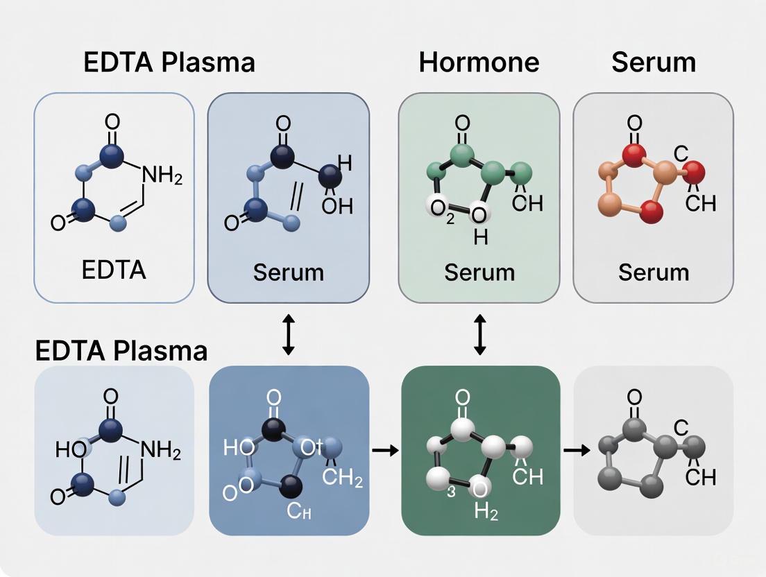
Diagram 1: EDTA Chelation and Hormone Assay Workflow. The top section illustrates the core anticoagulation mechanism where EDTA binds calcium. The bottom section outlines the experimental protocol for comparing hormone levels between sample matrices [9] [8].
Diagram 2: Troubleshooting Common EDTA Issues. This flowchart guides the diagnosis and resolution of two common problems in the lab: inhibition of enzymatic reactions and inaccuracies in hormone immunoassays, both linked to EDTA [9] [10] [11].
Documented Concentration Variances for Estradiol, Progesterone, Cortisol, and More
Troubleshooting Guides and FAQs
What is the core issue with using serum versus EDTA plasma for hormone testing?
The core issue is that the choice of sample matrix—serum or EDTA plasma—can significantly affect the measured concentration of certain hormones. These differences arise from factors such as the increased stability of some hormones in EDTA tubes and variations in how different assay methods interact with the sample matrix. Using an inappropriate matrix can lead to significant intra-individual variability or misclassification of a patient's status [14].
For which hormones does sample type cause the most significant differences?
Substantial differences have been documented for several key hormones:
- Parathyroid Hormone (PTH): Studies using the Advia Centaur platform show that EDTA plasma results can be significantly higher than serum results, with a mean difference of 13.8% and intra-individual differences as large as 25.0% observed on the same day [14].
- Insulin and C-Peptide: Recent evidence indicates that collecting blood in K₂EDTA tubes is suitable and can simplify pre-analytical handling. These analytes remain stable in EDTA whole blood at room temperature for up to 24 hours, which is particularly beneficial for resource-limited settings [13].
- Testosterone and other Steroid Hormones: While not always a direct plasma/serum difference, immunoassays for steroid hormones are notoriously susceptible to cross-reactivity from structurally similar molecules and interference from binding proteins like SHBG. This can cause inaccuracies in both serum and plasma [15] [16].
How can I prevent inaccurate hormone measurements in my study?
To ensure the reliability of your results, adhere to the following protocols:
- Define the Matrix: Choose a single, consistent sample type (e.g., EDTA plasma) for the entire study, especially for longitudinal assessments.
- Validate the Assay: Perform an on-site verification of the assay before measuring study samples. This is crucial for ensuring the method performs as expected in your specific laboratory and with your participant population [15].
- Control Pre-analytics: Standardize sample collection, processing times, and storage conditions. For PTH, delays in separation can exacerbate differences between serum and plasma [14].
- Use Quality Controls: Include internal quality controls that span the expected concentration range to monitor assay performance over time [15].
The table below summarizes documented concentration variances between EDTA plasma and serum for key analytes.
Table 1: Documented Variances Between EDTA Plasma and Serum
| Analyte | Documented Difference (EDTA Plasma vs. Serum) | Key Context / Platform |
|---|---|---|
| Intact Parathyroid Hormone (PTH) | Mean difference: +13.8% (EDTA plasma higher). Individual differences up to +25% [14]. | Advia Centaur immunoassay; difference attributed to greater stability in EDTA [14]. |
| Insulin and C-Peptide | EDTA plasma is a suitable matrix with stability at room temperature for 24 hours [13]. | Recommended for resource-limited settings; simplifies pre-analytical handling [13]. |
| Testosterone | Significant variability due to cross-reactivity and binding protein interference in immunoassays, affecting both matrices [15]. | LC-MS/MS methods are generally superior for specificity, though performance depends on laboratory expertise [15]. |
Experimental Protocol: Investigating PTH Stability
This protocol is based on a study investigating PTH differences in a routine clinical setting [14].
Objective
To assess the differences in intact-PTH concentration between serum and EDTA plasma samples using the Advia Centaur analytical platform.
Methodology
- Sample Collection: Paired blood samples are drawn from participants (e.g., patients with chronic renal failure) into both serum separator tubes and potassium EDTA tubes.
- Sample Processing: The time from sample collection to separation and freezing is recorded. In the referenced study, this time ranged from 10 to 231 minutes (median 85 minutes) to reflect routine practice.
- Analysis: Paired serum and EDTA plasma samples are analyzed in the same batch using a two-site sandwich immunoassay (e.g., Advia Centaur).
- Data Analysis:
- Perform Deming regression analysis to compare the two sample types.
- Calculate a percentage difference plot for individual samples [(Serum - EDTA) / Mean] to assess inter-individual variability.
Experimental Workflow Diagram
The following diagram illustrates the logical workflow for a method comparison experiment, as described in the experimental protocol.
Common Interference Mechanisms in Hormone Immunoassays
The diagram below outlines the primary sources of interference that can cause inaccuracies in hormone measurement, affecting both serum and plasma samples.
The Scientist's Toolkit: Research Reagent Solutions
Table 2: Essential Materials for Hormone Measurement Studies
| Item | Function in Research |
|---|---|
| K₂EDTA Tubes | Preserves blood for plasma collection; enhances stability for certain hormones like PTH and insulin [14] [13]. |
| Serum Separator Tubes | Contains a clot activator and gel for serum separation after centrifugation; commonly used but may be less stable for some hormones [14]. |
| Internal Quality Control (QC) Samples | Independent samples with known concentrations used to monitor the precision and accuracy of the assay over time [15]. |
| Liquid Chromatography-Tandem Mass Spectrometry (LC-MS/MS) | A highly specific analytical technique often considered superior to immunoassays for measuring steroid hormones due to minimal cross-reactivity [15]. |
| Automated Immunoassay Analyzer | Platform (e.g., Advia Centaur, Cobas) using antibody-based methods for high-throughput hormone testing. Susceptible to specific interferences [14] [16]. |
Frequently Asked Questions
FAQ 1: Why are my hormone concentration results different when I use EDTA plasma instead of serum? The differences are primarily due to the chemical interference of EDTA with the immunoassay process. EDTA is a powerful chelating agent that can bind to metallic ions used as tracers in many immunoassays. Furthermore, for some hormones, EDTA plasma offers greater stability, meaning the hormone degrades more slowly than in serum, which can lead to higher measured concentrations if there are delays in processing serum samples [9] [17].
FAQ 2: For which hormones is the difference between EDTA plasma and serum most pronounced? The magnitude of difference varies by hormone. Based on current evidence, the difference is particularly significant for progesterone, 17β-estradiol, testosterone, and cortisol [9] [17] [18]. The table below provides a detailed summary of the quantitative differences observed for specific hormones.
FAQ 3: Should I use serum or EDTA plasma for parathyroid hormone (PTH) measurement? EDTA plasma is strongly recommended for PTH. Multiple studies have demonstrated that PTH is significantly more stable in EDTA plasma than in serum, especially when samples are stored at room temperature for periods exceeding a few hours. This increased stability reduces pre-analytical variability and provides more reliable results [19] [20] [21].
FAQ 4: Can I overcome EDTA interference for specific hormone assays? For some hormones, interference can be mitigated. For instance, one study showed that the addition of magnesium chloride (MgCl₂) to EDTA-plasma samples negated the interference for cortisol measurements in a chemiluminescent enzyme immunoassay, bringing the values in line with those from serum. However, this corrective measure did not work for thyroxine (T4) [18]. Always consult the manufacturer's instructions for your specific assay kit.
The following table consolidates key findings from published research on the differences between hormone levels measured in EDTA plasma and serum.
Table 1: Observed Differences in Hormone Concentrations: EDTA Plasma vs. Serum
| Hormone | Population | Observed Difference (EDTA Plasma vs. Serum) | Key Findings & Statistical Significance | Source |
|---|---|---|---|---|
| 17β-Estradiol | Physically Active Females (n=25) | 44.2% higher in plasma (Median: 40.75 vs. 28.25 pg/ml) | Strong positive correlation (r=0.72); P < 0.001 | [9] |
| Progesterone | Physically Active Females (n=25) | 78.9% higher in plasma (Median: 1.70 vs. 0.95 ng/ml) | Strong positive correlation (r=0.89); P < 0.001 | [9] |
| Estradiol (E2) | Human Outpatients (n=30) | Markedly higher in plasma (Median: 2480 vs. 25.6 pg/ml) | Statistically significant (P < 0.05) | [17] |
| Testosterone | Human Outpatients (n=30) | Markedly higher in plasma (Median: 687 vs. 31.7 ng/dL) | Statistically significant (P < 0.05) | [17] |
| Progesterone | Human Outpatients (n=30) | Markedly higher in plasma (Median: 38 vs. 0.3 ng/mL) | Statistically significant (P < 0.05) | [17] |
| Cortisol | Dogs (n=50) | 51.2% higher in EDTA-plasma | P < 0.001 | [18] |
| Thyroxine (T4) | Dogs (n=50) | 43.7% higher in EDTA-plasma | P < 0.001 | [18] |
| Intact PTH | Humans with Chronic Renal Failure (n=26) | Plasma concentrations lower than serum | Deming regression: serum = 0.8927 EDTA – 0.447; Mean difference 13.8% | [20] |
Experimental Protocols for Key Studies
Study 1: 17β-Estradiol and Progesterone in Physically Active Females [9]
- Objective: To determine whether concentrations of 17β-estradiol and progesterone, as measured by immunoassay, differ between plasma and serum.
- Participant Cohort: 25 recreationally active/trained females, including 13 with a natural menstrual cycle and 12 using combined oral contraceptives.
- Blood Collection: Venous blood was sampled from an antecubital vein after 30 minutes of supine rest. Blood was drawn into paired EDTA (K2) and gold serum separator tubes (SST).
- Sample Processing:
- Plasma (EDTA tube): Centrifuged at 3500g at 4°C for 10 minutes immediately after collection.
- Serum (SST tube): Left to clot for 15 minutes at room temperature before being centrifuged.
- All aliquots were stored at -80°C until analysis.
- Hormone Analysis: 17β-estradiol and progesterone concentrations were determined in duplicate using competitive immunoenzymatic assays (Abcam: ab108667 and ab108670).
Study 2: Effect of Anticoagulants on Multiple Hormone Assays [17]
- Objective: To verify the effect of EDTA and sodium citrate on hormone assays performed by fluorometric (FIA) or immunofluorometric (IFMA) methods.
- Participant Cohort: 30 human outpatients (11 men, 19 women).
- Blood Collection: Blood was drawn into three Vacutainer tube types: serum separation tube (SST), K3 EDTA tube, and 0.129 mol/L buffered sodium citrate tube.
- Sample Processing: Serum or plasma was separated from blood cells immediately after drawing by centrifugation at 2000 g for 5 minutes.
- Hormone Analysis: Samples were analyzed on the automated AutoDelfia platform for 15 different hormones, including LH, FSH, prolactin, GH, TSH, insulin, estradiol, progesterone, and testosterone.
Workflow and Mechanism Diagrams
Sample Processing and Analysis Workflow
This diagram illustrates the parallel processing paths for serum and plasma samples in a typical comparative study.
Mechanism of EDTA Interference in Immunoassays
This diagram outlines the proposed mechanisms by which EDTA causes higher hormone readings in different assay types.
The Scientist's Toolkit: Essential Research Reagents & Materials
Table 2: Key Materials and Reagents for EDTA Plasma vs. Serum Hormone Studies
| Item | Function / Role in Research | Example from Literature |
|---|---|---|
| K₂ or K₃ EDTA Vacutainer Tubes | Anticoagulant blood collection tube; prevents clotting by chelating calcium. Essential for plasma preparation. | K2 EDTA vacutainers were used to collect plasma samples [9]. |
| Serum Separator Tubes (SST) | Contains a gel barrier and clot activator; used for clean serum separation after centrifugation. | Gold-top SST vacutainers were used for serum collection [9]. |
| Competitive Immunoenzymatic Assay Kits | Used to quantify specific hormone concentrations (e.g., 17β-estradiol, progesterone) in plasma and serum samples. | Abcam kits (ab108667 for E2, ab108670 for progesterone) [9]. |
| Automated Immunoassay Analyzer | Platform for performing fluoroimmunometric or chemiluminescent assays with high throughput and precision. | AutoDelfia (PerkinElmer) platform [17]; Immulite 1000 (Siemens) [18]. |
| Magnesium Chloride (MgCl₂) | Used in troubleshooting to counteract EDTA interference in certain chemiluminescent assays (e.g., for cortisol). | Added to EDTA-plasma to a final concentration of 5 mmol/L to negate interference [18]. |
Frequently Asked Questions (FAQs)
FAQ 1: Why are my progesterone concentrations significantly higher when I use EDTA plasma tubes compared to serum separator tubes?
Your observation is consistent with established research. A 2025 study that directly compared sample matrices found that median progesterone concentrations were 78.9% higher in EDTA plasma (1.70 ng/ml) compared to serum (0.95 ng/ml) [9] [22]. This is due to fundamental differences in tube chemistry and sample processing. Serum requires clot formation, which can trap some analytes or lead to proteolytic degradation, whereas EDTA plasma uses an anticoagulant to preserve the sample, potentially yielding more complete recovery of certain hormones [9].
FAQ 2: Can I use the same reference ranges for serum and EDTA plasma samples when classifying menstrual cycle phases?
No, you should not use the same reference ranges. The same study demonstrated that 17β-estradiol concentrations were 44.2% higher in EDTA plasma than in serum [9] [22]. Because hormone concentrations are systematically different between these matrices, applying serum-based reference ranges to plasma samples will lead to misclassification of menstrual cycle phases (e.g., follicular vs. luteal). Researchers must establish or use reference ranges specific to the sample matrix they are using to ensure accurate participant classification [9].
FAQ 3: My samples cannot be centrifuged immediately. Is EDTA plasma or serum more stable for hormone assays?
EDTA plasma is generally more tolerant of processing delays. For hormones like 17β-estradiol and progesterone, EDTA plasma is preferable if processing is not immediate [9]. Furthermore, stability studies for other hormones support the robustness of EDTA tubes. For instance, insulin and C-peptide in EDTA whole blood are stable at room temperature for up to 24 hours [13]. Adrenocorticotropic hormone (ACTH) in EDTA plasma is also stable at room temperature for at least 6 hours [23].
FAQ 4: Despite the concentration differences, are plasma and serum measurements at least correlated?
Yes, they show strong correlation. Although absolute concentrations differ, the measurements from the two matrices are highly correlated. For 17β-estradiol, the correlation coefficient (r) is 0.72, and for progesterone, it is 0.89 [9] [22]. This strong positive correlation indicates that both matrices are suitable for tracking relative hormonal changes and for biomarker analysis, provided the consistent bias is accounted for [9].
Troubleshooting Guides
Problem: Inconsistent Hormone Levels Leading to Incorrect Cycle Phase Classification
Potential Cause and Solution:
- Cause: The most likely cause is applying inclusion/exclusion criteria or reference ranges developed for serum to EDTA plasma samples, or vice-versa.
- Solution: Implement matrix-specific thresholds. The table below summarizes the quantitative biases to inform your criteria adjustments [9] [22]:
Table 1: Measured Bias Between EDTA Plasma and Serum Hormone Concentrations
| Hormone | Sample Matrix | Median Concentration | Measured Bias (Plasma vs. Serum) |
|---|---|---|---|
| 17β-estradiol | EDTA Plasma | 40.75 pg/mL | +44.2% |
| Serum | 28.25 pg/mL | ||
| Progesterone | EDTA Plasma | 1.70 ng/mL | +78.9% |
| Serum | 0.95 ng/mL |
Problem: High Background or Imprecise Results in Immunoassays
Potential Causes and Solutions:
- Cause 1: Improper reagent handling or plate washing.
- Solution: Ensure all reagents are fresh, properly mixed, and volumes are accurately dispensed. Check that the plate washer is functioning correctly, with no clogged tubes, to avoid uneven washing that causes high background noise [24].
- Cause 2: Inconsistent incubation conditions.
- Solution: Use plate sealers during incubation steps. Do not stack plates in the incubator, as this can create uneven temperature distribution and "edge effects." Incubate the substrate in the dark [24].
- Cause 3: Non-specific binding.
- Solution: Always include the recommended controls (e.g., Blank, Zero Concentration, and Non-Specific Binding controls) to assess background signal contributions. Use a high-quality blocking buffer [24].
Experimental Protocols
Core Protocol: Parallel Blood Collection for Serum and Plasma Hormone Comparison
This methodology is adapted from the 2025 study by Rowland et al. [9].
1. Materials and Reagents (Research Reagent Solutions)
Table 2: Essential Materials for Hormone Concentration Comparison Studies
| Item | Function | Example/Note |
|---|---|---|
| EDTA (K2) Vacutainers | Anticoagulant tube for plasma collection; chelates calcium to prevent clotting. | BD Vacutainer [9] [13]. |
| Serum Separator Tubes (SST) | Tube for serum collection; contains a gel separator. | Gold-top SST vacutainers [9]. |
| Competitive Immunoenzymatic Assay Kit | For quantifying hormone levels. | Kits from manufacturers like Abcam [9]. |
| Microplate Reader | To measure optical density (OD) in the assay. | - |
| Centrifuge | For separating plasma/serum from cells. | Capable of 3500g [9]. |
| -80°C Freezer | For long-term storage of sample aliquots. | - |
2. Step-by-Step Procedure
- Participant Preparation: After 30 minutes of supine rest, apply a tourniquet to the upper arm [9].
- Venous Blood Sampling: Draw blood from an antecubital vein via venepuncture. Collect blood into both EDTA and serum separator tubes [9].
- Sample Processing:
- EDTA Plasma: Centrifuge tubes at 3500g at 4°C for 10 minutes. Extract the plasma layer and aliquot it into cryovials. Store at -80°C [9].
- Serum: Allow the serum tube to clot at room temperature for 15 minutes. Centrifuge under the same conditions as plasma. Extract the serum, aliquot, and store at -80°C [9].
- Hormone Analysis: Measure hormone concentrations in duplicate using a competitive immunoenzymatic assay, strictly following the manufacturer's instructions for both plasma and serum samples [9].
- Data Analysis: Use statistical tests (e.g., Wilcoxon matched-pairs signed-rank test for non-normally distributed data) and Bland-Altman plots to assess the agreement and bias between the two matrices [9].
Workflow Diagram: Serum vs. EDTA Plasma Processing
From Theory to Practice: Selecting the Right Matrix for Your Hormone Assay
A technical support center for researchers navigating the complexities of biofluid matrix selection in endocrine research.
Frequently Asked Questions
Q1: Why does the choice between serum and plasma matter for hormone testing?
The choice of matrix (serum or plasma) is a critical pre-analytical variable that can significantly influence measured hormone concentrations. Different tube chemistries can affect the stability of the analyte, the presence of interfering substances, and the efficiency of the assay itself. Using an inappropriate matrix can lead to inaccurate results, potentially compromising study conclusions and diagnostic accuracy [16] [15].
Q2: My immunoassay kit says it is validated for both serum and plasma. Can I use them interchangeably in my study?
No, you should not assume interchangeability without conducting your own verification. While many commercial kits claim compatibility with multiple matrices, significant concentration differences have been documented. For instance, a 2025 study found that concentrations of 17β-estradiol and progesterone were markedly higher in EDTA plasma compared to serum [22]. To ensure data consistency, you must select a single matrix type for your entire study and validate the assay performance in that specific matrix [15].
Q3: What are the key factors to consider when selecting a collection tube for a specific hormone panel?
Your decision should be guided by the hormone's stability, the assay methodology, and your research question. Consider the following:
- Hormone Stability: Some hormones, like ACTH, are known to be labile. However, evidence shows that with appropriate preservatives like EDTA, ACTH in plasma is stable at room temperature for at least 6 hours [23].
- Assay Interference: The additives in blood collection tubes can interfere with certain assay chemistries. For example, EDTA can chelate metallic ions used as labels in some immunoassays, potentially suppressing the signal [16].
- Research Context: Always consult the latest literature for your specific hormone of interest. If your research involves participant classification based on hormone thresholds (e.g., for inclusion/exclusion criteria), the documented matrix bias must be accounted for to avoid misclassification [22].
Q4: I am seeing inconsistent results between my sample replicates. Could the collection tube be the cause?
Yes, inconsistent results can stem from pre-analytical factors. To troubleshoot:
- Verify Sample Processing: Ensure adherence to sample processing protocols, including centrifugation speed, time, and storage temperature [23].
- Check for Clots: In plasma tubes, the presence of micro-clots can cause variability. Ensure complete mixing with the anticoagulant immediately after collection.
- Audit Tube Lot Numbers: Variation between different lots of collection tubes, while uncommon, can occur. Note the lot numbers for all samples.
- Confirm Matrix Suitability: Re-evaluate the validation data for your specific assay and the chosen matrix. The problem may be due to an undetected matrix effect [15].
Troubleshooting Guides
Guide 1: Addressing Matrix-Induced Concentration Bias
Problem: Measured hormone concentrations are consistently and significantly biased when comparing data from studies that used different matrices, complicating meta-analyses and cross-study comparisons.
Investigation & Solution:
- Action 1: Literature Review: Before starting your study, investigate if a matrix bias has already been established for your target analytes. For example, a recent study provides clear quantitative data on the differences for reproductive hormones [22].
- Action 2: Conduct a Bridging Study: If no data exists, run a small validation experiment. Collect paired serum and plasma samples from a subset of participants (e.g., n=20-30) and measure the hormones in both matrices using the same assay.
- Action 3: Apply a Correction Factor (if justified): If a strong, consistent correlation and bias are found, you may develop a lab-specific correction factor for comparative purposes. However, this should be done with caution and clearly stated in all publications [22].
Table 1: Documented Concentration Differences Between EDTA Plasma and Serum
| Hormone | Median Concentration in Plasma | Median Concentration in Serum | Percentage Difference | P-value |
|---|---|---|---|---|
| 17β-estradiol | 40.75 pg/mL | 28.25 pg/mL | 44.2% Higher in Plasma | < 0.001 |
| Progesterone | 1.70 ng/mL | 0.95 ng/mL | 78.9% Higher in Plasma | < 0.001 |
Data adapted from Rowland et al. (2025), Exp Physiol [22].
Guide 2: Managing Hormone Stability in Clinical Settings
Problem: Strict laboratory handling requirements (e.g., immediate freezing) for unstable hormones like ACTH and renin limit testing to hospital settings and can lead to sample rejection [23].
Investigation & Solution:
- Action 1: Validate Extended Stability: Research indicates that with the correct preservatives, these hormones are more stable than previously thought.
- Action 2: Optimize Tube Type and Timeline: Use EDTA plasma for ACTH and serum gel tubes for renin and aldosterone. Data shows these analytes are stable at room temperature for up to 6 hours, allowing for transportation from outpatient or emergency rooms to a central lab [23].
- Action 3: Update SOPs: Implement a standard operating procedure that defines this 6-hour processing window, facilitating broader testing access.
Table 2: Room Temperature Stability of Key Hormones
| Hormone | Recommended Matrix | Stability at Room Temperature | Mean Change at 6h (95% CI) |
|---|---|---|---|
| ACTH | EDTA Plasma | ≥ 6 hours | -2.6% (-9.7 to 4.5) |
| Aldosterone | Serum Gel | ≥ 6 hours | +0.2% (-3.6 to 4.0) |
| Renin | Serum Gel | ≥ 6 hours | -1.9% (-7.0 to 3.2) |
Data from a study of 31 participants [23].
Experimental Protocols
Protocol: Method Comparison for Matrix Bias Investigation
Aim: To determine the concentration bias and agreement between serum and EDTA plasma matrices for the measurement of specific steroid hormones.
Materials:
- Research Reagent Solutions:
- EDTA Vacutainer tubes (e.g., Lavender top)
- Serum Gel Vacutainer tubes (e.g., Gold top)
- Equipment for venipuncture
- Centrifuge
- Freezer (-80°C)
- Competitive immunoenzymatic assay kit for target hormone(s)
Methodology:
- Participant Recruitment: Recruit participants matching your study population. The example study used 25 physically active females [22].
- Blood Collection: Draw venous blood from each participant into both EDTA and serum gel tubes simultaneously.
- Sample Processing:
- Plasma (EDTA tube): Centrifuge within 6 hours of collection. Aliquot the supernatant plasma and freeze at -80°C [23].
- Serum (Gel tube): Allow blood to clot, then centrifuge. Aliquot the supernatant serum and freeze at -80°C.
- Hormone Analysis: Analyze all samples (paired plasma and serum) in the same analytical run to minimize inter-assay variation. Use a validated competitive immunoenzymatic assay [22].
- Data Analysis:
- Use non-parametric tests (e.g., Wilcoxon signed-rank test) to assess if median concentration differences are statistically significant.
- Calculate Pearson's correlation coefficient (r) to evaluate the strength of the relationship.
- Perform Bland-Altman analysis to determine the mean bias and limits of agreement between the two matrices.
The workflow for this investigation is outlined below:
Method Comparison Workflow
The Scientist's Toolkit
Table 3: Essential Materials for Matrix Comparison Studies
| Item | Function in Experiment |
|---|---|
| EDTA Vacutainer Tubes | Contains anticoagulant (K2/K3 EDTA) to prevent clotting; produces plasma for analysis [22]. |
| Serum Gel Separator Tubes | Contains a clot activator and a gel barrier; produces serum after centrifugation [22]. |
| Competitive Immunoenzymatic Assay | A common method for quantifying small molecules like steroid hormones; used to measure concentrations in plasma and serum samples [22]. |
| -80°C Freezer | For long-term storage of processed plasma and serum aliquots to preserve hormone integrity. |
| Statistical Software (e.g., R, SPSS) | To perform correlation analyses (e.g., Pearson's r) and agreement statistics (e.g., Bland-Altman plots) [22]. |
The decision-making process for selecting the appropriate biofluid matrix is summarized in the following flowchart:
Matrix Selection Decision Guide
Frequently Asked Questions
1. Why do my hormone results differ between serum and plasma samples? Differences are often due to the chemical interaction between anticoagulants and the assay's detection method. EDTA, a powerful chelating agent, can bind to metallic ions that are constituents of chemiluminescent or fluorescent labels used in immunoassays. Furthermore, hormones like intact PTH, insulin, and C-peptide are more stable in EDTA plasma because the anticoagulant inhibits degrading enzymes, leading to more reliable results, especially when sample processing is delayed [14] [17] [13].
2. When is EDTA plasma recommended over serum for hormone testing? EDTA plasma is strongly recommended for specific tests and settings. Evidence supports its use for:
- Intact Parathyroid Hormone (PTH): EDTA provides increased stability, reducing intra-individual variability that can exceed 25% when compared to serum [14].
- Insulin and C-Peptide: These molecules are highly stable in K2EDTA whole blood, even when stored at room temperature for up to 24 hours, making EDTA tubes ideal for resource-limited settings [13].
- General Practice: To minimize the impact of common delays in sample separation and processing encountered in routine clinical practice [14].
3. My ELISA kit says it's validated for serum and plasma. Can I use the results interchangeably? No, you should not use the results interchangeably. Even if a kit is validated for both matrices, the results are specific to the sample type. Consistent use of the same sample type (either serum or plasma) is critical for the accurate serial monitoring of a patient. Switching between tube types for the same patient can introduce significant variability and lead to clinical misclassification [14].
4. How do I validate a sample type if it's not listed in the manufacturer's instructions? Perform a spike-and-recovery experiment. This involves spiking a known concentration of the recombinant target protein into your specific sample matrix (e.g., EDTA plasma) and into the matrix recommended by the kit (e.g., serum). After running the ELISA, calculate the percentage recovery in your sample. An average recovery of 80–120% generally indicates that components in your sample matrix are not interfering with the assay [25] [26].
Troubleshooting Guides
Problem: Inconsistent or Erratic Hormone Results
| Potential Cause | Diagnostic Steps | Recommended Solution |
|---|---|---|
| Incorrect Sample Type | Review patient records and tube types. Check if results are inconsistently high/low for specific patients or batches. | Standardize sample collection protocol. Use EDTA plasma for hormones like PTH, insulin, and C-peptide where evidence supports its superiority [14] [13]. |
| Delayed Sample Processing | Audit the time from sample collection to centrifugation and freezing. | Implement a strict processing protocol. For serum, ensure clotting time is minimized. For stability, EDTA plasma is more forgiving of delays [14] [27]. |
| Improper Sample Handling | Review freeze-thaw cycle records. Check storage temperature logs. | Aliquot samples to avoid repeated freeze-thaw cycles. Store at recommended temperatures (e.g., -20°C or lower). Enzymes are particularly susceptible to degradation over time [27]. |
| Matrix Interference | Perform a spike-and-recovery experiment in the sample matrix of interest. | If recovery is outside 80-120%, consider using a different kit or sample type. The assay's buffers may not be optimized for your specific matrix [25]. |
Problem: Poor Assay Performance or Validation Failure
| Potential Cause | Diagnostic Steps | Recommended Solution |
|---|---|---|
| Anticoagulant Interference | Compare standard curves generated in serum vs. plasma. Check for cross-reactivity data in the kit insert. | Adhere strictly to the manufacturer's validated sample types. Be aware that EDTA can cause falsely elevated values in FIA and lower results in IFMA methods [17]. |
| Low Precision (High %CV) | Calculate intra-assay and inter-assay Coefficient of Variation (CV). | Ensure consistent pipetting technique. Cover plates during incubations to prevent well drying. Maintain a stable incubation temperature. Intra- and inter-assay CV should ideally be <10% [25] [26]. |
| Non-Linear Dilutions | Serially dilute a high-concentration sample and plot measured vs. expected values. | Check for sample matrix effects. Ensure the diluent specified in the kit manual is used. Results for each dilution should be 70–130% of the expected value [25]. |
Experimental Data & Evidence
The following table summarizes key findings from studies investigating sample type effects on hormone measurements.
Table 1: Impact of Sample Type on Hormone Measurement Results
| Analyte | Assay Method | Key Finding: Serum vs. EDTA Plasma | Reference |
|---|---|---|---|
| Intact PTH | Advia Centaur (Chemiluminometric) | EDTA plasma results were more stable. A mean difference of 13.8% was observed, with intra-individual differences as large as 25%. | [14] |
| Insulin (INS) | Immunofluorometric (IFMA) | Results in EDTA plasma were significantly lower, often below the detection limit. | [17] |
| C-Peptide (CPEP) | Immunofluorometric (IFMA) | Results in EDTA plasma were significantly lower. | [17] |
| Estradiol (E2) | Fluorometric (FIA) | Results in EDTA plasma were drastically higher. | [17] |
| Testosterone | Fluorometric (FIA) | Results in EDTA plasma were drastically higher. | [17] |
| Insulin & C-Peptide | Immunoassay | No significant degradation in K2EDTA tubes stored at room temperature for 24 hours. Ideal for resource-limited settings. | [13] |
Detailed Protocol: Establishing Sample Type Suitability
This protocol is adapted from stability studies and ELISA validation principles [25] [13].
Objective: To verify the suitability of EDTA plasma for measuring a specific hormone (e.g., Insulin) compared to the standard serum sample.
Workflow Diagram
Materials:
- Participants: Consented adults.
- Blood Collection: Venous blood sample (e.g., 40ml).
- Collection Tubes: K2EDTA tubes and plain serum tubes.
- Equipment: Centrifuge, -20°C freezer, calibrated thermometer, cool box with ice packs.
- Assay Kit: Validated insulin/C-peptide immunoassay.
Method:
- Collection: Collect venous blood and dispense it into multiple K2EDTA and serum tubes. Invert tubes gently as per manufacturer instructions (e.g., 8-10 times for EDTA tubes).
- Baseline Processing: Immediately centrifuge a set of EDTA and serum tubes ("time zero"). For serum tubes, allow 30 minutes for clotting first. Aliquot and freeze at -20°C as a reference.
- Delayed Processing: Store the remaining EDTA and serum tubes under different conditions:
- Room Temperature
- Cool Box with ice packs (to simulate field conditions)
- Time-Point Sampling: At pre-defined time points (e.g., 2, 6, 12, and 24 hours), remove tubes from each storage condition, centrifuge, prepare aliquots, and store at -20°C.
- Batch Analysis: Analyze all aliquots from all time points and conditions in a single batch to minimize inter-assay variation.
- Data Analysis: Compare the measured hormone concentrations at each time point and storage condition against the baseline (time zero) concentration. Stability is generally defined as a change of less than 10-15% from baseline.
The Scientist's Toolkit
Table 2: Essential Research Reagents and Materials
| Item | Function in Sample Type Validation |
|---|---|
| K2EDTA Vacutainer Tubes | Anticoagulant blood collection tube that chelates calcium; preserves labile hormones like insulin and PTH by inhibiting enzymatic degradation. |
| Serum Separator Tubes (SST) | Tubes containing a clot activator and gel separator; yield serum, the traditional matrix for many hormone assays. |
| Standard Dilution Buffer | A defined matrix provided with ELISA kits; used to create the standard curve and dilute samples to check for linearity and parallelism. |
| Recombinant Target Protein | A purified form of the analyte; essential for performing spike-and-recovery experiments to test for matrix interference. |
| Temp-Chex / Data Loggers | Single-use or reusable temperature monitoring devices; critical for validating storage conditions during stability studies. |
Core Findings: EDTA Plasma vs. Serum Hormone Concentrations
For researchers investigating hormone levels, the choice of blood collection matrix is a critical pre-analytical factor. Evidence consistently shows that measured concentrations of key steroid hormones are significantly higher in EDTA plasma than in serum [9] [22].
The table below summarizes the quantitative differences observed for 17β-estradiol and progesterone.
Table 1: Comparison of Hormone Concentrations in EDTA Plasma vs. Serum
| Hormone | Median EDTA Plasma Concentration | Median Serum Concentration | Percentage Increase in Plasma | Statistical Significance (P-value) |
|---|---|---|---|---|
| 17β-estradiol | 40.75 pg/mL | 28.25 pg/mL | 44.2% higher | < 0.001 |
| Progesterone | 1.70 ng/mL | 0.95 ng/mL | 78.9% higher | < 0.001 |
Despite these concentration differences, strong positive correlations exist between the two matrices (Spearman's r = 0.72 for 17β-estradiol and r = 0.89 for progesterone, P < 0.001 for both), confirming that both are suitable for biomarker analysis [9]. However, the lack of statistical equivalence means that applying consistent inclusion/exclusion criteria across matrices could lead to misclassification of participants. Researchers must account for the systematically higher concentrations when using EDTA plasma.
Detailed Experimental Protocol: Direct Comparison of Plasma and Serum
The following methodology provides a template for experiments designed to compare hormone concentrations between different sample matrices [9].
Participant Cohort
- Population: Recruit a defined group (e.g., n=25 young, physically active females).
- Grouping: Include subgroups such as:
- Females with a regular, natural menstrual cycle (verified by calendar tracking and urinary luteinizing hormone surge tests).
- Females using combined monophasic oral contraceptive pills.
Blood Collection and Processing
- Collection: Draw venous blood from an antecubital vein after 30 minutes of supine rest.
- Tubes: Use matched EDTA (K2) and serum separator tubes (SST) for each draw.
- Processing:
- Plasma (EDTA tube): Centrifuge at 3500g at 4°C for 10 minutes. Extract and aliquot plasma promptly. Store at -80°C.
- Serum (SST tube): Allow blood to clot for 15 minutes at room temperature. Then centrifuge, aliquot, and store at -80°C.
Hormone Analysis
- Technique: Use competitive immunoenzymatic assays performed in duplicate.
- Example Assays: Commercial kits for 17β-estradiol (e.g., Abcam ab108667) and progesterone (e.g., Abcam ab108670).
- Quality Control: Report intra-assay coefficients of variation (e.g., 3.4-3.6% for 17β-estradiol, 2.4-3.0% for progesterone).
Pre-Analytical Stability of Hormones and Related Analytes
The stability of an analyte after blood draw but before analysis is a major source of variability. Adherence to defined stability windows is essential for data integrity.
Table 2: Stability of Hormones and Related Analytes in Whole Blood and Serum
| Analyte | Sample Type | Established Stability Conditions | Key Findings |
|---|---|---|---|
| ACTH | EDTA Plasma | Room Temperature (RT) | Stable for at least 6 hours in whole blood (mean change -2.6%) [23]. |
| Aldosterone & Renin | Serum Gel Tube | Room Temperature (RT) | Stable for at least 6 hours in whole blood (mean change +0.2% and -1.9%, respectively) [23]. |
| IGF-1 | Serum Gel Tube | Various Temperatures | Stable for at least 72 hours regardless of delayed centrifugation or storage temperature. Stability extends to 168 hours (7 days) at 4°C, and 672 hours (28 days) at -20°C [28]. |
| Lactate Dehydrogenase (LDH) | Serum | Time & Age-dependent | Significantly affected by 2h and 24h incubation at 20-24°C. Blood from older individuals (60-75 years) may be more vulnerable to preparation conditions than from younger individuals (20-35 years) [29]. |
Troubleshooting Guide & FAQs
Q1: My hormone values are consistently higher than expected. Could my sample type be the cause? Yes. If you are using EDTA plasma, your measured values for 17β-estradiol and progesterone are expected to be significantly higher (44-79% in one study) than if you were using serum [9]. First, verify your collection tube type and ensure your reference ranges are appropriate for your chosen matrix.
Q2: I need to batch process samples. What is the maximum time I can leave blood samples at room temperature before centrifuging them for hormone assay? The safe time window depends on the analyte:
- For ACTH, aldosterone, and renin, you have at least 6 hours at room temperature [23].
- For IGF-1, samples are stable for up to 24 hours at room temperature before centrifugation [28].
- As a general precaution, processing samples as soon as possible is always recommended to minimize potential degradation.
Q3: After centrifugation, how long can I store serum/plasma extracts for hormone testing? Stability is highly dependent on storage temperature.
- For short-term storage (up to 3 days), serum for IGF-1 analysis can be kept at 4°C, 20-25°C, or 30°C [28].
- For medium-term storage (up to 7 days), refrigerate serum for IGF-1 at 4°C [28].
- For long-term storage, freeze extracts at -20°C or lower. Serum IGF-1 is stable for at least 28 days at -20°C [28]. Always avoid repeated freeze-thaw cycles.
Q4: Are quick-clotting serum tubes reliable for hormone testing? Yes, for many analytes. A 2025 evaluation of a thrombin-based quick-clotting SST (VQ-Tube SST) found comparable performance to conventional SSTs for a broad panel of chemistry and immunology measurands after a 5-minute clotting time [30]. However, always validate the performance for your specific hormone assays.
Experimental Workflow Diagram
The following diagram illustrates the critical decision points in sample processing to ensure hormone stability.
The Scientist's Toolkit: Key Research Reagent Solutions
Table 3: Essential Materials for Hormone Stability and Comparison Studies
| Item | Function / Application | Example from Literature |
|---|---|---|
| K2 EDTA Tubes | Anticoagulant for plasma collection; yields higher concentrations of 17β-estradiol and progesterone compared to serum. | BD Vacutainer K2EDTA Tube [9] [30] |
| Serum Separator Tubes (SST) | Tube with clot activator and gel barrier for serum collection; the conventional matrix for many hormone immunoassays. | BD Vacutainer SST II Advance Tube [9] [30] |
| Quick-Clotting SST | SST containing thrombin to reduce clotting time to ~5 minutes, improving workflow efficiency. | VQ-Tube SST (Thrombin-based) [30] |
| Competitive Immunoenzymatic Assays | For quantitative measurement of steroid hormones (e.g., 17β-estradiol, progesterone) in plasma and serum. | Abcam Kits (ab108667, ab108670) [9] |
| Solid-Phase Extraction (SPE) Sorbents | Purification and enrichment of hormone extracts from complex matrices; removes interfering proteins and lipids. | Oasis-HLB Copolymer [31] |
| Derivatization Reagents | For GC-MS analysis; increases volatility and detection sensitivity of steroid hormones. | BSTFA + TMCS [31] |
Troubleshooting Guides
Guide 1: Addressing Cross-Signal Contribution (Cross-Talk) in LC-MS/MS
Problem: Unexpected peaks appear in the multiple reaction monitoring (MRM) channel of one analyte after injecting a different, supposedly pure compound. This can lead to inaccurate quantification, especially in multiplexed assays [32].
Solution Flowchart:
Detailed Troubleshooting Steps:
- Confirm Co-elution: Check retention times of the unexpected peak and the authentic standard of compound B. If they differ, the interference is not from B itself [32].
- Check Standard Purity: Inject a blank solvent to rule out carryover. Then, analyze the stock and working solutions of compound A for the presence of B. In one case study, morphine glucuronide standards were contaminated with the parent drug, requiring adjustment of the quantification range [32].
- Assess Structural Relationships: If A and B are related (e.g., a drug and its metabolite), in-source fragmentation or conversion can occur. Review source conditions (e.g., declustering potential) [32] [33].
- Evaluate Stable Isotope-Labeled Internal Standards (SIL-IS): A common issue is the unlabeled analyte contributing to the SIL-IS signal or vice versa due to insufficient purity of the SIL-IS or isotopic instability in storage solvents [32].
Guide 2: Mitigating Matrix Interference and Ion Suppression
Problem: Reduced or variable analyte signal caused by co-eluting matrix components, leading to poor sensitivity and inaccurate quantification [34] [35].
Solution Flowchart:
Detailed Troubleshooting Steps:
- Post-Column Infusion Study: During method development, continuously infuse analyte into the mass spectrometer while injecting a prepared blank matrix extract. This identifies regions of ion suppression in the chromatogram, allowing you to adjust the gradient to elute analytes in "clean" zones [33].
- Quantitative Matrix Effect Evaluation: Spike analyte into at least six different lots of matrix and compare the signal to that in a pure solvent. A signal difference >15% indicates a significant matrix effect. Use a stable isotope-labeled internal standard that co-elutes perfectly with the analyte to compensate for this effect [33].
- Simplify Sample Preparation: For complex matrices like avocados or blood, a simplified "dilute-and-shoot" protocol can be sufficient when using a robust LC-MS/MS system designed to handle dirtier samples, reducing preparation time and potential errors [34].
Frequently Asked Questions (FAQs)
FAQ 1: Why should I consider switching from immunoassay to LC-MS/MS for hormone testing?
LC-MS/MS offers superior specificity by separating and detecting analytes based on their mass, unlike immunoassays which rely on antibody binding and are susceptible to cross-reactivity with structurally similar compounds [36]. This is critical for accurately measuring small molecules like hormones (e.g., cortisol, estradiol) in complex matrices. LC-MS/MS also allows for simultaneous quantification of multiple analytes and has a wider dynamic range [35] [37].
FAQ 2: My immunoassay and LC-MS/MS results for the same hormone sample disagree. What is the likely cause?
This is a common issue. Immunoassays can overestimate concentrations due to cross-reacting substances. For example, a study on urinary free cortisol found that while immunoassays correlated strongly with LC-MS/MS, they showed a consistent positive bias, requiring method-specific cut-off values for accurate diagnosis [36]. LC-MS/MS provides more accurate results by physically separating these interferents.
FAQ 3: How does the choice of blood collection tube (e.g., serum vs. EDTA plasma) affect my hormone results?
The matrix itself can significantly influence measured concentrations. A 2025 study found that concentrations of 17β-estradiol and progesterone were 44.2% and 78.9% higher, respectively, in EDTA plasma compared to serum from the same individuals [9] [22]. This underscores that serum and plasma are not interchangeable matrices. Researchers must use matrix-specific reference ranges and consistently report the sample type used.
FAQ 4: What are the key quality control metrics I should monitor in every LC-MS/MS run to detect interference?
Continuously monitor these three data quality metrics [33]:
- Ion Ratios: The ratio of quantifier to qualifier ions for each analyte should be consistent.
- Internal Standard Area: Significant deviation can indicate a matrix effect or problem.
- Retention Time: Should be stable; shifts may suggest chromatographic issues.
Experimental Protocols
Protocol 1: Method for Comparing Hormone Concentrations in Serum vs. EDTA Plasma
This protocol is adapted from research comparing 17β-estradiol and progesterone levels [9] [22].
1. Sample Collection:
- Collect venous blood from participants using both EDTA (K2) and serum separator (SST) vacuum tubes.
- Invert tubes 10 times gently to mix additives.
2. Sample Processing:
- EDTA Plasma: Centrifuge at 3500g at 4°C for 10 minutes. Aliquot plasma immediately and store at -80°C.
- Serum: Allow the SST tube to clot at room temperature for 15-30 minutes. Centrifuge as above, aliquot serum, and store at -80°C.
3. Analysis:
- Analyze hormones using a validated competitive immunoenzymatic assay (or LC-MS/MS for higher specificity) according to manufacturer instructions.
- Perform all measurements in duplicate.
4. Data Analysis:
- Use Spearman's rank correlation to assess the relationship between plasma and serum concentrations.
- Apply Wilcoxon matched-pairs signed-rank test to evaluate significant differences between matrices.
- Perform Bland-Altman analysis to determine the mean bias and limits of agreement.
Protocol 2: LC-MS/MS Method for Simultaneous Quantification of Analytes in Microvolume Whole Blood
This protocol is adapted from a validated method for immunosuppressants [37] and can be adapted for hormone panels.
1. Sample Preparation:
- Use a minimal volume of whole blood (e.g., 2.8 μL).
- Add a stabilizing agent or internal standard solution.
- Perform protein precipitation or solid-phase extraction.
2. LC-MS/MS Analysis:
- Chromatography: Use a reversed-phase C18 column. Employ a gradient elution with mobile phases A (water/volatile buffer) and B (organic solvent like methanol or acetonitrile) to achieve optimal separation [35] [37].
- Mass Spectrometry:
- Ionization: Use electrospray ionization (ESI) in positive or negative mode, optimized for the target analytes.
- Data Acquisition: Operate in Multiple Reaction Monitoring (MRM) mode. Monitor at least two transitions per analyte for confirmation.
3. Data Processing and Quantification:
- Use a linear calibration curve with a coefficient of determination (R²) >0.99.
- Apply hematocrit correction if estimating plasma-equivalent concentrations from whole blood [37].
- Accept accuracy within ±15% and precision with relative standard deviations (RSDs) <10% for quality control samples.
Data Presentation
Table 1: Comparison of Hormone Concentrations in EDTA Plasma vs. Serum
Data from Rowland et al. 2025 (n=25 physically active females) [9] [22]
| Hormone | Median Concentration in EDTA Plasma | Median Concentration in Serum | Percentage Difference | Statistical Significance (P-value) |
|---|---|---|---|---|
| 17β-Estradiol | 40.75 pg/mL | 28.25 pg/mL | +44.2% | < 0.001 |
| Progesterone | 1.70 ng/mL | 0.95 ng/mL | +78.9% | < 0.001 |
Table 2: Diagnostic Performance of Urinary Free Cortisol Immunoassays vs. LC-MS/MS
Data from a study on Cushing's syndrome diagnosis (n=337) [36]
| Immunoassay Platform | Spearman's Correlation (r) with LC-MS/MS | Area Under the Curve (AUC) | Sensitivity (%) | Specificity (%) |
|---|---|---|---|---|
| Autobio A6200 | 0.950 | 0.953 | 89.7 - 93.1 | 93.3 - 96.7 |
| Mindray CL-1200i | 0.998 | 0.969 | 89.7 - 93.1 | 93.3 - 96.7 |
| Snibe MAGLUMI X8 | 0.967 | 0.963 | 89.7 - 93.1 | 93.3 - 96.7 |
| Roche 8000 e801 | 0.951 | 0.958 | 89.7 - 93.1 | 93.3 - 96.7 |
The Scientist's Toolkit: Research Reagent Solutions
Table 3: Essential Materials for Hormone Analysis and Interference Testing
| Item | Function/Application | Key Considerations |
|---|---|---|
| K2EDTA Tubes | Plasma collection for hormone analysis. | Yields higher hormone concentrations than serum; requires matrix-specific reference intervals [9] [30]. |
| Serum Separator Tubes (SST) | Serum collection for hormone analysis. | Requires clotting time (15-30 min); cleaner matrix but may have lower hormone recovery [9] [30]. |
| Stable Isotope-Labeled Internal Standards (SIL-IS) | Internal standard for LC-MS/MS quantification. | Use labels that don't impact chromatography (e.g., 13C, 15N) for optimal compensation of matrix effects [32] [33]. |
| Solid-Phase Extraction (SPE) Cartridges | Sample clean-up to remove matrix interferents. | Select sorbent chemistry (e.g., C18, mixed-mode) based on analyte properties to reduce ion suppression [35] [33]. |
| Volatile Buffers | Mobile phase additives for LC-MS/MS. | Ammonium acetate or formate; compatible with ESI and prevent source contamination [35]. |
Frequently Asked Questions (FAQs)
FAQ 1: Why is the choice between EDTA plasma and serum critical for hormone research? The choice of sample matrix (EDTA plasma vs. serum) is critical because certain anticoagulants, like EDTA, can chemically interfere with assay reagents, leading to significant over- or under-estimation of hormone concentrations. The direction and magnitude of the bias depend on the specific hormone and the assay method used [17]. Using an incorrect matrix can produce misleading data, potentially invalidating study conclusions related to endocrine function.
FAQ 2: For a study on insulin secretion involving oral contraceptive users, which sample matrix is recommended? EDTA plasma is a suitable and practical matrix for measuring insulin and C-peptide, especially in resource-limited settings. A 2025 study confirmed that insulin and C-peptide in EDTA whole blood remain stable at room temperature for up to 24 hours, simplifying sample transport and storage without the need for immediate centrifugation or refrigeration [13]. This stability is highly beneficial for multi-site clinical trials.
FAQ 3: My immunofluorometric (IFMA) assay results for insulin in EDTA plasma are unexpectedly low. What is the likely cause? This is a known interference. EDTA can chelate the Eu3+ ion (europium) used as a fluorescent tracer in IFMA methods. This chemical reaction disrupts the assay's detection system, leading to falsely low or even undetectable results for insulin, C-peptide, and other hormones like TSH and GH when measured in EDTA plasma [17]. For these specific assays, serum is the required matrix.
FAQ 4: How does oral contraceptive use affect endocrine study design? Oral contraceptives (OCs) introduce a distinct endocrine state by suppressing endogenous production of hormones like estradiol and progesterone and replacing them with synthetic versions [38] [39]. Researchers must treat OC users as a separate experimental group rather than grouping them with naturally cycling women. The phase of OC use (active vs. inactive pill week) should also be recorded and controlled for, as it represents different hormonal conditions [38].
Troubleshooting Guides
Problem: Inconsistent Hormone Results Between Sample Types
Description: Measurements of the same hormone (e.g., TSH, Estradiol, Testosterone) differ significantly when analyzed in EDTA plasma compared to serum, creating data inconsistency.
Impact: This can lead to incorrect clinical interpretations, invalidate longitudinal studies if sample types are mixed, and ultimately compromise research integrity [17].
Investigation & Resolution:
| Step | Action & Questions | Outcome-Based Next Step |
|---|---|---|
| 1. Identify | Confirm the sample type (EDTA plasma vs. serum) used for each discrepant result. Check assay manufacturer's instructions for approved sample types. | If the wrong matrix was used, the data point may be invalid. |
| 2. Theorize | Research known interferences. EDTA causes falsely low results in IFMA assays and falsely high results in many FIA assays [17]. | If the result direction (low/high) matches known interference, proceed to test the theory. |
| 3. Test | Re-assay the hormone using the manufacturer's recommended sample matrix (typically serum). If possible, run a split-sample comparison (serum vs. EDTA from the same donor). | If the discrepancy resolves with the correct matrix, the theory is confirmed. |
| 4. Resolve | Standardize sample collection protocols across your study. Use serum for FIA/IFMA hormone panels unless specific, validated protocols for EDTA exist for your analyte [17]. Document the chosen protocol meticulously. | - |
| 5. Verify | Re-run the assay with the corrected sample type to confirm results fall within the expected range. | - |
| 6. Document | Record the incident, the root cause (matrix interference), and the final validated protocol to prevent future occurrences. | - |
Problem: Sample Degradation in Field Collection for Insulin/C-Peptide
Description: In multi-site studies or field settings, it is logistically challenging to process blood samples (centrifugation and freezing) immediately after collection, risking analyte degradation.
Impact: Degraded samples provide inaccurate measurements of insulin and C-peptide, biasing study results on metabolic function [13].
Investigation & Resolution:
| Step | Action & Questions | Outcome-Based Next Step |
|---|---|---|
| 1. Identify | Note the time between sample collection and processing/freezing. Check if temperature was controlled during this period. | If the delay is >1 hour without stabilization, degradation is a risk. |
| 2. Theorize | The stability of insulin and C-peptide in whole blood is limited unless an anticoagulant like EDTA is used, which inhibits degrading enzymes [13]. | Theory: Using EDTA tubes and storing at room temperature will stabilize the analytes. |
| 3. Test | A 2025 study provides a validated protocol: collect blood in K2EDTA tubes and store them at room temperature (up to 24-30°C) for up to 24 hours before processing [13]. | Implement this protocol and re-check analyte stability in your own lab if possible. |
| 4. Resolve | Implement the use of K2EDTA tubes for insulin and C-peptide studies where immediate processing is not feasible. Establish a standard operating procedure (SOP) for room temperature storage and transport for up to 24 hours [13]. | - |
| 5. Verify | Compare analyte levels from samples processed immediately versus those processed after a 24-hour room-temperature delay using the new protocol. The values should remain stable. | - |
| 6. Document | Update study protocols to explicitly require K2EDTA tubes for these analytes and document the allowed storage conditions. | - |
Data & Protocol Summaries
Quantitative Data: Hormone Measurement Variations by Sample Matrix
The following table summarizes key findings from a study comparing hormone levels in EDTA and Citrate plasma against serum (the reference standard) using IFMA and FIA methods [17].
Table 1: Impact of Anticoagulants on Hormone Assay Results (vs. Serum)
| Hormone (Assay Type) | EDTA Plasma Effect | Citrate Plasma Effect | Recommended Matrix |
|---|---|---|---|
| Insulin (IFMA) | Falsely Low / Undetectable [17] | Falsely Low [17] | Serum |
| C-Peptide (IFMA) | Falsely Low [17] | Falsely Low [17] | Serum |
| TSH (IFMA) | Falsely Low [17] | Falsely Low [17] | Serum |
| Estradiol (FIA) | Falsely High [17] | Falsely High [17] | Serum |
| Testosterone (FIA) | Falsely High [17] | Falsely High [17] | Serum |
| Progesterone (FIA) | Falsely High [17] | Falsely High [17] | Serum |
| LH (IFMA) | No Significant Difference [17] | No Significant Difference [17] | Serum or Plasma |
| FSH (IFMA) | No Significant Difference [17] | No Significant Difference [17] | Serum or Plasma |
Experimental Protocol: Stability Testing for Insulin in EDTA Whole Blood
Objective: To validate the stability of Insulin and C-Peptide in K2EDTA whole blood stored at room temperature for up to 24 hours [13].
Methodology:
- Collection: Draw venous blood from consenting participants directly into multiple K2EDTA vacutainer tubes.
- Baseline Processing: Immediately centrifuge a set of tubes at 3000 RPM for 10 minutes. Aliquot the plasma and freeze at -20°C as baseline reference samples.
- Delayed Processing: Store the remaining K2EDTA tubes at ambient room temperature.
- Time-Points: At 2, 6, 12, and 24 hours post-collection, centrifuge the respective tubes and aliquot the plasma for freezing.
- Analysis: Measure insulin and C-peptide concentrations in all aliquots using a validated immunoassay.
- Comparison: Statistically compare the concentrations at each time point against the baseline (0-hour) reference to determine stability.
Visual Workflows and Diagrams
Sample Matrix Decision Pathway
Hormone Research Group Classification
The Scientist's Toolkit: Research Reagent Solutions
Table 2: Essential Materials for Hormone Research Studies
| Item | Function & Application |
|---|---|
| K2EDTA Vacutainer Tubes | Anticoagulant blood collection tube. Preferred for insulin/C-peptide studies where immediate processing is not possible, as it provides stability at room temperature for up to 24 hours [13]. |
| Serum Separation Tubes (SST) | Blood collection tube containing a gel separator. Used to obtain clean serum after centrifugation. It is the standard and required matrix for many hormone immunoassays to avoid anticoagulant interference [17]. |
| Immunoassay Kits (FIA/IFMA) | Kits for Fluorescent or Immunofluorometric Assays. Used for quantitative measurement of hormone concentrations. The choice of kit dictates the compatible sample matrix (serum vs. plasma) [17]. |
| Calibrated Temp-Chex | A single-use temperature monitoring device. Critical for verifying that samples stored in cool boxes or at room temperature remain within the validated range (e.g., 2-8°C or ambient) throughout the storage period [13]. |
| Algorithmic Grouping Protocol | A pre-defined study design framework for classifying participants into hormonally distinct groups (e.g., follicular, luteal, active/inactive OC) to control for hormonal variability [38] [39]. |
Troubleshooting Pre-Analytical Variables and Optimizing Data Quality
FAQs on Hormone Stability and Sample Handling
Q1: What are the maximum allowable pre-centrifugation delays for adrenocorticotropic hormone (ACTH) in whole blood?
ACTH stability in uncentrifuged EDTA whole blood varies significantly with storage temperature [40].
- At Room Temperature (RT ~22°C): ACTH remains stable (mean percentage difference <10% from baseline) for up to 6 hours [40].
- Under Refrigerated Conditions (4°C): ACTH remains stable for at least 8 hours. Some studies indicate stability may extend to 12 hours, but stability beyond 8 hours is less consistent and should be verified for your specific protocol [40].
- Beyond 24 hours: Storage for 24 hours, whether refrigerated or at room temperature, generally leads to significant degradation, with mean percentage differences exceeding the 10% threshold [40].
Q2: How does the sample matrix (EDTA plasma vs. serum) affect sex hormone measurements?
The choice of sample matrix introduces significant systematic bias in sex hormone immunoassays [9] [22].
- 17β-Estradiol: Median plasma concentrations were 44.2% higher than serum concentrations (Plasma: 40.75 pg/ml vs. Serum: 28.25 pg/ml) [9].
- Progesterone: Median plasma concentrations were 78.9% higher than serum concentrations (Plasma: 1.70 ng/ml vs. Serum: 0.95 ng/ml) [9].
Despite strong positive correlations between plasma and serum values, the concentrations are not statistically equivalent. Researchers must account for these differences, especially when defining inclusion/exclusion criteria or classifying menstrual cycle status based on established serum reference ranges [9].
Q3: What common sample quality issues interfere with hormone immunoassays?
Sample integrity is critical; several common pre-analytical issues can cause analytical interference [41] [42].
- Hemolysis: Causes overestimation of ferritin and TSH, and progressive underestimation of Vitamin B12 [41]. It can also interfere with anti-HIV antibody detection methods at varying free hemoglobin concentrations [41].
- Lipemia: Interferes with antigen-antibody precipitation, potentially leading to falsely high results. It has been shown to cause a significant negative bias in progesterone levels [41].
- Icterus (High Bilirubin): High bilirubin can cause spectral interference in immunoassays that use absorbance measurement readings around 450-460 nm, leading to decreased folic acid levels, for example [41].
Q4: Why is the timing of sample collection critical for female sex hormone testing?
Hormone levels in females of reproductive age exhibit significant physiological variation [41].
- Menstrual Cycle Phase: Estrogen and progesterone levels fluctuate dramatically during the menstrual cycle. For accurate interpretation, the exact day of the cycle must be documented [41].
- Reference Ranges: Normal ranges for hormones like progesterone are dependent on the menstrual cycle phase (e.g., follicular vs. luteal phase), age, and hormone replacement therapy status [43].
- Standardization: To minimize the confounding effect of cyclical fluctuations, some research designs standardize blood collection for female participants to the early follicular phase (days 1-4 of the cycle) when hormone levels are at their nadir [44].
The following table consolidates stability data for hormones in uncentrifuged whole blood, based on current literature. These timeframes represent stability before significant degradation (generally defined as a change of >10% from baseline).
Table 1: Stability of Hormones in Uncentrifuged Whole Blood
| Hormone | Stability at Room Temperature (RT) | Stability Refrigerated (2-8°C) | Key Notes & Evidence |
|---|---|---|---|
| ACTH | Up to 6 hours [40] | At least 8 hours [40] | Systematic review of 9 studies; EDTA tubes; stability defined as <10% mean percentage difference from baseline. |
| Kisspeptin (Kps) | Sample must be processed immediately after collection [41] | N/A | Rapid degradation in serum; use heparin with trasylol or EDTA as anticoagulant; citrate causes lower results [41]. |
| Testosterone | Varies by assay | Varies by assay | Levels show diurnal and seasonal variation; wide fluctuations possible even in the same patient [41]. |
Table 2: EDTA Plasma vs. Serum Concentration Bias in Sex Hormones
| Hormone | Median Concentration in EDTA Plasma | Median Concentration in Serum | Measured Bias (Plasma vs. Serum) |
|---|---|---|---|
| 17β-Estradiol | 40.75 pg/mL [9] | 28.25 pg/mL [9] | +44.2% [9] |
| Progesterone | 1.70 ng/mL [9] | 0.95 ng/mL [9] | +78.9% [9] |
Detailed Experimental Protocols
Protocol 1: Establishing ACTH Stability in Whole Blood
This protocol is adapted from the systematic review by Dong et al. and other studies included in the analysis [40].
- Objective: To determine the stability of ACTH in uncentrifuged EDTA whole blood stored at room temperature and under refrigerated conditions for various durations.
- Materials:
- Collection Tubes: K2EDTA or K3EDTA blood collection vacutainers.
- Equipment: Refrigerator (4°C), room temperature environment (22°C), calibrated centrifuge, -80°C freezer, ACTH immunoassay system.
- Methodology:
- Blood Collection: Draw venous blood from consented donors into EDTA tubes.
- Baseline Sample Processing (T=0): Immediately after collection, centrifuge a set of baseline tubes at recommended conditions (e.g., 2000xg for 15 min). Aliquot plasma and freeze at -80°C until analysis.
- Experimental Storage: Store the remaining uncentrifuged EDTA tubes under two conditions:
- Room Temperature (RT): e.g., 22°C.
- Refrigerated (Cold): e.g., 4°C.
- Time-Point Sampling: At pre-defined time points (e.g., 2h, 4h, 6h, 8h, 12h, 24h) for each storage condition, remove a subset of tubes, centrifuge them, aliquot the plasma, and freeze at -80°C.
- Batch Analysis: Analyze all samples (baseline and time-points) in a single batch using a validated ACTH immunoassay to minimize inter-assay variance.
- Data Analysis: Calculate the Percentage Difference (PD%) for each time-point relative to the baseline concentration:
PD% = [(Concentration_time - Concentration_baseline) / Concentration_baseline] * 100. Stability is typically defined as a mean PD% below a predetermined threshold, such as 10% [40].
Protocol 2: Comparing Sex Hormone Concentrations in EDTA Plasma vs. Serum
This protocol is based on the study by Rowland et al. (2025) [9] [22].
- Objective: To compare the measured concentrations of 17β-estradiol and progesterone in matched EDTA plasma and serum samples from the same venipuncture.
- Materials:
- Collection Tubes: Paired K2EDTA plasma tubes and Serum Separator Tubes (SST).
- Equipment: Centrifuge, -80°C freezer, competitive immunoenzymatic assay kits for 17β-estradiol and progesterone.
- Methodology:
- Participant Preparation: Recruit participants according to study criteria (e.g., young, physically active females). Record menstrual cycle status or contraceptive use.
- Paired Blood Collection: After a period of rest (e.g., 30 minutes supine), perform venipuncture and collect blood simultaneously into a K2EDTA tube and an SST.
- Sample Processing:
- EDTA Plasma: Centrifuge the EDTA tube promptly (e.g., 3500g at 4°C for 10 min). Aliquot plasma and freeze at -80°C.
- Serum: Allow the SST to clot at room temperature for a standardized time (e.g., 15-30 min). Centrifuge, aliquot serum, and freeze at -80°C.
- Hormone Analysis: Analyze all plasma and serum samples in duplicate using the same lot of commercial competitive immunoenzymatic assays. Ensure the assays are validated for both matrices.
- Statistical Analysis:
- Use Wilcoxon matched-pairs signed-rank test to assess significant differences between paired plasma and serum concentrations.
- Perform Spearman's correlation analysis to evaluate the relationship between plasma and serum values.
- Use Bland-Altman plots to assess the agreement and calculate the mean bias and limits of agreement between the two matrices [9].
Workflow and Decision Pathways
ACTH Sample Handling Pathway
Matrix Selection Decision Guide
The Scientist's Toolkit: Research Reagent Solutions
Table 3: Essential Materials for Hormone Stability Research
| Item | Specification / Function | Application Notes |
|---|---|---|
| Blood Collection Tubes | K2/K3EDTA for plasma; Serum Separator Tubes (SST) for serum. | Tube additive is a major experimental variable. Use matched tubes for matrix comparisons [9]. |
| Competitive Immunoenzymatic Assay Kits | Commercial kits for specific hormones (e.g., 17β-estradiol, progesterone). | Must be validated for the specific sample matrix (plasma/serum) used. Batch analysis is critical for stability studies [9]. |
| Temperature-Monitored Storage | Programmable refrigerators (4°C) and freezers (-80°C). | Essential for maintaining strict, documented temperatures during stability experiments and long-term sample storage [40]. |
| Centrifuge | Calibrated, capable of ~2000xg. | Standardized centrifugation force and time are critical for reproducible plasma/serum separation [40]. |
| Sample Aliquoting Tubes | Low-protein-binding cryovials. | Prevents analyte adhesion to tube walls, preserving concentration integrity during frozen storage [9]. |
FAQs: Understanding EDTA Interference in Hormone Assays
Q1: What is the fundamental mechanism by which excess EDTA causes interference in hormone immunoassays? EDTA is a powerful chelating agent that binds to metallic ions. In immunoassays that use a fluorescent tracer substance (Eu3+), EDTA can chelate the Eu3+ ion, altering its original structure and causing analytical variation. This effect manifests differently depending on the assay type: it typically causes falsely lower results in immunofluorometric assays (IFMA) and falsely higher values in fluoroimmunoassays (FIA) [17].
Q2: Are all hormone assays equally susceptible to interference from underfilled EDTA tubes? No, the susceptibility is highly assay-dependent. Research demonstrates that some hormones are significantly affected while others remain stable. The assay methodology (e.g., FIA vs. IFMA, chemiluminescent immunoassays) and the specific reporter enzymes used are major factors determining the degree of interference [17] [45].
Q3: For which hormones has underfilling of EDTA tubes been shown to cause clinically significant differences? Studies have identified clinically significant differences for several key hormones, summarized in the table below [17] [45].
Table 1: Hormone Measurement Variations Due to EDTA Tube Under-filling
| Hormone | Assay Type | Observed Effect vs. Serum | Clinical Significance |
|---|---|---|---|
| ACTH | Chemiluminescent Immunoassay | Significantly lower in underfilled tubes [45] | Yes; requires completely filled tubes [45] |
| Cortisol | Chemiluminescent Immunoassay (IMMULITE 1000) | Significantly higher in EDTA plasma; exacerbated by underfilling [45] | Yes; serum is the recommended sample type [45] |
| Total T4 (TT4) | FIA / Chemiluminescent Immunoassay | Significantly higher in EDTA plasma [17] [45] | Yes; serum is the recommended sample type [45] |
| Free T4 (FT4) | FIA | No significant difference seen [17] | No |
| TSH | IFMA / Chemiluminescent Immunoassay | Significantly lower in EDTA plasma [17] [45] | Yes; serum is recommended [45] |
| Estradiol (E2) | FIA | Significantly higher in EDTA plasma [17] | Yes |
| Testosterone | FIA | Significantly higher in EDTA plasma [17] | Yes |
| Insulin | IFMA | Significantly lower in EDTA plasma; can become undetectable [17] | Yes |
| LH, FSH, Prolactin | IFMA | No significant difference seen [17] | No |
Q4: What is the minimum recommended fill volume for an EDTA tube to avoid clinically significant bias in HbA1c testing? For HbA1c measurement in a standard 2 mL K3-EDTA tube, the tube should be filled to at least 50% of its capacity (≥1.0 mL) to avoid clinical variations. Tubes filled less than 25% (≤0.5 mL) can show a statistically significant positive bias, which is particularly impactful for results near the diagnostic cut-off of 6.5% [46] [47].
Q5: Why is serum often the matrix of choice for hormone immunoassays? Serum is considered the optimal matrix because it lacks anticoagulants, thereby avoiding interactions with assay reagents. Calibrators are often prepared in a matrix that closely mimics serum to minimize matrix effects and provide accurate reference points for patient sample comparison [48].
Troubleshooting Guides
Guide 1: Diagnosing EDTA Interference in Hormone Results
Unexpected or inconsistent hormone results may stem from pre-analytical errors involving EDTA tubes. The following workflow helps systematically investigate potential EDTA-related interference.
Steps:
- Check Sample Type: Verify the sample was collected in an EDTA tube and not misidentified as serum.
- Assess Tube Fill Volume: Visually inspect the tube or consult collection records. Underfilled tubes have a higher EDTA-to-blood ratio.
- Review Assay Methodology: Consult the manufacturer's package insert to determine if the assay is known to be susceptible to EDTA interference. Chemiluminescent assays using alkaline phosphatase (AP) as a reporter enzyme are often affected [45].
- Confirm with Re-measurement: If possible, request a new sample collected correctly (completely filled EDTA tube or serum tube) and re-measure. Correlation of results confirms the issue.
- Conclusion: A significant change in the hormone value upon re-measurement with a proper sample strongly points to EDTA interference.
Guide 2: Best Practices for Sample Collection to Prevent Interference
This guide outlines a protocol to standardize blood collection for hormone testing, minimizing pre-analytical variability linked to EDTA [17] [45].
Table 2: Protocol for Standardized Blood Collection for Hormone Assays
| Step | Action | Critical Point |
|---|---|---|
| 1. Patient Identification | Confirm patient identity using two identifiers. | Ensures sample integrity from the start. |
| 2. Tube Selection | Select the correct tube type per the test manufacturer's recommendation (serum or specific anticoagulant). | Serum is recommended for cortisol, TT4, FT4, and TSH on some platforms [45]. |
| 3. Venipuncture | Perform venipuncture using a standardized technique. | Avoids hemolysis and ensures smooth blood flow. |
| 4. Tube Filling | Allow the tube to fill until the vacuum is exhausted. | This is critical. Ensures the correct blood-to-additive ratio [45]. |
| 5. Mixing | Gently invert the tube 8-10 times immediately after collection. | Ensures proper mixing of blood with EDTA, preventing clot formation. |
| 6. Labeling | Label the tube accurately at the bedside. | Clearly indicate "plasma (EDTA)" to prevent sample type misidentification [17]. |
The Scientist's Toolkit: Research Reagent Solutions
Table 3: Essential Materials for Investigating EDTA Interference
| Item | Function in Research | Key Considerations |
|---|---|---|
| Serum Separation Tubes | Provides the reference matrix (serum) for comparison studies. | The inert gel barrier enables clean separation of serum from coagulum [17]. |
| K2 or K3 EDTA Tubes | The variable of interest for testing the effects of anticoagulant and its concentration. | Use spray-dried evacuated tubes for consistency. The standard concentration is ~1.5-2.2 mg/mL of blood [49]. |
| Automated Immunoassay Analyzer | Platform for performing hormone measurements under standardized conditions. | Examples include AutoDelfia, Siemens IMMULITE 1000, Cobas series, Abbott Alinity c, and Roche Cobas c303 [17] [50] [45]. |
| International Reference Preparations (IRP) | Used to calibrate assays and assign values to in-house calibrators, ensuring consistency. | Sourced from organizations like the WHO. Provides a traceable chain of measurement [48] [51]. |
| Reference Measurement Procedures (RMPs) | The highest standard for accurate measurement, used to assign true values to samples. | For steroids like testosterone and estradiol, RMPs often use liquid chromatography-tandem mass spectrometry (LC-MS/MS) [52] [51]. |
Experimental Protocol: Validating the Impact of Tube Under-filling
This detailed methodology is adapted from published studies investigating the effect of EDTA and tube fill volume [17] [45].
Objective: To quantify the analytical variation in hormone measurements between serum, plasma from correctly filled EDTA tubes, and plasma from underfilled EDTA tubes.
Materials:
- Study participants (with ethical approval and informed consent)
- Vacutainer Serum Separation Tubes (SST)
- Vacutainer Whole Blood Tubes with K2 or K3 EDTA
- Venipuncture equipment (21-gauge needle, syringe/tourniquet)
- Centrifuge
- Automated immunoassay analyzer (e.g., Siemens IMMULITE 1000, AutoDelfia)
Procedure:
- Sample Collection: Draw blood from each participant and distribute it into three different tubes:
- Tube A (Serum): Serum separation tube (SST), filled to capacity.
- Tube B (EDTA-Plasma): EDTA tube, filled to capacity (100%).
- Tube C (Underfilled EDTA-Plasma): EDTA tube, filled to approximately 50% of its capacity.
- Sample Processing: Centrifuge all tubes at 2000 g for 5 minutes to separate serum or plasma immediately after collection.
- Sample Analysis: Analyze all samples in a single batch on the automated immunoassay analyzer for the hormones of interest (e.g., ACTH, cortisol, TSH, TT4, FT4, insulin, estradiol, testosterone) according to the manufacturer's instructions.
- Data Analysis:
- Perform statistical analysis (e.g., Kruskal-Wallis with Dunn's test, Passing-Bablok regression, Bland-Altman plots) to compare results from the three sample types.
- A statistically significant difference (p < 0.05) between serum and EDTA-plasma groups indicates an effect of the anticoagulant.
- A significant difference between the 100% filled and 50% filled EDTA tubes demonstrates the impact of variable EDTA concentration.
Context Within Broader Research on Plasma vs. Serum
This research on tube under-filling is an integral part of a larger thesis investigating the adjustments required when measuring hormones in EDTA plasma versus serum. The core finding is that these sample matrices are not interchangeable for many hormone tests, especially when modern, automated immunoassays are used [45].
The variability introduced by underfilling tubes exacerbates the inherent differences between plasma and serum, leading to greater measurement uncertainty. This underscores the critical need for standardized pre-analytical protocols across research and clinical settings. The ultimate goal of this field is to achieve standardized and traceable hormone measurements, where results are consistent and comparable regardless of the sample matrix or laboratory method, often through the use of higher-order reference methods like isotope dilution-mass spectrometry (ID-MS) [52] [51]. Understanding and controlling for the variable of EDTA concentration is a fundamental step in this standardization process.
FAQs: Understanding EDTA Interference and Magnesium Chloride Correction
Q1: How does EDTA cause interference in chemiluminescent enzyme immunoassays (CEIA)?
EDTA interference primarily affects immunoassays that use alkaline phosphatase (AP) as a reporter enzyme. AP is a metalloenzyme that requires zinc (Zn²⁺) and magnesium (Mg²⁺) as cofactors for its activity. EDTA, being a potent chelating agent, binds to these divalent cations, effectively removing them from the enzyme. This chelation inactivates or significantly reduces AP activity. In competitive immunoassays like cortisol measurement, this inactivation leads to falsely elevated hormone readings. In two-site immunometric assays, the same interference can result in falsely low values [18] [53].
Q2: For which hormones has magnesium chloride been proven effective in countering EDTA interference?
Magnesium chloride (MgCl₂) has been experimentally proven to effectively neutralize EDTA interference for cortisol measurements in canine samples when using CEIA. However, the same corrective effect was not observed for thyroxine (T4), where MgCl₂ addition did not resolve the interference. This indicates that the effectiveness of MgCl₂ is hormone and potentially assay-specific [18].
Q3: What is the critical EDTA concentration at which interference begins to significantly affect cortisol results?
Interference becomes statistically significant at EDTA concentrations ≥ 5.1 mmol/L. This concentration can be reached when standard blood collection tubes are underfilled. For example, filling a tube to only 80% of its optimal capacity can result in an EDTA concentration of 5.1 mmol/L, while filling to 60% can increase it to 6.8 mmol/L, causing more pronounced interference [53].
Q4: What is the recommended working concentration of magnesium chloride to counteract EDTA interference?
A final concentration of 5 mmol/L MgCl₂ has been successfully used to negate the effects of EDTA in cortisol immunoassays. This concentration was found to reverse interference for EDTA concentrations up to approximately 8.1 mmol/L without affecting the assay's performance in untreated samples [18] [53].
Q5: Are serum and EDTA-plasma samples interchangeable for hormone testing by CEIA?
No, serum and EDTA-plasma are not interchangeable for hormone testing via CEIA. Multiple studies have demonstrated clinically significant differences in measured concentrations for cortisol, thyroxine, and other hormones between these sample types. It is crucial to use the sample type specified by the assay manufacturer or validating laboratory to ensure result accuracy [45].
Troubleshooting Guides
Guide 1: Diagnosing EDTA Interference in Immunoassay Results
| Observation | Potential Cause | Confirmatory Steps |
|---|---|---|
| Unexplained, consistently elevated cortisol values (CEIA) | EDTA interference from underfilled tubes or improper sample type | 1. Verify sample type and tube fill volume.2. Re-assay with serum sample for comparison.3. Add MgCl₂ to suspect sample and re-test. |
| Discrepancy between clinical presentation and lab results | Pre-analytical error (e.g., wrong tube type, underfilling) | 1. Review patient history and medication list.2. Confirm sample collection protocol was followed.3. Contact lab to discuss possible interferents. |
| Inconsistent hormone values between different sample types from the same patient | Sample matrix effects (serum vs. plasma) | 1. Re-check sample labels and types.2. Ensure consistent use of a single, validated sample type.3. Re-draw and test using the correct sample type. |
Guide 2: Implementing a Magnesium Chloride Protocol
Scenario: You are working with cortisol samples known to be collected in underfilled EDTA tubes.
Objective: Neutralize EDTA interference to obtain a valid cortisol measurement.
Materials:
- Magnesium Chloride (MgCl₂) solution, 1 M stock
- Precision pipettes and tips
- Sample cups for your immunoassay analyzer
- SpeedVac concentrator (optional, for dried-down method)
Procedure:
- Prepare MgCl₂ Treatment:
- Liquid Addition: Add 7.5 µL of 0.1 M MgCl₂ stock solution directly to the sample cup. Then, add 150 µL of the EDTA-plasma sample. This achieves a final MgCl₂ concentration of approximately 5 mmol/L. Vortex briefly to mix [53].
- Dried-Down Method (For Automated Labs): Add 7.5 µL of 0.1 M MgCl₂ containing a trace of food dye (e.g., 1:100 vol:vol) to empty sample cups. Dry completely using a SpeedVac concentrator. The dye allows for visual confirmation of reconstitution. When the plasma sample (150 µL) is added, the MgCl₂ is redissolved, ensuring accurate treatment [53].
Run the Assay: Process the treated sample according to the standard operating procedure for your CEIA cortisol assay.
Interpretation: The cortisol value from the MgCl₂-treated EDTA-plasma sample should be comparable to a value obtained from a serum sample, thus correcting for the EDTA artifact.
Data Presentation: Quantitative Effects of EDTA and Magnesium Chloride
Table 1: Effect of Increasing EDTA Concentration on Apparent Cortisol Concentration in Canine Serum Pools (Measured by CEIA) [53]
| EDTA Concentration (mmol/L) | Simulated Tube Fill Level | Low Cortisol Pool | High Cortisol Pool |
|---|---|---|---|
| 0.0 (Baseline) | N/A (Serum) | Baseline (No significant change) | Baseline (No significant change) |
| 4.1 | 100% (Optimal) | No significant change | No significant change |
| 5.1 | ~80% | Significant Increase | No significant change |
| 6.8 | ~60% | Significant Increase | Significant Increase |
| 10.0 | ~40% | Significant Increase | Significant Increase |
Table 2: Effectiveness of 5 mmol/L MgCl₂ in Correcting EDTA-Induced Interference [18] [53]
| Hormone | Sample Type | Without MgCl₂ | With 5 mmol/L MgCl₂ | Corrected to Serum Level? |
|---|---|---|---|---|
| Cortisol | EDTA-Plasma (from 50 dogs) | 51.2% higher than serum | Not significantly different from serum | Yes |
| Cortisol | Serum + 8.1 mmol/L EDTA | Significantly increased (218 vs 183 nmol/L) | Not significantly different from baseline (192 nmol/L) | Yes |
| Thyroxine (T4) | EDTA-Plasma (from 50 dogs) | 43.7% higher than serum | Remained significantly different from serum | No |
Experimental Protocols
Protocol 1: Establishing an EDTA Interference Curve
Purpose: To determine the threshold of EDTA interference for a specific hormone assay in your laboratory.
Methodology:
- Prepare Serum Pools: Create a low, medium, and high-concentration pool of human or animal serum for the hormone of interest.
- Spike with EDTA: Aliquot each pool and add a stock K₂EDTA or K₃EDTA solution to create a series of samples with final EDTA concentrations of 0, 2.0, 4.1, 5.1, 6.8, and 10.0 mmol/L.
- Assay Samples: Run all samples in duplicate on your CEIA system.
- Data Analysis: Plot the measured hormone concentration against the EDTA concentration. Use statistical analysis (e.g., one-way ANOVA) to identify the EDTA concentration at which a significant deviation from the baseline (0 mmol/L) occurs [53].
Protocol 2: Validating a Magnesium Chloride Correction Protocol
Purpose: To confirm that adding 5 mmol/L MgCl₂ effectively neutralizes EDTA interference without affecting untreated samples.
Methodology:
- Sample Preparation: Prepare the following sets from a single serum pool:
- Set A: Baseline serum (no additives).
- Set B: Serum spiked with a problematic EDTA concentration (e.g., 8.1 mmol/L).
- Set C: Serum spiked with the same EDTA concentration and 5 mmol/L MgCl₂.
- Set D: Serum with only 5 mmol/L MgCl₂ added.
- Assay and Compare: Measure the hormone concentration in all samples. Statistically compare:
Signaling Pathways and Workflows
Mechanism of EDTA Interference and MgCl₂ Correction
The Scientist's Toolkit: Research Reagent Solutions
Table 3: Essential Reagents for Investigating EDTA Interference
| Reagent / Material | Function / Role | Example from Literature |
|---|---|---|
| K₂EDTA or K₃EDTA | To spike serum/plasma samples for creating controlled interference models. | Used to create concentrations from 4.1 to 20 mmol/L in serum pools [53]. |
| Magnesium Chloride (MgCl₂) | The primary corrective additive to replenish Mg²⁺ cations chelated by EDTA. | A 1M stock solution was diluted to deliver a final concentration of 5 mmol/L in the sample [18] [53]. |
| Control Serum Pools | To provide a baseline for measuring interference and correction efficacy. | Canine serum pools with low, medium, and high cortisol concentrations were used [53]. |
| Chemiluminescent Immunoassay System | The analytical platform for quantifying hormone levels and measuring interference. | Studies utilized the Siemens Immulite 1000 system with AP-based assays [18] [45] [53]. |
| SpeedVac Concentrator | For preparing dried-down MgCl₂ in sample cups to standardize the correction protocol. | Used to dry 7.5 µL of 0.1 M MgCl₂ in sample cups before adding the patient sample [53]. |
Frequently Asked Questions: Matrix Effects on Hormone Measurement
Q1: We use both serum and plasma tubes for hormone assays. Do I really need separate reference ranges for each matrix?
Yes, absolutely. Research demonstrates that the blood collection matrix significantly impacts measured hormone concentrations. A 2025 study found that median plasma concentrations of 17β-estradiol and progesterone were 44.2% and 78.9% higher than serum concentrations, respectively [9]. Despite strong correlations between matrices, the concentrations were not statistically equivalent [9]. Using a single reference range for both matrices risks misclassifying participant hormonal status.
Q2: What is the evidence that matrix-specific reference ranges are necessary?
Multiple recent studies provide compelling evidence:
Table 1: Matrix-Related Differences in Hormone Concentrations [9]
| Hormone | Matrix Comparison | Percentage Difference | Statistical Significance |
|---|---|---|---|
| 17β-Estradiol | Plasma vs. Serum | 44.2% higher in plasma | P < 0.001 |
| Progesterone | Plasma vs. Serum | 78.9% higher in plasma | P < 0.001 |
Table 2: Multi-Laboratory Comparison of Etonogestrel Measurements [54]
| Sample Type | Inter-Laboratory Correlation | Intra-Laboratory Agreement | Key Finding |
|---|---|---|---|
| Prepared Samples | Kendall’s Tau-B 0.80-0.88 | High precision (CV ≤15%) | Positive plasma-serum association |
| Clinical Samples | Kendall’s Tau-B 0.76-0.95 | Kendall’s Tau-B 0.92-0.96 | Good agreement within labs |
Q3: How does the sample matrix cause these measurement differences?
The matrix effect is a well-documented phenomenon in analytical chemistry where the sample's components interfere with the detection and quantification of an analyte [55]. In the context of hormone immunoassays:
- Serum tubes are allowed to clot, removing clotting factors and other proteins, which provides a "cleaner" matrix [9].
- EDTA plasma tubes contain an anticoagulant that preserves different components of whole blood [9]. These different compositions can affect antibody binding in immunoassays or ionization efficiency in mass spectrometry, leading to different measured concentrations even for the same original blood sample [55].
Q4: What practical steps should I take to establish matrix-specific reference ranges?
- Standardize Collection Protocols: Use the same tube type (either serum or EDTA plasma) throughout your entire study [9].
- Validate Methods for Each Matrix: If you must use both matrices, perform a method comparison study with at least 40 samples covering the measuring range [9].
- Establish Separate Reference Ranges: Develop and apply distinct reference intervals for serum and plasma results [9] [54].
- Single Laboratory Utilization: For multi-site studies, use a single central laboratory to minimize inter-laboratory variability [54].
Troubleshooting Guide: Addressing Matrix Effect Problems
Problem: Inconsistent hormone values between different sample types.
Solution:
- Cause: Matrix effects from different blood collection tubes or sample processing methods [9] [55].
- Action: Validate your assay for each specific sample type (serum, EDTA plasma, etc.) and establish separate reference ranges. Do not rely on manufacturer's reference ranges without verifying them for your specific matrix [9].
Problem: Poor precision between duplicate samples in ELISA.
Solution:
- Cause: Likely airborne contamination of kit reagents or work surfaces from concentrated analyte sources [56].
- Action:
- Work in a clean area separate from where concentrated samples are handled
- Use aerosol barrier pipette tips
- Clean all work surfaces thoroughly before assay setup
- Do not talk or breathe over uncovered microtiter plates [56]
Problem: High background or non-specific binding in ELISA.
Solution:
- Cause: Incomplete washing of wells or contamination of reagents [56].
- Action:
- Review and optimize washing technique - ensure complete aspiration between washes
- Use only the diluted wash concentrate provided with the kit
- Check for substrate contamination (particularly with PNPP substrate)
- Avoid washing plates more than 4 times or allowing extended soak times [56]
Problem: Inaccurate quantification of samples at concentration extremes.
Solution:
- Cause: Use of inappropriate curve-fitting routines [56].
- Action:
- Avoid linear regression for immunoassay data
- Use Point-to-Point, Cubic Spline, or 4-Parameter curve fitting methods
- Validate your curve fit by "back-fitting" your standards as unknowns [56]
Experimental Protocols for Matrix Comparison Studies
Protocol 1: Method Comparison Between Serum and Plasma
This protocol is adapted from the study design used to investigate 17β-estradiol and progesterone differences [9].
Diagram: Sample Processing Workflow
Materials:
- EDTA vacutainer tubes (K2) - anticoagulant for plasma separation [9]
- Serum separator tubes (SST) - for clean serum collection [9]
- Competitive immunoenzymatic assays - for hormone quantification [9]
- -80°C freezer - for sample preservation [9]
Procedure:
- Collect venous blood from antecubital vein after 30 minutes of supine rest [9].
- Split blood collection between EDTA and serum SST vacutainers [9].
- Process plasma: centrifuge EDTA tubes at 3500g at 4°C for 10 minutes [9].
- Process serum: allow SST tubes to clot for 15 minutes at room temperature before centrifuging [9].
- Aliquot supernatants and store at -80°C until analysis [9].
- Analyze all samples in the same assay batch to minimize inter-assay variability [9].
- Analyze data using Spearman's correlation, Wilcoxon matched-pairs test, and Bland-Altman plots for agreement [9].
Protocol 2: Multi-Laboratory Method Validation
Adapted from the etonogestrel comparison study across six laboratories [54].
Materials:
- Prepared quality control samples at six known concentrations [54]
- Clinical samples from participants using relevant medications [54]
- Stable-isotope labeled standards - for mass spectrometry methods [55]
Procedure:
- Prepare blinded samples of known concentrations for accuracy assessment [54].
- Include clinical samples for real-world method comparison [54].
- Distribute samples to all participating laboratories for simultaneous analysis [54].
- Analyze data using:
The Scientist's Toolkit: Essential Research Reagents & Materials
Table 3: Key Reagents for Hormone Assay Validation
| Reagent/Material | Function | Application Notes |
|---|---|---|
| EDTA Vacutainer Tubes | Anticoagulant blood collection for plasma | Yields higher hormone concentrations than serum; requires specific reference ranges [9] |
| Serum Separator Tubes (SST) | Clot-activated blood collection for serum | Cleaner matrix but requires prompt processing; different hormone profile vs. plasma [9] |
| Competitive Immunoenzymatic Assays | Hormone quantification | Validate for each matrix; strong plasma-serum correlations but different absolute values [9] |
| Stable-Isotope Labeled Standards | Matrix effect compensation in LC-MS | Correct for ionization suppression/enhancement; improve accuracy in untargeted metabolomics [55] |
| Assay-Specific Diluents | Sample dilution for linearity | Maintain matrix similarity to standards; critical for accurate recovery in dilutional linearity [56] |
| PNPP Substrate | Alkaline phosphatase detection in ELISA | Susceptible to environmental contamination; aliquot carefully to avoid high backgrounds [56] |
Decision Framework for Matrix Selection
Diagram: Matrix Selection Decision Tree
This framework incorporates findings that EDTA plasma may tolerate processing delays better than serum [9], and that LC-MS methods often benefit from stable-isotope standards for matrix effect compensation [55].
Within the context of research comparing hormone concentrations in EDTA plasma versus serum, mitigating cross-matrix contamination is not merely a procedural detail but a foundational requirement for data integrity. Errors introduced during the pre-analytical phase can account for 46-68% of all laboratory errors [57], compromising the validity of critical comparisons between sample matrices. This guide provides targeted troubleshooting advice to help researchers identify, prevent, and resolve common contamination issues that can skew analytical results in endocrinology research and drug development.
FAQs: Fundamental Concepts in Contamination Control
Q1: What are the primary consequences of EDTA contamination in serum or heparinized plasma samples?
EDTA contamination, often occurring from improperly handled collection tubes, has profound and varied effects on analytical results. The consequences are method-dependent but can lead to factitious or misleading clinical chemistry profiles [58] [59]. The table below summarizes the key interferences.
Table 1: Primary Effects of EDTA Contamination on Common Analytes
| Analyte | Effect of EDTA Contamination | Primary Mechanism |
|---|---|---|
| Potassium | Spurious Hyperkalemia | Direct addition of potassium from K2EDTA or K3EDTA salts [58]. |
| Calcium | Spurious Hypocalcemia | Chelation of calcium ions, interfering with colorimetric assays and ion-selective electrodes [58]. |
| Alkaline Phosphatase (ALP) | Abnormally Low Activity | Depletion of zinc and magnesium cofactors required for enzymatic activity [58]. |
| Magnesium | Spurious Hypomagnesemia (Assay-dependent) | Chelation of magnesium ions; affects xylidyl blue methods more than isocitrate dehydrogenase methods [59]. |
| Iron | Spurious Hypoferremia (Assay-dependent) | Chelation of iron ions; affects ferrozine methods more than ferene methods [59]. |
| Hormone Immunoassays | Signal Interference | Chelation of metallic ion labels (e.g., Europium) or enzyme cofactors [16]. |
Q2: How does cross-contamination occur during sample collection, and how can it be prevented?
Cross-contamination primarily happens when the order of draw is not followed, leading to the transfer of tube additives (like EDTA or clot activators) between samples [57]. A typical recommended order of draw is:
- Blood Culture Tubes / Sterile Media
- Sodium Citrate Tubes
- Serum Tubes (with or without gel)
- Lithium Heparin Tubes
- EDTA Tubes [57]
Prevention Strategies:
- Adhere to Order of Draw: Strictly follow the validated sequence for your laboratory [57].
- Avoid Reusing Equipment: Never transfer blood from a syringe to an evacuated tube using a needle, and never transfer blood from one tube type to another [57].
- Proper Sample Identification: Pre-labeling tubes before collection increases the risk of putting the wrong patient's sample into a pre-labeled tube, which is a form of patient identification contamination [57].
Q3: What are the key differences in pre-analytical stability between insulin and C-peptide, and how does tube matrix affect this?
While both molecules are susceptible to degradation, their stability varies, influencing the choice of sample matrix. C-peptide is generally more stable than insulin because it does not undergo significant hepatic extraction [13]. A 2025 study demonstrated that K2EDTA whole blood tubes are suitable for both insulin and C-peptide measurement and that samples can be stored at room temperature for up to 24 hours without significant degradation, which is particularly useful for resource-limited settings [13]. This stability in EDTA tubes helps mitigate pre-analytical variability when comparing plasma and serum matrices.
Q4: Beyond tube additives, what other sources of contamination threaten low-biomass hormone studies?
For sensitive studies, particularly those involving low-level biomarkers, contamination can arise from the laboratory environment and reagents.
- Reagent and Kit Contamination: Microbial DNA in extraction kits and reagents can contaminate samples [60].
- Well-to-Well Contamination: In high-throughput 96-well plate setups, a shared plate seal can lead to cross-contamination between adjacent wells during processing steps like cell lysis [61].
- Operator and Environment: Human skin, hair, aerosol droplets, and laboratory surfaces are significant contamination sources [60].
Mitigation Strategies:
- Use Appropriate Controls: Include negative controls (e.g., blank collection vessels, swabs of PPE) processed alongside samples [60].
- Employ Advanced Methods: The "Matrix method," which uses individual barcoded tubes instead of 96-well plates for sample accession and lysis, has been shown to significantly reduce well-to-well contamination [61].
- Use Personal Protective Equipment (PPE): Gloves, masks, and lab coats act as barriers to human-derived contamination [60].
Troubleshooting Guides
Problem 1: Suspected EDTA Contamination in a Serum Sample
Symptoms: Unexplained hyperkalemia coupled with hypocalcemia and/or abnormally low alkaline phosphatase activity in a clinically stable patient [58].
Confirmatory Investigation:
- Check Stoichiometry: Calculate the expected relationship. For example, each 1.0 mmol/L of K2EDTA contamination should increase potassium by ~2.0 mmol/L and decrease calcium proportionally [58].
- Re-collect Sample: The most definitive action is to collect a new sample following the correct order of draw.
- Measure EDTA: If available, some laboratories can directly measure EDTA concentration in the serum sample to confirm contamination. A concentration ≥0.1 mmol/L is typically considered significant [58].
Corrective Action:
- Discard the contaminated sample and issue a corrected report.
- Retrain phlebotomy staff on the critical importance of the order of draw and proper sample handling techniques.
Problem 2: Inconsistent Hormone Results Between Immunoassay and Mass Spectrometry
Symptoms: Discordant results, particularly for steroid hormones like testosterone, when measured by different platforms.
Root Cause Analysis:
- Immunoassay Cross-reactivity: Immunoassays rely on antibody binding and are notorious for cross-reactivity with structurally similar molecules (e.g., DHEAS in testosterone assays) or drug metabolites [15] [16]. This is a key consideration when validating assays for matrix comparisons.
- Binding Protein Interference: Immunoassays can be affected by high or low concentrations of binding proteins (e.g., SHBG), leading to inaccurate total hormone measurements, especially in pregnant women or patients with liver disease [15].
- Biotin Interference: High doses of biotin supplements can interfere with immunoassays that use a biotin-streptavidin capture system, causing falsely high or low results [57] [16].
Corrective and Preventive Actions:
- Withhold Biotin: Advise patients to withhold biotin-containing supplements for at least one week before testing [57].
- Use Superior Methods: For steroid hormones, liquid chromatography-tandem mass spectrometry (LC-MS/MS) is superior to immunoassays due to its high specificity and minimal cross-reactivity [15] [62].
- Verify Assays: Perform thorough on-site verification of any new assay, ensuring it performs adequately within the specific context of your study population [15].
Problem 3: High Background or Contamination in Low-Biomass Samples
Symptoms: Detection of unexpected microbial signals or hormone levels in samples with very low native biomass (e.g., in microbiome studies of blood or fetal tissues).
Root Cause: Contamination from reagents, kits, or the laboratory environment is proportionally much larger than the true signal in low-biomass samples [60].
Mitigation Workflow: The following diagram outlines a proactive workflow to prevent and identify contamination in low-biomass studies.
Detailed Experimental Protocol: Evaluating Hormone Stability in EDTA Plasma vs. Serum
This protocol is designed to generate reliable data for a thesis investigating pre-analytical variables in matrix comparison.
Objective: To determine the stability of specific hormones (e.g., Insulin, C-peptide) in paired EDTA plasma and serum samples under various pre-analytical storage conditions.
The Scientist's Toolkit: Essential Materials
Table 2: Key Research Reagents and Materials
| Item | Function/Description |
|---|---|
| K2EDTA Tubes | Anticoagulant tube for plasma collection; chelates calcium to prevent clotting [13]. |
| Serum Tubes (Plain or Gel) | Tubes without anticoagulant for serum collection; may contain a clot activator [13]. |
| CapitainerB qDBS Cards | Volumetric dried blood spot devices for precise capillary blood sampling; allows for alternative matrix analysis [63]. |
| Luminex xMAP Technology | Multiplex immunoassay platform enabling simultaneous quantification of multiple hormones from a single sample [63]. |
| LC-MS/MS System | Gold-standard method for steroid hormone analysis due to high specificity and sensitivity; minimizes immunoassay interference [15] [62]. |
| DNA Degradation Solution (e.g., Bleach) | Used to decontaminate surfaces and equipment by destroying contaminating DNA [60]. |
| Matrix Tubes | Individual barcoded tubes used in the "Matrix method" to replace 96-well plates, reducing well-to-well contamination during nucleic acid extraction [61]. |
Methodology:
- Sample Collection: After obtaining informed consent, collect venous blood from participants using a butterfly needle and syringe. Dispense blood into multiple K2EDTA and plain serum tubes [13]. Gently invert tubes as per manufacturer's instructions.
- Initial Processing (Time Zero): Centrifuge a set of paired EDTA and serum tubes at 3000 RPM for 10 minutes. For serum tubes, allow 30 minutes for clotting first. Aliquot the plasma/serum and store immediately at -20°C or -80°C as reference specimens [13].
- Stability Testing:
- Store additional paired tubes at room temperature (e.g., 23°C) and in a refrigerated cool box (2-8°C).
- At pre-defined time points (e.g., 2, 6, 12, and 24 hours), centrifuge the tubes and prepare aliquots for storage at -80°C [13].
- Hormone Quantification:
- Analyze all aliquots (reference and stability time points) in a single batch to minimize inter-assay variation.
- Use validated methods such as multiplex immunoassays (Luminex) or LC-MS/MS [63].
- Include appropriate quality controls and standards in every run.
- Data Analysis:
- Calculate the percentage recovery for each hormone at each time point/temperature condition compared to the Time Zero reference.
- Stability can be defined as a recovery within ±15% of the baseline value or within the assay's allowable performance limit.
This structured approach ensures that comparisons between EDTA plasma and serum are based on a clear understanding of pre-analytical stability, thereby strengthening the conclusions of your research.
Ensuring Accuracy: Validation Strategies and Cross-Method Comparisons
Core Concepts: Why Correlation is Not Enough for Method Comparison
Q: In my thesis research on EDTA plasma versus serum hormone concentrations, why is a high correlation coefficient (r) between the two matrices considered insufficient evidence of agreement?
A high correlation coefficient indicates that as values in one matrix increase, values in the other tend to increase as well. However, it does not mean the two methods provide identical results. A new method could consistently produce values 20% higher than the established method, yet the correlation could be perfect (r=1.0). Correlation assesses the strength of a linear relationship, not the actual agreement between two measurement techniques [64].
For assessing the comparability of two quantitative methods—like hormone concentrations in EDTA plasma versus serum—the statistical approach recommended is Bland-Altman analysis [64]. This method quantifies the agreement by focusing on the differences between paired measurements, providing an estimate of the average discrepancy (bias) and the range within which most differences between the two methods will fall [64] [65].
The Bland-Altman Method: A Step-by-Step Guide
Q: What is the Bland-Altman analysis, and how do I perform it for my plasma/serum comparison data?
Bland-Altman analysis is a method to quantify the agreement between two quantitative measurement techniques. It involves calculating the difference between each pair of measurements and plotting these differences against the average of the two measurements. The key outputs are the average bias and the limits of agreement [64].
Experimental Protocol for Bland-Altman Analysis:
- Data Collection: Collect paired samples from the same individuals. For your research, this means drawing blood from each participant and processing it to obtain both EDTA plasma and serum.
- Measurement: Measure the concentration of your hormone(s) of interest in all plasma and serum samples using the same analytical platform.
- Data Calculation:
- For each pair of results, calculate the difference (e.g., Plasma - Serum).
- For each pair, calculate the average ( (Plasma + Serum) / 2 ).
- Statistical Analysis:
- Visualization: Create a Bland-Altman plot with the averages on the x-axis and the differences on the y-axis. Plot the mean bias line and the upper and lower limits of agreement.
The following diagram illustrates this workflow and the key components of the final plot:
Interpreting Your Bland-Altman Plot
Q: How do I interpret the results from the Bland-Altman plot in a clinically or biologically meaningful way?
Interpreting a Bland-Altman plot involves answering several key questions [65]:
- How big is the average bias? You must interpret this value clinically. Is the average discrepancy between plasma and serum concentrations large enough to impact clinical decision-making or your research conclusions? This is not a statistical question, but a practical one.
- How wide are the limits of agreement? A wide interval indicates that the differences between the two methods for an individual sample can be large, leading to ambiguity. If the limits are narrow and the bias is negligible, the methods can be considered equivalent.
- Is there a trend? Check if the differences become larger or smaller as the average concentration increases. This suggests a proportional, rather than a constant, difference between the methods.
- Is the variability consistent? Observe whether the scatter of the differences around the bias line remains constant across all concentration levels.
The table below summarizes a real-world example from a study comparing metabolite concentrations in plasma and serum, illustrating how to present quantitative Bland-Altman results [66].
Table 1: Example Metabolite Concentration Differences Between Plasma and Serum
| Metric | Finding in Metabolomics Study [66] |
|---|---|
| Overall Pattern | Metabolite profiles were "clearly distinct." |
| Typical Bias | 104 of 163 metabolites showed "significantly higher concentrations in serum." |
| Magnitude of Difference | 9 metabolites had relative concentration differences > 20%. |
| Correlation | Despite absolute differences, overall correlation was high (mean r = 0.81 ± 0.10), indicating proportional changes. |
Application in Hormone Stability Research
Q: Can you provide an example of how Bland-Altman analysis has been used in studies similar to my thesis, specifically for hormones?
A 2025 study directly used Bland-Altman plots to assess the utility of leftover EDTA whole blood for analyzing various hormones and clinical chemistry tests after storage, comparing the results to serum [67]. The study pre-defined performance specifications for acceptable bias and then used the analysis to see if the biases between matrices and over time fell within these limits.
Table 2: Bland-Altman Results for Hormone Assays: Serum vs. EDTA Plasma (Day 6) [67]
| Analyte | Calculated Bias (Serum vs. EDTA Plasma) | Pre-defined Performance Specification (Allowable Bias) | Within Acceptable Limits? |
|---|---|---|---|
| Cobalamin | 1.9% | 14% | Yes |
| fT4 | 2.6% | 3.5% | Yes |
| fT3 | 0.4% | 3.6% | Yes |
| TSH | -2.1% | 15% | Yes |
| Ferritin | -4.5% | 7.4% | Yes |
| Homocysteine | 41% | 13% | No |
This table demonstrates how Bland-Altman analysis, combined with pre-defined clinical goals, can objectively determine whether plasma and serum results are interchangeable for specific assays.
Essential Research Reagents and Materials
Q: What are the key reagents and materials required for a method comparison study between plasma and serum for hormone analysis?
Table 3: Research Reagent Solutions for Plasma vs. Serum Studies
| Reagent / Material | Function in the Experiment |
|---|---|
| EDTA Tubes | Collection tubes containing ethylenediaminetetraacetic acid (EDTA) as an anticoagulant to obtain plasma. |
| Serum Tubes | Tubes without anticoagulant (or with a clot activator) to obtain serum. |
| Centrifuge | Essential equipment for separating cells and clotting factors from plasma or serum after blood collection. |
| Hormone Assay Kits | Validated immunoassay kits (e.g., ELISA, CLIA) for the quantitative measurement of specific hormones (e.g., TSH, fT4, fT3). |
| Saliva Collection Kits | An alternative, non-invasive method for obtaining samples for hormone level analysis (e.g., for estradiol, progesterone, testosterone) [68]. |
| Statistical Software | Software capable of performing Bland-Altman analysis and generating difference plots (e.g., GraphPad Prism [65]). |
Troubleshooting Common Issues
Q: What are some common pitfalls when performing a Bland-Altman analysis, and how can I avoid them?
- Pitfall: Relying only on correlation. As established, a high r value does not imply agreement.
- Solution: Always perform a difference-based analysis like Bland-Altman when comparing methods [64].
- Pitfall: Not defining acceptable limits a priori.
- Pitfall: Misinterpreting the limits of agreement.
- Solution: Remember that the 95% limits show the range where most (about 95%) of the differences between the two methods will lie for individual future measurements. It is a range of likely discrepancies, not a range for the true value.
- Pitfall: Ignoring trends or patterns in the plot.
- Solution: Carefully inspect your plot for proportional bias or non-uniform variability, as these can inform how the two methods differ and may require data transformation or more complex modeling [65].
What is matrix bias and why does it matter in hormone research? Matrix bias refers to the systematic difference in measured analyte concentrations that arises from the type of biological sample used (e.g., serum vs. plasma). In the context of hormone research, the choice between EDTA plasma and serum collection tubes can significantly influence the reported concentrations of steroid hormones like 17β-estradiol and progesterone, even when using the same immunoassay kit with the same reference ranges [9]. Failing to account for this pre-analytical variable can lead to misclassification of menstrual cycle status, inaccurate participant inclusion/exclusion, and ultimately, invalid research conclusions.
What is the fundamental takeaway for researchers? Studies consistently demonstrate that EDTA plasma yields higher measured concentrations for key ovarian steroid hormones compared to serum from the same individuals. Therefore, these matrices are not interchangeable, and researchers must account for this systematic offset in their experimental design and data reporting [9].
The table below summarizes key quantitative findings from a study specifically designed to assess the bias between EDTA plasma and serum for hormone concentrations in physically active females [9].
Table 1: Measured Concentrations of Ovarian Hormones in EDTA Plasma vs. Serum
| Hormone | Median EDTA Plasma Concentration | Median Serum Concentration | Percentage Increase in Plasma | Statistical Significance (P-value) | Correlation (Spearman's r) |
|---|---|---|---|---|---|
| 17β-Estradiol | 40.75 pg/mL | 28.25 pg/mL | 44.2% higher | < 0.001 | 0.72 |
| Progesterone | 1.70 ng/mL | 0.95 ng/mL | 78.9% higher | < 0.001 | 0.89 |
Table 2: Bland-Altman Analysis for Agreement Between Plasma and Serum Matrices
| Hormone | Mean Bias (Plasma vs. Serum) | Lower Limit of Agreement | Upper Limit of Agreement |
|---|---|---|---|
| 17β-Estradiol | 12.5 pg/mL | -20.6 pg/mL | 45.5 pg/mL |
| Progesterone | 1.01 ng/mL | -5.6 ng/mL | 7.6 ng/mL |
Frequently Asked Questions (FAQs)
Q1: I've found an assay kit that lists the same reference ranges for both plasma and serum. Can I use them interchangeably? No, you should not. The study by Rowland et al. (2025) explicitly tested this scenario and found that despite assays permitting the use of different biofluids with similar reference ranges, the measured concentrations between EDTA plasma and serum were not statistically equivalent [9]. The observed positive bias means that a value considered "normal" in serum might be artificially inflated in plasma, potentially leading to incorrect physiological interpretations.
Q2: My centrifuge is scheduled for maintenance, and there will be a several-hour delay in processing my serum samples. Will this affect my hormone results? Yes, processing delays can be a significant source of pre-analytical error. While serum samples require clotting and can be sensitive to processing time, EDTA plasma may be preferable if you anticipate processing delays, as it appears to tolerate short delays better than serum [9]. Always validate stability under your specific laboratory conditions.
Q3: The bias for progesterone seems large. Is the relationship between plasma and serum values at least consistent? Yes. While the absolute bias is substantial, the strong positive correlation (r=0.89) between plasma and progesterone concentrations indicates that the relationship is consistent and predictable [9]. This suggests that with appropriate calibration or statistical correction, data from both matrices could be compared, though they should not be used raw and interchangeably.
Q4: Beyond hormones, does the choice of collection tube affect other types of analyses? Absolutely. The impact of blood collection tube chemistry is a broad pre-analytical concern. Studies using techniques like NMR spectroscopy have shown statistically significant alterations in the metabolomic and lipoprotein profiles across serum, EDTA plasma, and citrate plasma tubes [69]. This highlights that the matrix effect is a fundamental consideration for all biochemical analyses, not just hormone assays.
Detailed Experimental Protocol: Comparing Matrices
The following workflow details the methodology adapted from a study comparing 17β-estradiol and progesterone concentrations between EDTA plasma and serum [9].
Diagram: Experimental workflow for matrix comparison studies.
Title: Participant Recruitment Detail: The protocol should clearly define participant inclusion and exclusion criteria. The cited study recruited recreationally active/trained females (n=25), including both eumenorrhoeic women (with natural, ovulatory cycles verified by urinary luteinizing hormone surge testing) and users of combined oral contraceptives. This allows for the assessment of hormone bias across different physiological states (e.g., early follicular phase, mid-luteal phase, active/inactive pill phases) [9].
Title: Blood Collection & Sample Processing Detail: Following a period of supine rest, venous blood is sampled from an antecubital vein.
- Collection: Blood is drawn simultaneously into both EDTA (K2) vacutainers and serum separator tubes (SST) [9].
- Processing (EDTA Plasma): Centrifuge at 3500g at 4°C for 10 minutes. Extract plasma and store at -80°C [9].
- Processing (Serum): Allow the tube to clot for 15 minutes at room temperature. Centrifuge, aliquot serum, and store at -80°C [9].
Title: Hormone Analysis via Immunoassay Detail: Measure hormone concentrations in duplicate using competitive immunoenzymatic assays according to the manufacturer's instructions. The cited study used kits for 17β-estradiol (Abcam, ab108667) and progesterone (Abcam, ab108670). Record the intra-assay coefficients of variation to ensure precision [9].
Title: Data & Statistical Analysis Detail:
- Test for Normality: Use the Shapiro-Wilk test.
- Correlation: Use Spearman's rank correlation to assess the relationship between plasma and serum concentrations.
- Paired Comparison: Use the Wilcoxon matched-pairs signed-rank test to evaluate median concentration differences.
- Agreement Analysis: Construct Bland-Altman plots to visualize the mean bias and limits of agreement between the two matrices [9].
The Scientist's Toolkit
Table 3: Essential Materials and Reagents for Matrix Comparison Studies
| Item | Function / Specification | Example from Literature |
|---|---|---|
| EDTA Vacutainers | Anticoagulant tube (K2 or K3 EDTA) that chelates calcium to prevent clotting, producing plasma. | K2 EDTA tubes (BD Vacutainer) [9] [30]. |
| Serum Separator Tubes (SST) | Tube containing a clot activator and a gel barrier; produces serum after clotting and centrifugation. | Gold SST vacutainers (BD) [9]. |
| Competitive Immunoenzymatic Assay Kits | Pre-packaged kits for quantifying specific hormones (e.g., 17β-estradiol, progesterone). | Abcam kits: ab108667 (17β-estradiol) & ab108670 (progesterone) [9]. |
| Low-Temperature Freezer | For long-term storage of processed plasma and serum aliquots to preserve analyte stability. | -80°C freezer [9] [69]. |
| Refrigerated Centrifuge | For separating plasma or serum from cellular components under controlled temperature. | Centrifugation at 4°C [9] [69]. |
| Statistical Software | For performing correlation, hypothesis testing, and agreement analyses (e.g., Bland-Altman). | GraphPad Prism v.10.1.2 [9]. |
Troubleshooting Common Scenarios
Scenario: You are designing a multi-center trial, and some sites only have the capability to supply EDTA plasma, while others supply serum. Solution: Do not pool the raw data. Your analysis plan must account for the sample matrix as a key covariate. Prior to the main study, conduct a pilot method comparison study to precisely quantify the bias between matrices for your specific assays. You can then use the regression parameters from the Bland-Altman or correlation analyses to statistically adjust the values from one matrix to be comparable with the other in your final dataset [9] [69].
Scenario: Your hormone measurements in the early follicular phase or pill phase are below the detection limit of your assay. Solution: This is a common challenge, as noted in the cited study where many 17β-estradiol samples were undetectable in these low-hormone phases [9]. To mitigate this:
- Confirm Assay Sensitivity: Ensure your chosen kit has a low enough detection limit for your population (e.g., the cited study's limit was 8.68 pg/mL for 17β-estradiol).
- Sample Volume: If possible, and ethically approved, consider collecting a larger initial blood volume to allow for sample concentration if needed.
- Reporting: Transparently report the number of samples below the detection/quantification limit and how these were handled statistically (e.g., exclusion, imputation).
Scenario: You need to report your findings in a manuscript. How should you address the matrix bias? Solution: Transparency is key. Adhere to the following:
- Methods: Explicitly state the type of blood collection tube used (including the specific anticoagulant for plasma, e.g., K2EDTA).
- Results: When presenting hormone concentrations, always specify the matrix (e.g., "plasma 17β-estradiol" or "serum progesterone").
- Discussion: Acknowledge the systematic bias described in the literature as a limitation of your study or of cross-study comparisons. Cite relevant methodological studies like those discussed here to contextualize your work [9] [69].
Troubleshooting Guides
FAQ 1: Why do my hormone concentration results differ between EDTA plasma and serum, and how can I adjust for this?
Issue: Unexplained discrepancies in hormone measurements when using EDTA plasma compared to serum references.
Explanation: The differences arise from fundamental differences in sample composition and processing. Serum is obtained from clotted blood, during which platelets release factors including certain hormones and cytokines. EDTA plasma is obtained from blood mixed with an anticoagulant, which prevents clotting but can introduce different matrix effects. Furthermore, the clotting process itself removes certain proteins like fibrinogen from serum, altering the final composition [70] [1]. These pre-analytical variations can significantly impact the absolute concentration of many hormones and other biomarkers.
Solution:
- Establish Separate Reference Ranges: Do not use serum-based reference intervals for plasma results. Validate your method and establish specific reference ranges for EDTA plasma [70].
- Standardize Sample Processing: Ensure consistent processing protocols across all samples. In a method comparison study, processing time and centrifugation force must be identical for all matched samples to minimize introduced variability. The SPIROMICS protocol, for instance, processed serum and EDTA plasma tubes within one hour of collection using centrifugation at 1100–1300 RCF for 10 minutes [70].
- Verify Anticoagulant Compatibility: Ensure your assay's antibodies or MS-ion pairs are not interfered with by EDTA. While many immunoassays and LC-MS/MS methods work with EDTA plasma, some may show non-specific binding or signal suppression.
- Apply a Correlation Factor (if validated): For a specific analyte, a mathematical correction factor can be derived from a robust method comparison experiment. However, this requires comparing paired serum and plasma samples from a sufficient number of donors (e.g., n≥40) across the measuring interval using a Deming or Passing-Bablok regression. Caution: Such factors are analyzer and reagent-lot specific.
FAQ 2: My new LC-MS/MS method for steroids shows poor precision at low concentrations after multiple freeze-thaw cycles. How can I improve analyte stability?
Issue: Unacceptable coefficient of variation (%CV) or significant concentration drift for low-concentration steroid hormones in samples that have undergone repeated freezing and thawing.
Explanation: Repeated freeze-thaw cycles can degrade labile hormones, leading to inaccurate quantification. This is particularly critical for low-concentration analytes and for large cohort studies where repeated analysis of biobanked samples is common. A 2025 stability study found that while some endocrine analytes like cortisol, androstenedione, and 17-OH progesterone were stable after four freeze-thaw cycles, others like free thyroxine showed significant changes [71].
Solution:
- Minimize Freeze-Thaw Cycles: Aliquot samples into single-use volumes before the initial freezing to avoid repeated thawing of the primary specimen [70] [71].
- Consider P100 Plasma Tubes: For specific protein biomarkers or peptide hormones, use collection tubes containing proteinase inhibitors (e.g., BD P100 tubes). A study on COPD biomarkers noted that while P100 plasma showed a modestly increased CV for some analytes, it may offer superior stability for certain targets by inhibiting proteolysis [70].
- Validate Stability for Your Assay: Conduct a stability study for your specific analytes and matrix. The protocol from Tjernvoll et al. (2025) involved creating pooled serum and plasma aliquots, subjecting them to 1-4 freeze-thaw cycles, and comparing results to fresh (T0) and once-frozen (T1) samples, using criteria based on allowable bias from biological variation [71].
- Optimize Sample Preparation for LC-MS/MS: For steroid hormones, a reliable in-house LC-MS/MS method can be developed using optimized protein precipitation combined with solid-phase extraction (SPE) for sample clean-up, which improves precision and accuracy, especially at lower concentrations [72].
FAQ 3: How do I validate a new in-house LC-MS/MS method for hormones against a commercial immunoassay?
Issue: Need to demonstrate the comparative performance of a new, specific LC-MS/MS method against an established, but potentially less specific, immunoassay.
Explanation: Immunoassays are widely used but can suffer from cross-reactivity with structurally similar molecules, leading to overestimation. LC-MS/MS offers superior specificity and sensitivity, allowing for simultaneous, precise quantification of multiple steroids [72]. However, method comparisons often reveal proportional and constant biases.
Solution: Follow a structured method comparison protocol:
- Sample Cohort: Use a set of authentic and pooled human plasma/serum samples (e.g., n=208) that cover the clinically relevant range for each hormone [72].
- Statistical Analysis:
- Use Passing-Bablok regression and Bland-Altman plots to assess agreement and identify constant and proportional biases.
- Calculate Intraclass Correlation Coefficients (ICCs) to evaluate consistency. A study comparing LC-MS/MS to immunoassay reported ICCs >0.90 overall, but with improved accuracy for LC-MS/MS at lower concentrations for hormones like testosterone and progesterone [72].
- Assay Validation: Rigorously validate the new LC-MS/MS method per guidelines (e.g., CLSI EP guidelines) for:
- Report: Clearly state the limitations of the comparator method (immunoassay cross-reactivity) and emphasize the enhanced specificity of the LC-MS/MS method in the results.
Key Experimental Data and Protocols
Quantitative Differences Between Serum and Plasma
The table below summarizes documented differences for selected analytes across sample matrices, illustrating why method comparison is critical.
Table 1: Analyte Performance Across Different Blood Collection Matrices
| Analyte / Metric | Serum | EDTA Plasma | P100 Plasma | Key Findings & Recommendations |
|---|---|---|---|---|
| General Metabolomics (NMR) [1] | ||||
| ♢ Number of significantly different metabolites (vs. EDTA Plasma) | 452 | (Baseline) | Not Reported | Heparin plasma most similar to serum. ACD/Citrate tubes show severe interference. |
| ♢ Amino Acid Levels | Higher | Lower | Not Reported | Significant for Heparin, EDTA, Fluoride plasma. |
| Multiplex Immunoassays (Luminex) [70] | ||||
| ♢ Number of Analytes with Higher Reliability | 11 | 12 | Similar to EDTA | Choose sample type based on analyte-specific validation. |
| ♢ Coefficient of Variation (CV) | Analyte-dependent | Analyte-dependent | Modestly increased for 8 analytes | Multiplexing may not be ideal if large reliability differences exist across analytes. |
| Endocrine Analyte Stability (Freeze-Thaw) [71] | ||||
| ♢ Stable for 4 cycles | 17-OH Progesterone, Aldosterone, Androstenedione, AMH, Cortisol, DHEAS, C-peptide, SHBG | Aldosterone, Cortisol | Not Tested | Consider stability when designing studies with biobanked samples. |
| ♢ Unstable for 4 cycles | Free Thyroxine | Free Thyroxine | Not Tested | Minimize freeze-thaw cycles for these analytes. |
Detailed Experimental Protocol: Method Comparison for Hormone Assays
This protocol is designed for comparing the performance of a new analytical method (e.g., in-house LC-MS/MS) against an established reference method (e.g., commercial immunoassay or LC-MS/MS) using paired serum and plasma samples.
Workflow Diagram: Method Comparison Study Design
Step-by-Step Instructions:
Study Design and Cohort Selection:
- Recruit a sufficient number of participants (e.g., 20-40) to ensure the results are statistically robust. The cohort should include individuals with a range of expected values to cover the low, normal, and high clinical ranges for the hormones of interest [72].
- Standardize pre-analytical conditions. Participants should fast overnight, and blood should be drawn at a consistent time of day to account for diurnal variation in hormone levels [73] [1].
Paired Blood Collection and Processing:
- Collect blood from each participant into both serum tubes (e.g., BD Vacutainer Plus plastic serum tube) and EDTA plasma tubes (e.g., BD lavender-stoppered K₂EDTA tube) [70] [73].
- Process tubes according to manufacturer specifications and a strict, standardized protocol:
- Critical Step: Record the exact processing time for each tube to monitor and control for this variable.
Aliquot and Storage:
Sample Analysis:
- Analyze all paired samples on both the new and the reference platform in the same batch, or in a randomized order across batches to avoid batch effects.
- Include quality control (QC) samples at low, medium, and high concentrations in every run to monitor assay performance and reproducibility [72].
Data and Statistical Analysis:
- For each analyte and sample type (serum vs. plasma), perform the following:
- Passing-Bablok Regression: Used to evaluate constant and proportional bias without assuming a Gaussian distribution of errors. The 95% confidence interval for the intercept and slope should be examined.
- Bland-Altman Plot: Plots the difference between the two methods against their average. This helps visualize the mean bias and the limits of agreement, revealing any concentration-dependent bias [67].
- Intraclass Correlation Coefficient (ICC): Assesses the consistency and absolute agreement between the two measurements. ICCs >0.9 indicate excellent agreement [72].
- Use statistical software (e.g., R, MedCalc, SAS) for these analyses.
- For each analyte and sample type (serum vs. plasma), perform the following:
The Scientist's Toolkit: Research Reagent Solutions
Table 2: Essential Materials for EDTA Plasma vs. Serum Method Comparison Studies
| Item | Function & Rationale |
|---|---|
| Serum Collection Tubes (e.g., BD Vacutainer Plus plastic serum tube) | Contains no additives. Provides the reference "gold standard" matrix for many legacy clinical assays. Allows clot formation, which alters the analyte profile compared to plasma [70] [1]. |
| K₂EDTA Plasma Collection Tubes (e.g., BD lavender-stoppered tube) | Contains spray-coated K₂EDTA, a potent anticoagulant that chelates calcium. The preferred matrix for many molecular and immunoassays; minimizes platelet activation and release of cellular components [70] [73]. |
| P100 Plasma Collection Tubes (e.g., BD P100 tube) | Contains K₂EDTA and a proprietary cocktail of proteinase inhibitors. Ideal for stabilizing labile protein and peptide biomarkers by inhibiting enzymatic degradation during processing and storage [70]. |
| Liquid Chromatography Tandem Mass Spectrometry (LC-MS/MS) | Analytical platform offering high specificity and sensitivity for simultaneous quantification of multiple steroid hormones. Superior to immunoassays by minimizing cross-reactivity, especially at low concentrations [72]. |
| Multiplex Immunoassay Platform (e.g., Luminex xMAP, Myriad RBM) | Intermediate coverage platform allowing measurement of dozens of analytes simultaneously from a small sample volume. Useful for biomarker discovery, but requires careful validation as analyte performance can be highly variable within a single plex [70]. |
| Deep Eutectic Solvents (DES) / Molecularly Imprinted Polymers (MIPs) | Green microextraction sorbents used in sample preparation for LC-MS/MS. They enhance selectivity and sensitivity for target hormone families (e.g., steroids), while reducing organic solvent waste [74]. |
Within the framework of research on EDTA plasma versus serum hormone concentration adjustments, the evaluation of innovative microsampling techniques is crucial. Volumetric Dried Blood Spots (qDBS) represent a significant advancement in bio-sampling, offering a minimally invasive alternative to traditional venous blood collection. This technical support center provides detailed troubleshooting and methodological guidance for scientists and drug development professionals integrating qDBS into their analytical workflows, particularly for hormone and therapeutic drug monitoring assays where sample matrix effects are a critical consideration.
Quantitative Data Comparison: qDBS vs. Venous Sampling
The following table summarizes key performance data from recent studies comparing qDBS and traditional venous sampling methods, providing a clear overview of their analytical correlation and practical performance.
Table 1: Quantitative Comparison of qDBS and Venous Sampling Performance
| Analyte / Application | Sampling Method | Correlation / Agreement | Key Findings | Citation |
|---|---|---|---|---|
| Ganciclovir (GCV) TDM | Mitra (VAMS), Capitainer (qDBS) vs. Plasma | Strong correlation between capillary blood (qDBS/VAMS) and plasma levels | Method validated per ICH M10; accuracies and precisions within 15%; suitable for home-based self-sampling in pediatric patients. | [75] |
| SARS-CoV-2 Serology | CapitainerB qDBS (Home) vs. Venous Blood (Clinic) | r² = 0.96 (p < 0.0001) | High correlation in ELISA for IgG anti-S antibodies; home sampling increased diagnostic access in rural areas. | [76] |
| Retinol & α-Tocopherol | Volumetric DBS | Recovery >90% for retinol | 10 μL sample volume; stability improved to 30 days at -80°C; >1000x less storage space than liquid blood. | [77] |
| Intact Parathyroid Hormone (PTH) | Serum vs. EDTA Plasma (Advia Centaur) | Deming regression: Serum = 0.8927(EDTA) – 0.447 | Mean difference of 13.8%; intra-individual differences up to 25%; EDTA plasma recommended due to greater stability. | [20] |
| Cortisol, TT4, FT4, TSH | Serum vs. EDTA Plasma (Siemens IMMULITE) | Poor agreement, clinically significant differences | Sample types are not interchangeable; serum recommended for cortisol, TT4, FT4, TSH; EDTA plasma for ACTH. | [45] |
Experimental Protocols for Method Comparison
Protocol 1: Method Validation for Drug Monitoring (e.g., Ganciclovir)
This protocol is adapted from validated LC-MS/MS methods for quantifying drugs in microsamples [75].
- Sample Collection:
- qDBS/VAMS: Collect capillary blood via finger-prick using a volumetric device (e.g., Mitra VAMS or CapitainerB qDBS) to absorb a precise volume (e.g., 10-20 μL).
- Venous Reference: Collect paired venous samples in appropriate tubes (e.g., K₂EDTA for plasma, serum separator tubes). Centrifuge to isolate plasma or serum.
- Sample Storage and Transport:
- Air-dry qDBS cards at room temperature for a minimum of 3 hours.
- Store dried samples with desiccant in vapor-proof bags at -20°C or below until analysis.
- Freeze liquid plasma/serum samples at -80°C or below.
- Sample Preparation and Analysis:
- qDBS: Punch out a precise disc from the dried spot or use the entire spot. Perform liquid extraction with a suitable solvent containing an internal standard (IS).
- Plasma/Serum: Precipitate proteins with an organic solvent containing IS.
- Analysis: Analyze extracts using a validated LC-MS/MS method. The calibration range for GCV was 0.01–25 mg/L, with accuracies and precisions within 15% [75].
- Data Analysis:
- Use regression analysis (e.g., Deming regression) and bias estimation plots (e.g., Bland-Altman) to assess the agreement between concentrations measured in qDBS and plasma/serum.
Protocol 2: Serology Testing for Immunoglobulins (e.g., SARS-CoV-2 IgG)
This protocol is based on studies using qDBS for large-scale serology surveillance [76].
- Sample Collection:
- qDBS Home Sampling: Participants use a mailed kit with a CapitainerB device for finger-prick capillary blood collection.
- Venous Sampling: Phlebotomists collect venous blood at clinics.
- Sample Elution:
- Punch the entire qDBS spot into a microcentrifuge tube.
- Elute antibodies by incubating with a buffered solution (e.g., PBS with 0.05% Tween 20) for a defined period with shaking.
- Centrifuge to collect the eluate.
- Immunoassay:
- Analyze the qDBS eluate and paired serum samples in the same batch using a standardized ELISA protocol.
- Coat plates with the target antigen (e.g., SARS-CoV-2 spike protein).
- Add samples and controls, followed by enzyme-conjugated detection antibodies and substrate.
- Measure absorbance and interpolate concentrations from a standard curve.
- Clinical Validation:
- Compare seroconversion results and quantitative values (if available) between the two sample types to establish clinical concordance.
Troubleshooting Guides and FAQs
FAQ 1: How do I mitigate the impact of hematocrit (HCT) on my qDBS results?
Answer: Traditional DBS is highly susceptible to HCT bias, affecting spot size, homogeneity, and extraction recovery [78]. Volumetric microsampling devices like CapitainerB and Mitra are designed to collect a precise volume of blood, overcoming the spot size and volume variation issues [78]. However, HCT can still affect extraction recovery. To minimize this:
- Use a volumetric device to ensure a known starting volume.
- Optimize the extraction protocol by assessing analyte recoveries at different HCT levels.
- Consider whole-spot analysis instead of a sub-punch to avoid inhomogeneity.
- Use an appropriate internal standard (IS). For small molecules, co-spiking the IS with the extraction solvent can help correct for recovery variations, though it may not fully nullify HCT-based recovery bias [78].
FAQ 2: Can I use qDBS and venous plasma/serum interchangeably for hormone assays?
Answer: No, they are not automatically interchangeable. The choice of matrix (serum vs. EDTA plasma) can significantly impact results, and this extends to comparisons with capillary blood.
- Parathyroid Hormone (PTH): Studies show a mean difference of 13.8% between serum and EDTA plasma, with differences up to 25% in the same patient on the same day. EDTA plasma is recommended due to the greater stability of PTH [20].
- ACTH, Cortisol, and Thyroid Hormones: For chemiluminescent immunoassays (e.g., Siemens IMMULITE), serum and EDTA plasma show poor agreement and are not interchangeable [45].
- Use serum for measuring cortisol, total T4 (TT4), free T4 (FT4), and TSH.
- Use EDTA plasma from completely filled tubes for measuring ACTH. Underfilled tubes can lead to clinically significant errors [45].
- Key Takeaway: A thorough method comparison and clinical validation must be performed for each specific analyte and assay platform before substituting one matrix for another.
FAQ 3: My qDBS samples are yielding high background or inconsistent results in my immunoassay. What could be wrong?
Answer: This is a common issue in ELISA-based analysis of DBS samples. The following troubleshooting table addresses these problems.
Table 2: Troubleshooting Common qDBS and ELISA Issues
| Problem | Possible Cause | Solution |
|---|---|---|
| Weak or No Signal | Reagents not at room temperature; incorrect storage; expired reagents. | Allow all reagents to reach room temp (15-20 mins) before use; check storage conditions (usually 2-8°C); confirm expiration dates [79]. |
| High Background | Insufficient washing; substrate exposed to light; long incubation times. | Follow recommended washing procedure; ensure substrate is stored in the dark; adhere to protocol-specified incubation times [79]. |
| Poor Replicate Data | Inconsistent spotting or drying; insufficient washing. | Ensure homogeneous application of blood; use consistent and thorough washing steps after each incubation [79]. |
| Inconsistent Assay-to-Assay Results | Inconsistent incubation temperature; variable elution efficiency. | Maintain stable incubation temperature as per protocol; optimize and standardize the elution step (time, buffer, shaking) [79]. |
| Edge Effects | Uneven temperature across the plate; evaporation. | Avoid stacking plates during incubation; always use a plate sealer during incubations to prevent evaporation [79]. |
The Scientist's Toolkit: Essential Research Reagents and Materials
Table 3: Key Materials for qDBS-Based Research
| Item | Function | Example Products / Notes |
|---|---|---|
| Volumetric Microsampler | Collects a precise volume of capillary blood, minimizing HCT-related volume bias. | CapitainerB (qDBS), Mitra (VAMS), HemaPEN [78]. |
| Blood Lancets | To perform a safe, standardized finger-prick for capillary blood collection. | Single-use, controlled-depth lancets. |
| Desiccant | Protects dried samples from moisture during storage and transport, preserving analyte stability. | Silica gel desiccant packs. |
| Vapor-proof Bags | For storing and shipping dried samples with desiccant, protecting them from humidity and environmental contaminants. | Plastic zip bags with a foil lining. |
| LC-MS/MS System | The gold-standard for sensitive and specific quantitative analysis of small molecules (drugs, metabolites) from microsamples. | Used for method validation and routine analysis [75]. |
| ELISA Kits/Components | For immunodetection and quantification of proteins, antibodies (e.g., IgG), and hormones. | Requires validation for use with qDBS eluates [76]. |
| Plate Washer | Automated instrument for consistent and thorough washing of ELISA plates, critical for reducing background. | Calibrate tips to avoid scratching well bottoms [79]. |
| Plate Reader | To measure absorbance, fluorescence, or luminescence in microplate-based assays. | Ensure it is set to the correct wavelength for your substrate [79]. |
Experimental Workflow and Decision Pathway
The following diagram visualizes the key steps and decision points involved in implementing and validating a qDBS method.
Troubleshooting Guides
Guide 1: Addressing Erratic Hormone Recovery in Immunoassays
Problem: Measured hormone concentrations are unexpectedly high, low, or demonstrate poor reproducibility, potentially due to matrix effects or interference.
Solution: Perform a spiking experiment to calculate percent recovery and identify the source of interference [80].
Investigation Procedure:
Prepare Samples:
- Test Sample: Spike a known quantity of the pure hormone standard into your patient sample (e.g., EDTA plasma or serum).
- Control Sample: Spike the same quantity of standard into an appropriate dilution buffer.
- Background Sample: Include an unspiked aliquot of the patient sample.
Run Assay: Measure the concentration in all three samples using your standard immunoassay protocol.
Calculate Percent Recovery: Use the formula below to quantify interference.
Percent Recovery = (Spiked Sample Concentration - Unspiked Sample Concentration) / Spiked Standard Diluent Concentration × 100[80]Interpret Results:
- Acceptable Recovery: 80-120% [80]. Assay performance is likely acceptable.
- Poor Recovery (<80% or >120%): Indicates significant matrix interference.
Corrective Actions:
- Dilute the Sample: Dilute the patient sample with the assay's recommended dilution buffer to reduce the concentration of interfering substances. Re-assay and remember to apply the dilution factor to the final result [80].
- Use Matrix-Matched Calibration: Prepare standard curves in the same matrix as your samples (e.g., normal serum for serum samples) to balance out matrix-induced variations [80].
- Verify Sample Integrity: Confirm that pre-analytical conditions (tube type, storage temperature, time to processing) adhere to the analyte's stability requirements [23] [13] [16].
Guide 2: Managing Pre-analytical Stability for Hormone Testing in Remote Settings
Problem: Samples from satellite clinics or outpatient settings show degraded hormone levels due to delayed processing or improper storage, complicating the EDTA plasma vs. serum comparison.
Solution: Implement validated, room temperature stability protocols for specific hormones to ensure sample integrity.
Stability Protocols Based on Recent Evidence:
- For ACTH (in EDTA Plasma): Process samples within 6 hours of collection when stored at room temperature. Research shows mean change at 6 hours is -2.6% (95% CI -9.7 to 4.5), indicating stability [23].
- For Renin & Aldosterone (in Serum Gel Tubes): Process samples within 6 hours of collection when stored at room temperature. Studies show mean changes of -1.9% (renin) and +0.2% (aldosterone) at 6 hours [23].
- For Insulin & C-peptide (in K₂EDTA Tubes): Whole blood samples can be stored at room temperature for up to 24 hours before processing and freezing without significant degradation [13].
Corrective Actions:
- Update SOPs: Revise standard operating procedures to reflect these evidence-based, extended stability timelines.
- Select Appropriate Tubes: Use EDTA plasma tubes for ACTH, insulin, and C-peptide; use serum gel tubes for renin and aldosterone to leverage this stability [23] [13].
- Train Satellite Staff: Ensure proper sample collection and room temperature transport protocols are followed, facilitating accurate testing from remote locations [23].
Frequently Asked Questions (FAQs)
FAQ 1: What are the most common causes of interference in hormone immunoassays, and how can I detect them?
Interference stems from both endogenous and exogenous substances [16]. Common culprits include:
- Heterophile antibodies: Human antibodies that interact with assay antibodies.
- Biotin: High concentrations from supplements can interfere with assays using biotin-streptavidin separation.
- Cross-reacting molecules: Metabolites, precursor hormones, or drugs with structural similarity to the target analyte [16].
- Matrix effects: Components like phospholipids, proteins, or salts in plasma or serum can alter ionization or antibody binding [80].
Detection requires vigilance. Suspect interference when clinical findings and lab results are discordant, or when results are implausible. Techniques like spiking experiments (see Troubleshooting Guide 1), using alternative methodologies (e.g., LC-MS/MS), or serial dilution tests can help identify the issue [80] [16].
FAQ 2: Our lab is transitioning from immunoassay to LC-MS/MS for steroid hormones. How can we manage matrix effects in this new platform?
Matrix effects (ion suppression or enhancement) are a major challenge in LC-MS/MS [81]. Key strategies to manage them include:
- Effective Sample Cleanup: Incorporate a solid-phase extraction (SPE) or liquid-liquid extraction step to remove phospholipids and other interferents prior to analysis [82] [81].
- Use of Stable Isotope-Labeled Internal Standards (SIL-IS): This is the gold standard. SIL-IS co-elute with the analyte and experience identical matrix effects, effectively compensating for them in the quantification [81].
- Chromatographic Optimization: Adjust the LC method to separate the analyte from major co-eluting interferences [81].
- Matrix-Matched Calibration: Prepare calibration standards in the same biological matrix as the samples (e.g., charcoal-stripped serum) to mimic the sample background [81].
Data Presentation: Hormone Stability in Different Matrices
The following table summarizes key quantitative stability data from recent studies, providing a reference for quality assurance protocols.
Table 1: Room Temperature Stability of Hormones in Different Sample Tubes
| Hormone | Sample Tube Type | Demonstrated Stability at Room Temperature | Mean Change at Key Timepoint (vs. baseline) | Citation |
|---|---|---|---|---|
| ACTH | EDTA Plasma | Up to 6 hours | -2.6% at 6 hours (95% CI: -9.7 to 4.5) | [23] |
| Aldosterone | Serum Gel | Up to 6 hours | +0.2% at 6 hours (95% CI: -3.6 to 4.0) | [23] |
| Renin | Serum Gel | Up to 6 hours | -1.9% at 6 hours (95% CI: -7.0 to 3.2) | [23] |
| Insulin | K₂EDTA | Up to 24 hours | Stable (no significant degradation) | [13] |
| C-peptide | K₂EDTA | Up to 24 hours | Stable (no significant degradation) | [13] |
| Levonorgestrel | K₂EDTA Whole Blood | Up to 25 hours | Stable | [83] |
| Etonogestrel | K₂EDTA Whole Blood | Up to 25 hours | Stable | [83] |
| Medroxyprogesterone Acetate | K₂EDTA Whole Blood | Up to 25 hours | Stable | [83] |
| Norethisterone | K₂EDTA Whole Blood | Up to 25 hours | Stable | [83] |
Experimental Protocols
Protocol: Verification of Hormone Stability in EDTA Plasma vs. Serum
Objective: To empirically verify the stability of a target hormone in paired EDTA plasma and serum samples over time under defined storage conditions, supporting research on matrix-specific concentration adjustments.
Materials:
- Research Reagent Solutions:
- K₂EDTA Vacutainer tubes
- Serum Gel Vacutainer tubes
- Calibrated Temp-Chex temperature monitors
- Ice packs and cool box
- -20°C freezer for sample storage
- Hormone-specific immunoassay or LC-MS/MS kit
Methodology: [13]
- Sample Collection: After obtaining informed consent, collect venous blood from participants using a syringe and butterfly needle. Dispense blood into multiple K₂EDTA and serum gel tubes. Invert tubes gently as per manufacturer recommendations (e.g., 8-10 times for EDTA, 5-6 times for serum tubes).
- Baseline Processing (T=0): Centrifuge four tubes of each type at 3000 RPM for 10 minutes (allow serum tubes to clot for 30 minutes first). Aliquot the supernatant and store at -20°C as reference specimens.
- Stability Timepoints: For the remaining tubes, establish two storage conditions:
- Room Temperature: Store tubes at ambient temperature.
- "Cold Chain" Simulation: Store tubes in a cool box with ice packs, monitoring the temperature.
- Delayed Processing: At pre-defined timepoints (e.g., 2h, 6h, 12h, 24h), remove and process two tubes from each condition and matrix type. Centrifuge, prepare aliquots, and store at -20°C.
- Analysis: Batch analyze all aliquots (baseline and stability timepoints) using a validated hormone assay. Compare concentrations at each timepoint and condition to the T=0 baseline to determine stability.
Workflow Diagram: Hormone Stability Verification
The Scientist's Toolkit: Research Reagent Solutions
Table 2: Essential Materials for Hormone Assay Verification Studies
| Item | Function in Research | Example Application in this Context |
|---|---|---|
| K₂EDTA Tubes | Anticoagulant that chelates calcium, prevents clotting, and preserves certain hormones. | Stability studies for ACTH, insulin, C-peptide, and synthetic progestins [23] [13] [83]. |
| Serum Gel Tubes | Contains a clot activator and a gel separator. After centrifugation, it provides a stable serum matrix. | Comparative studies for renin, aldosterone, and other hormones typically measured in serum [23]. |
| Stable Isotope-Labeled Internal Standards (SIL-IS) | Added to samples prior to processing; corrects for losses during extraction and matrix effects in LC-MS/MS. | Essential for achieving accurate quantification in multiplexed LC-MS/MS assays for progestins and endogenous steroids [83] [81]. |
| Charcoal-Stripped Serum | Serum processed to remove endogenous hormones and other small molecules. | Used to prepare matrix-matched calibration standards for immunoassays and LC-MS/MS [81]. |
| Solid Phase Extraction (SPE) Plates (96-well) | High-throughput platform for semi-automated sample cleanup, reducing phospholipids and matrix effects. | Used in sensitive LC-MS/MS methods for salivary steroids to achieve low limits of detection [82]. |
| Liquid Chromatography-Tandem Mass Spectrometry (LC-MS/MS) | Highly specific and sensitive analytical platform; considered a reference method for hormone quantification. | Used to validate immunoassay results, measure panels of steroids/progestins, and overcome antibody cross-reactivity [84] [83]. |
Conclusion
The choice between EDTA plasma and serum is not merely a procedural detail but a critical pre-analytical factor that directly impacts the accuracy and validity of hormone concentration data. A consistent finding across recent studies is that EDTA plasma yields significantly higher concentrations for many hormones compared to serum, necessitating matrix-specific reference intervals and careful interpretation of results. Future efforts must focus on developing standardized reporting guidelines for the matrix used, fostering the adoption of more specific technologies like LC-MS/MS, and creating robust correction algorithms. For the research and drug development community, a proactive and informed approach to matrix selection and validation is paramount for generating reliable, reproducible, and clinically translatable endocrine data.
References
- 1. A Comparison of Serum and Plasma Blood Collection ... [https://www.frontiersin.org/journals/molecular-biosciences/articles/10.3389/fmolb.2021.682134/full]
- 2. Comparability of serum, plasma, and urinary estrogen and ... [https://pmc.ncbi.nlm.nih.gov/articles/PMC6447065/]
- 3. How to be a Better Troubleshooter in Your Laboratory [https://www.goldbio.com/blogs/articles/how-to-be-a-better-troubleshooter?srsltid=AfmBOoqAenI0_ZGU-my3zysoXtv_tK9eHauBlbgO3T-JRAE3nTvXRjMF]
- 4. How to Troubleshoot Experimental Protocols in the Lab [https://www.linkedin.com/advice/1/what-some-strategies-troubleshooting-experimental-jekfe]
- 5. Troubleshooting Protocols - Biological Sciences: Summer ... [https://guides.library.cmu.edu/biologicalsciencesSRI/troubleshooting]
- 6. Evaluating 27 metals in serum, heparine-plasma-, EDTA ... [https://www.sciencedirect.com/science/article/abs/pii/S0946672X2500080X]
- 7. Ethylenediaminetetraacetic Acid (EDTA) - StatPearls - NCBI [https://www.ncbi.nlm.nih.gov/books/NBK565883/]
- 8. Ethylenediaminetetraacetic acid [https://en.wikipedia.org/wiki/Ethylenediaminetetraacetic_acid]
- 9. Comparing 17β‐estradiol and progesterone concentrations in ... [https://pmc.ncbi.nlm.nih.gov/articles/PMC12314634/]
- 10. Troubleshooting Common Issues with Restriction Digestion ... [https://www.thermofisher.com/us/en/home/brands/thermo-scientific/molecular-biology/molecular-biology-learning-center/molecular-biology-resource-library/spotlight-articles/7-common-issues-with-restriction-digestion-reactions-and-how-to-avoid-them.html]
- 11. EM-seq Troubleshooting Guide [https://www.neb.com/en-us/tools-and-resources/troubleshooting-guides/em-seq-troubleshooting-guide?srsltid=AfmBOopGS9w0aYh8UjxQ790pSFHZS12te3yLemMWYn5RBU5lV1Vxdrgk]
- 12. Interactions between ethylenediaminetetraacetic acid ... [https://www.sciencedirect.com/science/article/abs/pii/S027869150800447X]
- 13. EDTA tubes are suitable for insulin and C-peptide ... [https://journals.plos.org/plosone/article?id=10.1371/journal.pone.0312065]
- 14. Differences between serum and plasma for intact parathyroid hormone measurement in patients with chronic renal failure in routine clinical practice - PMC [https://www.ncbi.nlm.nih.gov/pmc/articles/PMC1770825/]
- 15. Improving Science by Overcoming Laboratory Pitfalls With ... [https://pmc.ncbi.nlm.nih.gov/articles/PMC7993596/]
- 16. Hormone Immunoassay Interference: A 2021 Update - PMC [https://pmc.ncbi.nlm.nih.gov/articles/PMC8368230/]
- 17. Effects of EDTA and Sodium Citrate on hormone ... [https://pmc.ncbi.nlm.nih.gov/articles/PMC115861/]
- 18. Effect of EDTA on measurement of cortisol and thyroxine ... [https://pmc.ncbi.nlm.nih.gov/articles/PMC7377623/]
- 19. comparison on non-centrifuged EDTA and serum samples ... [https://pubmed.ncbi.nlm.nih.gov/28099122/]
- 20. Differences between serum and plasma for intact parathyroid ... [https://pmc.ncbi.nlm.nih.gov/articles/PMC1770825/]
- 21. Parathyroid hormone is more stable in EDTA plasma than ... [https://www.semanticscholar.org/paper/Parathyroid-hormone-is-more-stable-in-EDTA-plasma-Glendenning-Laffer/d76f74bbef3ebc5e2be65f2d371373a4badfe874]
- 22. Comparing 17β-estradiol and progesterone concentrations ... [https://pubmed.ncbi.nlm.nih.gov/40077907/]
- 23. Current laboratory requirements for adrenocorticotropic ... [https://www.sciencedirect.com/science/article/pii/S1470211824023054]
- 24. Troubleshooting common problems during ELISA ... [https://www.mybiosource.com/learn/troubleshooting-common-problems-during-elisa-experiments/]
- 25. ELISA Kit Validation and Quality Testing [https://www.thermofisher.com/us/en/home/life-science/antibodies/immunoassays/elisa-kits/elisa-kit-validation-quality-testing.html]
- 26. Calculating & Analyzing ELISA Data [https://www.assaygenie.com/calculating-analyzing-elisa-data/?srsltid=AfmBOopTlpInqIfAXwOkvCWlcaRbASet9vAqjO_sQnn-9ASWwwH8nFAw]
- 27. Quality assessment from biobank plasma and serum ... [https://pmc.ncbi.nlm.nih.gov/articles/PMC12514999/]
- 28. Short- and medium-term pre-analytical stability of human .. ... [https://www.degruyterbrill.com/document/doi/10.1515/cclm-2025-0868/html?srsltid=AfmBOopgDzigofooGu3fH1OXGQd8pncAw8BkGd7HBz4wzDdopqz4jbSJ]
- 29. Biochemical analytes in centrifuged blood samples could ... [https://pmc.ncbi.nlm.nih.gov/articles/PMC12364043/]
- 30. Comparative Evaluation of Serum Separator V-Tube™, VQ ... [https://www.mdpi.com/2075-4418/15/14/1775]
- 31. A Simple, Efficient, Eco-Friendly Sample Preparation ... [https://www.mdpi.com/2304-8158/11/19/3095]
- 32. Cross-Signal Contribution as a Challenge in LC-MS ... [https://pmc.ncbi.nlm.nih.gov/articles/PMC12268833/]
- 33. Interference Testing and Mitigation in LC-MS/MS Assays [https://myadlm.org/cln/articles/2017/august/interference-testing-and-mitigation-in-liquid-chromatography-tandem-mass-spectrometry-assays]
- 34. Overcoming Matrix Interference in LC-MS/MS [https://www.sepscience.com/overcoming-matrix-interference-in-lc-ms-ms-12015]
- 35. Optimizing LC‑MS/MS Sensitivity: Strategies to Overcome ... [https://biotech-spain.com/en/articles/optimizing-lc-ms-ms-sensitivity-strategies-to-overcome-ion-suppression-and-boost-robustness-in-bioanalysis/]
- 36. Comparative evaluation of four new immunoassays and LC ... [https://www.sciencedirect.com/science/article/pii/S235255172500037X]
- 37. Analytical Validation of an LC-MS/MS Method for ... [https://www.mdpi.com/1422-0067/26/13/6358]
- 38. Oral contraceptive pill use and menstrual cycle phase are ... [https://pmc.ncbi.nlm.nih.gov/articles/PMC4113343/]
- 39. Hormone-based models for comparing menstrual cycle ... [https://www.sciencedirect.com/science/article/pii/S0091302222000590]
- 40. Is the stability of ACTH in whole blood a genuine concern ... [https://pmc.ncbi.nlm.nih.gov/articles/PMC11838715/]
- 41. Causes of Preanalytical Interferences on Laboratory ... [https://pmc.ncbi.nlm.nih.gov/articles/PMC7109499/]
- 42. Preanalytical Errors in Clinical Laboratory Testing at a ... [https://pmc.ncbi.nlm.nih.gov/articles/PMC10981510/]
- 43. Hormone Testing 101: How to Test and Interpret Your Results [https://www.rupahealth.com/post/hormone-testing-101-how-to-test-and-interpret-your-results]
- 44. HORMONAL FACTORS - PMC [https://pmc.ncbi.nlm.nih.gov/articles/PMC2547876/]
- 45. Effects of EDTA on chemiluminescent immunoassay ... [https://www.sciencedirect.com/science/article/abs/pii/S073972402100103X]
- 46. Undefilled blood tube containing EDTA: Is it an ... [https://pmc.ncbi.nlm.nih.gov/articles/PMC9927728/]
- 47. Underfilled blood tube containing EDTA: Is it an ... [https://www.biochemia-medica.com/en/journal/33/1/10.11613/BM.2023.010901]
- 48. Standards, Calibrators, and Controls in Immunoassays [https://dcndx.com/insights/standards-calibrators-controls-immunoassays/]
- 49. Preanalytical Impact of Incomplete K 2 EDTA Blood Tube ... [https://www.mdpi.com/2075-4418/14/17/1934]
- 50. Assay-dependent Effects of EDTA Contamination on ... [https://www.medrxiv.org/content/10.1101/2025.03.06.25323264v1.full-text]
- 51. Standardization of hormone determinations [https://www.sciencedirect.com/science/article/abs/pii/S1521690X13001577]
- 52. Hormones Reference Laboratory | CSP [https://www.cdc.gov/clinical-standardization-programs/php/hormones/hormone-reference-laboratory.html]
- 53. Excess EDTA interferes with cortisol measurement using a ... [https://pmc.ncbi.nlm.nih.gov/articles/PMC6505827/]
- 54. Comparison of etonogestrel bioanalytical assay results in ... [https://www.sciencedirect.com/science/article/pii/S0010782424004281]
- 55. Matrix Effects in Untargeted LC-MS Metabolomics [https://www.sciencedirect.com/science/article/pii/S0021967325008520]
- 56. Troubleshooting & FAQs [https://www.cygnustechnologies.com/resources/guidelines?srsltid=AfmBOop_pyHj6vp1tJC0n4_HQvMGypwCBC1gjRirA9RosDe13cIovDWc]
- 57. Preparation for blood tests: what can go wrong before the ... [https://pmc.ncbi.nlm.nih.gov/articles/PMC12377977/]
- 58. Potassium ethylenediaminetetraacetic acid (kEDTA) sample ... [https://jlpm.amegroups.org/article/view/9375/html]
- 59. Assay-Dependent Effects of EDTA Contamination on ... [https://www.sciencedirect.com/science/article/pii/S0009898125003328]
- 60. Guidelines for preventing and reporting contamination in ... [https://www.nature.com/articles/s41564-025-02035-2]
- 61. Clearing the plate: a strategic approach to mitigate well- ... [https://pmc.ncbi.nlm.nih.gov/articles/PMC11494942/]
- 62. Clinical utility of laboratory developed mass spectrometry ... [https://www.sciencedirect.com/science/article/pii/S2667145X23000068]
- 63. Multiplex quantification of endocrine proteins in volumetric ... [https://pmc.ncbi.nlm.nih.gov/articles/PMC12063380/]
- 64. Understanding Bland Altman analysis - PMC [https://pmc.ncbi.nlm.nih.gov/articles/PMC4470095/]
- 65. Interpreting results: Bland-Altman [https://www.graphpad.com/guides/prism/latest/statistics/bland-altman_results.htm]
- 66. Differences between Human Plasma and Serum Metabolite ... [https://journals.plos.org/plosone/article?id=10.1371/journal.pone.0021230]
- 67. serum vs. plasma for nine clinical chemistry analyses [https://pubmed.ncbi.nlm.nih.gov/40253713/]
- 68. The impact of sex hormone concentrations on decision ... [https://pmc.ncbi.nlm.nih.gov/articles/PMC4220662/]
- 69. Serum or Plasma (and Which Plasma), That Is the Question [https://pmc.ncbi.nlm.nih.gov/articles/PMC8981325/]
- 70. of Comparison , serum and P100 EDTA for... plasma plasma [https://translational-medicine.biomedcentral.com/articles/10.1186/1479-5876-12-9]
- 71. Stability of 17 endocrine analytes in serum or plasma after ... [https://pubmed.ncbi.nlm.nih.gov/40243258/]
- 72. Reliable in-house LC-MS/MS method for steroid hormone ... [https://pubmed.ncbi.nlm.nih.gov/40946765/]
- 73. Global biochemical analysis of plasma , serum and whole blood... [https://pmc.ncbi.nlm.nih.gov/articles/PMC8031419/]
- 74. Green and Emerging Microextraction Strategies for ... [https://www.mdpi.com/1420-3049/30/22/4471]
- 75. The comparison of two volumetric microsampling devices ... [https://www.sciencedirect.com/science/article/pii/S092809872500199X]
- 76. Major study finds that home sampling for serology ... [https://capitainer.com/major-study-finds-that-home-sampling-for-serology-surveillance-with-capitainer-dbs-device-greatly-increases-access-to-covid-19-diagnostics-in-rural-sweden/]
- 77. Volumetric dried blood spot microsampling [https://www.sciencedirect.com/science/article/pii/S104620232500180X]
- 78. Revolutionizing Blood Collection: Innovations, Applications ... [https://pmc.ncbi.nlm.nih.gov/articles/PMC10818866/]
- 79. ELISA Troubleshooting Guide [https://www.thermofisher.com/us/en/home/life-science/protein-biology/protein-biology-learning-center/protein-biology-resource-library/pierce-protein-methods/overview-elisa/elisa-troubleshooting-guide.html]
- 80. Managing Matrix Interference in Immunoassays: Tips and ... [https://www.bio-connect.nl/news/managing-matrix-interference-in-immunoassays-tips-and-solutions/]
- 81. Compensate for or Minimize Matrix Effects? Strategies ... [https://pmc.ncbi.nlm.nih.gov/articles/PMC7412464/]
- 82. Non-invasive assessment of free steroid hormones [https://pmc.ncbi.nlm.nih.gov/articles/PMC12367928/]
- 83. Development and validation of a multiplexed assay for ... [https://www.sciencedirect.com/science/article/abs/pii/S0731708523000900]
- 84. Development and Validation of a Serum Total Testosterone ... [https://pubmed.ncbi.nlm.nih.gov/40340881/]