Cortisol Awakening Response (CAR) Measurement: A Comprehensive Guide for Biomedical Research and Drug Development
This article provides a comprehensive overview of Cortisol Awakening Response (CAR) measurement for researchers and drug development professionals.
Cortisol Awakening Response (CAR) Measurement: A Comprehensive Guide for Biomedical Research and Drug Development
Abstract
This article provides a comprehensive overview of Cortisol Awakening Response (CAR) measurement for researchers and drug development professionals. It covers the foundational physiology of CAR and its role as a biomarker of HPA axis integrity, explores standardized methodological protocols for reliable assessment, addresses common troubleshooting and optimization challenges, and reviews validation strategies and comparative clinical applications. The content synthesizes current evidence, including recent 2025 findings challenging the traditional CAR concept, updated 2022 expert consensus guidelines, and comparative assay performance data to support rigorous study design and interpretation in clinical research settings.
The Biology and Significance of the Cortisol Awakening Response
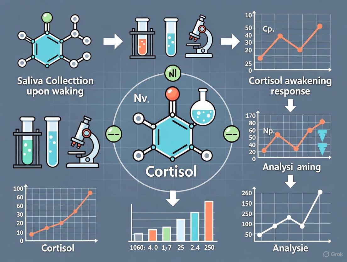
The Cortisol Awakening Response (CAR) is traditionally defined as a sharp 38–75% increase in cortisol levels that peaks 30–45 minutes after awakening in the morning [1]. This phenomenon is superimposed upon the endogenous circadian rise in cortisol that occurs in the early morning hours [1]. For decades, CAR was largely considered a distinct physiological response to the act of waking up, hypothesized to prepare the body for the anticipated stressors of the upcoming day [2]. However, recent high-resolution studies have challenged this paradigm, suggesting that the cortisol increase observed after waking may be a continuation of the circadian rhythm rather than a direct consequence of awakening itself [3] [4]. This application note delineates the physiological mechanisms and circadian interactions of CAR, providing detailed protocols and resources for researchers and drug development professionals engaged in HPA-axis research.
Physiological Mechanisms and Circadian Interactions
The core physiological system governing cortisol secretion is the hypothalamic-pituitary-adrenal (HPA) axis. The traditional view posits that the hippocampus plays a pivotal role in regulating CAR, potentially to activate prospective memory representations and enable orientation for the day ahead [1]. This process is thought to be modulated by the suprachiasmatic nucleus (SCN), the body's central circadian clock [1].
Emerging evidence from 2025, utilizing continuous in vivo microdialysis, indicates that the rate of cortisol increase does not change in the hour after awakening compared to the hour before it [3] [4]. This finding challenges the concept of CAR as a discrete "response" and instead positions it as part of a broader circadian rhythm. The peak of this rhythm occurs at a circadian phase corresponding to approximately 3:40–3:45 a.m., with no detectable CAR during circadian phases corresponding to the afternoon [2].
Table 1: Key Characteristics of the Cortisol Awakening Response
| Characteristic | Traditional Understanding | Insights from Recent Evidence (2025) |
|---|---|---|
| Core Definition | A distinct ~50% increase in cortisol peaking 30-45 minutes after awakening [1]. | A manifestation of the circadian cortisol rise; the rate of secretion does not accelerate upon waking [3] [4]. |
| Primary Driver | Response to the event of awakening, potentially mediated by the hippocampus [1]. | Endogenous circadian system, with peak activity at a phase corresponding to ~3:40 AM [2] [4]. |
| Key Modulators | Anticipated stress, workdays, time of awakening, and sleep duration [1]. | Sleep duration and regularity of wake time; maximum cortisol increase occurs before waking in long sleepers and after in short sleepers [3]. |
| Response to Forced Awakening | Blunted or absent response when participants are forcibly awoken at night [3]. | Supports the role of circadian anticipation rather than the sleep-wake transition itself as the key driver. |
Visualizing the Cortisol Rhythm and Experimental Paradigms
The following diagrams illustrate the shift in the theoretical model of CAR and the design of a key forced desynchrony protocol used to isolate circadian effects.
Detailed Experimental Protocols
This section outlines specific methodologies from seminal studies, enabling researchers to replicate and build upon this work.
In-Vivo Microdialysis Protocol for Continuous Cortisol Monitoring
This protocol, derived from Klaas et al. (2025), allows for continuous, high-fidelity measurement of free cortisol in interstitial fluid in a naturalistic home setting [3] [4].
- Objective: To measure the rate of change of tissue-free cortisol levels immediately before and after awakening without disrupting natural sleep.
- Materials: Refer to Table 3 for specific research reagents.
- Procedure:
- Participant Preparation: Recruit healthy volunteers (e.g., n=201, aged 18-68). Insert a linear microdialysis probe subcutaneously in abdominal tissue under local anesthetic.
- Device Setup: Connect the probe to a portable, automated microdialysis pump and collection unit secured around the participant's waist.
- Sample Collection: Program the device to collect interstitial fluid samples at fixed 20-minute intervals over a 24-hour period while participants remain in their homes.
- Sleep/Wake Logging: Participants self-report their precise sleep and wake times.
- Sample Analysis: Analyze adrenal steroids, including cortisol, using ultrasensitive liquid chromatography coupled with tandem mass spectroscopy (LC-MS/MS).
- Data Analysis: Calculate the rate of cortisol increase (slope) for the 60 minutes preceding and the 60 minutes following the self-reported wake time. Compare these rates using appropriate statistical models (e.g., linear mixed-effects models).
Laboratory-Based Forced Desynchrony Protocol
This protocol, as used in earlier foundational studies, is designed to separate the influence of the endogenous circadian system from behavioral sleep/wake cycles [2].
- Objective: To assess the endogenous circadian rhythm of CAR independent of sleep and other behaviors.
- Materials: Controlled laboratory environment with dim light, salivary cortisol collection kits, actigraphy, and polysomnography equipment.
- Procedure:
- Pre-Study Stabilization: Instruct participants to maintain a constant self-selected 8-hour sleep schedule for at least one week prior to the lab study, verified by actigraphy and sleep diaries.
- Laboratory Admission: Admit participants to a controlled laboratory environment where light, food intake, and activity can be regulated.
- Forced Desynchrony: Implement one of two complementary protocols:
- Protocol 1: 10 identical consecutive 5-hour 20-minute sleep/wake cycles.
- Protocol 2: 5 identical consecutive 18-hour sleep/wake cycles. Throughout these protocols, all behaviors (including sleep opportunities) are uniformly distributed across all circadian phases.
- Circadian Phase Marking: Use salivary melatonin samples to determine each participant's dim light melatonin onset (DLMO), defined as circadian phase 0°.
- Cortisol Sampling: During each scheduled awakening period, collect salivary cortisol samples immediately upon waking and again 50 minutes later. The change in cortisol level is defined as the CAR for that cycle.
- Data Analysis: Align all CAR measurements to the individual's circadian phase (based on DLMO). Use cosinor analysis to detect a significant circadian rhythm in the CAR across the 24-hour cycle.
Table 2: Key Quantitative Findings from CAR Studies
| Study / Parameter | Protocol | Key Finding | Statistical / Quantitative Detail |
|---|---|---|---|
| Klaas et al. (2025) [3] [4] | In-vivo microdialysis at home (n=201) | No difference in the rate of cortisol increase before vs. after awakening. | The maximum rate of increase occurred 97 min before waking in long sleepers (mean 548 min) and 12 min after waking in short sleepers (mean 369 min). |
| Forced Desynchrony Study [2] | 10 cycles of 5h20m sleep/wake (n=17) | A clear circadian rhythm in CAR was observed. | CAR peaked at a circadian phase corresponding to 3:40–3:45 a.m., with no detectable CAR in the afternoon. |
| Forced Desynchrony Study [2] | 5 cycles of 18h sleep/wake (n=18) | Confirmed circadian rhythm in CAR, independent of sleep structure. | Total sleep time was associated with CAR in one protocol, but REM/NREM sleep percentages were not. |
The Scientist's Toolkit: Research Reagent Solutions
The following table details essential materials and their applications for conducting CAR research, based on the cited methodologies.
Table 3: Essential Research Reagents and Materials for CAR Studies
| Item | Function/Application | Example from Search Results |
|---|---|---|
| Salivary Cortisol Collection Kit | Non-invasive collection of free cortisol for measurement by immunoassay or LC-MS/MS. Used in forced desynchrony and ambulatory studies [2]. | Protocols measuring cortisol upon awakening and 30-50 minutes later to calculate CAR [2] [1]. |
| Portable Microdialysis System | Continuous, automated sampling of tissue-free cortisol in interstitial fluid from ambulatory participants in their homes, minimizing intrusion [3]. | System with abdominal probe and portable collector used by Klaas et al. (2025) for 20-min interval sampling over 24 hours [3] [4]. |
| Liquid Chromatography Tandem Mass Spectrometry (LC-MS/MS) | Gold-standard, highly sensitive analytical method for the precise quantification of adrenal steroids, including cortisol, in biological samples [3]. | Validation method for cortisol levels in microdialysis fluid and plasma samples in the ULTRADIAN trial [3]. |
| Salivary Melatonin Assay | Determination of Dim Light Melatonin Onset (DLMO), the gold-standard marker for internal circadian phase in human studies [2]. | Used in forced desynchrony protocols to align cortisol measurements to the endogenous circadian cycle rather than clock time [2]. |
| Actigraphy Device | Objective monitoring of participant sleep/wake cycles and physical activity during ambulatory and pre-study stabilization phases [2]. | Used to verify participant adherence to a fixed 8-hour sleep schedule before laboratory admission [2]. |
| Dexamethasone | A synthetic glucocorticoid used in suppression tests to probe the sensitivity of the HPA axis negative feedback loop [1]. | Low-dose dexamethasone strongly inhibits the ACTH release that creates CAR [1]. |
CAR as a Biomarker of HPA Axis Function and Stress System Integrity
The Cortisol Awakening Response (CAR) is defined as the dynamic increase in cortisol secretion that occurs during the first 30-60 minutes after awakening. This specific neuroendocrine phenomenon has attracted significant research interest as a potential biomarker of Hypothalamic-Pituitary-Adrenal (HPA) axis function and stress system integrity [3]. A properly functioning CAR is hypothesized to prepare individuals for anticipated energy demands and stressors of the forthcoming day, making it a valuable indicator of adaptive physiological preparedness.
Recent research has prompted important questions about the fundamental nature of CAR. A groundbreaking 2025 study using continuous microdialysis sampling in home settings demonstrated that the rate of increase in cortisol secretion did not change when participants awoke compared with the preceding hour when they were asleep [3]. This finding challenges the long-standing assertion that CAR is a distinctive post-awakening response superimposed on an endogenous cortisol rhythm, suggesting instead that cortisol secretion during initial waking appears to be more tightly regulated by intrinsic circadian rhythmicity than by the awakening process itself [3]. Despite this paradigm shift, CAR measurement remains a valuable tool for understanding HPA axis dynamics, particularly when interpreted with consideration of underlying circadian influences.
Key Quantitative Findings in CAR Research
Table 1: Key Factors Influencing Cortisol Awakening Response Variability
| Factor | Effect on CAR Dynamics | Study Population | Citation |
|---|---|---|---|
| Sleep Duration | Short sleep (~6h): maximal cortisol increase 12min AFTER wakingLong sleep (~9h): maximal cortisol increase 97min BEFORE waking | 201 healthy volunteers | [3] |
| Wake Time Consistency | Aligned sleepers (<1h variation): maximum rate 12min after wakingMisaligned sleepers (>1h variation): maximum rate 68min before waking | 201 healthy volunteers | [3] |
| Perceived Stress | No consistent association with CAR found | 229 predominantly Latino adolescents | [5] |
| Chronic Stress | Leads to HPA axis dysregulation, impaired feedback, glucocorticoid receptor resistance | Clinical and experimental studies | [6] |
Table 2: Technical Considerations for CAR Measurement Methodologies
| Method | Temporal Resolution | Key Advantages | Key Limitations |
|---|---|---|---|
| In Vivo Microdialysis | 20-minute samples over 24h | Continuous sampling in naturalistic environment; measures tissue-free cortisol | Potential lag between interstitial and plasma cortisol; averaging over 20-min intervals [3] |
| Salivary Cortisol | Discrete time points (awakening, +30min, +60min, evening) | Non-invasive; suitable for home collection; reflects free cortisol | No pre-awakening measurements; dependent on participant compliance [5] |
| Serum Cortisol | Single time point (typically morning) | High accuracy; clinical standard | Single snapshot; invasive collection; doesn't capture dynamics [5] |
Experimental Protocols for CAR Assessment
Protocol 1: Comprehensive Salivary CAR Assessment in Free-Living Conditions
Purpose: To measure the cortisol awakening response through salivary cortisol sampling in participants' natural environments.
Materials and Reagents:
- Salivette saliva collection devices (Salimetrics)
- Portable freezer for sample storage
- Electronic timing device (e.g., smartphone application) for compliance monitoring
- Salivary cortisol ELISA kit (Salimetrics, Inc.)
- Centrifuge for sample processing
- -80°C freezer for long-term storage
Procedure:
- Participant Training: Conduct thorough training on sampling procedure, emphasizing strict timing and contamination prevention.
- Sample Collection Schedule:
- Time 1: Immediately upon awakening (before getting out of bed)
- Time 2: 30 minutes after awakening
- Time 3: 60 minutes after awakening (optional for detailed kinetics)
- Time 4: Evening sample (before bedtime, for diurnal slope calculation)
- Collection Protocol: Participants should refrain from eating, drinking, brushing teeth, or smoking for at least 30 minutes before each sample. They should record exact collection times.
- Compliance Monitoring: Use electronic timestamp verification (e.g., smartphone photos with timestamps) to ensure adherence.
- Sample Handling: Participants temporarily store samples in home freezers until transfer to research facility. Centrifuge salivettes upon receipt, aliquot saliva, and store at -80°C until assay.
- Assay Procedure: Use validated salivary cortisol ELISA according to manufacturer specifications. Include quality control samples with each batch.
Calculation:
- CAR = (Cortisol at 30min - Cortisol at awakening)
- Diurnal Cortisol Slope (DCS) = (Evening cortisol - Awakening cortisol)
- Total Daily Cortisol (TDC) = Area under the curve calculated using trapezoid method [5]
Protocol 2: Continuous Cortisol Monitoring via In Vivo Microdialysis
Purpose: To obtain continuous measurements of tissue-free cortisol before and after awakening using microdialysis.
Materials and Reagents:
- Linear microdialysis probe for subcutaneous abdominal tissue implantation
- Portable microdialysis pump and collection device
- Ultrasensitive liquid chromatography coupled with tandem mass spectroscopy (LC-MS/MS) system
- Calibration standards for adrenal steroids
Procedure:
- Probe Insertion: Insert sterile linear microdialysis probe into subcutaneous abdominal tissue under local anesthesia.
- System Setup: Secure portable collection device around waist, allowing free ambulation and normal daily activities.
- Sampling Parameters: Collect interstitial fluid samples automatically every 20 minutes over a 24-hour period.
- Sleep/Wake Monitoring: Participants self-report sleep and wake times using standardized sleep diary.
- Sample Analysis: Analyze adrenal steroids, including cortisol, using ultrasensitive LC-MS/MS.
- Validation: In subset of participants, correlate tissue-free cortisol levels with simultaneous plasma measurements [3].
Data Analysis:
- Calculate rate of cortisol increase in the hour preceding awakening and the hour following awakening
- Compare pre- and post-awakening rates using appropriate statistical tests
- Account for between-subject variability factors (sleep duration, wake time alignment) [3]
Signaling Pathways and Physiological Context
HPA Axis and CAR Regulation: This diagram illustrates the neuroendocrine pathway regulating cortisol secretion, showing integration of circadian inputs from the SCN with stress responses. The dotted lines indicate debated awakening-specific activation in light of recent research [3] [6].
The Scientist's Toolkit: Essential Research Reagents
Table 3: Essential Research Reagents for CAR Measurement Studies
| Reagent/Equipment | Specific Function | Application Notes |
|---|---|---|
| Salivette Collection Devices (Salimetrics) | Passive drool saliva collection for cortisol measurement | Preferred over cotton-based swabs for better recovery; compatible with standard ELISA protocols [5] |
| Salivary Cortisol ELISA Kits | Quantitative measurement of cortisol in saliva | Provides sensitivity to 0.007-0.095 μg/dL; validate for salivary matrix; inter-assay CV <10% [5] |
| Electronic Compliance Monitoring | Verification of exact sampling times | Smartphone applications with timestamped photos; critical for CAR validity given sensitivity to timing [5] |
| Microdialysis System | Continuous sampling of interstitial fluid cortisol | Linear subcutaneous probes with portable pump; 20-min sampling resolution; measures tissue-free cortisol [3] |
| LC-MS/MS System | High-sensitivity steroid analysis | Gold standard for specificity; required for microdialysis samples due to low concentrations [3] |
| Portable Freezers (-20°C) | Temporary sample storage in field studies | Maintain sample integrity between collection and transfer to -80°C storage [5] |
Interpretation Guidelines and Methodological Considerations
When interpreting CAR data, researchers should consider several critical methodological factors. First, substantial between-subject variability is consistently observed, with sleep duration and wake time alignment explaining significant portions of this variability [3]. Short sleepers (~6 hours) and those with aligned sleep schedules typically show maximal cortisol increases shortly after waking, while long sleepers (~9 hours) and those with misaligned schedules may peak before waking.
The relationship between perceived stress and CAR appears complex and inconsistent. Recent research in adolescent populations found no significant associations between Perceived Stress Scale (PSS) scores and CAR, despite finding some associations with other cortisol biomarkers [5]. This suggests CAR may reflect different aspects of stress physiology than subjective stress measures.
Under conditions of chronic stress, the HPA axis undergoes significant alterations including impaired feedback mechanisms, glucocorticoid receptor resistance, and potential adrenal exhaustion [6]. These changes can result in paradoxical cortisol dysregulation that may manifest as blunted or exaggerated CAR patterns.
When designing CAR studies, researchers should prioritize electronic compliance monitoring given the sensitivity of CAR to exact sampling times. Additionally, consideration should be given to the emerging evidence that CAR may represent a continuation of circadian rhythmicity rather than a purely awakening-dependent phenomenon [3].
The cortisol awakening response (CAR) is a distinct phenomenon within the human circadian rhythm, characterized by a sharp increase in cortisol secretion during the first 30-45 minutes after morning awakening [7]. This dynamic response is considered a non-invasive biomarker for the health and reactivity of the hypothalamic-pituitary-adrenal (HPA) axis, the body's central stress response system [8] [9]. The CAR is typically quantified by measuring the increase in cortisol concentration from the moment of awakening (sample 1) to its peak, which usually occurs 30-45 minutes post-awakening (sample 2 and 3), with the cortisol level rising by approximately 38% to 75% in healthy individuals [7] [9].
A growing body of research indicates that an aberrant CAR—either blunted (hypocortisolic) or heightened (hypercortisolic)—is associated with a range of disorders [7] [8] [9]. This application note synthesizes current evidence on the associations between CAR and psychiatric, metabolic, and pain disorders. It provides researchers and clinicians with structured data and detailed protocols for investigating the CAR as a biomarker in clinical populations, framed within the broader context of cortisol awakening response measurement research.
Clinical Associations with Disorder Subtypes
Alterations in the cortisol awakening response serve as a sensitive indicator of HPA axis dysregulation across various clinical conditions. The tables below summarize key quantitative associations, highlighting the direction of CAR change and its clinical significance.
Table 1: CAR Associations with Psychiatric Disorders
| Disorder | Typical CAR Alteration | Key Clinical and Research Correlations |
|---|---|---|
| Major Depressive Disorder (MDD) | Blunted CAR in severe or chronic depression [8] [9]. Potentially heightened CAR in mild to moderate cases [8] [9]. | A blunted CAR at hospital admission predicted higher depression severity 6 months post-discharge (r = -0.223, p < 0.05) [8]. A higher CAR is considered an "index of one's overall vulnerability to depression" [9]. |
| Chronic Stress & Burnout | Blunted CAR [9]. | A study of students pre-exam showed a blunted CAR, most pronounced in those with the highest perceived stress [9]. Associated with PTSD, caregiver stress, and chronic fatigue syndrome [9]. |
| Daily Stress Reactivity | Higher CAR [10]. | A higher CAR was associated with greater reactivity to daily perceived stress and higher subsequent daily negative affect [10]. |
Table 2: CAR Associations with Metabolic Disorders
| Disorder | Typical CAR Alteration | Key Clinical and Research Correlations |
|---|---|---|
| Obesity | Blunted CAR [9]. | A significant inverse association exists between CAR and both BMI and waist circumference [9]. Obese children also show a flat CAR, with severity correlating to weight [9]. |
| Type 2 Diabetes | Blunted CAR [9]. | HPA axis dysregulation is associated with diabetes. A blunted CAR may precede the development of the disease, suggesting a potential risk marker [9]. |
Detailed Experimental Protocols for CAR Assessment
Adherence to standardized protocols is critical for obtaining reliable and valid CAR measurements. The following section outlines a core sampling protocol and specific methodologies for clinical research applications.
Core Salivary CAR Sampling Protocol
This protocol is adapted from established guidelines and is suitable for most clinical and research applications [7] [8].
Objective: To accurately capture the dynamic change in free, biologically active cortisol levels in response to morning awakening.
Materials:
- Saliva collection kits (e.g., Salivettes).
- Cool bag or refrigerator for sample storage.
- Freezer (-20°C or lower) for long-term storage.
- Electronic timing device or diary for participants.
- Laboratory capable of conducting salivary cortisol immunoassays.
Procedure:
- Participant Instruction: Train participants thoroughly. Emphasize the importance of adherence to the timing protocol. Provide a simplified instruction sheet.
- Sampling Schedule: Collect saliva samples at four time points:
- Sample 1 (S1): Immediately upon awakening (0 minutes).
- Sample 2 (S2): 15 minutes post-awakening.
- Sample 3 (S3): 30 minutes post-awakening.
- Sample 4 (S4): 45 minutes post-awakening.
- Sample Collection: Participants should not eat, drink (except water), smoke, or brush their teeth until after the final sample is collected. They should record the exact time of awakening and each sample collection.
- Sample Handling: Participants should store samples in their personal refrigerator or a provided cool bag immediately after collection. Researchers should centrifuge samples and store them at -20°C or lower until analysis.
Data Analysis: The CAR can be quantified using several indices, chosen based on the research question:
- Area Under the Curve with respect to Increase (AUCi): Reflects the total cortisol secretion over the CAR period, sensitive to changes from the first sample [7]. This is the preferred method for capturing the dynamic response.
- Mean Increase (MnInc): The average of the increases of all post-awakening samples (S2, S3, S4) relative to S1 [7].
- Peak Change: The simple difference between the peak cortisol value (max of S2, S3, S4) and the awakening value (S1) [7].
Protocol for Investigating CAR in Major Depressive Disorder
This protocol is designed for longitudinal studies assessing CAR as a predictor of treatment outcome or symptom trajectory.
Objective: To determine if the CAR measured at clinical intake predicts depression severity at follow-up points after treatment.
Study Design:
- Population: Inpatients with a primary diagnosis of MDD, displaying moderate to severe symptoms [8].
- Exclusion Criteria: Glucocorticoid medication use, comorbid addiction disorders, psychosis, and specific medical conditions (e.g., autoimmune thyroiditis, respiratory disease) [8].
- CAR Assessment: Follow the core salivary sampling protocol (S1 at 0, 15, 30, 45 min) on the first two days after hospital admission.
- Clinical Assessment:
- Administer a standardized depression inventory (e.g., Beck Depression Inventory-II, BDI-II) at intake.
- Re-administer the same inventory at discharge, 6 weeks post-discharge, and 6 months post-discharge.
- Statistical Analysis: Perform correlation analysis (e.g., Pearson's r) between the CAR (e.g., AUCi or AUCg) at intake and BDI-II scores at each follow-up time point [8].
Protocol for Investigating CAR in Chronic Stress
This protocol uses a case-control design to examine the effect of prolonged stress exposure on HPA axis function.
Objective: To compare the CAR between a group experiencing chronic stress and a matched control group.
Study Design:
- Population: A group under chronic stress (e.g., students during a major exam period, chronically stressed caregivers) and a matched control group without such stressors [9].
- CAR Assessment: Follow the core salivary sampling protocol across three consecutive typical days (e.g., weekdays for the exam group).
- Psychometric Assessment: Administer validated self-report questionnaires for perceived stress (e.g., Perceived Stress Scale) and anxiety at the end of the sampling period.
- Statistical Analysis: Use an independent samples t-test or ANOVA to compare the CAR (AUCi) and self-reported stress levels between the two groups. A blunted CAR is expected in the chronic stress group [9].
Visualizing the CAR Workflow and Regulatory Pathways
The following diagrams, generated using Graphviz DOT language, illustrate the experimental workflow for CAR assessment and its underlying neuroendocrine pathways.
Diagram 1: CAR Sampling & Analysis Workflow.
Diagram 2: Neuroendocrine Regulation of CAR.
The Scientist's Toolkit: Key Research Reagents & Materials
The following table details essential materials and reagents required for conducting CAR research in accordance with the protocols described above.
Table 3: Essential Research Reagents and Materials for CAR Studies
| Item | Function/Application | Key Considerations |
|---|---|---|
| Saliva Collection Device (e.g., Salivette, plain cotton swabs) | Collection of saliva samples for cortisol analysis. | Must be inert and not interfere with the immunoassay. Swabs should not contain citric acid or other stimulants [9]. |
| Salivary Cortisol Immunoassay Kit | Quantification of free cortisol levels in saliva samples. | Choose a kit with high sensitivity and specificity, validated for saliva. Common methods include ELISA and LC-MS/MS [3]. |
| Electronic Diary or Timer | For participants to record exact awakening and sampling times. | Critical for monitoring and ensuring protocol adherence, which is a major source of measurement error [7] [8]. |
| Low-Temperature Freezer (-20°C to -80°C) | Long-term storage of saliva samples to preserve cortisol integrity. | Essential for maintaining sample stability before batch analysis. |
| Laboratory Centrifuge | Processing of saliva samples after collection to separate saliva from swabs and debris. | Ensures clear samples for accurate assay results. |
| Validated Psychometric Scales (e.g., BDI-II, Perceived Stress Scale) | Quantification of clinical symptoms and subjective stress. | Allows for correlation between biological (CAR) and psychological measures [8] [9]. |
The Cortisol Awakening Response (CAR), defined as the marked increase in cortisol secretion occurring in the first 30-45 minutes after morning awakening, has long been a cornerstone of psychoneuroendocrine stress research [11]. It is widely investigated as a biomarker for stress reactivity in various disorders, from depression to post-traumatic stress disorder [3]. The traditional hypothesis posits that the act of waking triggers a distinct, superimposed endocrine response, preparing the individual for the anticipated demands of the coming day [3]. However, the very foundation of this concept—that the CAR is a discrete response to awakening—is now being rigorously challenged by recent high-resolution studies. This application note synthesizes the emerging evidence questioning the CAR's existence as a unique phenomenon, provides a detailed protocol for a pivotal recent study, and offers tools to navigate this evolving methodological landscape.
The Core Debate: Endogenous Rhythm vs. Distinct Response
The central debate revolves around whether the post-awakening rise in cortisol is a direct consequence of the transition from sleep to wakefulness or merely a continuation of an underlying circadian rhythm that begins its ascent hours before awakening [3]. The traditional view supports the former, but a groundbreaking 2025 study by Klaas et al. provides compelling evidence for the latter.
A quantitative evaluation of methodological adherence reveals significant shortcomings in the field. An analysis of studies published in Psychoneuroendocrinology between 2018 and 2020 showed that only 9.3% implemented the critical guideline of objectively verifying both awakening and sampling times, a factor essential for reliable CAR measurement [11]. This widespread methodological limitation may have historically obscured the true nature of cortisol dynamics around wakefulness.
Table 1: Key Quantitative Findings from the Klaas et al. (2025) Microdialysis Study
| Parameter | Finding | Implication for CAR Concept |
|---|---|---|
| Rate of Cortisol Increase | No difference between the first hour after awakening and the preceding hour. | Challenges the idea that waking itself accelerates cortisol secretion. |
| Peak Cortisol Timing | At a population level, cortisol levels peaked within the first hour of being awake, but the rise began well before waking. | Suggests the peak is part of a pre-programmed rhythm, not a response to an event. |
| Key Predictor of Post-Awakening Rise | The cortisol level reached in the hour preceding awakening. | Indicates the circadian phase is a stronger driver than the waking event. |
| Effect of Sleep Duration (Short vs. Long) | Short sleepers (~6h): Maximal rate of increase 12 minutes after waking. Long sleepers (~9h): Maximal rate 97 minutes before waking. | Demonstrates significant between-subject variability based on sleep habits. |
| Effect of Wake Time Alignment | Aligned sleepers (<1h variation): Max rate 12 minutes after waking. Misaligned sleepers (>1h variation): Max rate 68 minutes before waking. | Shows that regularity of sleep schedule dramatically shifts cortisol dynamics. |
Experimental Protocol: In-Vivo Microdialysis for High-Resolution Cortisol Assessment
The following protocol is adapted from the innovative methodology employed by Klaas et al. (2025) and Upton et al. (2023) [3], which enabled the continuous, at-home measurement of tissue-free cortisol.
Protocol: Continuous Ambulatory Cortisol Microdialysis
Objective: To measure the dynamic profile of tissue-free cortisol in interstitial fluid continuously for 24 hours, including the pre- and post-awakening periods, in a naturalistic home setting.
Materials and Reagents:
- Linear Microdialysis Probe: For subcutaneous insertion into abdominal tissue.
- Portable Automated Microdialysis Device: A waist-worn system for continuous sample collection (e.g., as described in Upton et al., 2023).
- Collection Vials: For 20-minute interval sampling over 24 hours.
- Ultrasensitive Liquid Chromatography Coupled with Tandem Mass Spectroscopy (LC-MS/MS) System: For analysis of adrenal steroids, including cortisol.
- Validated Sleep/Wake Diary or Electronic Participant Log: For self-reporting of sleep and wake times.
Procedure:
- Participant Preparation: Recruit healthy adult volunteers (e.g., aged 18-68). Exclude individuals with conditions or medications known to significantly influence HPA axis function.
- Probe Insertion: Insert a sterile linear microdialysis probe into the subcutaneous tissue of the participant's abdomen. This procedure should be performed by trained clinical staff.
- System Calibration & Attachment: Calibrate the portable microdialysis device and secure it around the participant's waist, ensuring it allows for free ambulation and normal daily activities.
- At-Home Sampling: Instruct the participant to go about their normal routine, including sleep, at their home. The system will automatically collect interstitial fluid samples at 20-minute intervals for a 24-hour period.
- Event Logging: The participant must meticulously self-report their actual wake-up time and the timing of each sample, if not automatically recorded by the device.
- Sample Recovery and Analysis: After the 24-hour period, recover the collection vials. Analyze cortisol concentrations using LC-MS/MS.
- Data Processing: Align cortisol measurements with the recorded awakening time. Calculate the rate of cortisol increase for the 60 minutes before and the 60 minutes after awakening for statistical comparison.
Visualizing the Experimental Workflow
The following diagram illustrates the key stages of the microdialysis protocol for assessing cortisol dynamics.
The Scientist's Toolkit: Research Reagent Solutions
Table 2: Essential Materials for High-Resolution CAR Research
| Item | Function & Application Note |
|---|---|
| Ambulatory Microdialysis System | Enables continuous, real-time collection of biologically active, tissue-free cortisol from interstitial fluid in a participant's natural environment, overcoming the limitations of discrete saliva or blood sampling [3]. |
| Linear Microdialysis Probe | A subcutaneous probe that allows for the diffusion of analytes across a semi-permeable membrane. Its linear design is suited for abdominal tissue insertion and comfortable for 24-hour ambulatory use [3]. |
| LC-MS/MS System | Provides ultrasensitive and highly specific quantification of cortisol and other adrenal steroids from low-volume microdialysis samples, minimizing cross-reactivity issues found in immunoassays [3]. |
| Objective Awakening Verification | Electronic timers (e.g., TrackCap) or integrated sensors in microdialysis devices that verify the exact moment of awakening. This is critical for valid pre- and post-awakening phase alignment, a major source of error in CAR studies [11]. |
| CAR Methodological Checklist | A consensus-based checklist (e.g., Stalder et al., 2022) to ensure adherence to best practices in participant instruction, sampling timing, compliance verification, and data reporting, thereby improving reproducibility [11]. |
Conceptualizing the Paradigm Shift in CAR Research
The debate between the traditional and emerging views of CAR necessitates a new conceptual model for designing and interpreting studies. The following diagram contrasts the two frameworks and highlights the role of key moderating variables identified by recent evidence.
The recent evidence challenging the CAR concept, particularly from high-resolution microdialysis studies, necessitates a significant shift in the interpretation of post-awakening cortisol dynamics. The findings that the rate of cortisol secretion does not accelerate upon waking and is heavily influenced by pre-awakening circadian levels and sleep patterns suggest that the "response" may be an emergent property of the circadian system rather than a discrete event [3].
For researchers and drug development professionals, this paradigm shift has critical implications:
- Measurement and Interpretation: CAR values from single time points or short sampling windows post-awakening should be interpreted with extreme caution, as they may reflect circadian phase more than a stress response.
- Study Design: Future research must account for key moderating variables like sleep duration and wake time regularity. Objective verification of awakening is non-negotiable for valid CAR assessment [11].
- Therapeutic Context: In clinical trials where HPA axis function is an endpoint, a more sophisticated model of cortisol dynamics is required to avoid misattributing drug effects or disease correlates.
The field is moving beyond simply quantifying the CAR to understanding the origins of the profound individual differences in cortisol dynamics and their true relevance for health and disease.
Best Practices in CAR Assessment: Protocols and Analytical Approaches
The cortisol awakening response (CAR) is a distinct aspect of hypothalamic-pituitary-adrenal (HPA) axis activity, characterized by a marked increase in cortisol secretion during the first 30–45 minutes after morning awakening [11]. As a key biomarker in psychoneuroendocrinological research, obtaining reliable CAR data requires meticulous attention to methodological detail, particularly in sampling protocol design. The ecological validity of measuring CAR in participants' home settings is a significant advantage, but this lack of direct researcher oversight introduces critical methodological challenges [12]. This application note synthesizes current expert consensus and empirical evidence to establish rigorous, evidence-based sampling protocols for CAR assessment, framed within the broader context of cortisol awakening response measurement research for scientific and drug development professionals.
Expert Consensus Guidelines on Sampling Protocols
Core Sampling Parameters
The International Society of Psychoneuroendocrinology (ISPNE) expert panel has established clear consensus guidelines for CAR assessment to promote methodological rigor and reproducibility [12]. The fundamental parameters for reliable sampling protocols are summarized in Table 1.
Table 1: Core CAR Sampling Protocol Parameters
| Parameter | Recommendation | Rationale | Key References |
|---|---|---|---|
| Sampling Duration | 30-45 minutes post-awakening | Captures the dynamic increase period of cortisol secretion | [11] |
| Sampling Frequency | 3-4 samples within first hour (at awakening, +30 min, +45 min, optionally +60 min) | Accurately characterizes the response trajectory and peak | [11] [13] |
| Sampling Days | ≥2 consecutive days (preferably 3+ days) | Accounts for day-to-day variability and improves reliability | [14] |
| Awakening Time Verification | Objective monitoring (e.g., electronic containers, headband sensors) | Critical for accuracy; self-reporting is unreliable | [15] [11] [16] |
| Sample Timing Precision | Exact recording of each sample time | CAR is time-sensitive; small deviations affect measurements | [11] [17] |
Sampling Protocol Specifics
The recommended sampling protocol involves collecting saliva samples immediately upon awakening (before getting out of bed), then at 30 minutes and 45 minutes after awakening [11] [13]. Some protocols include an additional sample at 60 minutes post-awakening to better characterize the decline phase. Participants should refrain from eating, drinking, smoking, or brushing teeth until after all samples are collected, as these activities can contaminate samples or influence cortisol levels [12].
Multiple days of sampling are essential because considerable day-to-day and between-subject variability exists in CAR patterns [3]. Research indicates that single-day assessments capture only 30-40% of between-person variance in CAR, while 2-6 days are needed to achieve reliability coefficients of 0.80 or higher [11]. For most research applications, sampling across 3-5 consecutive days represents an optimal balance between reliability and participant burden.
Quantitative Data Synthesis
Methodological Adherence and Variability Factors
Recent evaluations of methodological quality in CAR research reveal significant gaps in implementing consensus guidelines. Quantitative analysis shows that only 9.3% of recent studies implemented the crucial guideline of objectively verifying both awakening and sampling times [15] [11]. This methodological shortcoming substantially compromises data reliability and represents a critical area for improvement in future research.
Table 2: Factors Influencing CAR Variability and Methodological Recommendations
| Factor | Impact on CAR | Methodological Recommendation | Evidence |
|---|---|---|---|
| Sleep Duration | Short sleep (~6h): maximal cortisol increase 12min post-awakening. Long sleep (~9h): maximal increase 97min pre-awakening | Record and control for sleep duration in analysis | [3] |
| Awakening Time Consistency | >1h variation: maximal cortisol increase 68min pre-awakening. <1h variation: maximal increase 12min post-awakening | Standardize wake times or account for variability | [3] |
| Anticipated Stress | Higher anticipated stress predicts increased next-day CAR magnitude | Control for anticipated demands in study design | [16] |
| Participant Apprehension | Research participation itself increases apprehension, affecting mood, cognition, and sleep | Include habituation days, simplify protocols | [17] |
| Sampling Adherence | Delays in sampling significantly alter CAR trajectory and parameters | Use objective adherence monitoring, clear instructions | [11] [14] |
Detailed Experimental Protocols
Standard Salivary CAR Assessment Protocol
Purpose: To reliably measure the cortisol awakening response in participants' natural environments for psychobiological research and clinical studies.
Materials:
- Salivette collection devices or similar saliva collection aids
- Cooler or refrigerator for sample storage
- Electronic monitoring devices (e.g., MEMS tracks or similar)
- Laboratory equipment for cortisol analysis (LC-MS/MS or immunoassay)
- Participant instructions and sleep/wake diaries
Procedure:
- Participant Training: Conduct comprehensive training sessions emphasizing the critical importance of precise sampling times. Specifically clarify that the "moment of awakening" means when eyes open, before any physical movement from bed [17].
- Sample Collection Sequence:
- Sample 1: Immediately upon awakening (while still in bed)
- Sample 2: 30 minutes (±2 minutes) after awakening
- Sample 3: 45 minutes (±2 minutes) after awakening
- Adherence Monitoring: Utilize electronic trackers to record exact sampling times. These devices track when collection tubes are opened, providing objective verification of protocol adherence [11].
- Sample Handling: Participants should refrigerate samples immediately after collection and return them to researchers within 1-2 weeks (or according to laboratory specifications).
- Protocol Duration: Implement sampling for 3-5 consecutive days, typically including weekdays to capture work-related anticipation effects [16].
Quantification:
- Calculate both the Area Under the Curve with respect to ground (AUCg), representing total cortisol secretion
- Calculate Area Under the Curve with respect to increase (AUCi), representing the dynamic response component [18]
Microdialysis Protocol for High-Resolution CAR Assessment
Purpose: To obtain continuous, high-temporal resolution measurements of tissue-free cortisol levels before and after awakening, circumventing limitations of discrete salivary sampling.
Materials:
- Linear microdialysis probe for subcutaneous abdominal tissue implantation
- Portable automated collection device worn around waist
- LC-MS/MS equipment for adrenal steroid analysis
- Data recording equipment for sleep and wake times
Procedure:
- Probe Insertion: Insert microdialysis probe subcutaneously in abdominal tissue under controlled conditions [3].
- Sample Collection: Program portable device to automatically collect interstitial fluid samples every 20 minutes over a 24-hour period.
- Sleep/Wake Monitoring: Participants self-report sleep and wake times while maintaining relatively normal daily activities.
- Cortisol Analysis: Analyze adrenal steroids, including cortisol, using ultrasensitive liquid chromatography coupled with tandem mass spectroscopy.
Applications: This continuous sampling approach is particularly valuable for investigating the fundamental nature of CAR, specifically for determining whether the cortisol increase following awakening represents a distinct response or merely reflects continuation of pre-awakening circadian rhythms [3].
Experimental Sleep Restriction Protocol
Purpose: To systematically examine the effects of controlled sleep restriction on CAR magnitude and dynamics.
Materials:
- Controlled sleep laboratory environment
- Polysomnography equipment for sleep monitoring
- Saliva collection kits
- Cortisol assay reagents
Procedure:
- Baseline Phase: Participants acclimatize to laboratory environment with 9 hours time in bed for 1-2 nights.
- Experimental Manipulation: Randomly assign participants to different time-in-bed conditions (5h, 6h, 7h, 8h, or 9h) for 5-7 consecutive nights.
- Sample Collection: Collect saliva samples at fixed clock times (e.g., 08:00, 08:30, and 08:45) across multiple days including baseline, experimental days, and recovery days.
- Control Factors: Maintain identical waking times across conditions to control for circadian influences on cortisol measurement.
Key Findings: Implementation of this protocol has demonstrated that mild to moderate sleep restriction (5-7 hours time in bed) does not significantly affect CAR compared to 8-9 hours time in bed, suggesting CAR robustness to moderate sleep perturbations [13].
Signaling Pathways and Workflow Diagrams
CAR Assessment Workflow
CAR Assessment Workflow Diagram
HPA Axis Regulation Neural Circuitry
HPA Axis Neural Regulation Diagram
The Scientist's Toolkit: Research Reagent Solutions
Table 3: Essential Materials for CAR Research
| Item | Function/Application | Specification Notes |
|---|---|---|
| Salivette Collection Devices | Saliva sample collection | Synthetic swab or passive drool format; avoid cotton if using immunoassays |
| Electronic Monitoring Devices (MEMS) | Objective verification of sampling times | Track tube opening times; essential for adherence documentation |
| Portable Microdialysis System | Continuous cortisol sampling in interstitial fluid | Allows 20-min sampling intervals over 24h; measures tissue-free cortisol [3] |
| Cortisol Assay Kits | Quantitative cortisol measurement | LC-MS/MS preferred for specificity; immunoassays require validation |
| Sleep Monitoring Headbands | Objective awakening time verification | Provides precise awakening time data complementary to self-report [16] |
| Temperature-Controlled Storage | Sample preservation | Maintain samples at -20°C to -80°C until analysis |
| Participant Diaries | Contextual data collection | Record sleep quality, stress, medication, and protocol deviations |
Robust assessment of the cortisol awakening response demands meticulous attention to sampling protocols, particularly regarding timing precision, frequency, and duration. The expert consensus guidelines provide a critical framework for obtaining reliable, reproducible CAR data, though current adherence to these standards remains concerningly low. Implementation of objective adherence monitoring, multi-day sampling protocols, and appropriate quantification methods is essential for advancing our understanding of CAR as a biomarker in basic research and clinical applications. The continuous evolution of assessment technologies, including microdialysis and electronic monitoring, offers promising avenues for enhancing methodological rigor in future CAR research.
The accurate assessment of the cortisol awakening response (CAR), defined as the dynamic increase in cortisol concentration within the first 30-60 minutes after awakening, serves as a critical biomarker in psychoneuroendocrinology for investigating stress reactivity and hypothalamic-pituitary-adrenal (HPA) axis functionality [3]. Its measurement relies on biospecimens collected through various methods, each with distinct advantages and limitations. While salivary cortisol measurement has been the predominant method in community and biobehavioral research due to its non-invasive nature [19], and serum cortisol offers a direct measure of circulating hormone levels [5], a paradigm shift is underway. Emerging evidence from 2025 challenges the fundamental concept that waking itself stimulates a distinct cortisol response, suggesting instead that post-awakening cortisol levels may simply reflect the continuation of the underlying circadian rhythm that begins increasing hours earlier [20] [21]. This revelation, largely enabled by ambulatory microdialysis techniques, underscores the profound influence of sample collection methodology on biological interpretation and highlights the necessity for researchers to critically evaluate their methodological choices within the context of their specific research questions.
Established Sampling Methodologies
Salivary Cortisol Sampling
Salivary cortisol measurement is a mainstay in biobehavioral research conducted in community settings. Its popularity stems from the method's non-invasiveness, ease of handling and storage, and suitability for repeated sampling in short intervals by participants in their home environments [19].
- Collection Protocol: A typical robust protocol for assessing the CAR involves collecting saliva samples immediately upon awakening (S1), then at 30 minutes (S2), 45 minutes (S3), and 60 minutes (S4) post-awakening. To achieve reliable trait data, this collection is often repeated across two consecutive days [22]. Participants must be thoroughly instructed to refrain from eating, drinking, smoking, or brushing their teeth before completing the sample series to avoid contamination. The exact timing of each sample must be recorded to ensure validity [22].
- Data Analysis: The raw cortisol concentrations (nmol/L) from the two days are typically averaged for each time point. The CAR can be quantified using several parameters:
- R30: The absolute change in cortisol level 30 minutes after awakening (R30 = S2 - S1) [22].
- Area Under the Curve with respect to increase (AUCi): This provides a measure of the total cortisol output relative to the waking value, calculated as: AUCi = [(S1 + S2)0.5/2 + (S2 + S3)0.25/2 + (S3 + S4)*0.25/2] - S1 [22].
- Considerations: Researchers should note the high prevalence of negative CAR values (a decrease in cortisol after waking), which may not be mere measurement errors but potentially indicative of underlying health conditions or environmental exposures, such as elevated blood lead levels or inflammatory processes [23]. Excluding these values without further investigation is not recommended.
Blood-Based Serum/Plasma Sampling
Blood collection provides a direct measurement of circulating cortisol, often considered the gold standard for single time-point assessments, particularly in clinical settings.
- Collection Protocol: Blood is drawn via venipuncture, typically in a fasted state during the morning (e.g., between 7:30 and 9:30 AM) [5]. For CAR assessment, this would require multiple draws immediately upon waking and at subsequent intervals, which is highly intrusive and impractical in ambulatory settings. It is therefore primarily used in controlled laboratory studies or for obtaining a single morning reference value.
- Analysis: Serum or plasma is separated by centrifugation and cortisol concentrations are commonly measured using commercially available immunoassays, such as ELISA [5].
Table 1: Comparison of Established Cortisol Sampling Methods
| Feature | Salivary Cortisol | Blood-Based Cortisol |
|---|---|---|
| Specimen Type | Saliva | Serum or Plasma |
| Measurement | Free (biologically active) cortisol | Total cortisol (protein-bound + free) |
| Collection | Non-invasive, self-administered | Invasive, requires phlebotomist |
| Setting | Ideal for ambulatory, home-based studies | Best suited for clinical or lab settings |
| Key Advantage | Enables frequent, ecologically valid sampling | Direct measure of systemic concentration |
| Key Limitation | Timing compliance and potential contamination | Impractical for dense CAR sampling; stressful |
An Emerging Technique: Ambulatory Microdialysis
Recent technological advances have introduced in vivo microdialysis as a powerful method for continuous hormone monitoring. This technique was pivotal in a landmark 2025 study that challenged the traditional CAR paradigm by measuring tissue-free cortisol both before and after waking [20] [3].
Protocol for Ambulatory Microdialysis
The following workflow details the protocol based on the ULTRADIAN study [20] [3]:
Key Technical Steps:
- Probe Insertion: A linear microdialysis probe is inserted subcutaneously into the abdominal tissue. This is generally well-tolerated and allows for free ambulation [3].
- Automated Sampling: The probe is connected to a portable automated collection device, which is secured around the participant's waist. The system is programmed to collect interstitial fluid samples at fixed intervals (e.g., every 20 minutes) over a 24-hour period, generating 72 individual samples [20].
- Ambulatory Monitoring: Participants continue their normal activities, including sleep, in their home environment. They self-report their exact sleep and wake times in an activity diary [20] [3].
- Laboratory Analysis: The collected microdialysis samples are analyzed for free cortisol and other adrenal steroids using ultrasensitive methods like liquid chromatography-tandem mass spectrometry (LC-MS/MS) [20] [3].
- Data Processing: Cortisol measurements are timestamped and realigned relative to each participant's individual wake time (t=0). This allows for direct comparison of hormone trajectories in the pre- and post-awakening periods [20].
Comparative Analysis of Methodologies
The choice of sampling method directly dictates the type of scientific questions that can be addressed, particularly in light of new findings on the CAR.
Table 2: Comprehensive Comparison of Cortisol Sampling Methodologies
| Parameter | Salivary Cortisol | Blood-Based Cortisol | Ambulatory Microdialysis |
|---|---|---|---|
| Biomarker Measured | Free cortisol | Total serum cortisol (sCOR) | Tissue-free cortisol in interstitial fluid |
| Temporal Resolution | Discrete samples (e.g., 4 points over 1h) | Discrete samples | Continuous (e.g., 20-min intervals) |
| Pre-awakening Assessment | Not feasible | Possible only in lab setting with sleep monitoring | Yes, key advantage - automated during sleep |
| Ecological Validity | High (home setting) | Low (lab setting) | Very High (home setting, 24h monitoring) |
| Primary Application | Community biobehavioral research; large cohorts [19] | Clinical diagnostics; mechanistic lab studies [5] | High-resolution dynamics; circadian rhythm research [20] |
| Key Finding Enabled | Established the common CAR pattern (post-awakening rise) | Corroborated post-awakening peak | Revealed no change in cortisol secretion rate at awakening [20] [21] |
Impact on Biological Interpretation
The methodological differences between these techniques have led to a significant evolution in the understanding of the CAR:
- The Traditional View (Saliva/Blood): Measurements confined to the post-awakening period supported the hypothesis that the act of waking is a stimulus that triggers a distinct surge in cortisol secretion, a phenomenon thought to prepare the body for the upcoming day [3].
- The New View (Microdialysis): By capturing the continuous cortisol rhythm before and after waking, microdialysis studies have demonstrated that the rate of cortisol increase does not change at the moment of awakening [20] [21]. The peak observed shortly after waking appears to be the culmination of a circadian rise that began in the early hours of the morning, challenging the notion of the CAR as a discrete response to awakening.
Furthermore, microdialysis has revealed substantial interindividual variability in cortisol dynamics, which can be attributed to factors such as sleep duration and wake-time consistency. For instance, in individuals with long sleep duration, the maximal rate of cortisol release can occur over 90 minutes before waking, whereas in short sleepers, it occurs just after waking [3].
The Scientist's Toolkit: Essential Reagents & Materials
Table 3: Key Research Reagent Solutions for Cortisol Measurement
| Item | Function/Application | Example(s) |
|---|---|---|
| Salivette Collection Device | For hygienic and standardized saliva sample collection; consists of a cotton swab stored in a plastic tube [22]. | Sarstedt Salivette [22] |
| Portable Microdialysis System | Automated, ambulatory system for continuous sampling of interstitial fluid over 24 hours in a home setting [20]. | System as used in ULTRADIAN study [20] |
| LC-MS/MS (Liquid Chromatography-Tandem Mass Spectrometry) | High-sensitivity analytical method for the precise quantification of cortisol and cortisone in microdialysis and other samples [20] [3]. | - |
| Enzyme-Linked Immunosorbent Assay (ELISA) | Immunoassay for measuring cortisol concentrations in saliva or serum; more accessible but potentially less specific than LC-MS/MS. | Salimetrics ELISA, DRG ELISA, Alpco ELISA [22] [5] |
| Cortisol & Cortisone Standards | Calibrators and controls used with LC-MS/MS to ensure accurate analyte identification and quantification. | - |
The field of cortisol research, particularly concerning the CAR, is at a methodological crossroads. While salivary sampling remains a valid and powerful tool for large-scale biobehavioral studies in naturalistic contexts, researchers must interpret post-awakening measurements with caution, acknowledging they may reflect circadian rhythm rather than a distinct waking response. Blood-based measurements continue to provide critical data in controlled settings. The emergence of ambulatory microdialysis represents a significant technological leap, enabling unprecedented temporal resolution and the ability to capture the pre-awakening cortisol trajectory. This new capability has directly fueled a paradigm shift in our physiological understanding of the morning cortisol peak. Moving forward, the choice of sampling method must be carefully aligned with the research hypothesis, with microdialysis offering a robust approach for investigating high-resolution dynamics and unraveling the profound interindividual variability in HPA axis function.
The cortisol awakening response (CAR) is defined as the marked increase in cortisol secretion that occurs in the first 30-45 minutes after morning awakening [11]. As a unique aspect of hypothalamus-pituitary-adrenal (HPA) axis activity, it serves as a crucial biomarker in psychoneuroendocrinological research, combining features of both a reactivity index and circadian regulation [11]. However, the ecological validity of CAR measurement—typically assessed through saliva samples collected in participants' domestic settings—critically depends on one fundamental factor: precise timing of sample collection [24].
This application note establishes the critical importance of objective time verification in CAR research. We detail specific protocols and methodologies to ensure temporal precision, addressing the concerning finding that, despite established guidelines, only 9.3% of recent CAR studies published in a leading journal adhered to the core recommendation of objectively verifying both awakening and sampling times [11]. The procedures outlined herein are designed to help researchers overcome this methodological gap, thereby enhancing the reliability, reproducibility, and scientific validity of future CAR studies.
Quantitative Assessment of Current Methodological Adherence
A quantitative evaluation was conducted to assess adherence to CAR methodological guidelines by comparing studies published in Psychoneuroendocrinology during a three-year period before (2013-2015) and after (2018-2020) the publication of the expert consensus guidelines [11]. The results reveal limited improvement and persistent critical shortcomings.
Table 1: Adherence to Key CAR Methodological Guidelines in Published Research
| Methodological Guideline | Pre-Guidelines (2013-2015) | Post-Guidelines (2018-2020) |
|---|---|---|
| Objective verification of awakening time | 2.4% (1 of 41 studies) | 9.3% (4 of 43 studies) |
| Objective verification of sampling times | 2.4% (1 of 41 studies) | 9.3% (4 of 43 studies) |
| Use of objective adherence control methods | 4.9% (2 of 41 studies) | 11.6% (5 of 43 studies) |
| Assessment on consecutive days | 78.0% (32 of 41 studies) | 83.7% (36 of 43 studies) |
| Exclusion of non-adherent samples | 63.4% (26 of 41 studies) | 65.1% (28 of 43 studies) |
The data demonstrates that while adherence to some procedural guidelines (e.g., consecutive-day assessment) is high, implementation of the most critical recommendation—objective time verification—remains alarmingly low. This fundamental methodological flaw threatens the validity of a substantial portion of contemporary CAR research and underscores the urgent need for standardized electronic monitoring protocols.
Detailed Experimental Protocols for Electronic Monitoring
Core Sampling Protocol
The following procedure must be followed for each day of CAR assessment.
Materials:
- Pre-labeled salivettes or saliva collection tubes
- Timer or stopwatch (electronic, integrated into monitoring device is preferable)
- Cool bag/box for sample storage (4°C)
- Freezer for long-term storage (-20°C or below)
- Electronic monitoring device (e.g., trackCap, MEMS)
Procedure:
Pre-Awakening Preparation (Evening Before):
- Place the electronic monitoring device and saliva collection kit within easy reach of the bed.
- Ensure the monitoring device is fully functional and activated.
At Awakening (Sample S1):
- Immediately upon awakening, press the event marker button on the electronic monitoring device. Do not get out of bed, check your phone, or engage in other activities first.
- Record the self-reported awakening time in the provided log.
- Collect the first saliva sample (S1) immediately. Place the sample into the monitoring device to register the time.
Post-Awakening Samples:
- Set a timer for 30 minutes (±2 minutes). During this period, remain in a relaxed state; you may get up for quiet activities (e.g., bathroom use) but avoid vigorous activity, eating, drinking (except water), brushing teeth, or smoking.
- At 30 minutes (±2 minutes) post-awakening, collect the second saliva sample (S2). Ensure the sample is registered by the electronic monitor.
- Depending on the study protocol, a third sample (S3) may be collected at 45 minutes post-awakening.
Post-Collection Handling:
- Store all samples immediately in a cool bag or domestic refrigerator (4°C).
- Within 24 hours, transfer samples to a -20°C freezer for long-term storage.
Protocol for Objective Time Verification using an Electronic Monitoring Device
This protocol uses a dedicated electronic monitoring system (e.g., trackCap, MEMS) to validate participant adherence.
Table 2: Key Parameters for Electronic Monitoring Validation
| Parameter | Target Value | Acceptable Deviation | Action for Non-Adherence |
|---|---|---|---|
| Awakening to S1 interval | Immediate | < 2 minutes | Flag for review; exclude if >5 min |
| S1 to S2 interval | 30 minutes | ± 2 minutes | Exclude if deviation > ±5 min |
| Sample Collection Duration | 1.5 - 2 minutes | N/A | Flag as potentially problematic if <1 min or >5 min |
| Ambient Temperature | Consistent with refrigeration post-collection | N/A | Flag for potential sample degradation if high post-collection temps are recorded |
Implementation and Data Processing:
- Device Initialization: Program each monitoring device with a unique participant/kit ID and the target sampling times for the study protocol.
- Data Collection: The device automatically records the date and time (to the second) of each bottle opening event, which is used as a proxy for sample collection.
- Data Download: Upon device return, download the electronically recorded timestamps.
- Adherence Analysis:
- Calculate the intervals between recorded events (S1, S2).
- Compare electronic timestamps with self-reported times from participant diaries.
- Apply the validation criteria from Table 2 to classify samples as "adherent" or "non-adherent."
- Data Exclusion: Pre-define in the statistical analysis plan that CAR data derived from non-adherent sampling periods will be excluded from the primary analysis.
Workflow Visualization
Electronic CAR Sampling Workflow
The Researcher's Toolkit: Essential Materials for Electronic Monitoring
Successful implementation of a CAR study with objective time verification requires specific tools and reagents.
Table 3: Essential Research Reagents and Materials for Electronic CAR Assessment
| Item | Function / Purpose | Specification / Notes |
|---|---|---|
| Electronic Monitoring Device | Objective verification of sampling time adherence. Records exact time of sample tube opening. | e.g., trackCap, MEMS Cap. Must have event marker button for awakening time. |
| Saliva Collection Kit | Biological sample acquisition and storage. | Salivettes or similar passive drool tubes. Must be compatible with the electronic monitor. |
| Cold Chain Logistics Kit | Preserves sample integrity from collection to lab analysis. | Includes cool bag, freezer packs, and access to a -20°C freezer. |
| Participant Instruction Materials | Ensures standardized protocol understanding and execution. | Include simplified visual aids, Do's/Don'ts list, and emergency contact. |
| Data Logging & Analysis Software | Manages, processes, and analyzes downloaded electronic timestamp data and cortisol assays. | Vendor-specific software for device data extraction and statistical software (e.g., R, SPSS). |
| Cortisol Assay Kit | Quantifies cortisol concentration in saliva samples. | High-sensitivity immunoassay (e.g., ELISA, LC-MS). Must be validated for saliva. |
The integration of objective electronic monitoring is not an optional enhancement but a methodological necessity for rigorous CAR research. The protocols and tools detailed in this document provide a clear roadmap for achieving superior temporal data quality. As the field advances, leveraging these technologies to verify adherence will be paramount in generating reliable, valid, and reproducible data that can truly advance our understanding of HPA axis dynamics and its relationship to health and disease.
The cortisol awakening response (CAR) is defined as the marked increase in cortisol secretion that occurs during the first 30-45 minutes after morning awakening [7]. This phenomenon is a distinct feature of the hypothalamus-pituitary-adrenal (HPA) axis and is considered a crucial biomarker in psychoneuroendocrinological research for assessing stress reactivity, HPA axis function, and their relationship with various physical and mental health conditions [7] [25]. Accurate quantification of the CAR is therefore essential, with the Area Under the Curve with respect to ground (AUCg) and Area Under the Curve with respect to increase (AUCi) representing two fundamental but distinct analytical approaches for interpreting cortisol data [7]. This protocol outlines standardized methodologies for calculating these parameters and interpreting CAR data within clinical and research settings, framed within the broader context of CAR measurement research.
Physiological Basis of the Cortisol Awakening Response
The CAR is embedded within the circadian rhythm of cortisol secretion but is regulated by a unique dual-mechanism. While the overall circadian rhythm is governed by the hypothalamic-pituitary-adrenal (HPA) axis, the CAR is additionally fine-tuned by a direct neural pathway from the suprachiasmatic nucleus (SCN) to the adrenal cortex via the sympathetic nervous system [7]. This direct input enhances adrenal sensitivity specifically during the post-awakening period, making the CAR a more sensitive marker of central biological clock function than general HPA axis activity alone [7].
A recent groundbreaking study using in vivo microdialysis has challenged the long-standing view of the CAR as a distinct response to awakening, demonstrating that the rate of cortisol increase after awakening was not significantly different from the rate of increase during the hour preceding awakening [3]. This suggests that cortisol secretion during initial waking may be more tightly regulated by intrinsic circadian rhythmicity than by the waking process itself. The study also revealed substantial between-subject variability, influenced by factors such as sleep duration and the timing of waking relative to the previous morning [3].
Figure 1: Regulatory Pathways and Modulating Factors of the Cortisol Awakening Response (CAR). The diagram illustrates the dual regulatory input to the CAR, involving both the standard HPA axis for circadian rhythm and a direct sympathetic nervous system pathway from the suprachiasmatic nucleus (SCN) that fine-tunes the adrenal response upon awakening. Recent evidence also highlights the influence of pre-awakening cortisol secretion and various lifestyle factors on the measured CAR [3] [7].
Experimental Protocol for CAR Assessment
Adherence to a rigorous sampling protocol is critical for obtaining reliable CAR data. The following guidelines are based on expert consensus and recent methodological reviews [24] [11].
Sample Collection Workflow
The following procedure should be followed for a minimum of two consecutive weekdays to ensure reliable data, as single-day measurements can be significantly influenced by situational factors [26].
Figure 2: Experimental Workflow for CAR Assessment. The protocol mandates strict timing for the first three samples to capture the dynamic CAR, with additional samples to contextualize the diurnal rhythm. Objective time verification is essential, as poor adherence to sampling times is a major source of measurement error [24] [11] [27].
Sampling Protocol Details
Table 1: Standardized Sampling Protocol for CAR Assessment
| Sample | Timing | Critical Procedures | Rationale |
|---|---|---|---|
| 1 (T0) | Immediately upon waking (within 5 min) | No eating, drinking (except water), or tooth brushing for at least 2 hours prior. Rinse mouth with water before collection. | Establishes reliable baseline cortisol level at awakening [28] [27]. |
| 2 (T1) | 30 minutes (±2 min) after T0 | Remain in a fasted state. Collect sample quickly (within 5-minute window). | Captures the expected peak of the CAR [7] [28]. |
| 3 (T2) | 60 minutes (±2 min) after T0 | Final fasted sample. | Documents the return of cortisol toward baseline or the progression of the response [28] [27]. |
| Additional Samples | Before lunch, dinner, bedtime | Maintain 2-hour fast before each collection. | Provides data for the full diurnal cortisol curve [27]. |
The Scientist's Toolkit: Essential Research Reagents and Materials
Table 2: Key Research Reagents and Materials for CAR Assessment
| Item | Specification/Function |
|---|---|
| Saliva Collection Device | Synthetic swab or passive drool kit (e.g., Salivette). Must not interfere with immunoassay [24]. |
| Electronic Monitoring Device | Time-stamping container (e.g., Medication Event Monitoring System - MEMS) or ambulatory device to objectively verify sampling adherence [11]. |
| Storage Supplies | -80°C or -20°C freezer; appropriate cryogenic tubes for sample integrity [27]. |
| Cortisol Assay Kit | High-sensitivity immunoassay (ELISA, CLIA) or liquid chromatography-tandem mass spectrometry (LC-MS/MS). LC-MS/MS is preferred for high accuracy and specificity [3]. |
| Participant Documentation | Standardized forms for self-reporting wake time, sleep quality, medication use, and health status [24]. |
Data Analysis and Calculation of CAR Parameters
The CAR is a dynamic response, and its quantification requires specific metrics that are sensitive to the change over time. It is critical to distinguish between parameters that measure the total hormone output versus the dynamic change of the response [7].
Formulas for AUCg and AUCi
The most robust measures for analyzing CAR data are the Area Under the Curve with respect to ground (AUCg) and with respect to increase (AUCi), calculated using the trapezoidal formula [7]. For a standard three-sample protocol (T0=0 min, T1=30 min, T2=60 min), the formulas are as follows:
AUCg (Area Under the Curve with respect to ground): Represents the total cortisol secretion during the CAR measurement period, reflecting overall hormone output.
AUCg = [(T0 + T1) / 2 * (t1 - t0)] + [(T1 + T2) / 2 * (t2 - t1)]In a simplified form for equal intervals:AUCg = (T0/2 + T1 + T2/2) * 30[7]AUCi (Area Under the Curve with respect to increase): Reflects the dynamic change in cortisol secretion after awakening, relative to the baseline (T0) level. It is a measure of the sensitivity of the HPA axis response.
AUCi = [(T1 - T0) / 2 * (t1 - t0)] + [(T2 - T0) / 2 * (t2 - t1)]In a simplified form for equal intervals:AUCi = ((T1 - T0) + (T2 - T0)) * 15[7]
Worked Example and Data Interpretation
Table 3: Example CAR Data Calculation for a Single Participant
| Parameter | Time Point | Cortisol (nmol/L) | Calculation | Result | Interpretation |
|---|---|---|---|---|---|
| Sample 1 (T0) | 0 min (Awakening) | 8.5 | - | - | Baseline level at awakening |
| Sample 2 (T1) | +30 min | 16.2 | - | - | Post-awakening level |
| Sample 3 (T2) | +60 min | 12.1 | - | - | Post-awakening level |
| AUCg | Total Output | - | (8.5/2 + 16.2 + 12.1/2) * 30 |
673.5 nmol/L*min | Moderate total cortisol output |
| AUCi | Dynamic Increase | - | ((16.2-8.5) + (12.1-8.5)) * 15 |
169.5 nmol/L*min | Positive CAR magnitude |
Alternative CAR Metrics
Table 4: Alternative Indices for Quantifying the Cortisol Awakening Response
| Metric | Calculation | Interpretation | Considerations |
|---|---|---|---|
| Mean Increase (MnInc) | Average of (T1 - T0) and (T2 - T0) | Measures the average rise in cortisol across the CAR period. | Less sensitive to a single peak value than AUCi [7]. |
| Peak Change | Peak CAR value (T1 or T2) - T0 | Represents the maximum amplitude of the response. | Simple but may be influenced by a single outlying measurement [7]. |
| Awakening Level | Cortisol value at T0 | Baseline secretion level at the moment of awakening. | Subject to its own regulatory mechanisms and shows different stability than the CAR itself [26]. |
Interpretation of CAR Parameters in a Clinical and Research Context
The interpretation of AUCg and AUCi must be conducted with a clear understanding of their physiological correlates and in the context of potential confounding factors.
AUCg vs. AUCi: The AUCg provides a measure of the total cortisol output during the first hour after awakening. An elevated AUCg may indicate general HPA axis overactivity during this period. In contrast, the AUCi specifically captures the phasic response to awakening. A blunted AUCi is often interpreted as a marker of HPA axis dysregulation and has been consistently associated with conditions like chronic stress, burnout, post-traumatic stress disorder (PTSD), and chronic fatigue syndrome [7] [28]. Conversely, an elevated AUCi can be found in individuals experiencing major depressive episodes or under conditions of work overload and chronic worrying [28].
Impact of Methodological Factors: Failure to objectively verify sampling time adherence is a primary source of unreliable data and can lead to misinterpretation of both AUCg and AUCi [11]. The time of awakening also plays a role; the CAR is significantly larger following morning awakenings compared to afternoon awakenings and is absent after evening naps [7]. Furthermore, intra-individual variability is substantial, necessitating multiple days of sampling (2-6 days) to derive a reliable trait-like measure of a person's CAR [26] [29].
Association with Health Outcomes: Beyond psychiatric conditions, a blunted CAR profile (low AUCi) has been linked to systemic hypertension, functional gastrointestinal diseases, autoimmune conditions, and a higher risk of upper respiratory illnesses [28]. When interpreting results, researchers must account for key covariates known to influence CAR, including age, sex, medication use (especially corticosteroids), oral contraceptive use in women, and smoking status [24] [11].
Overcoming Methodological Challenges in CAR Research
Addressing Participant Compliance and Sampling Inaccuracy
The cortisol awakening response (CAR), defined as the marked increase in cortisol secretion during the first 30-45 minutes after morning awakening, serves as a critical biomarker in psychoneuroendocrinological research [30]. Its assessment provides valuable insights into hypothalamus-pituitary-adrenal (HPA) axis functioning and its relationship with psychosocial, physical, and mental health parameters [18]. However, obtaining reliable CAR data presents significant methodological challenges due to its sensitivity to participant compliance and sampling inaccuracy [11]. The ecological validity of at-home saliva sampling is compromised when participants fail to adhere precisely to sampling protocols, potentially biasing CAR estimates and undermining data quality [17] [30]. This application note examines the sources and impacts of compliance issues in CAR assessment and provides evidence-based strategies to enhance methodological rigor, ensuring more reliable and reproducible research outcomes in both basic and clinical studies.
Methodological Challenges in CAR Assessment
The Compliance Problem
Participant non-compliance with CAR sampling protocols manifests in several forms, including incorrect sampling timing, omission of samples, and inaccurate documentation of collection times [30]. The fundamental challenge lies in the rapid dynamics of cortisol secretion during the post-awakening period; cortisol levels typically peak approximately 30 minutes after wake-time, requiring precise temporal sampling to capture an accurate response curve [31]. Research demonstrates that even minor deviations from protocol can significantly alter CAR measurements, with delays exceeding 15 minutes resulting in blunted CAR and steeper diurnal slope estimates [31].
Quantitative evaluations reveal disappointing adherence to methodological standards in CAR research. A systematic assessment of studies published in Psychoneuroendocrinology between 2018-2020 found that only 9.3% implemented objective verification of both awakening and sampling times, despite this being a central recommendation in expert consensus guidelines [11] [15]. This lack of methodological rigor contributes to inconsistent findings across studies and reduces the reliability of CAR as a biomarker.
Participant Experiences and Challenges
Understanding participant perspectives provides crucial insights into compliance barriers. Qualitative research reveals that individuals participating in CAR studies experience apprehension, cognitive burden, and disruptions to sleep patterns and morning routines [17]. Participants report heightened consciousness about the sampling protocol, with one noting, "I was more conscious than usual, I wanted to get it right" [17]. This anxiety can paradoxically influence the very physiological processes researchers aim to measure.
Additionally, participants often struggle to identify the precise "moment of awakening," leading to hesitancy in determining when to collect the first sample [17]. This ambiguity introduces uncertainty in the critical initial measurement point upon which the entire CAR calculation depends. These challenges are particularly pronounced in developmental populations, where children and adolescents face additional complications due to school routines, sleep/wake pattern changes, and varying degrees of parental supervision [31].
Monitoring Techniques and Compliance Verification
Objective Compliance Monitoring
Effective CAR research requires implementing robust systems to verify participant adherence to sampling protocols. The table below summarizes the primary objective monitoring techniques available to researchers:
Table 1: Objective Monitoring Techniques for CAR Assessment
| Technique | Methodology | Advantages | Limitations |
|---|---|---|---|
| Electronic Monitoring | Use of electronic devices (e.g., MEMS Caps) that record date and time of container opening [30] | Direct documentation of sampling times; relatively inexpensive | Does not verify actual awakening time; only confirms container manipulation |
| Accelerometry | Tri-axle accelerometers detect postural changes (supine to upright) as proxy for awakening [31] | Objective verification of wake-time; continuous monitoring | Requires specialized equipment; data processing complexity |
| Integrated Systems | Combination of electronic sampling monitors with accelerometry or other wake-time verification [11] | Comprehensive verification of both awakening and sampling times | Higher cost and participant burden |
| Microdialysis | Continuous sampling of interstitial fluid cortisol via subcutaneous probe [3] | Eliminates participant compliance issues; continuous cortisol measurement | Highly invasive; requires specialized expertise; costly |
Compliance Workflow Diagram
The following diagram illustrates a comprehensive compliance verification system integrating multiple monitoring approaches:
Diagram 1: Compliance verification workflow integrating accelerometry and electronic monitoring.
Accelerometry provides objective wake-time verification by detecting the postural change from lying down (supine) to sitting up or standing, which is considered a valid proxy for awakening in sleep research [31]. When combined with electronic monitoring of sample collection times, researchers can precisely quantify the delay between awakening and first sample collection, enabling data quality assessment and exclusion of non-compliant samples.
Optimizing Sampling Protocols
Evidence-Based Sampling Recommendations
The expert consensus guidelines for CAR assessment provide specific recommendations for sampling protocols to balance methodological rigor with participant burden [30]. Based on empirical investigations of sampling intensity, researchers can implement abbreviated protocols that maintain measurement accuracy while enhancing feasibility:
Table 2: CAR Sampling Protocol Recommendations
| Protocol Aspect | Recommendation | Evidence Base |
|---|---|---|
| Number of Samples | Minimum 2 samples (awakening + 30-45 min post-awakening); ideal 4-5 samples over first hour [32] | 2-sample protocol closely approximates CAR from intensive sampling [32] |
| Sampling Timing | First sample immediately upon awakening; subsequent samples at precisely timed intervals (e.g., +30, +45, +60 min) [30] | Accurate timing critical due to rapid cortisol dynamics [31] |
| Sampling Days | Multiple days (minimum 2, ideally more) to account for day-to-day variability [30] | CAR shows substantial day-to-day variability; single-day assessment insufficient [30] |
| Awakening Verification | Objective verification via accelerometry or integrated electronic systems [11] | Self-reported awakening times often inaccurate [31] [17] |
Participant Instruction and Support
Effective participant instruction is crucial for protocol adherence. Qualitative research reveals that participants benefit from clear, detailed explanations of the importance of precise timing, along with practical strategies for integrating sampling into morning routines [17]. Researchers should:
- Provide explicit instructions about what constitutes the "moment of awakening" [17]
- Emphasize the importance of collecting the first sample before any other activities (including getting out of bed) [30]
- Acknowledge potential anxieties and normalize initial difficulties to reduce performance pressure [17]
- Implement practice sessions or demonstration videos to familiarize participants with procedures
- Establish clear communication channels for participants to report problems or uncertainties
For special populations, additional considerations apply. In children, developmental factors influence sampling compliance due to school routines, sleep/wake patterns, and age-related cortisol changes [31]. In these populations, parental supervision and involvement significantly enhance compliance, though this must be balanced against potential influences on the child's stress response.
Implementation Guidelines and Future Directions
Integrated Protocol Recommendations
Implementing a comprehensive approach to addressing compliance and sampling inaccuracy requires systematic attention to methodological details. The following diagram outlines an optimal CAR assessment protocol:
Diagram 2: Optimal CAR assessment protocol from recruitment to reporting.
Beyond the sampling protocol itself, researchers should carefully account for relevant covariates that may influence CAR measurements, including sleep duration and quality, wake time variability, medication use, oral contraceptive use, smoking status, age, and sex [30]. For studies involving multiple groups or longitudinal assessments, it is essential to control for time of awakening itself, as earlier awakening times are generally associated with larger CAR [30].
The Scientist's Toolkit: Essential Research Reagents and Materials
Table 3: Essential Materials for CAR Research
| Item | Specification | Function/Application |
|---|---|---|
| Salivette Cortisol Sampler | Synthetic swab with polypropylene tube [31] | Standardized saliva collection; improves participant compliance and sample stability |
| Electronic Monitoring Device (MEMS) | Date/time-stamping container opening [30] | Objective verification of sample collection timing |
| Tri-axial Accelerometer | Postural change detection capability [31] | Objective verification of awakening time via posture change (supine to upright) |
| Cortisol Immunoassay Kit | High-sensitivity, saliva-optimized (e.g., DELFIA) [31] [32] | Quantitative cortisol analysis with appropriate sensitivity for salivary concentrations |
| Temperature-Controlled Storage | -20°C freezer or temporary refrigeration [32] | Preservation of sample integrity prior to analysis |
| Participant Diaries | Standardized recording forms for self-report [31] | Documentation of sampling times, awakening, covariates, and protocol deviations |
Emerging Methodological Innovations
Recent technological advances offer promising avenues for enhancing CAR assessment. Microdialysis techniques now enable continuous measurement of tissue-free cortisol levels in interstitial fluid, eliminating participant compliance issues entirely [3]. While currently resource-intensive, this approach provides unprecedented temporal resolution and reveals that the rate of cortisol increase may not actually change at awakening compared to the preceding hour, challenging fundamental assumptions about the CAR [3].
Additionally, the field is moving toward greater methodological transparency. In response to persistent deficiencies in methodological reporting, Psychoneuroendocrinology has implemented a requirement for authors to submit a methodological checklist based on consensus guidelines alongside CAR manuscripts [11] [15]. This policy aims to increase transparency and enable reviewers to better assess data quality, potentially driving improvements in methodological rigor across the field.
Future research should continue to develop and validate less burdensome compliance monitoring systems that can be more widely implemented, particularly in large-scale studies where resource-intensive methods are impractical. Integration of smartphone technology with built-in accelerometers and reminder systems represents a promising direction for balancing methodological rigor with practical feasibility in diverse research contexts.
Impact of Sleep Duration, Timing, and Schedule Irregularities
The cortisol awakening response (CAR), defined as the sharp increase in cortisol levels within the first 30-60 minutes after awakening, is a critical biomarker for assessing the integrity of the hypothalamic-pituitary-adrenal (HPA) axis [33] [34]. Its accurate measurement is essential for research spanning psychoneuroendocrinology, metabolic disorders, and drug development. Recent evidence challenges the traditional view of the CAR as purely a response to awakening, instead highlighting it as a point on a pre-existing circadian cortisol rhythm that is exquisitely sensitive to modulation by sleep parameters [3] [21] [4]. This application note provides a detailed framework for investigating the effects of sleep duration, timing, and schedule consistency on the CAR, offering standardized protocols for researchers and drug development professionals.
Theoretical Background and Current Evidence
The Circadian Nature of the CAR
The longstanding paradigm that waking itself triggers a distinct cortisol surge has been recently challenged. A seminal 2025 study using continuous in vivo microdialysis found no increase in the rate of cortisol secretion in the hour after waking compared to the hour before waking [3] [21] [4]. This indicates that the observed morning cortisol peak is more accurately characterized as the culmination of a circadian rhythm that begins hours before awakening, rather than a direct response to the waking process. This fundamental shift in understanding underscores the necessity of considering pre-awakening cortisol dynamics and circadian phase in all CAR research [35].
Impact of Sleep Parameters on CAR Dynamics
Sleep parameters do not operate in isolation but interact with the circadian system and lifestyle factors to modulate the CAR. The key relationships are summarized in the table below.
Table 1: Impact of Sleep Parameters and Moderators on the Cortisol Awakening Response (CAR)
| Parameter | Impact on CAR | Key Evidence | Proposed Mechanism |
|---|---|---|---|
| Short Sleep Duration | Augmented CAR, particularly when combined with high physical activity [33]. | Actigraphy-measured short sleep with high physical activity predicted an elevated next-day CAR [33]. | Resource mobilization to meet anticipated energy demands under conditions of sleep debt and high expenditure. |
| Sleep Timing & Circadian Misalignment | Blunted or altered CAR profile. Peak cortisol secretion rate occurs before waking in long sleepers, but after waking in short sleepers [3] [35]. | Forced desynchrony protocols show a robust circadian rhythm in CAR magnitude, peaking at a biological night phase corresponding to ~3:45 AM [35]. | Misalignment between the endogenous circadian phase and the sleep-wake cycle, leading to a suboptimal preparation for daytime stressors. |
| Poor Sleep Quality | Inconsistent findings, with effects heavily moderated by physical activity [34]. | In insufficiently active police officers, poor sleep quality was associated with a significantly reduced CAR; this association was absent in active officers [34]. | Physical activity may confer resilience to HPA axis dysregulation from poor sleep, potentially via improved stress adaptation. |
| Schedule Irregularity | Associated with a dysregulated CAR profile. | "Misaligned" sleepers (wake time variation >1 hour) showed a maximal cortisol increase rate before waking, unlike "aligned" sleepers [3]. | Inconsistent sleep-wake timing causes instability in the entrainment of the central circadian pacemaker to environmental cues. |
Detailed Experimental Protocols
The following protocols are designed for the rigorous collection of CAR and sleep data in human studies, accounting for the latest findings in the field.
Protocol 1: Comprehensive Assessment of Sleep and CAR in Ambulatory Settings
This protocol is ideal for observational studies and clinical trials requiring ecologically valid data.
Objective: To characterize the relationship between objectively measured sleep parameters, physical activity, and the CAR in a naturalistic environment.
Workflow Diagram: Ambulatory Assessment Protocol
Materials:
- Actigraphy Device (e.g., ActiGraph wGT3X-BT): For objective, 24/7 measurement of sleep parameters (duration, awakenings) and physical activity levels [33].
- Salivary Cortisol Collection Kit (e.g., Salivettes): Non-invasive method to collect free, biologically active cortisol. Requires clear instructions to avoid contamination [34].
- Electronic Diary (e.g., smartphone app): For self-reported wake time, anticipated stress for the day, and confirmation of sample collection times [16].
- Freezer (-20°C or lower): For stable storage of saliva samples until assay.
- High-Sensitivity Cortisol Assay (e.g., LC-MS/MS, ELISA): For precise quantification of cortisol concentrations. LC-MS/MS is considered the gold standard [3].
Procedure:
- Participant Preparation: Exclude individuals on psychotropic or steroid-based medications (excluding birth control). Train participants thoroughly on the use of all devices and the importance of adherence to the saliva sampling protocol [33].
- Data Collection:
- Participants wear the actigraph continuously for a minimum of 4 consecutive days [33].
- On each day, immediately upon waking (before getting out of bed), participants provide the first saliva sample and note the exact time.
- The second saliva sample is provided 30 minutes after the recorded wake time.
- Participants complete an electronic diary each evening, rating their anticipated stress for the following day on a standardized scale [16].
- Data Processing:
- Process actigraphy data using validated algorithms to extract total sleep time, wake after sleep onset (WASO), and physical activity volume.
- Calculate the CAR using the area under the curve with respect to increase (AUCI) or the mean increase from waking to 30 minutes post-awakening [33] [34].
- Analyze data using mixed-effects models to account for nested data (days within individuals), testing for interactions between sleep duration and physical activity on CAR [33].
Protocol 2: Laboratory-Based Forced Desynchrony Protocol
This high-control protocol is used to dissociate the endogenous circadian influence on CAR from the effects of sleep and behavior.
Objective: To isolate the contribution of the endogenous circadian system to the CAR, independent of sleep and environmental cues.
Workflow Diagram: Circadian Rhythm Isolation Protocol
Materials:
- Circadian Laboratory Facility: Equipped with controlled light (<3 lux during wakefulness), temperature, and sound.
- Polysomnography (PSG): For detailed monitoring of sleep architecture (e.g., REM, NREM sleep) [35].
- Salivary Collection for Melatonin and Cortisol: To measure the circadian phase marker (dim light melatonin onset - DLMO) and the CAR simultaneously.
Procedure:
- Baseline Stabilization: Participants maintain a fixed 8-hour sleep schedule for at least one week before the lab study, verified by actigraphy and sleep diaries [35].
- Forced Desynchrony: Participants are placed on an abnormal sleep-wake cycle (e.g., 5-hour 20-minute cycles or 18-hour "days") in dim light for multiple cycles. This distributes sleep and wake behaviors evenly across all circadian phases [35].
- Sampling: At each scheduled awakening, saliva samples are collected immediately upon waking and 50 minutes later to measure the CAR. Saliva is also collected to determine DLMO and establish each participant's circadian phase [35].
- Analysis: Cosinor analysis is used to fit a 24-hour curve to the CAR data, aligned to the individual's circadian phase (0° = DLMO), to determine if a significant endogenous circadian rhythm in the CAR exists [35].
Signaling Pathways and Conceptual Framework
The following diagram integrates the key physiological pathways and moderating factors that regulate cortisol secretion around awakening, based on current evidence.
Conceptual Framework: CAR Regulation
The Scientist's Toolkit: Research Reagent Solutions
Table 2: Essential Materials for CAR and Sleep Research
| Item | Function/Application | Key Considerations |
|---|---|---|
| Wrist Actigraph (e.g., ActiGraph) | Objective, 24/7 measurement of sleep parameters (duration, efficiency, timing) and physical activity levels in free-living conditions [33]. | Ensure sufficient battery life for study duration. Use validated algorithms for sleep-scoring. |
| Salivary Cortisol Collection Kit | Non-invasive collection of free, biologically active cortisol. Ideal for ambulatory sampling upon waking [34]. | Train participants to avoid contaminating samples with food, drink, or blood. Provide dedicated freezer storage. |
| High-Sensitivity LC-MS/MS | Gold-standard method for precise quantification of cortisol concentrations in saliva, minimizing cross-reactivity issues [3]. | More expensive and complex than immunoassays but offers superior specificity and accuracy. |
| Portable Microdialysis System | Continuous measurement of tissue-free cortisol levels in interstitial fluid, allowing pre- and post-awakening assessment without disrupting sleep [3] [4]. | Emerging technology; requires specialized training for probe insertion and operation. |
| Electronic Diary Platform | Capture self-reported wake times, anticipated stress [16], and confirm protocol adherence in real-time. | Use time-stamped, compliant applications to ensure data integrity. |
| Circadian Phase Assay Kits (Melatonin) | Determine individual circadian phase (e.g., DLMO) via saliva or blood, critical for interpreting CAR in the context of circadian timing [35]. | Sampling must occur in dim light. |
The cortisol awakening response (CAR), defined as the sharp increase in cortisol secretion that typically occurs in the first 30-45 minutes after morning awakening, is a critical biomarker in psychoneuroendocrinology for investigating hypothalamus-pituitary-adrenal (HPA) axis dynamics [11]. Its measurement provides valuable insights into stress-related pathophysiology, cognitive functioning, and vulnerability to various disorders [36] [18]. However, obtaining reliable and valid CAR data is methodologically challenging, as numerous confounding factors can significantly influence cortisol levels and patterns. This application note synthesizes current evidence and expert consensus to outline controlled protocols for CAR assessment, with particular emphasis on controlling for the confounding effects of medications, health status, and oral health. Proper management of these confounders is essential for both research validity and the potential application of CAR measures in clinical trials and drug development.
Key Confounding Factors in CAR Assessment
The accurate measurement of the CAR can be influenced by a range of factors which, if not properly controlled, can introduce significant variability and compromise data interpretation. The table below summarizes the primary confounding factors, their documented effects, and recommended control strategies.
Table 1: Key Confounding Factors and Control Strategies in CAR Research
| Factor Category | Specific Factor | Documented Effect on CAR | Recommended Control Method |
|---|---|---|---|
| Health Status | Chronic Disease/Health Problems | Elevated initial cortisol levels and altered profile with smaller increase [37] | Screen participants; exclude those with uncontrolled chronic conditions. |
| Medications | Oral Contraceptives | Accounts for ~1-4% of total variance [37] | Document use; stratify groups or exclude based on research question. |
| Glucocorticoid-based Medication (e.g., Dexamethasone) | Blunts or suppresses the CAR [36] [38] | Strict exclusion criterion or require appropriate washout period. | |
| Serotonin Receptor Agonists (e.g., Sumatriptan) | Reduces cortisol levels indirectly [38] | Document use; consider exclusion based on mechanism. | |
| Awakening Timing | Early vs. Late Awakening | More pronounced CAR in early awakeners [37] | Statistically control for awakening time or restrict awakening time window. |
| Sleep & Circadian | Sleep Duration | Alters cortisol dynamics; short sleep linked to post-awaking maxima [3] [39] | Record sleep duration and quality (e.g., actigraphy). |
| Circadian Phase | CAR exhibits a robust endogenous circadian rhythm [35] | For shift workers, note recent schedule; consider circadian phase assessment. | |
| Methodological | Objective Time Verification | Non-adherence leads to unreliable sampling [11] | Use electronic devices (e.g., timestamps, MEMS caps). |
| Sampling Protocol Compliance | Inaccurate self-reporting of awakening and sampling times [11] | Use supervised sampling or objective compliance verification. |
Experimental Protocols for Controlled CAR Measurement
Core Sampling Protocol and Participant Instruction
This protocol is adapted from expert consensus guidelines to ensure reliable CAR assessment in home or ambulatory settings [11].
Primary Materials:
- Salivary cortisol sample collection kits (e.g., Salivettes).
- Portable cooler or freezer for sample storage.
- Electronic monitoring device (e.g., MEMS Cap, smartphone app with timestamp functionality).
Detailed Procedure:
- Pre-study Screening: Recruit participants based on strict health and medication criteria (refer to Section 4). Obtain informed consent.
- Participant Training: Train participants on the sampling procedure, emphasizing strict adherence to timing and the importance of not deviating from the protocol.
- Sampling Schedule: On the sampling day, participants must provide saliva samples:
- Immediately upon awakening (Sample 1): Before any activity, including getting out of bed, eating, or drinking.
- +15 minutes after awakening (Sample 2)
- +30 minutes after awakening (Sample 3)
- +45 minutes after awakening (Sample 4)
- +60 minutes after awakening (Sample 5) - This is optional but recommended for capturing the full response [11].
- Objective Compliance Monitoring: Participants must use an electronic monitoring device to record the exact time of each sample. This is a critical step to verify protocol adherence [11].
- Sample Storage: Participants must immediately store samples in their personal freezer or a provided portable cooler and return them to the lab at the end of the sampling day or series.
- Diary Entries: Participants must complete a diary for each sampling day, noting their awakening time, sleep quality, health status, medication intake, and any deviations from the protocol.
Protocol for Pharmacological Manipulation of CAR
This protocol describes a method for experimentally suppressing the CAR using dexamethasone (DXM), as used in mechanistic studies [36].
Primary Materials:
- Dexamethasone (e.g., 0.5 mg tablets) and matched placebo.
- Materials for fMRI scanning (if applicable).
- Psychological tasks (e.g., Emotional Face Matching Task).
Detailed Procedure:
- Blinded Administration: In a double-blinded design, randomly assign participants to either the DXM group or a placebo control group.
- Intervention: The DXM group administers a single, low dose of dexamethasone (e.g., 0.5 mg) at 2300h the night before the CAR assessment. The control group takes a matched placebo.
- CAR Measurement: The following morning, measure the CAR using the core sampling protocol (Section 3.1). DXM is expected to significantly blunt the CAR via negative feedback on the HPA axis [36].
- Outcome Assessment: Following CAR measurement, participants can perform subsequent outcome measures (e.g., fMRI during an emotional task) to investigate the consequences of a suppressed CAR [36].
Diagrams of Workflows and Pathways
HPA Axis and CAR Signaling Pathway
The diagram below illustrates the core hypothalamic-pituitary-adrenal (HPA) axis signaling pathway that regulates cortisol secretion, including the potential sites of action for common confounding factors.
Experimental Protocol Workflow
This workflow outlines the key steps for conducting a controlled CAR study, from participant screening to data analysis.
The Scientist's Toolkit: Essential Research Reagents and Materials
Successful and reliable CAR measurement requires careful selection of materials and reagents. The following table details key solutions for implementing the protocols described in this document.
Table 2: Essential Research Reagents and Materials for CAR Studies
| Category | Item | Specific Function/Example | Protocol Relevance |
|---|---|---|---|
| Sample Collection | Salivary Cortisol Collection Kit (e.g., Salivettes) | Non-invasive collection of free, bioavailable cortisol. | Core Sampling Protocol (3.1) |
| Portable Cooler/Freezer | Maintains sample stability from collection to lab analysis. | Core Sampling Protocol (3.1) | |
| Compliance Monitoring | Electronic Monitoring Device (e.g., MEMS Cap) | Electronically timestamps sample tube opening, objectively verifying sampling time [11]. | Core Sampling Protocol (3.1) |
| Timestamping Smartphone App | Provides alternative objective verification of awakening and sampling times. | Core Sampling Protocol (3.1) | |
| Pharmacological Agents | Dexamethasone | Synthetic glucocorticoid used to experimentally suppress the HPA axis and blunt the CAR [36]. | Pharmacological Manipulation (3.2) |
| Matched Placebo | Inert substance to serve as a control in blinded pharmacological studies. | Pharmacological Manipulation (3.2) | |
| Biochemical Analysis | Immunoassay Kits (e.g., ELISA) | Standard method for quantifying cortisol concentration in saliva samples. | Standard for all protocols |
| Liquid Chromatography-Tandem Mass Spectrometry (LC-MS/MS) | Gold-standard, high-sensitivity method for steroid analysis [3]. | High-precision studies | |
| Adjunct Measurement | Actigraphs | Objective monitoring of sleep/wake patterns and sleep duration, a key covariate [3] [35]. | Covariate assessment |
| Validated Questionnaires | Assess health status, chronic stress, resilience, and other psychological covariates [37] [18]. | Screening & covariate assessment |
The integrity of research on the cortisol awakening response is fundamentally dependent on the rigorous control of confounding factors. Medications, health status, and awakening timing are not merely peripheral variables but can profoundly alter the CAR phenotype. Adherence to standardized protocols, including objective compliance monitoring and careful participant screening, is paramount. As research continues to evolve—with recent studies challenging the very nature of the CAR as a distinct awakening response and highlighting its strong circadian underpinnings [3] [39] [35]—the need for methodological rigor becomes even more critical. By implementing the controlled procedures and considerations outlined in this application note, researchers and drug development professionals can enhance the reliability, validity, and interpretability of their CAR data, thereby advancing our understanding of HPA axis function in health and disease.
Real-Time Analysis Strategies for Handling Delayed Samples
The following tables summarize key parameters and methodological strategies for handling delayed samples, synthesized from current research and analytical best practices.
Table 1: Characterization of Reporting Delay Parameters in Biological Sampling
| Parameter | Typical Range / Value | Impact on Real-Time Analysis | Data Source / Measurement Method |
|---|---|---|---|
| Average Reporting Delay | Weeks to months [40] | Severely decreases precision near present time; can cause extreme biases [40] | Historical time-stamp analysis of sample collection vs. database deposition [40] |
| Delay Distribution Variability | Location, time, and lineage-specific [40] | Requires adaptable, non-uniform correction models [40] | Analysis of population-level reporting lag times [40] |
| System In-Process Time Delay | Target: ≤1 minute (tap to analyzer) [41] | Inferior process control if underestimated; renders analyzer readings irrelevant [41] | System-wide calculation of cumulative sample travel and processing time [41] |
Table 2: Comparison of Real-Time Analysis Strategies for Delayed Samples
| Strategy | Core Principle | Advantages | Limitations |
|---|---|---|---|
| Incorporating Reporting Probabilities [40] | Integrates historical reporting delay distributions into the sampling intensity model. | Mitigates bias from missing recent samples; improves precision and coverage near present time [40]. | Requires detailed, sequence-level delay data from historical records [40]. |
| Systemic Time Delay Reduction [41] | Minimizes physical delay by optimizing probe, transport, and conditioning systems. | Provides more recent samples, improving relevance and accuracy of readings [41]. | Requires engineering analysis and potential hardware modifications [41]. |
| Continuous Microdialysis Sampling [3] | Uses in vivo microdialysis for continuous, automated fluid collection in a naturalistic setting. | Reduces operational delays; allows assessment of pre-event baselines; minimizes intrusion [3]. | Potential lag in interstitial fluid levels vs. plasma; averages measurements over time windows (e.g., 20 min) [3]. |
Detailed Experimental Protocols
Protocol: Incorporating Reporting Delays in Phylodynamic Analysis
This protocol adapts a state-of-the-art Bayesian method to account for delays between sample collection and data availability, crucial for accurate real-time estimation of dynamic biological processes [40].
- Application: For analyzing time-stamped genetic sequence data or other sequentially reported samples where recent data is incomplete.
- Key Materials:
- Computing Environment: R or Python environment with Bayesian inference libraries (e.g., Stan, PyMC).
- Input Data:
- Primary Data: Time-stamped samples (e.g., pathogen sequences, cortisol measurements).
- Historical Delay Data: A dataset of past time differences between sample collection and reporting for the population of interest [40].
- Procedure:
- Model Specification:
- Define a Bayesian hierarchical model. The base layer should model the effective population size trajectory (e.g., using a Gaussian Process or a piecewise constant model).
- Incorporate a sampling model that relates the sampling intensity at a given time to the effective population size at that time (preferential sampling) [40].
- Integrate Delay Information:
- Using the historical delay data, model the reporting delay distribution. This distribution can be non-stationary to account for changes over time [40].
- Incorporate this distribution into the sampling model as a reporting probability. This explicitly models the likelihood that a sample collected at time t has been reported by the analysis cut-off date [40].
- Posterior Inference:
- Use Markov Chain Monte Carlo (MCMC) methods to sample from the joint posterior distribution of the model parameters, including the effective population size trajectory and the parameters of the reporting delay distribution.
- Validate model convergence using diagnostic statistics (e.g., $\hat{R}$).
- Model Specification:
- Validation: The method's performance should be tested on simulated data where the ground truth is known, demonstrating lower bias and better coverage than methods that ignore reporting delays [40].
Protocol: High-Resolution Cortisol Assessment with Minimal Operational Delay
This protocol details a method for continuous cortisol monitoring in interstitial fluid, designed to capture the cortisol awakening response (CAR) with high temporal resolution in a naturalistic setting, thereby reducing operational and reporting delays inherent in discrete sampling [3].
- Application: For continuous, high-fidelity measurement of cortisol dynamics in free-living participants.
- Key Materials:
- Microdialysis System: Linear microdialysis probe for subcutaneous abdominal tissue, connected to a portable, automated collection device [3].
- Analytical Equipment: Ultrasensitive liquid chromatography system coupled with tandem mass spectroscopy (LC-MS/MS) [3].
- Data Logger: Device for participant self-reporting of precise sleep and wake times [3].
- Procedure:
- Participant Preparation & Probe Insertion:
- Recruit healthy volunteers. Insert a sterile linear microdialysis probe subcutaneously in abdominal tissue.
- Secure the portable collection device around the participant's waist.
- Continuous Sampling:
- Program the device to collect interstitial fluid samples automatically at fixed intervals (e.g., every 20 minutes) over a 24-hour period that encompasses the waking cycle [3].
- Instruct participants to go about their normal daily activities and to accurately log their sleep and wake times.
- Sample Analysis:
- Analyze the collected microdialysis samples for cortisol concentration using LC-MS/MS [3].
- Data Integration and Analysis:
- Align the cortisol concentration time series with the self-reported wake time.
- Calculate the rate of change of cortisol secretion in the hour preceding and the first hour following awakening. Compare these rates statistically (e.g., using a paired t-test or linear mixed model) to test for a significant CAR [3].
- Participant Preparation & Probe Insertion:
- Validation: The method can be validated in a subset of participants by comparing tissue-free cortisol levels from microdialysis with concurrent measurements from blood plasma, which should show a strong correlation [3].
Signaling Pathways, Workflows, and Logical Diagrams
Research Reagent Solutions
Table 3: Essential Materials for High-Resolution Cortisol Sampling and Analysis
| Item | Function/Application | Key Characteristics |
|---|---|---|
| Linear Microdialysis Probe | Continuous sampling of tissue-free cortisol from interstitial fluid in a naturalistic setting [3]. | Subcutaneous insertion; compatible with portable collection device; minimal tissue trauma [3]. |
| Portable Automated Microdialysis Device | Automatically collects microdialysate samples at fixed intervals over 24+ hours while participants are ambulatory [3]. | Portable, waist-secured; programmable sampling frequency (e.g., 20-min intervals); well-tolerated [3]. |
| Liquid Chromatography Tandem Mass Spectrometry (LC-MS/MS) | Ultrasensitive quantification of cortisol levels in microdialysate samples [3]. | Provides high specificity and sensitivity for low-concentration analytes in small sample volumes [3]. |
| Thermal Camera (for ancillary stress measurement) | Objectively captures task-induced sympathetic arousal via perinasal perspiration in controlled experiments [42]. | High thermal and spatial resolution (e.g., 640x512 pixels); virtual tissue tracker for motion compensation [42]. |
Assay Validation and Clinical Applications Across Populations
Within cortisol awakening response (CAR) measurement research, the selection of an analytical method is a critical determinant of data reliability and biological interpretation. CAR, defined as the sharp increase in cortisol secretion within the first 30-45 minutes after awakening, serves as a dynamic index of hypothalamic-pituitary-adrenal (HPA) axis activity and is influenced by circadian timing, sleep quality, and psychological stress [43]. The accurate quantification of cortisol is thus paramount for understanding its role in health and disease.
This application note provides a detailed comparative analysis of immunoassay and liquid chromatography-tandem mass spectrometry (LC-MS/MS) methodologies for cortisol measurement, framed within the specific context of CAR research. We present experimental protocols, performance characteristics, and practical guidance to enable researchers to select and implement the most appropriate analytical approach for their specific investigative needs.
Experimental Protocols
Sample Collection Protocol for CAR Assessment
Principle: Salivary cortisol measurement is the preferred method for CAR assessment due to its non-invasive nature, which allows for repeated self-collection in ecological settings and closely approximates biologically active, unbound plasma cortisol [44].
Materials:
- Salivette cortisol sampling devices (cotton swab or similar)
- Refrigerated centrifuge
- -80°C freezer for sample storage
- Laboratory timer or stopwatch
- Participant diary for recording sleep/wake times and covariates
Procedure:
- Participant Preparation: Instruct participants to avoid drinking, eating, brushing teeth, or smoking for at least 30 minutes before each sample collection [44].
- Sample Collection Schedule:
- T0: Immediately upon awakening (before getting out of bed)
- T1: 15 minutes after awakening
- T2: 30 minutes after awakening
- T3: 45 minutes after awakening (optional, for extended profile)
- Sample Handling: Participants should store samples in their home refrigerator immediately after collection.
- Transport and Processing: Collect samples from participants within 24 hours. Centrifuge Salivettes at 2000 × g for 5 minutes to obtain clear, low-viscosity saliva. Transfer aliquots to cryovials and store at -80°C until analysis [44].
- Covariate Documentation: Record exact awakening time, sleep duration, medication use, perceived stress, and day of week for each collection [3].
Liquid Chromatography-Tandem Mass Spectrometry (LC-MS/MS) Protocol
Principle: LC-MS/MS separates cortisol from interfering compounds chromatographically before detection via mass spectrometry, offering high specificity and sensitivity for low-concentration salivary cortisol [45].
Materials:
- UPLC system coupled to tandem mass spectrometer
- Oasis PRiME HLB solid-phase extraction cartridges (60 mg, 3 mL)
- Cortisol and cortisone reference standards
- Deuterated internal standards (d4-cortisol, d8-cortisone)
- LC-MS grade methanol, formic acid, and water
Procedure:
- Sample Preparation:
- Thaw saliva samples on ice and centrifuge at 10,000 × g for 5 minutes.
- Aliquot 200 µL of clear saliva into a clean tube.
- Add 20 µL of internal standard working solution (1 µg/mL in water).
- Dilute with 800 µL water and mix thoroughly.
- Solid-Phase Extraction:
- Condition Oasis PRiME HLB cartridge with 1 mL methanol followed by 1 mL water.
- Load diluted sample onto cartridge.
- Wash with 500 µL water:methanol (95:5, v/v).
- Elute cortisol/cortisone with 2 × 500 µL methanol.
- Evaporate eluate to dryness under gentle nitrogen stream at 40°C.
- Reconstitute dried extract in 100 µL water:methanol (50:50, v/v) with 0.1% formic acid.
- LC-MS/MS Analysis:
- Chromatography: Reverse-phase C18 column (100 × 2.1 mm, 1.7 µm); mobile phase A: 0.1% formic acid in water, B: 0.1% formic acid in methanol; gradient elution from 30% B to 95% B over 5 minutes; flow rate 0.3 mL/min.
- Mass Spectrometry: Positive electrospray ionization; multiple reaction monitoring (MRM) transitions: cortisol m/z 363.2→121.2, cortisone m/z 361.2→163.2, d4-cortisol m/z 367.2→121.2, d8-cortisone m/z 369.2→163.2.
- Quantification: Generate calibration curve using analyte/internal standard peak area ratios versus concentration. Include quality control samples throughout analytical batch [44].
Immunoassay Protocol
Principle: Immunoassays utilize antibody-antigen binding to detect cortisol, with newer automated platforms eliminating the need for organic solvent extraction while maintaining good analytical performance [46].
Materials:
- Automated immunoassay system (e.g., Roche Cobas e801, Mindray CL-1200i)
- Manufacturer-matched cortisol reagent kit, calibrators, and controls
- Sample cups or plates compatible with the system
Procedure:
- Sample Preparation:
- Thaw saliva samples completely and mix by gentle inversion.
- Centrifuge at 10,000 × g for 10 minutes to remove particulates.
- System Preparation:
- Load reagents, calibrators, and controls according to manufacturer instructions.
- Perform calibration as required by laboratory protocol.
- Analysis:
- Program instrument with sample sequence including appropriate controls.
- Load samples and initiate analysis.
- Typical incubation times range from 18-30 minutes depending on platform.
- Data Review:
- Verify control values within established ranges.
- Report cortisol concentrations for unknown samples [46].
Performance Comparison
Analytical Performance Characteristics
Table 1: Method Comparison for Cortisol Measurement
| Parameter | Immunoassays | LC-MS/MS |
|---|---|---|
| Analytical Sensitivity | Varies by platform; functional sensitivity typically 0.5-1.0 nmol/L | Lower limit of quantitation: 1.40 nmol/L for cortisol, 2.13 nmol/L for cortisone [45] |
| Specificity | Subject to cross-reactivity with cortisol metabolites (e.g., cortisone) and synthetic steroids [44] | High specificity; distinguishes cortisol from cortisone and other structurally similar steroids [43] |
| Precision | Intra-assay CV <5% for newer platforms [46] | Intra-assay CV <5% for both cortisol and cortisone [45] |
| Throughput | High (up to hundreds of samples per hour) | Moderate (minutes per sample) |
| Sample Volume | Low (10-50 µL) | Moderate (100-200 µL) |
| Multiplexing Capability | Limited to single analyte | Simultaneous quantification of cortisol, cortisone, and other steroids [44] |
Diagnostic Performance in Clinical Applications
Table 2: Diagnostic Performance for Hypercortisolism Screening
| Assay Method | Correlation with LC-MS/MS | Area Under Curve (AUC) | Sensitivity | Specificity | Recommended Cut-off |
|---|---|---|---|---|---|
| Autobio A6200 | r = 0.950 [46] | 0.953 [46] | 89.66-93.10% [46] | 93.33-96.67% [46] | 178.5-272.0 nmol/24h (UFC) [46] |
| Mindray CL-1200i | r = 0.998 [46] | 0.969 [46] | 89.66-93.10% [46] | 93.33-96.67% [46] | 178.5-272.0 nmol/24h (UFC) [46] |
| Roche e801 | r = 0.951 [46] | 0.958 [46] | 89.66-93.10% [46] | 93.33-96.67% [46] | 178.5-272.0 nmol/24h (UFC) [46] |
| Roche Elecsys gen I | Proportional bias: +32.5% (basal), +6.1% (post-DST) [47] | N/A | 79.6% (at 50 nmol/L) [48] | 94.2% (at 50 nmol/L) [48] | 41 nmol/L (post-DST) [47] |
| Beckman Access | Proportional bias: -4.7% (basal), -5.9% (post-DST) [47] | N/A | 82.3% (at 50 nmol/L) [48] | 99.3% (at 50 nmol/L) [48] | 33 nmol/L (post-DST) [47] |
Methodological Workflow and Decision Pathway
The following diagrams illustrate the methodological workflows for cortisol analysis and provide guidance for method selection based on research objectives.
Figure 1: Cortisol Analysis Workflow. This diagram illustrates the complete analytical process from sample collection to final quantification, highlighting the parallel paths for LC-MS/MS and immunoassay methodologies.
Figure 2: Method Selection Decision Pathway. This flowchart provides a systematic approach for selecting the most appropriate analytical method based on specific research requirements and constraints.
The Scientist's Toolkit: Essential Research Reagents and Materials
Table 3: Essential Materials for Cortisol Measurement Research
| Item | Function/Application | Examples/Specifications |
|---|---|---|
| Saliva Collection Device | Non-invasive sample collection for CAR studies | Salivette cortisol devices (cotton or polyester swabs) [44] |
| Internal Standards | Quantification accuracy in LC-MS/MS | Deuterated standards (d4-cortisol, d8-cortisone) [44] |
| Solid-Phase Extraction Cartridges | Sample clean-up and concentration for LC-MS/MS | Oasis PRiME HLB (60 mg, 3 mL) [44] |
| LC-MS/MS System | High-specificity separation and detection of steroids | UPLC system coupled to tandem mass spectrometer [44] |
| Automated Immunoassay System | High-throughput cortisol analysis | Roche Cobas e801, Mindray CL-1200i, Snibe MAGLUMI X8 [46] |
| Cortisol Standards | Method calibration and quantification | Certified reference materials in methanol [44] |
| Quality Control Materials | Method validation and quality assurance | Commercial controls at multiple concentrations |
Application in CAR Research
The methodological considerations for cortisol quantification are particularly relevant for CAR research, where precise measurement of dynamic changes is essential. Recent investigations using innovative microdialysis approaches challenge conventional interpretations of CAR, demonstrating that the rate of cortisol increase does not necessarily change upon awakening compared to the preceding hour, suggesting that cortisol secretion during initial waking may be more tightly regulated by intrinsic circadian rhythmicity than by the waking process itself [3].
Nevertheless, psychological factors significantly influence CAR measurements. A longitudinal pilot study demonstrated that anticipated stress for the upcoming day predicts next-day CAR magnitude, with higher anticipated stress associated with increased cortisol levels at post-awakening time points [16]. This finding underscores the importance of controlling for psychosocial variables in CAR study design and interpretation.
When designing CAR studies, researchers should consider that salivary cortisone may offer advantages over cortisol measurement in some contexts. Cortisone demonstrates similar morning patterns to cortisol but with somewhat lower day-to-day variability and potentially less susceptibility to state-related covariates [44]. The simultaneous measurement of both cortisol and cortisone via LC-MS/MS provides a more comprehensive assessment of HPA axis activity without significant additional cost or analytical time [44].
Both immunoassay and LC-MS/MS methodologies offer viable approaches for cortisol quantification in CAR research, with distinct advantages and limitations. LC-MS/MS provides superior specificity and the ability to simultaneously measure multiple steroids, making it ideal for research requiring the highest analytical accuracy. Modern immunoassays offer excellent throughput and convenience while maintaining strong correlation with LC-MS/MS, particularly for clinical applications.
The selection between these methodologies should be guided by specific research objectives, available resources, and the need for multiplexing capability. As CAR research continues to evolve, with emerging evidence challenging traditional concepts of the awakening response, the implementation of rigorous methodological standards and appropriate analytical techniques becomes increasingly important for advancing our understanding of HPA axis dynamics in health and disease.
The Cortisol Awakening Response (CAR) is a distinct and dynamic period of increased cortisol secretion that occurs within the first 30-60 minutes after morning awakening [7]. This phenomenon is a genuine response to awakening and is considered a key biomarker for the integrity of the hypothalamic-pituitary-adrenal (HPA) axis [28]. The CAR is typically characterized by a 50% or greater increase in cortisol levels from the point of awakening, peaking around 30-45 minutes post-awakening [2] [9]. A growing body of evidence indicates that alterations in the CAR—either a blunted or heightened response—are associated with various endocrine disorders and may be influenced by substance use and psychotropic medications [49] [28]. This application note details the diagnostic utility of the CAR and provides standardized protocols for its measurement in clinical research settings, with a specific focus on applications in endocrine and substance use treatment research.
CAR Alterations in Endocrine and Metabolic Disorders
Abnormalities in the CAR pattern serve as a sensitive indicator of HPA axis dysregulation, which is frequently observed in a range of endocrine and metabolic conditions. A blunted CAR is often associated with states of chronic stress and HPA axis exhaustion, whereas an elevated CAR may indicate heightened anticipatory stress or altered circadian rhythmicity [28].
Table 1: CAR Alterations in Endocrine and Metabolic Disorders
| Disorder/Condition | CAR Alteration | Clinical and Research Implications |
|---|---|---|
| Obesity | Blunted CAR [9] | Inverse association with BMI and waist circumference; potential risk factor for metabolic disease [9]. |
| Type 2 Diabetes | Blunted CAR (findings mixed, but trend toward blunting) [9] | Associated with insulin resistance; may precede disease onset, suggesting a role in early detection [9]. |
| Cushing's Syndrome/Disease | Blunted CAR [28] | Result of chronic hypercortisolism and HPA axis negative feedback [28]. |
| Addison's Disease | Blunted CAR [28] | Result of primary adrenal insufficiency and an inability to mount a cortisol response [28]. |
| Chronic Stress & Fatigue Syndromes | Blunted CAR [9] [28] | Marker of HPA axis burnout; associated with PTSD, chronic fatigue, and burnout [9] [28]. |
CAR in the Context of Substance Use and Pharmacotherapy
Psychotropic medications, often encountered in populations with substance use disorders, can significantly confound the interpretation of the CAR. Understanding these effects is critical for research design and data analysis in substance use treatment studies.
Table 2: Effects of Common Psychotropic Medication Classes on Cortisol Secretion
| Medication Class | Effect on Basal Cortisol | Effect on CAR | Research Considerations |
|---|---|---|---|
| Antidepressants (SSRIs, SNRIs, TCAs) | Most studies report a reduction [49] | Less consistent; may normalize with treatment response [49] [28] | CAR may increase in patients who achieve remission with SSRIs, suggesting utility in monitoring therapy [28]. |
| Antipsychotics (Typical & Atypical) | Reduction in most studies [49] | Data limited and inconsistent [49] | A primary confound in psychosis spectrum research; necessitates careful participant stratification. |
| Psychostimulants | Increase or no change [49] | Data limited and inconsistent [49] | May mimic or exacerbate a stress-like physiological state, potentially elevating cortisol. |
Detailed Experimental Protocols for CAR Assessment
Accurate measurement of the CAR is highly sensitive to methodological rigor. The following protocol is optimized for reliability in clinical research settings.
Sample Collection Protocol
Core Principle: The dynamic nature of the CAR requires multiple samples in the first hour after awakening to capture the peak response accurately [28].
Materials:
- Saliva collection kits (e.g., Salivettes).
- Cool bag or refrigerator for sample storage.
- Participant diary or electronic reminder system.
- Freezer (-20°C or lower) for long-term sample storage.
Procedure:
- Sampling Schedule: Collect saliva samples at three time points:
- T0: Immediately upon waking (within first 5 minutes).
- T+30: 30 minutes after waking.
- T+60: 60 minutes after waking [28].
- Diurnal Curve: For a full diurnal assessment, additional samples should be collected before lunch, before dinner, and at bedtime [28].
- Participant Instructions: Provide standardized instructions emphasizing:
- No eating, drinking (except water), smoking, or brushing teeth until after the T+30 sample is collected.
- Record exact sampling times in a provided log.
- Refrain from vigorous exercise prior to sample collection.
- Note medication use, sleep quality, and stress levels in the log.
- Sample Handling: Store samples in a personal refrigerator or cool bag immediately after collection. Transfer to a -20°C freezer within 24 hours until assay.
- Sampling Schedule: Collect saliva samples at three time points:
Laboratory Analysis Protocol
- Analytical Method: Employ enzyme-linked immunosorbent assay (ELISA) or radioimmunoassay (RIA) kits validated for salivary cortisol.
- Quantification: The CAR is typically quantified using one or more of the following indices [7]:
- Area Under the Curve with respect to Increase (AUCi): Reflects the total cortisol secretion over the CAR period, sensitive to the change from the first sample.
- Mean Increase (MnInc): The average of the increases in the post-awakening samples (T+30 and T+60) relative to T0.
- Peak Change: The simple difference between the peak cortisol value (usually at T+30) and the T0 value.
The Scientist's Toolkit: Research Reagent Solutions
Table 3: Essential Materials for CAR Research
| Item | Function/Application | Examples/Notes |
|---|---|---|
| Salivary Cortisol Kit | Enzyme-linked immunosorbent assay (ELISA) for quantifying free cortisol in saliva. | Commercially available kits from vendors such as Salimetrics, IBL International, and Demeditec. |
| Saliva Collection Device | Hygienic and standardized collection of saliva samples. | Salivettes (polyester swabs); plain cotton swabs; passive drool into a tube. |
| Participant Diary/App | Logging exact sample times, medication, sleep, and stress. | Critical for adherence monitoring and covariate analysis. Paper logs or dedicated mobile applications. |
| Freezer (-20°C to -80°C) | Preservation of biological samples prior to batch analysis. | Prevents degradation of cortisol. |
| Electronic Reminder System | Timely prompting for sample collection to improve protocol adherence. | Alarms on smartphones or dedicated pagers. |
Visualizing the CAR Workflow and Regulatory Pathways
The following diagrams illustrate the experimental workflow for CAR assessment and the underlying neuroendocrine regulatory pathways.
CAR Assessment Workflow
Neuroendocrine Regulation of the CAR
The Cortisol Awakening Response (CAR) is defined as the dynamic increase in cortisol secretion that occurs in the first 30-60 minutes after morning awakening [7]. As a distinct component of the hypothalamic-pituitary-adrenal (HPA) axis diurnal rhythm, it is theorized to provide an "allostatic boost" that prepares the brain for anticipated challenges and energy demands of the forthcoming day [3] [7]. Research increasingly links CAR patterns to emotional processing, stress-related psychiatric disorders, and specific alterations in brain structure and function, positioning it as a crucial biomarker at the intersection of neuroendocrinology and clinical research [36] [8]. This Application Note synthesizes current evidence on the neurobiological correlates of CAR, providing structured data and detailed experimental protocols for researchers and drug development professionals.
Key Neurobiological Correlates of CAR
The following table summarizes the principal associations identified between CAR and measures of brain function and structure.
Table 1: Neurobiological Correlates of the Cortisol Awakening Response
| Brain Region | Functional/Structural Correlation | Associated CAR Phenotype | Methodological Approach |
|---|---|---|---|
| Amygdala-Prefrontal Circuitry | Increased functional connectivity between the amygdala and dorsolateral prefrontal cortex (DLPFC) during negative emotion processing [36]. | Pharmacologically suppressed CAR. | Pharmaco-fMRI (Dexamethasone suppression), Emotional Face Matching Task. |
| Fronto-Limbic Network | Altered moment-to-moment regulation of responses to emotionally charged stimuli; impaired discrimination of negative facial expressions [36]. | Suppressed CAR. | fMRI, Psychophysiological Interaction (PPI) analysis, behavioral accuracy measures. |
| Hippocampus | Implicated in the CAR's proposed role in anticipatory preparation; part of the proactive modulation network [36]. | Not specified. | fMRI, task-based functional connectivity. |
| Global Brain Structure | CAR serves as a non-invasive biomarker of HPA axis dysregulation, reflecting accumulated stress [8]. | Blunted CAR associated with greater depression severity post-therapy. | Longitudinal clinical cohort studies, area under the curve (AUC) analysis of CAR. |
Detailed Experimental Protocols
This section outlines core methodologies for investigating the neurobiological correlates of CAR.
Protocol for CAR Assessment and fMRI of Emotional Processing
This protocol is adapted from a pharmaco-fMRI study examining the causal role of CAR in emotional brain function [36].
A. Study Design and Participant Inclusion
- Design: Double-blinded, placebo-controlled, between-groups study.
- Participants: Healthy adults (e.g., 36 in active group, 31 in placebo control). Studies may be restricted to a single sex (e.g., males) to control for hormonal variability.
- Inclusion Criteria: Good general health, right-handedness, normal or corrected-to-normal vision.
- Exclusion Criteria: Current or past neurological or psychiatric disorders, psychoactive medication use, MRI contraindications.
B. Pharmacological Manipulation of CAR
- Intervention Group (DXM): Administers a cortisol-repressive agent (e.g., dexamethasone, 4 mg) orally at 23:00 on the night preceding the fMRI scan.
- Control Group (Placebo): Administers an identical placebo pill on the same schedule.
C. Cortisol Awakening Response Measurement
- Timing: Samples collected at immediate awakening (0 min), 30 minutes, and 45 minutes post-awakening.
- Medium: Saliva samples collected using Salivette collection devices.
- Protocol Adherence: Participants are instructed not to eat, drink, smoke, or brush teeth until after the final sample. Electronic monitoring (e.g., Medication Event Monitoring System, MEMS) is used to verify sampling times.
- Biochemical Analysis: Saliva samples are assayed for cortisol concentration using high-sensitivity enzyme-linked immunosorbent assay (ELISA) or liquid chromatography-tandem mass spectroscopy (LC-MS/MS).
- CAR Calculation: The dynamic response is calculated as the Area Under the Curve with respect to increase (AUCi) [7].
D. fMRI Task: Emotional Face Matching
- Task: The Emotional Face Matching Task (EFMT) is performed during afternoon fMRI scanning.
- Experimental Condition ("Emotion"): Participants view a trio of angry or fearful faces and select one of two bottom faces that matches the top target face.
- Control Condition ("Shape"): Participants view a trio of simple geometric shapes and perform a matching task, controlling for sensorimotor components.
- fMRI Acquisition: T2*-weighted echo-planar imaging (EPI) sequences on a 3T scanner.
- Analysis: Data preprocessing (realignment, normalization, smoothing) is followed by generalized linear model (GLM) analysis to identify task-related activation. Psychophysiological Interaction (PPI) analysis is used to investigate condition-specific functional connectivity with seed regions like the amygdala.
Protocol for In Vivo Microdialysis of Cortisol Dynamics
This protocol details an innovative approach for continuous cortisol monitoring, challenging the traditional CAR concept [3].
A. Participant and Setup
- Participants: Healthy volunteers (e.g., n=201) studied in their home environment.
- Device: A linear microdialysis probe is inserted subcutaneously in abdominal tissue, connected to a portable collection device.
B. Continuous Sampling
- The portable device automatically collects interstitial fluid samples at fixed intervals (e.g., every 20 minutes) over a 24-hour period.
- Participants self-report their sleep and wake times.
C. Biochemical Analysis and Data Processing
- Adrenal steroids in the microdialysis fluid are analyzed using ultrasensitive liquid chromatography coupled with tandem mass spectroscopy (LC-MS/MS).
- Cortisol levels are time-aligned to each participant's reported wake time.
- The rate of change in cortisol secretion is calculated for the hour preceding and the hour following awakening for comparison.
Visualizations of Pathways and Workflows
CAR and Fronto-Limbic Emotion Processing
Experimental CAR-fMRI Protocol Workflow
The Scientist's Toolkit: Research Reagent Solutions
Table 2: Essential Research Reagents and Materials for CAR-Brain Research
| Item | Function/Application | Examples & Specifications |
|---|---|---|
| Salivary Cortisol Collection Device | Non-invasive collection of saliva for cortisol assay. | Salivette (Sarstedt); ensures clean sample and easy centrifugation [36] [8]. |
| High-Sensitivity Cortisol Assay | Precise quantification of cortisol concentration from biological samples. | Enzyme-Linked Immunosorbent Assay (ELISA) kits; Liquid Chromatography-Tandem Mass Spectrometry (LC-MS/MS) for highest sensitivity [3] [36]. |
| Dexamethasone | Synthetic glucocorticoid used to pharmacologically suppress the endogenous CAR for causal experiments. | Typically 4 mg administered orally the night before testing [36]. |
| fMRI-Compatible Emotion Task | Standardized paradigm to elicit and measure neural correlates of emotional processing in the scanner. | Emotional Face Matching Task (EFMT) with angry/fearful faces from standardized databases like NimStim [36]. |
| In Vivo Microdialysis System | Continuous sampling of tissue-free cortisol in interstitial fluid for high-resolution circadian profiling. | Portable microdialysis system with subcutaneous abdominal probe and automated collector (e.g., as used in Upton et al., 2023) [3]. |
| Electronic Compliance Monitor | Verifies participant adherence to salivary sampling protocols, critical for data validity. | Medication Event Monitoring System (MEMS) caps for sample tubes [8]. |
The cortisol awakening response (CAR), defined as the dynamic increase in cortisol secretion that occurs in the first 30–60 minutes after awakening, is a critical neuroendocrine phenomenon under intense investigation [3]. It is theorized to prepare the individual for the anticipated demands of the upcoming day by proactively regulating brain circuitry involved in stress and emotion processing [36]. Accurate measurement and interpretation of CAR profiles are therefore essential for understanding stress physiology and its role in health and disease. This Application Note provides a structured comparison of CAR patterns across different populations and details the experimental protocols required for its rigorous assessment in clinical research, framed within a broader thesis on CAR measurement.
Quantitative Comparison of CAR Profiles
Disturbances in the CAR are associated with a range of stress-related disorders, making it a potential biomarker for disease risk and resilience [3]. The following tables summarize key quantitative findings from recent research, highlighting both population-level trends and the substantial between-subject variability that characterizes the CAR.
Table 1: Key CAR Characteristics in Healthy and Clinical Populations
| Population | Key CAR Feature | Reported Magnitude / Pattern | Associated Factors |
|---|---|---|---|
| Healthy Adults | Peak cortisol levels typically reached within first hour of awakening [3]. | Rate of cortisol increase not significantly different from pre-awakening period [3]. | Sleep duration, regularity of wake time [3]. |
| CAR-Suppressed (DXM) | Pharmacologically suppressed CAR via dexamethasone [36]. | Impaired accuracy in discriminating negative facial expressions in the afternoon [36]. | Increased amygdala-dlPFC connectivity during emotion processing [36]. |
| General Clinical | Disturbances in post-awakening cortisol secretion [3]. | Associated with depression, PTSD, and other stress-related disorders [3]. | Proposed as a biomarker of stress reactivity [3]. |
Table 2: Factors Explaining Between-Subject Variability in CAR (from Klaas et al., 2025)
| Factor | Group | Effect on Cortisol Secretion Dynamics |
|---|---|---|
| Sleep Duration | Long Sleepers (~9 hours) | Maximal rate of cortisol release occurred 97 minutes before waking [3]. |
| Short Sleepers (~6 hours) | Maximal rate of cortisol release occurred 12 minutes after waking [3]. | |
| Wake Time Variation | Aligned (<1h variation) | Maximal rate of cortisol increase occurred 12 minutes after waking [3]. |
| Misaligned (>1h variation) | Maximal rate of cortisol increase occurred 68 minutes before waking [3]. |
Experimental Protocols for CAR Assessment
Robust measurement of the CAR is methodologically challenging. The following protocols detail two advanced approaches for capturing the pre-awakening and post-awakening cortisol dynamics essential for valid CAR interpretation.
Protocol: In Vivo Microdialysis for Continuous Interstitial Fluid Cortisol Measurement
This protocol, based on the ULTRADIAN study, allows for continuous, minimally intrusive sampling of free cortisol in a participant's naturalistic home environment [3].
- Participant Preparation: Recruit eligible participants (e.g., 201 healthy volunteers as in Klaas et al., 2025). Exclude individuals with conditions or medications known to significantly influence HPA axis function. Obtain informed consent.
- Microdialysis System Setup:
- Insert a sterile, linear microdialysis probe subcutaneously into the abdominal tissue.
- Connect the probe to a portable, automated microdialysis pump and fraction collector secured around the participant's waist.
- Program the system to collect interstitial fluid samples at fixed intervals (e.g., every 20 minutes) over a 24-hour period.
- At-Home Data Collection:
- Instruct participants to maintain their normal daily activities and sleep-wake cycles in their homes.
- Provide participants with a sleep diary or electronic device to self-report their precise sleep and wake times.
- Sample Analysis:
- Analyze the collected microdialysate samples for cortisol concentration using ultrasensitive liquid chromatography coupled with tandem mass spectroscopy (LC-MS/MS).
- Data Processing:
- Align cortisol measurements with the self-reported awakening time for each participant.
- Calculate the rate of cortisol increase for the 60-minute period before awakening and the 60-minute period after awakening for direct comparison.
Protocol: Pharmacological Manipulation of CAR and fMRI Assessment
This protocol assesses the causal, proactive effects of the CAR on brain function and emotional processing using a double-blinded, placebo-controlled design [36].
- Participant Recruitment and Randomization: Recruit eligible participants (e.g., healthy male adults). Randomly assign them to either an experimental (DXM) group or a placebo (PLC) group.
- Pharmacological Manipulation:
- DXM Group: Administer a dose of the cortisol-repressing synthetic glucocorticoid, dexamethasone (e.g., 0.5 mg), at a specified time the night before testing (e.g., 23:00) [36].
- PLC Group: Administer an identical placebo pill at the same time.
- CAR Confirmation and fMRI Session:
- The next day, confirm CAR suppression in the DXM group by collecting saliva or blood samples at immediate awakening and 30-45 minutes post-awakening.
- In the afternoon, participants perform an Emotional Face Matching Task (EFMT) during functional magnetic resonance imaging (fMRI) scanning.
- The EFMT should include blocks of emotion matching (e.g., matching fearful or angry faces) and a sensorimotor control condition (e.g., matching geometric shapes) [36].
- Data Acquisition and Analysis:
- Behavioral Data: Record and analyze accuracy and reaction times for the EFMT tasks.
- fMRI Data: Acquire blood-oxygen-level-dependent (BOLD) signals during the task. Preprocess the data and conduct whole-brain analysis.
- Psychophysiological Interaction (PPI): Seed regions of interest (e.g., the amygdala) to examine task-dependent functional connectivity with other brain regions (e.g., the prefrontal cortex) [36].
Signaling Pathways and Neurobiological Workflows
The CAR is hypothesized to proactively modulate brain systems to prepare for upcoming challenges. The following diagram illustrates the proposed neurobiological pathway through which a suppressed CAR impacts emotional processing, based on pharmacological fMRI findings.
Diagram 1: CAR suppression effects on emotion processing.
The overall workflow for conducting a comprehensive CAR study, from participant recruitment to final data interpretation, involves multiple critical stages. The diagram below outlines this end-to-end experimental process.
Diagram 2: End-to-end experimental workflow.
The Scientist's Toolkit: Essential Research Reagents and Materials
The following table catalogues key reagents, materials, and technologies essential for implementing the rigorous CAR assessment protocols described in this document.
Table 3: Key Research Reagent Solutions for CAR Studies
| Item Name | Function / Application | Critical Specifications |
|---|---|---|
| Portable Microdialysis System | Continuous, ambulatory sampling of tissue-free cortisol in interstitial fluid in a home setting [3]. | Programmable fraction collector; subcutaneous probe; portable pump. |
| LC-MS/MS System | Ultrasensitive quantification of cortisol concentrations in microdialysate, saliva, or plasma samples [3]. | High specificity and sensitivity for adrenal steroids. |
| Dexamethasone | A synthetic glucocorticoid used for pharmacological suppression of the CAR to investigate its causal effects [36]. | Pharmaceutical grade; precise low-dose formulation (e.g., 0.5 mg). |
| fMRI Scanner | Measurement of task-dependent brain activity (BOLD signal) and functional connectivity [36]. | High-field strength (e.g., 3T); compatible task presentation system. |
| Emotional Face Matching Task | A standardized paradigm to probe neural circuitry involved in emotion processing during fMRI [36]. | Includes blocks of emotion matching (e.g., fearful faces) and a sensorimotor control condition. |
| Salivary Cortisol Kit | Non-invasive collection of saliva for cortisol assay, useful for confirming CAR suppression. | High-compliance salivettes; suitable for storage and transport. |
Conclusion
CAR measurement remains a valuable but methodologically complex tool for assessing HPA axis dynamics in clinical research and drug development. Successful implementation requires strict adherence to updated consensus guidelines, particularly regarding electronic time verification and standardized protocols. While recent evidence challenges the traditional view of CAR as a distinct awakening-triggered response, it highlights the importance of considering underlying circadian rhythms and intersubject variability. Future directions should focus on establishing population-specific reference ranges, leveraging technological advances like continuous microdialysis, and exploring CAR's predictive value for treatment outcomes across psychiatric, metabolic, and neurological disorders. For researchers, methodological rigor remains paramount to producing reliable, reproducible data that can effectively inform diagnostic strategies and therapeutic development.
References
- 1. - Wikipedia Cortisol awakening response [https://en.wikipedia.org/wiki/Cortisol_awakening_response]
- 2. The circadian system modulates the cortisol in... awakening response [https://pmc.ncbi.nlm.nih.gov/articles/PMC9669756/]
- 3. The cortisol awakening response: Fact or fiction? - PMC [https://pmc.ncbi.nlm.nih.gov/articles/PMC12035071/]
- 4. Waking up is not stressful, study finds [https://www.sciencedaily.com/releases/2025/01/250114204144.htm]
- 5. Associations between perceived stress and cortisol ... [https://www.nature.com/articles/s41598-025-95603-9]
- 6. Chronic Stress and Autoimmunity: The Role of HPA Axis and ... [https://pmc.ncbi.nlm.nih.gov/articles/PMC12563903/]
- 7. Cortisol Awakening Response - an overview [https://www.sciencedirect.com/topics/medicine-and-dentistry/cortisol-awakening-response]
- 8. The cortisol awakening response at admission to hospital ... [https://pmc.ncbi.nlm.nih.gov/articles/PMC9597636/]
- 9. The Cortisol Awakening Response Test - DiagnosTechs, Inc. [https://www.diagnostechs.com/2024/08/02/the-cortisol-awakening-response-test/]
- 10. The cortisol : predicting self-reported daily... awakening response [https://pubmed.ncbi.nlm.nih.gov/40506117/]
- 11. Evaluation and update of the expert consensus guidelines ... [https://www.sciencedirect.com/science/article/abs/pii/S0306453022002876]
- 12. Assessment of the cortisol awakening response [https://westminsterresearch.westminster.ac.uk/item/9w301/assessment-of-the-cortisol-awakening-response-expert-consensus-guidelines]
- 13. Mild to Moderate Sleep Restriction Does Not Affect the ... [https://www.mdpi.com/2624-5175/4/4/54]
- 14. Rigor and Reproducibility: A Systematic Review of Salivary ... [https://pmc.ncbi.nlm.nih.gov/articles/PMC6700901/]
- 15. Evaluation and update of the expert consensus guidelines ... [https://pubmed.ncbi.nlm.nih.gov/36252387/]
- 16. Anticipated stress predicts the cortisol awakening response [https://www.sciencedirect.com/science/article/pii/S030105112400111X]
- 17. Experiences of participating in cortisol awakening ... [https://www.sciencedirect.com/science/article/pii/S2666497624000523]
- 18. Post-awakening cortisol and resilience: unravelling their ... [https://link.springer.com/article/10.1007/s00702-025-02958-4]
- 19. Measuring salivary cortisol in biobehavioral research [https://www.sciencedirect.com/science/article/pii/S266635462400214X]
- 20. Awakening not associated with an increased rate of cortisol ... [https://pmc.ncbi.nlm.nih.gov/articles/PMC11732391/]
- 21. 2025: sleep-stress | Bristol Neuroscience Research Network [https://www.bristol.ac.uk/neuroscience/news/2025/sleep-stress.html]
- 22. Salivary cortisol sampling and cortisol analysis [https://bio-protocol.org/exchange/minidetail?id=18584673&type=30]
- 23. Negative cortisol awakening responses (CAR) may be ... [https://www.sciencedirect.com/science/article/abs/pii/S0306453025001404]
- 24. Assessment of the cortisol awakening response: Expert ... [https://pubmed.ncbi.nlm.nih.gov/26563991/]
- 25. The cortisol awakening response (CAR): facts and future ... [https://pubmed.ncbi.nlm.nih.gov/18854200/]
- 26. Assessing the daily stability of the cortisol awakening ... [https://bmcpsychology.biomedcentral.com/articles/10.1186/s40359-016-0107-6]
- 27. Cortisol Awakening Response Profile by ZRT Laboratory [https://www.rupahealth.com/lab-tests/zrt-laboratory-cortisol-awakening-response-profile]
- 28. How to Use the Cortisol Awakening Response (CAR) in ... [https://www.zrtlab.com/blog/archive/cortisol-awakening-response/]
- 29. Long-term stability of the cortisol awakening response over ... [https://www.sciencedirect.com/science/article/pii/S0306453012002223]
- 30. Assessment of the cortisol awakening response [https://www.sciencedirect.com/science/article/abs/pii/S0306453015009580]
- 31. Sampling compliance for cortisol upon awakening in ... [https://pmc.ncbi.nlm.nih.gov/articles/PMC5730429/]
- 32. testing the impact of sampling intensity on diurnal cortisol ... [https://pmc.ncbi.nlm.nih.gov/articles/PMC9420386/]
- 33. The impact of prior day sleep and physical activity on ... [https://www.sciencedirect.com/science/article/abs/pii/S0306453021000056]
- 34. Sleep quality and the cortisol awakening response (CAR) ... [https://pmc.ncbi.nlm.nih.gov/articles/PMC6401560/]
- 35. The circadian system modulates the cortisol awakening ... [https://www.frontiersin.org/journals/neuroscience/articles/10.3389/fnins.2022.995452/full]
- 36. Cortisol awakening response proactively modulates fronto- ... [https://www.sciencedirect.com/science/article/pii/S105381192500254X]
- 37. Awakening cortisol responses are influenced by health ... [https://www.sciencedirect.com/science/article/abs/pii/S0306453002000082]
- 38. Conditioning of the Cortisol Awakening Response in ... [https://www.researchprotocols.org/2023/1/e38087/]
- 39. Awakening not associated with an increased rate of cortisol ... [https://pubmed.ncbi.nlm.nih.gov/39809311/]
- 40. Accounting for reporting delays in real-time phylodynamic analyses with preferential sampling | PLOS Computational Biology [https://journals.plos.org/ploscompbiol/article?id=10.1371/journal.pcbi.1012970]
- 41. 4 Areas to Inspect When Measuring Time Delay in Sampling Systems | Swagelok [https://www.swagelok.com/en/blog/4-areas-inspect-measuring-time-delay-sampling-systems]
- 42. Experimental Design [https://bio-protocol.org/exchange/minidetail?id=208638&type=30]
- 43. Circadian Biomarkers in Humans: Methodological Insights into ... [https://pmc.ncbi.nlm.nih.gov/articles/PMC12293921/]
- 44. Study of temporal variability of salivary cortisol and ... [https://www.nature.com/articles/s41598-019-55571-3]
- 45. Development and validation of salivary cortisol ... [https://www.endocrine-abstracts.org/ea/0109/ea0109p30]
- 46. Comparative evaluation of four new immunoassays and LC ... [https://pubmed.ncbi.nlm.nih.gov/40584217/]
- 47. Impact of old and current immunoassays on the 1 mg ... [https://pubmed.ncbi.nlm.nih.gov/40659360/]
- 48. Is the 18 ng/ml cortisol cut-off at the 1 mg overnight ... [https://www.endocrine-abstracts.org/ea/0110/ea0110p181]
- 49. Psychotropic medication effects on cortisol: Implications for ... [https://www.sciencedirect.com/science/article/abs/pii/S0920996419302440?utm_source=google&utm_medium=cpc&utm_content=GEN_Odyssey_Advertorials_June2025_061625&utm_campaign=SCM%7CGoogle%7CSearch%7CJolie%7CConsolidated%7CAdvertorialTest%7CSH&utm_term=jolie&utm_id=120214311613320300&ad_id=120228440494850300&adset_id=120228439786720300&placement=Instagram_Stories&site_source_name=ig&fbclid=PAZXh0bgNhZW0BMABhZGlkAaskyebNSvwBpyPN_nGVtjd-YZpEsw8Ulc2FEGonmx--xO4uRCBBRXdUC-M18QnmVhDkaLN6_aem_DAXSsrcq52hHZqA9EwOjiA&gad_source=1&gclid=EAIaIQobChMItvbG5vrNiwMVfQIMAh2syShXEAAYASABEgKO1_D_BwE&gbraid=0AAAAA9ZzO1ApH43Odga1Im5MM7gbO1AVs]