Comparative Fracture Risk of Osteoporosis Treatments: Efficacy, Safety, and Future Directions
This article provides a comprehensive analysis of the comparative effectiveness of available osteoporosis medications in reducing fracture risk, tailored for researchers, scientists, and drug development professionals.
Comparative Fracture Risk of Osteoporosis Treatments: Efficacy, Safety, and Future Directions
Abstract
This article provides a comprehensive analysis of the comparative effectiveness of available osteoporosis medications in reducing fracture risk, tailored for researchers, scientists, and drug development professionals. It synthesizes evidence from randomized controlled trials, large-scale observational studies, and indirect treatment comparisons to evaluate the efficacy of bisphosphonates, biologics, and anabolic agents across vertebral, non-vertebral, and hip fracture sites. The review further explores methodological challenges in comparative effectiveness research, strategies for optimizing treatment sequencing and managing adverse effects, and examines emerging therapeutic targets and novel agents poised to shape future osteoporosis management. By integrating foundational knowledge with current clinical evidence and future perspectives, this analysis aims to inform both clinical practice and the trajectory of future drug development.
Understanding the Osteoporosis Treatment Landscape and Fracture Burden
The Growing Global Burden of Osteoporosis and Fragility Fractures
Osteoporosis, a systemic metabolic disease characterized by decreased bone density, impaired bone microstructure, and increased bone fragility, represents a massive and growing global health challenge [1]. Often called a "silent disease" because bone loss occurs without symptoms until fractures happen, osteoporosis affects over 200 million individuals worldwide with an estimated global prevalence of 18.3%—significantly higher in women (23.1%) than men (11.7%) [1]. The personal and economic consequences are staggering, with 1 in 3 women and 1 in 5 men over 50 suffering osteoporotic fractures [1]. The disease creates substantial economic burdens, with annual costs in Australia alone projected to exceed $3.82 billion, where fracture-related expenses account for 67% of total costs [1]. This review examines the comparative effectiveness of current osteoporosis treatments within the context of this expanding global burden, providing researchers and drug development professionals with evidence-based comparisons of therapeutic options.
Pathophysiology and Diagnostic Approaches
Osteoporosis results from a bone remodeling imbalance where bone resorption exceeds formation [1]. Multiple factors contribute to this imbalance, including age-related changes, hormonal deficiencies (particularly post-menopausal estrogen deficiency), genetic factors, nutritional status, lifestyle factors, and secondary causes such as medications (glucocorticoids, proton pump inhibitors, antiepileptics) and medical conditions (hyperparathyroidism, thyroid disorders, chronic kidney disease) [1].
Diagnosis commonly involves bone mineral density (BMD) measurement using dual-energy X-ray absorptiometry (DXA) to generate a T-score, with osteoporosis defined by a T-score of -2.5 or lower according to WHO criteria [1]. Fracture risk assessment tools like FRAX incorporate clinical risk factors alongside BMD, though recent evidence indicates the predictive value of FRAX without BMD is poor and potentially inferior to simpler tools like the Osteoporosis Self-Assessment Tool and the Osteoporosis Risk Assessment Instrument [2].
Bone Remodeling Pathways and Therapeutic Targets
The following diagram illustrates the key cellular pathways and molecular targets in bone remodeling, which form the basis for pharmacological interventions in osteoporosis treatment:
- Figure 1. Molecular Pathways in Bone Remodeling and Therapeutic Targets. This diagram illustrates the key signaling pathways regulating bone formation (Wnt/β-catenin, green) and bone resorption (RANK/RANKL, blue), along with their natural inhibitors (Sclerostin, Dickkopf-1, red) and other modulators (PTH, TGF-β, cytokines). Most osteoporosis therapies work by targeting these specific pathways. Sclerostin inhibitors block the inhibitory effect on Wnt signaling, promoting bone formation. Denosumab is a monoclonal antibody that binds RANKL, inhibiting osteoclast formation and activity. PTH analogs directly stimulate bone formation. The crosstalk between these pathways enables the development of dual-action therapies.
Comparative Efficacy of Osteoporosis Pharmacotherapies
Fracture Risk Reduction and Bone Density Outcomes
Treatment strategies for osteoporosis have evolved substantially beyond traditional therapies, with an expanding array of pharmacological options offering different mechanisms of action, efficacy profiles, and safety considerations [1]. The table below provides a comprehensive comparison of key osteoporosis medications based on recent clinical evidence:
Table 1. Comparative Efficacy of Osteoporosis Pharmacotherapies
| Medication | Mechanism Class | Fracture Risk Reduction | BMD Improvement | Key Comparative Findings |
|---|---|---|---|---|
| Alendronate [1] [3] | Antiresorptive (Bisphosphonate) | Vertebral: ~50% [3]Non-vertebral: ~30% [3] | Significant in lumbar spine & hip [1] | Lowest fracture rates in real-world studies (1.54/100 person-years) [3] |
| Raloxifene [3] | Antiresorptive (SERM) | Vertebral: ~50% [3] | Moderate [1] | Lowest real-world fracture rate (1.24/100 person-years) [3] |
| Zoledronic Acid [4] [3] | Antiresorptive (Bisphosphonate) | Vertebral: ~50% [3]Hip: ~40% [1] | Significant in lumbar spine & hip [1] | Higher real-world fracture rate (1.98/100 person-years) than oral bisphosphonates [3] |
| Denosumab [5] [4] | Antiresorptive (RANKL Ab) | Vertebral: ~70% [1]Hip: ~40% [1] | Superior to zoledronic acid in lumbar spine, hip, and femoral neck (p<0.05) [4] | Lower fracture risk vs. zoledronic acid in meta-analyses [4] |
| Teriparatide [5] [3] | Anabolic (PTH Analog) | Vertebral: ~65% [1]Non-vertebral: ~35% [1] | Significant BMD gains, especially vertebral [1] | Highest real-world fracture rate (3.90/100 person-years) [3] |
| Romosozumab [1] | Anabolic (Sclerosin Ab) | Vertebral: ~73% [1]Non-vertebral: ~25% [1] | Rapid, significant BMD increases [1] | Dual-action: increases bone formation & decreases resorption [1] |
Combination Therapy Strategies
Emerging evidence supports combination and sequential treatment strategies, particularly for high-risk patients. A 2025 meta-analysis demonstrated that combining teriparatide with denosumab significantly increased BMD at the lumbar spine (+3.40%), femoral neck (+4.00%), and total hip (+4.25%) compared to teriparatide monotherapy [5]. This combination also produced bidirectional effects on bone turnover markers, with bisphosphonate combinations suppressing P1NP (40%-80% reduction versus monotherapy) while denosumab preserved osteocalcin levels [5].
For patients at very high fracture risk, current evidence supports initiating therapy with a potent anabolic agent (teriparatide, abaloparatide, or romosozumab) followed by an antiresorptive agent (bisphosphonate or denosumab) to maximize and maintain bone mineral density gains [1]. The DATA-HD randomized controlled trial demonstrated that combination denosumab and high-dose teriparatide in postmenopausal osteoporosis resulted in significant BMD improvements [1].
Experimental Protocols and Methodologies
Clinical Trial Design for Osteoporosis Therapeutics
Well-designed clinical trials are essential for evaluating osteoporosis treatments. The following diagram outlines a standardized workflow for phase 3 clinical trials in osteoporosis drug development:
- Figure 2. Phase 3 Clinical Trial Workflow for Osteoporosis. This standardized protocol outlines the key stages in evaluating osteoporosis therapeutics. Patient populations typically include postmenopausal women or men with confirmed osteoporosis, often with high fracture risk. Randomization is stratified by key risk factors. Primary outcome is typically new radiographic vertebral fracture incidence over 12-36 months. Key secondary endpoints include BMD changes at lumbar spine and hip, bone turnover marker (BTM) levels, and non-vertebral fracture risk.
Key Research Reagent Solutions
The following table details essential research reagents and materials used in osteoporosis research and clinical trials:
Table 2. Essential Research Reagents for Osteoporosis Investigation
| Reagent/Category | Primary Function | Research Application |
|---|---|---|
| Bone Turnover Markers (BTMs) [5] [4] | Quantify bone formation/resorption rates | P1NP (formation), β-CTX (resorption), OC monitoring treatment response |
| Cell Culture Assays [1] | Study osteoblast/osteoclast biology | In vitro models of bone formation and resorption using primary cells or cell lines |
| Animal Models [1] | Preclinical efficacy/safety testing | Ovariectomized rodents for postmenopausal osteoporosis; genetically modified mice |
| DXA (Dual-energy X-ray Absorptiometry) [1] | Gold standard BMD measurement | Primary efficacy endpoint in clinical trials; T-score calculation |
| FRAX Tool [1] [2] | Fracture risk assessment | Incorporates clinical risk factors with/without BMD for risk stratification |
| qCT/QMR [1] | 3D bone structure analysis | Advanced imaging for bone microarchitecture assessment beyond areal BMD |
Discussion
Clinical Implications and Future Directions
The osteoporosis treatment landscape has evolved significantly with novel therapies offering new mechanisms of action, enhanced efficacy, and potential for personalized treatment approaches [1]. The comparative effectiveness data presented in this review enables more informed treatment decisions, particularly for high-risk patients. Despite therapeutic advances, osteoporosis remains substantially underdiagnosed and undertreated, with fewer than 25% of patients who experience an osteoporotic fracture receiving appropriate treatment [1]. This treatment gap is particularly pronounced in men, younger postmenopausal women, and those with glucocorticoid-induced osteoporosis.
Future research directions should focus on establishing long-term safety and durable efficacy for novel therapies, conducting comparative effectiveness trials of novel agents and optimal sequential therapy strategies, investigating mechanisms underlying cardiovascular safety signals with certain drug classes, developing personalized medicine markers for risk and response prediction, and defining optimal strategies for therapy transitions [1]. Particularly important is addressing the rebound effect after denosumab discontinuation, which requires prompt follow-up with antiresorptive therapy to prevent rapid bone loss [1].
Novel therapeutic targets under investigation include Wnt pathway modulators beyond sclerostin, cytokine inhibitors that address the inflammatory component of osteoporosis, bone cell metabolism regulators, and advanced drug delivery systems and gene therapies [1]. These emerging approaches promise to further expand the therapeutic arsenal against this debilitating disease, potentially offering more personalized and effective strategies for fracture prevention.
The growing global burden of osteoporosis and fragility fractures necessitates continued innovation in both therapeutic development and clinical implementation strategies. While traditional bisphosphonates remain foundational in osteoporosis management, particularly as first-line therapy for many patients, the expanding array of treatment options—including anabolic agents and targeted biologics—provides opportunities for more personalized and effective fracture prevention strategies, especially for high-risk populations. The integration of combination and sequential treatment approaches, coupled with systematic case-finding through Fracture Liaison Services, represents the most promising avenue for reducing the substantial personal and economic costs of this prevalent metabolic bone disease.
Osteoporosis is a systemic skeletal disease characterized by an imbalance in bone remodeling, where bone resorption exceeds bone formation, leading to bone loss, degradation of bone microarchitecture, and increased fracture risk [6]. The two primary pharmacological strategies for treating osteoporosis—antiresorptive and anabolic therapies—target this imbalance through fundamentally different, yet complementary, mechanisms of action [7]. Antiresorptive agents primarily slow the breakdown of bone by inhibiting osteoclast activity, thereby preserving existing bone mass. In contrast, anabolic agents stimulate new bone formation by promoting the activity and proliferation of osteoblasts [6] [7]. Understanding these distinct core mechanisms is critical for researchers and drug development professionals aiming to optimize fracture prevention strategies and develop novel therapeutics. This guide provides a detailed, evidence-based comparison of these drug classes, focusing on their molecular mechanisms, efficacy data from key experimental trials, and relevant research methodologies.
Core Mechanisms of Action
Antiresorptive Agents: Inhibiting Bone Breakdown
Antiresorptive agents work primarily by reducing the activity and lifespan of osteoclasts, the cells responsible for bone resorption.
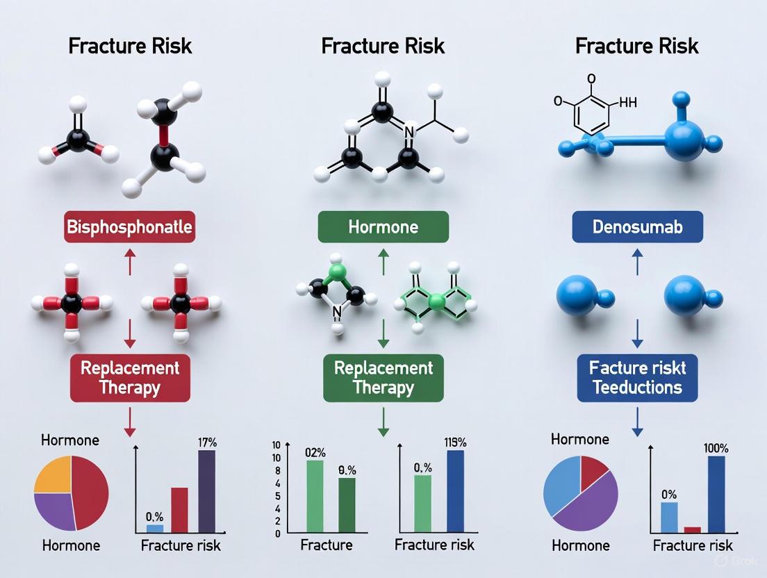
- Bisphosphonates: As the most widely used antiresorptive class, bisphosphonates have a high affinity for bone mineral, particularly at active resorption sites. They are internalized by osteoclasts and disrupt key intracellular pathways. Nitrogen-containing bisphosphonates (e.g., alendronate, zoledronic acid) inhibit the enzyme farnesyl pyrophosphate (FPP) synthase in the mevalonate pathway. This inhibition prevents the prenylation of small GTPase signaling proteins (such as Rac, Ras, and Rho), which are essential for osteoclast cytoskeletal organization, survival, and function, ultimately leading to osteoclast apoptosis [7]. Non-nitrogen-containing bisphosphonates are metabolized into cytotoxic analogs of ATP that induce osteoclast apoptosis [7].
- Anti-RANKL Antibodies (e.g., Denosumab): This class targets the Receptor Activator of Nuclear Factor-κB Ligand (RANKL) system, a pivotal pathway for osteoclast differentiation, activation, and survival. Denosumab, a monoclonal antibody, binds to RANKL, preventing its interaction with the RANK receptor on osteoclast precursors and mature osteoclasts. This inhibition results in reduced osteoclast formation and function, thereby decreasing bone resorption [8] [9].
- Other Antiresorptives: Additional classes include Selective Estrogen Receptor Modulators (SERMs), which mimic estrogen's protective effect on bone in some tissues, and Calcitonin, which directly inhibits osteoclast activity [7] [9].
Anabolic Agents: Stimulating Bone Formation
Anabolic agents directly stimulate osteoblast activity, leading to the formation of new bone and improved bone microarchitecture.
- Parathyroid Hormone Receptor Agonists (e.g., Teriparatide, Abaloparatide): These synthetic peptides (Teriparatide is PTH(1-34); Abaloparatide is a PTHrP analog) act as agonists of the PTH1 receptor. Their anabolic effect is achieved through intermittent administration, which differs from the persistent elevation of PTH in hyperparathyroidism (which is catabolic to bone). The mechanism involves the activation of multiple signaling pathways, including upregulation of Wnt signaling and increased production of insulin-like growth factors (IGFs) and fibroblast growth factors (FGFs) in osteoblasts. This leads to increased osteoblast proliferation, differentiation, and activity, and a reduction in osteoblast apoptosis. Critically, this creates an "anabolic window" where the increase in bone formation markers precedes and exceeds the subsequent increase in bone resorption markers [6].
- Anti-Sclerostin Antibodies (e.g., Romosozumab): This novel class targets sclerostin, a glycoprotein produced primarily by osteocytes that acts as a potent negative regulator of bone formation. Sclerostin inhibits the canonical Wnt/β-catenin signaling pathway by binding to the LRP5/6 co-receptors. Romosozumab, a humanized monoclonal antibody, neutralizes sclerostin, thereby releasing this inhibition and permitting Wnt signaling to proceed. This results in a dual effect: a potent stimulation of bone formation and a moderate reduction in bone resorption [6] [9]. The bone formation induced by sclerostin inhibition is believed to be primarily modeling-based (de novo bone formation), unlike the remodeling-based formation stimulated by PTH analogs [6].
The following diagram illustrates the key signaling pathways targeted by these anabolic and antiresorptive agents.
Comparative Efficacy: Fracture Risk Reduction and BMD Changes
Key Clinical Trial Designs and Patient Populations
The efficacy data for osteoporosis therapies are primarily derived from large, randomized, double-blind, placebo-controlled or active-comparator trials. Key trials include:
- Fracture Endpoint Trials: These are typically long-term (3-5 year) studies with primary endpoints being the incidence of new vertebral fractures (assessed radiographically) and non-vertebral fractures (e.g., hip, wrist). Examples include the Fracture Intervention Trial (FIT) for alendronate, the HORIZON trial for zoledronic acid, and the FREEDOM trial for denosumab [7] [10].
- BMD and Biomarker Trials: Shorter-term studies (1-2 years) often use BMD change (measured by Dual-Energy X-ray Absorptiometry, DXA) and bone turnover markers (BTMs) like P1NP (formation) and CTX (resorption) as surrogate endpoints for fracture efficacy [11] [9].
- High-Risk Patient Populations: Recent trials, such as ARCH (romosozumab vs. alendronate) and VERO (teriparatide vs. risedronate), specifically enrolled postmenopausal women with severe osteoporosis and prior fractures to compare the efficacy of anabolic versus antiresorptive agents in high-risk populations [6] [10].
Quantitative Comparison of Anti-Fracture Efficacy
The following table summarizes the relative risk reduction for fractures associated with different drug classes, as reported in pivotal clinical trials and meta-analyses.
Table 1: Comparative Anti-Fracture Efficacy of Osteoporosis Therapies
| Drug Class / Agent | Vertebral Fracture Risk Reduction vs. Placebo/Control | Non-Vertebral Fracture Risk Reduction vs. Placebo/Control | Key Supporting Trial / Meta-Analysis |
|---|---|---|---|
| Bisphosphonates (e.g., Zoledronic Acid) | ~70% reduction [7] | ~41% reduction (hip) [7] | HORIZON [7] |
| Anti-RANKL (Denosumab) | 68% reduction at 3 years [10] | 40% reduction (hip) at 3 years [10] | FREEDOM [10] |
| PTH Analog (Teriparatide) | 65-69% reduction [6] | 53-54% reduction [6] | Neer et al. [6] |
| PTH Analog (Abaloparatide) | 86% reduction [10] | 43% reduction [10] | ACTIVE [10] |
| Anti-Sclerostin (Romosozumab) | 73% reduction at 1 year [9] | 25% reduction at 1 year (non-significant trend) [9] | FRAME [9] |
| Head-to-Head: Teriparatide vs. Risedronate | 12% (Risedronate) vs. 5.4% (Teriparatide) incidence [6] | Clinical fractures: 9.8% (Risedronate) vs. 4.8% (Teriparatide) [6] | VERO [6] |
Quantitative Comparison of Bone Mineral Density (BMD) Changes
BMD gains, particularly at the hip, are a strong surrogate for anti-fracture efficacy. The table below shows typical BMD increases from key trials and a recent network meta-analysis.
Table 2: Comparative Bone Mineral Density (BMD) Changes
| Drug Class / Agent | Lumbar Spine BMD Change | Femoral Neck BMD Change | Duration | Source |
|---|---|---|---|---|
| Anti-Sclerostin (AS) Antibody | +13.30% (95% CI: 9.15-17.45) [9] | +6.00% (95% CI: 3.34-8.66) [9] | 12 months | Network Meta-Analysis [9] |
| Anti-RANKL (AR) Antibody | +9.49% (95% CI: 6.60-12.38) [9] | +5.67% (95% CI: 2.61-8.74) [9] | 36 months | Network Meta-Analysis [9] |
| PTH Analog (Teriparatide) | +9.7% (20 mcg dose) [6] | +2.8% (20 mcg dose) [6] | 18 months | Neer et al. [6] |
| Bisphosphonates (e.g., Zoledronic Acid) | +4-6% (typical range) [7] | +2-4% (typical range) [7] | 36 months | HORIZON [7] |
Research Reagent Solutions for Investigating Mechanisms
To study the mechanisms described, researchers require specific reagents and tools. The following table details essential materials for experimental protocols in bone biology.
Table 3: Key Research Reagents for Osteoporosis Mechanism Studies
| Reagent / Assay | Primary Function / Application | Relevance to Drug Class |
|---|---|---|
| HR-pQCT (High-Resolution Peripheral Quantitative Computed Tomography) | In vivo 3D assessment of bone microarchitecture (trabecular and cortical) at peripheral sites (radius, tibia). | Evaluates anabolic drug effects on cortical porosity and trabecular structure [12]. |
| Bone Turnover Markers (BTMs): • P1NP (Procollagen type 1 N-terminal propeptide) • CTX (C-terminal telopeptide of type I collagen) | P1NP: Serum marker of bone formation.CTX: Serum marker of bone resorption. | Monitors pharmacodynamic response. Anabolics rapidly increase P1NP. Antiresorptives rapidly decrease CTX [11]. |
| Finite Element Analysis (FEA) | Computational modeling based on CT/HR-pQCT data to estimate bone strength. | Used in clinical trials to predict bone strength changes from anabolic therapy beyond BMD [12]. |
| In Vitro Osteoclastogenesis Assay | Differentiate osteoclasts from primary (e.g., PBMCs) or immortalized precursor cells; assess osteoclast number, size, and resorptive activity. | Fundamental for testing antiresorptive agents (Bisphosphonates, Denosumab) [7]. |
| SOST/Sclerostin ELISA & Neutralizing Antibodies | Quantify sclerostin levels; use anti-sclerostin antibodies to probe Wnt pathway function in cell models. | Core for developing and testing anti-sclerostin therapeutics like romosozumab [6] [9]. |
| PTH1R Agonists & Cell-Based cAMP Assays | Test potency and efficacy of PTH analogs (e.g., teriparatide, abaloparatide) via receptor activation and downstream signaling (e.g., cAMP production). | Essential for screening and characterizing anabolic PTH receptor agonists [6]. |
The distinct core mechanisms of antiresorptive and anabolic agents underlie their different efficacy profiles and clinical applications. Evidence from pivotal trials and meta-analyses confirms that anabolic agents provide superior gains in BMD and more rapid fracture risk reduction in high-risk patients compared to antiresorptives [6] [9] [10]. However, the bone-forming effects of anabolics are transient, plateauing after 12-24 months [6]. This has led to the critical concept of sequential therapy: initiating treatment with an anabolic agent to rebuild bone, followed by an antiresorptive (e.g., denosumab or a bisphosphonate) to consolidate and maintain the gains achieved [11] [10]. Studies have consistently shown that this "anabolic-first" sequence results in greater BMD increases than the reverse sequence or monotherapy [11] [10]. For drug development, future directions include exploring new anabolic targets beyond the PTH and Wnt pathways, optimizing treatment sequences, and personalizing therapy based on individual patient risk profiles, bone turnover status, and comorbidities such as diabetes, which presents a unique bone fragility phenotype [12]. A deep understanding of these core mechanisms remains the foundation for these advancements.
In the field of osteoporosis research, the efficacy of pharmacologic interventions is rigorously assessed through predefined fracture outcomes, which are categorized primarily as vertebral, non-vertebral, and hip fractures [13]. These endpoints serve as critical indicators of treatment success and are fundamental to clinical trial design and drug development. Vertebral fractures, the most common osteoporotic fracture, often manifest as compression fractures of the anterior column and can be asymptomatic, yet they significantly predict future fracture risk and mortality [13] [14]. Non-vertebral fractures encompass a broad group of fragility fractures at other major sites, including the wrist, humerus, pelvis, and others, while hip fractures, though less frequent, carry the most severe consequences in terms of morbidity, mortality, and healthcare costs [15] [16]. This guide provides a structured comparison of how different osteoporosis treatments perform against these fracture outcomes, synthesizing current clinical data, detailed methodologies from pivotal trials, and the underlying molecular mechanisms of action.
Comparative Efficacy of Osteoporosis Treatments on Fracture Outcomes
The landscape of osteoporosis management has evolved from traditional antiresorptives to anabolic and dual-action therapies. The tables below summarize the relative risk reduction for vertebral, non-vertebral, and hip fractures from key clinical trials of established and novel treatments.
Table 1: Relative Risk Reduction of Key Antiresorptive Therapies vs. Placebo
| Treatment (Class) | Vertebral Fracture Risk Reduction | Non-Vertebral Fracture Risk Reduction | Hip Fracture Risk Reduction | Key Trial / Source |
|---|---|---|---|---|
| Alendronate (Bisphosphonate) | ~50% | Reported significant reduction | Reported significant reduction | Various RCTs [1] |
| Zoledronic Acid (Bisphosphonate) | Significant reduction | Significant reduction | Significant reduction | HORIZON Trial [16] |
| Denosumab (RANKL inhibitor) | 68% | 20% | 40% | FREEDOM Trial [17] |
| Selective Estrogen Receptor Modulators | Significant reduction | Not consistently significant | Not consistently significant | Various RCTs [1] |
Table 2: Relative Risk Reduction of Anabolic and Dual-Action Therapies vs. Control
| Treatment (Class) | Vertebral Fracture Risk Reduction | Non-Vertebral Fracture Risk Reduction | Hip Fracture Risk Reduction | Key Trial / Source |
|---|---|---|---|---|
| Teriparatide (PTH analog) | Significant reduction | Significant reduction | Data limited | VERO Trial [1] |
| Romosozumab (Sclerostin inhibitor) | Superior risk reduction vs. control | Superior risk reduction vs. control | Superior risk reduction vs. control | FRAME Trial [1] [18] |
| Abaloparatide (PTHrP analog) | Significant reduction | Significant reduction | Data limited | ACTIVE Trial [18] |
For high-risk patients, sequential therapy, typically starting with an anabolic agent like teriparatide or romosozumab followed by an antiresorptive (e.g., denosumab or a bisphosphonate), has demonstrated greater gains in bone mineral density (BMD) and superior fracture risk reduction compared to monotherapy [1]. A direct head-to-head trial, the VERO study, found that teriparatide was superior to risedronate in reducing new vertebral and clinical fractures in postmenopausal women with severe osteoporosis [1].
Cost-effectiveness analyses show that while generic alendronate is often first-line, denosumab is a cost-effective alternative, particularly for high-risk patients. A 2025 US analysis found 10-year denosumab treatment was cost-effective versus a 5-year alendronate regimen followed by a drug holiday, with an incremental cost-effectiveness ratio of $97,574 per quality-adjusted life-year (QALY) gained [17].
Methodologies for Fracture Outcome Assessment in Clinical Trials
Defining and Ascertaining Fracture Endpoints
- Vertebral Fractures: In clinical trials, vertebral fractures are primarily identified radiologically rather than based on clinical symptoms. The Genant semi-quantitative method is the standard for classifying osteoporotic vertebral fractures (OVFs) on lateral radiographs of the spine [13] [19]. This method grades fractures (Genant grades 1-3) based on the degree of height loss (20-25% mild, 25-40% moderate, >40% severe) and morphological change (wedge, biconcave, crush) [13]. Centralized reading by expert radiologists blinded to treatment allocation is critical to minimize ascertainment bias, especially since many OVFs are asymptomatic.
- Hip and Non-Vertebral Fractures: These are typically confirmed clinically and radiographically following reports of incident fractures from trial participants. Adjudication committees, blinded to treatment group, review source documents (e.g., radiographs, surgical reports, discharge summaries) to verify the fracture site and whether it resulted from low-trauma or fragility [16] [20]. Non-vertebral fractures are usually a composite endpoint including humerus, wrist, pelvis, and other sites, but excluding fractures of the face, fingers, toes, and those due to high trauma [17].
Clinical Trial Design and Statistical Analysis
Pivotal trials are typically randomized, double-blind, and placebo- or active-controlled. The FREEDOM trial for denosumab is a classic example: a 3-year, multicenter, randomized, double-blind, placebo-controlled study involving 7,808 postmenopausal women with osteoporosis [17]. The primary efficacy endpoint was the incidence of new radiographic vertebral fractures at 36 months. Key secondary endpoints were time to first non-vertebral fracture and time to first hip fracture [17].
Analysis is performed on an intent-to-treat (ITT) basis. Time-to-event data for hip and non-vertebral fractures are analyzed using Cox proportional hazards models, yielding hazard ratios (HR) and confidence intervals (CI). The cumulative incidence of new vertebral fractures is compared between groups using logistic regression or Cochran-Mantel-Haenszel tests [17]. Network meta-analyses (NMAs) have emerged to compare multiple interventions simultaneously, using both direct and indirect evidence when head-to-head trials are limited [18].
Diagram 1: Standard workflow for a pivotal osteoporosis clinical trial, from randomization to outcome analysis.
Molecular Pathways and Therapeutic Mechanisms
The pharmacological agents used to reduce fracture risk target specific pathways in bone remodeling, the continuous cycle of resorption by osteoclasts and formation by osteoblasts.
- Antiresorptive Agents (Bisphosphonates, Denosumab): These work by inhibiting osteoclast activity. Bisphosphonates (e.g., alendronate, zoledronic acid) incorporate into bone mineral and are internalized by osteoclasts, disrupting the mevalonate pathway and inducing apoptosis [1]. Denosumab is a fully human monoclonal antibody that binds with high affinity to RANKL (Receptor Activator of Nuclear Factor-kappa B Ligand), a key cytokine produced by osteoblasts that is essential for osteoclast formation, activation, and survival [17]. By inhibiting RANKL, denosumab profoundly reduces bone resorption.
- Anabolic Agents (PTH analogs, Sclerostin inhibitors): These directly stimulate bone formation. Teriparatide and abaloparatide are analogs of Parathyroid Hormone (PTH) and PTH-related peptide. They act on the PTH1 receptor on osteoblasts to promote bone formation, with evidence showing they also enhance vertebral microarchitecture [18]. Romosozumab is a monoclonal antibody that inhibits sclerostin, a product of the SOST gene secreted by osteocytes that negatively regulates the Wnt signaling pathway, a key promoter of bone formation [1]. By blocking sclerostin, romosozumab simultaneously increases bone formation and decreases bone resorption, a dual effect that leads to rapid gains in bone mass and strength.
Diagram 2: Key molecular pathways targeted by anabolic and antiresorptive osteoporosis therapies.
The Scientist's Toolkit: Essential Research Reagents and Materials
Table 3: Key Reagents and Materials for Osteoporosis and Fracture Research
| Tool / Reagent | Primary Function in Research | Application Example |
|---|---|---|
| Dual-energy X-ray Absorptiometry (DXA) | Non-invasive measurement of areal Bone Mineral Density (BMD). | Primary endpoint in clinical trials; diagnosing osteoporosis (T-score ≤ -2.5) [15]. |
| Genant Semi-Quantitative Score | Standardized visual grading of vertebral fracture severity from spinal radiographs. | Centralized, blinded adjudication of incident vertebral fractures in trials [13] [19]. |
| Serum CTX (C-terminal telopeptide) | Biomarker of bone resorption; measures collagen breakdown products from osteoclast activity. | Monitoring treatment response and adherence to antiresorptive therapies [14]. |
| Serum PINP (Procollagen type 1 N-terminal propeptide) | Biomarker of bone formation; measures collagen synthesis by osteoblasts. | Monitoring treatment response to anabolic therapies [14]. |
| Micro-CT (Micro-computed Tomography) | High-resolution, 3D imaging of bone microarchitecture (trabecular and cortical). | Pre-clinical research to quantify changes in bone structure and strength in animal models. |
| FRAX Tool | Algorithm calculating a patient's 10-year probability of hip & major osteoporotic fracture. | Risk stratification for enrolling high-risk patients into clinical trials [15]. |
Defining and accurately assessing vertebral, non-vertebral, and hip fractures remains the cornerstone of evaluating osteoporosis treatments. The field has moved beyond simply comparing fracture rates to understanding the molecular pathways that therapies modulate, allowing for more targeted drug development. While antiresorptives like bisphosphonates and denosumab provide robust fracture reduction, anabolic agents offer a powerful alternative for high-risk patients, with sequential strategies optimizing long-term outcomes. Future research will focus on personalized medicine approaches, novel targets beyond the Wnt and RANKL pathways, and mitigating treatment gaps through models like Fracture Liaison Services to ensure that therapeutic advances translate into reduced fracture burdens for patients.
Establishing the Clinical and Economic Imperative for Comparative Analysis
Osteoporosis, a systemic metabolic disease characterized by decreased bone density, mass, and microarchitectural deterioration, presents a substantial global health burden with over 200 million affected individuals worldwide [1]. The disease significantly increases fracture susceptibility, with approximately one in three women and one in five men over age 50 experiencing osteoporotic fractures [1]. The economic impact is substantial, with annual costs in Australia alone exceeding $3.82 billion, of which fracture-related expenses account for 67% of total costs [1]. This clinical and economic burden creates an imperative for rigorous comparative analysis of osteoporosis treatments to optimize patient outcomes and healthcare resource allocation.
The fundamental pathophysiology of osteoporosis stems from an imbalance in bone remodeling, where bone resorption exceeds formation [1] [21]. This imbalance results from multiple factors including age-related changes, hormonal deficiencies, genetic factors, nutritional status, medications, and comorbid conditions [1]. Treatment strategies primarily target this imbalance through antiresorptive agents that inhibit osteoclast activity or anabolic agents that promote bone formation [1] [21]. The expanding therapeutic landscape, which now includes bisphosphonates, biologics like denosumab, anabolic agents such as teriparatide, and novel therapies including romosozumab, necessitates comprehensive comparative evaluation to establish their respective positions in clinical practice [1].
Comparative Clinical Efficacy of Osteoporosis Treatments
Fracture Risk Reduction Across Therapeutic Classes
Table 1: Comparative Fracture Risk Reduction for Osteoporosis Pharmacotherapies
| Treatment Class | Specific Agent | Vertebral Fracture Risk Reduction (HR/RR/OR) | Non-vertebral Fracture Risk Reduction (HR/RR/OR) | Hip Fracture Risk Reduction (HR/RR/OR) |
|---|---|---|---|---|
| Bisphosphonates | Zoledronate | HR 0.38 (95% CrI: 0.28-0.49) [22] | HR 0.71 (95% CrI: 0.61-0.81) [22] | Data paucity noted [22] |
| Risedronate | Not specified | HR 0.70 (95% CrI: 0.53-0.84) [22] | Not specified | |
| Alendronate | Not specified | HR 0.86 (95% CrI: 0.51-1.21) [17] | Not specified | |
| Biologics | Denosumab | 68% reduction [17] | 20% reduction [17] | 40% reduction [17] |
| Anabolic Agents | Teriparatide | Not specified | Not specified | Not specified |
| Combination Therapy | Teriparatide + Denosumab | OR 0.93 (95% CI: 0.12-6.93) [5] | OR 0.68 (95% CI: 0.31-1.46) [5] | Not specified |
Bone Mineral Density Improvements
Table 2: Comparative Bone Mineral Density (BMD) Changes Across Therapies
| Treatment | Lumbar Spine BMD Change | Femoral Neck BMD Change | Total Hip BMD Change | Duration |
|---|---|---|---|---|
| Zoledronate | Not specified | MD 4.02 (95% CrI: 3.2-4.84) [22] | Not specified | 3 years [22] |
| Teriparatide + Denosumab | +3.40% (95% CI: 0.44-6.36) [5] | +4.00% (95% CI: 1.96-6.04) [5] | +4.25% (95% CI: 3.20-5.29) [5] | 24 months [5] |
| Teriparatide + Bisphosphonates | Not significant | Not significant | +1.81% (95% CI: 0.65-2.97) [5] | <24 months [5] |
| Denosumab (10-year) | Continued increase [17] | Continued increase [17] | Continued increase [17] | 10 years [17] |
Network meta-analyses of bisphosphonates have established zoledronate as the most effective in preventing vertebral fractures (HR 0.38; 95% CrI, 0.28-0.49) and among the most effective for non-vertebral fractures, alongside risedronate [22]. The 10-year FREEDOM extension study demonstrated continued increases in BMD at the lumbar spine, total hip, femoral neck, and one-third radius with long-term denosumab treatment, alongside low fracture incidence [17]. Combination therapy approaches, particularly teriparatide with denosumab, have demonstrated superior BMD outcomes compared to monotherapy, although fracture risk reduction benefits remain less clearly established [5].
Bone Turnover Marker Responses
Combination therapies differentially regulate bone turnover markers. Bisphosphonate combinations with teriparatide suppress P1NP (40%-80% reduction versus monotherapy), while denosumab combinations with teriparatide preserve OC levels (-8% to 16% versus monotherapy) [5]. This bidirectional regulation of bone turnover markers highlights distinct mechanisms of action between antiresorptive classes when combined with anabolic therapy.
Economic Evaluations in Osteoporosis Management
Cost-Effectiveness Thresholds and Treatment Algorithms
Table 3: Cost-Effectiveness Analysis of Osteoporosis Treatments
| Comparison | Incremental Cost-Effectiveness Ratio (ICER) | Quality-Adjusted Life Years (QALYs) | Key Assumptions |
|---|---|---|---|
| 10-year Denosumab vs. 5-year Alendronate | $97,574 per QALY gained [17] | 8.035 (denosumab) vs. 7.977 (alendronate) [17] | US third-party payer perspective, lifetime horizon [17] |
| Generic Bisphosphonate vs. No Treatment | Cost-effective at 3% 10-year hip fracture probability [23] | Varies by baseline risk | $600/year drug cost, 35% fracture reduction, 5-year treatment [23] |
Economic evaluations consistently demonstrate that osteoporosis treatment becomes cost-effective when the 10-year hip fracture probability reaches approximately 3% [23]. Although the relative risk at which treatment becomes cost-effective varies markedly between genders and race/ethnicity groups, the absolute 10-year hip fracture probability threshold remains consistent across populations [23]. Application of the WHO FRAX algorithm to identify individuals exceeding this 3% threshold facilitates efficient osteoporosis treatment allocation [23].
Recent analyses indicate that 10-year denosumab treatment would be cost-effective compared with 5 years of alendronate, followed by a 2-year drug holiday and 3 additional years of alendronate, with an incremental cost-effectiveness ratio of $97,574 per QALY gained at a $150,000 threshold [17]. Probabilistic sensitivity analysis demonstrated denosumab was cost-effective in 62.1% of simulations [17].
Methodological Framework for Comparative Studies
Experimental Designs and Outcome Measures
Randomized controlled trials (RCTs) remain the gold standard for evaluating osteoporosis treatments. Key methodological considerations include:
- Study Duration: Most trials extend 1-3 years for fracture outcomes, with extension studies (e.g., FREEDOM 10-year extension) providing long-term data [17]
- Primary Endpoints: Radiographically-confirmed vertebral fractures, clinically-apparent non-vertebral fractures, hip fractures [22] [17]
- Secondary Endpoints: BMD changes measured by dual-energy X-ray absorptiometry (DXA), bone turnover markers (BTMs), safety parameters [5] [22]
- Statistical Considerations: Intent-to-treat analysis, Cox proportional hazards models for time-to-fracture data, mixed models for repeated measures of BMD
Network meta-analyses have emerged as powerful tools for comparative effectiveness research, enabling simultaneous comparison of multiple interventions across different trials [22]. Recent applications have included 25 additional trials with a total population of 47,007 participants, substantially enhancing precision of comparative estimates [22].
Bone Biology Signaling Pathways in Osteoporosis Therapeutics
Research Reagent Solutions for Osteoporosis Studies
Table 4: Essential Research Reagents for Osteoporosis Investigations
| Reagent/Category | Primary Function | Specific Examples/Applications |
|---|---|---|
| Cell Culture Models | In vitro simulation of bone remodeling | Primary osteoblasts/osteoclasts, MC3T3-E1 pre-osteoblastic cells, RAW 264.7 osteoclast precursors |
| Bone Turnover Assays | Quantification of bone formation/resorption | ELISA for P1NP (formation), CTX (resorption), RANKL/OPG ratios |
| Molecular Biology Tools | Pathway manipulation and analysis | siRNA for Sclerostin/Wnt pathway, RANKL overexpression vectors |
| Imaging Reagents | Bone structure and quality assessment | Micro-CT contrast agents, calcein double-labeling for dynamic histomorphometry |
| Animal Models | In vivo fracture healing and therapeutic testing | Ovariectomized rodents (postmenopausal model), aged animals (senile osteoporosis) |
Comparative Research Analysis Workflow
Discussion and Future Directions
The expanding therapeutic landscape for osteoporosis, including novel agents targeting sclerostin, Wnt pathway, and cathepsin K, underscores the continuing need for rigorous comparative analysis [1]. Future research priorities include establishing long-term safety and durable efficacy for novel therapies, conducting comparative effectiveness trials of optimal sequential therapy strategies, and developing personalized medicine markers for risk and response prediction [1].
Overcoming the treatment gap remains a significant challenge, with fewer than 25% of patients who experience an osteoporotic fracture receiving appropriate treatment [1]. This gap is particularly pronounced in men, younger postmenopausal women, and those with glucocorticoid-induced osteoporosis [1]. Implementation of Fracture Liaison Services (FLS) has demonstrated effectiveness in identifying and managing high-risk patients, providing a systematic approach to bridge this gap [1].
The evolving paradigm in osteoporosis management emphasizes initial potent anabolic or dual-action therapy followed by antiresorptives for high-risk patients, moving beyond traditional approaches of indefinite antiresorptive monotherapy [1]. This approach, supported by emerging comparative evidence, promises to enhance fracture reduction outcomes while maintaining acceptable cost-effectiveness profiles in an increasingly constrained healthcare economic environment.
Research Methodologies for Comparing Treatment Efficacy in Real-World Practice
Evidence from Randomized Placebo-Controlled Trials (RPCTs) and Their Limitations
Osteoporosis, a systemic metabolic bone disease characterized by low bone mass and microarchitectural deterioration, poses a significant global health burden with an estimated prevalence exceeding 200 million individuals worldwide [1]. The clinical consequence of this "silent disease" is an increased susceptibility to fragility fractures, with approximately one in three women and one in five men over 50 experiencing osteoporotic fractures [1]. The development of effective pharmacological treatments has relied heavily on evidence generated from randomized placebo-controlled trials (RPCTs), which represent the gold standard for establishing efficacy and safety.
These trials have been fundamental in building the therapeutic arsenal for osteoporosis, which primarily includes antiresorptive agents (such as bisphosphonates and denosumab) that inhibit bone breakdown, and anabolic agents (such as teriparatide, abaloparatide, and romosozumab) that promote bone formation [1]. RPCTs have provided the critical evidence required for regulatory approval by demonstrating that these treatments significantly reduce the incidence of vertebral, non-vertebral, and hip fractures compared to placebo. This article provides a comprehensive comparison of the fracture risk reduction efficacy of various osteoporosis treatments based on evidence from RPCTs and meta-analyses, while critically examining the inherent limitations of this evidence base and its implications for clinical practice and drug development.
Comparative Efficacy of Osteoporosis Treatments from RPCTs and Meta-Analyses
Systematic reviews and network meta-analyses of RPCTs provide the most robust comparative effectiveness data for osteoporosis treatments. A landmark 2023 network meta-analysis published in The BMJ, which synthesized results from 69 randomized clinical trials involving over 80,000 postmenopausal women, offers key insights into the relative performance of different drug classes [24].
Table 1: Fracture Risk Reduction of Osteoporosis Treatments from Network Meta-Analysis
| Treatment Class | Clinical Fractures (vs. Placebo) | Vertebral Fractures (vs. Placebo) | Key Comparative Effectiveness Findings |
|---|---|---|---|
| Bisphosphonates | Protective effect [24] | Protective effect [24] | Less effective than PTH receptor agonists for clinical fractures (OR 1.49) [24] |
| Denosumab | Protective effect [24] | Protective effect [24] | More effective than oral bisphosphonates for vertebral fractures; less effective than PTH agonists and romosozumab for clinical fractures [24] |
| PTH Receptor Agonists | Protective effect [24] | Protective effect [24] | More effective than bisphosphonates and denosumab for clinical fractures [24] |
| Romosozumab | Protective effect [24] | Protective effect [24] | More effective than denosumab for clinical fractures (OR 0.64 for denosumab vs. romosozumab) [24] |
| SERMs (Raloxifene) | Data from analysis | Data from analysis | -- |
The data consistently demonstrate that all major treatment classes provide significant fracture protection compared to placebo. However, important differences emerge in head-to-head comparisons: bone anabolic treatments (romosozumab and PTH receptor agonists) generally show superior efficacy over bisphosphonates in preventing clinical and vertebral fractures [24]. This superior efficacy of anabolic agents is particularly evident in patients at high fracture risk.
Real-World Evidence and Active Comparator Studies
While RPCTs establish efficacy versus placebo, real-world evidence and active comparator studies provide complementary insights into relative effectiveness in clinical practice. A 2025 comparative effectiveness study using a nationwide Japanese database found that romosozumab was associated with a 20% lower incidence of major osteoporotic fractures over one year compared to teriparatide (HR: 0.80) [25]. This real-world data aligns with clinical trial evidence suggesting particularly potent fracture reduction with romosozumab.
Conversely, a large US database study (2018) found minimal differences in fracture rates between most oral osteoporosis medications in routine care, with raloxifene and alendronate showing the lowest unadjusted fracture rates [3]. This discrepancy with RCT findings may reflect channeling bias, where higher-risk patients are prescribed more potent injectable therapies, or differences in adherence patterns in real-world settings.
Table 2: Real-World Fracture Incidence from Cohort Studies
| Medication | Fracture Incidence (per 100 person-years) | Study Characteristics |
|---|---|---|
| Raloxifene | 1.24 [3] | Retrospective cohort (MarketScan database 2008-2012) [3] |
| Alendronate | 1.54 [3] | Retrospective cohort (MarketScan database 2008-2012) [3] |
| Risedronate | 1.57 [3] | Retrospective cohort (MarketScan database 2008-2012) [3] |
| Ibandronate | 1.66 [3] | Retrospective cohort (MarketScan database 2008-2012) [3] |
| Zoledronic Acid | 1.98 [3] | Retrospective cohort (MarketScan database 2008-2012) [3] |
| Teriparatide | 3.90 [3] | Retrospective cohort (MarketScan database 2008-2012) [3] |
| Romosozumab | 7.01 (1-year incidence) [25] | Prospective cohort (Japanese database, 2019-2021) [25] |
| Teriparatide | 10.14 (1-year incidence) [25] | Prospective cohort (Japanese database, 2019-2021) [25] |
Key Methodologies in Osteoporosis RPCTs
Standardized Experimental Protocols
Robust RPCTs in osteoporosis share common methodological elements designed to generate high-quality, reproducible evidence.
Patient Population and Recruitment: Most RPCTs focus on postmenopausal women with osteoporosis, defined by specific bone mineral density (BMD) criteria (typically T-score ≤ -2.5 at lumbar spine or hip) and/or presence of pre-existing vertebral fractures [24]. Participants are often stratified based on additional risk factors such as age, prior fracture history, and baseline BMD. Increasingly, trials are enrolling "high-risk" and "very high-risk" patients to demonstrate efficacy in populations most likely to benefit from potent anabolic therapies [1].
Intervention and Comparator Arms: Trials typically compare active drug against placebo or an active comparator, often with double-blind, double-dummy designs to maintain blinding [24]. Standardized administration protocols are used for each drug class, including specific dosing intervals (e.g., daily for teriparatide, weekly/oral for many bisphosphonates, every 6 months for denosumab, and monthly for romosozumab) [24] [25]. Most trials include calcium and vitamin D supplementation in both arms to ensure nutritional adequacy.
Outcome Measures and Follow-up: The primary endpoints typically include:
- Vertebral fracture incidence: Assessed using standardized radiograph or densitometer-based morphometry at predefined intervals (usually annually) [24].
- Non-vertebral and hip fractures: Confirmed radiographically and adjudicated by blinded endpoint committees [24].
- Bone Mineral Density (BMD) changes: Measured by dual-energy X-ray absorptiometry (DXA) at lumbar spine, total hip, and femoral neck [1].
- Safety parameters: Comprehensive monitoring of adverse events, including drug-specific concerns like osteonecrosis of the jaw (ONJ), atypical femoral fractures (AFF), and cardiovascular events [1].
Typical trial duration ranges from 1-3 years, which is considered sufficient to detect meaningful differences in fracture outcomes between treatment groups [24].
Advanced Imaging and Biomechanical Assessment
Beyond standard DXA, advanced imaging technologies are increasingly used in RPCTs to provide deeper insights into drug mechanisms and treatment effects on bone quality.
High-Resolution Peripheral Quantitative CT (HR-pQCT) is a sophisticated imaging technique that enables three-dimensional assessment of bone microarchitecture at peripheral sites like the distal radius and tibia [26]. This method provides detailed information on trabecular and cortical compartments, including volumetric BMD, cortical thickness and porosity, trabecular number, thickness, and separation [26]. HR-pQCT parameters have been shown to improve fracture prediction beyond DXA alone and can detect specific microstructural changes in response to different osteoporosis treatments [26] [27].
Finite Element Analysis (FEA) uses HR-pQCT images to computationally estimate bone strength by simulating biomechanical loading conditions [26]. This technique can provide insights into how different treatments affect bone biomechanical properties beyond what BMD measurements alone can reveal. For instance, FEA has demonstrated that teriparatide and abaloparatide increase bone strength by expanding periosteal and endosteal perimeters, creating a larger, structurally sound bone, while antiresorptives primarily increase endocortical bone by improving mineralization [28].
Limitations of RPCT Evidence
Methodological and Generalizability Constraints
While RPCTs provide the highest level of evidence for drug efficacy, they have significant limitations that affect the translation of their findings to routine clinical practice.
Strict Inclusion/Exclusion Criteria used in RPCTs create homogeneous study populations that often don't reflect real-world complexity. Many trials exclude patients with significant comorbidities, those on multiple medications, and individuals with various forms of secondary osteoporosis [3]. This limits the generalizability of findings to the broader patient population seen in clinical practice, particularly older adults with multiple chronic conditions.
The relatively short duration of most osteoporosis RPCTs (typically 1-3 years) fails to capture long-term treatment effects, safety concerns, and the need for sequential therapy strategies [1]. This is particularly relevant for chronic conditions like osteoporosis that require management over decades. The field lacks robust evidence regarding optimal treatment duration, drug holidays, and sequencing of different agents.
Use of Surrogate Endpoints like BMD changes, while practical for clinical trials, do not perfectly correlate with fracture risk reduction. Similarly, radiographic vertebral fractures detected in clinical trials often differ clinically from the symptomatic fractures that bring patients to medical attention [28]. This can lead to overestimation of treatment effects that may not translate to meaningful clinical benefits for patients.
Specific Evidence Gaps and Biases
Beyond generalizability issues, RPCT evidence contains substantial gaps that impact clinical decision-making.
There is a paucity of head-to-head trials comparing active treatments, particularly between newer anabolic agents. Most comparisons are made indirectly through network meta-analyses or observed in real-world studies with potential channeling bias [25]. For instance, the comparative effectiveness of romosozumab versus teriparatide has primarily been studied in observational settings rather than large-scale RPCTs [25].
Publication and reporting biases may lead to overestimation of efficacy and underestimation of harms. Trials with positive results are more likely to be published, and safety signals may only emerge after longer follow-up or through post-marketing surveillance [1]. Additionally, most RPCTs are industry-sponsored, potentially introducing further bias in study design and reporting.
The efficacy-effectiveness gap is substantial in osteoporosis treatment. While RCTs demonstrate 50-70% relative risk reduction in vertebral fractures, effectiveness in clinical practice is often lower due to issues with adherence, persistence, and suboptimal monitoring [3]. Real-world studies suggest that between 2-26% of compliant patients sustain a fracture each year while being treated with osteoporosis medications [3].
Research Reagents and Methodological Tools
Table 3: Key Research Reagent Solutions in Osteoporosis Trials
| Reagent/Technology | Primary Application | Key Features and Functions |
|---|---|---|
| Dual-energy X-ray Absorptiometry (DXA) | Bone Mineral Density (BMD) measurement [28] | Areal BMD assessment; Diagnostic T-score calculation; WHO diagnostic classification [28] |
| High-Resolution Peripheral Quantitative CT (HR-pQCT) | Bone microarchitecture assessment [26] | 3D visualization of trabecular and cortical compartments; In vivo resolution of 61-82 μm; Low radiation dose (3-5 μSv) [26] |
| Finite Element Analysis (FEA) | Bone strength estimation [26] | Computational modeling of biomechanical properties; Predicts bone stiffness and failure load [26] |
| Bone Turnover Markers (BTMs) | Treatment monitoring [1] | Biochemical markers of bone formation (P1NP) and resorption (CTX); Early response assessment [1] |
| FRAX Algorithm | Fracture risk assessment [1] | Calculates 10-year probability of major osteoporotic fracture; Incorporates clinical risk factors with/without BMD [1] |
The landscape of osteoporosis treatment continues to evolve beyond traditional therapies. Novel therapeutic targets including modulators of the Wnt pathway beyond sclerostin, cytokine inhibitors, and bone cell metabolism regulators are under investigation [1]. Additionally, emerging technologies such as oral formulations of anabolic agents (e.g., SIK inhibitors) and advanced drug delivery systems aim to improve treatment adherence and accessibility [29].
Future research should focus on addressing critical evidence gaps through long-term comparative effectiveness trials, personalized medicine approaches using genetic and imaging biomarkers, and optimal sequencing strategies [1]. The growing recognition of the importance of initiating treatment with potent anabolic or dual-action therapies followed by antiresorptives for high-risk patients represents a paradigm shift in osteoporosis management [1].
In conclusion, while RPCTs have provided fundamental evidence for the efficacy of osteoporosis treatments in reducing fracture risk, they have important limitations in generalizability, duration, and comparative data. The integration of RPCT evidence with real-world data, advanced imaging technologies, and understanding of drug mechanisms provides the most comprehensive approach to treatment selection. As the therapeutic arsenal expands, future research should focus on personalized treatment strategies and direct comparisons between active therapies to optimize patient outcomes across the spectrum of fracture risk.
The Role of Large-Scale Observational and Database Studies
Osteoporosis, characterized by compromised bone strength and an increased risk of fragility fractures, represents a significant global health burden with profound clinical and economic consequences [30] [31]. While randomized controlled trials (RCTs) remain the gold standard for establishing drug efficacy, large-scale observational and database studies have emerged as indispensable tools for comparing the real-world effectiveness of osteoporosis treatments across diverse patient populations and practice settings. These studies fill critical evidence gaps left by RCTs, which often employ strict inclusion criteria that may limit generalizability to complex patients encountered in routine clinical practice [32]. The comparative fracture risk profile of different pharmacological interventions constitutes a central focus of this research, providing essential insights for clinicians, researchers, and drug development professionals seeking to optimize patient outcomes.
Database studies leverage routinely collected health information to evaluate treatment performance outside experimental conditions, capturing how medications perform across broad populations with varying compliance patterns, comorbidities, and concomitant treatments. This article systematically examines how these methodological approaches have advanced our understanding of osteoporosis treatment effectiveness, with particular emphasis on comparative fracture outcomes between key drug classes, including bisphosphonates, denosumab, and teriparatide.
Core Methodologies in Database Osteoporosis Research
Large-scale database research in osteoporosis typically utilizes several distinct data sources and methodological approaches, each with specific strengths and limitations:
- Administrative Claims Databases: Sources like the MarketScan Commercial Claims and Encounters database contain de-identified demographic information and paid claims for inpatient and outpatient medical services and prescriptions for commercial populations [32]. These databases enable researchers to identify large patient cohorts, track medication initiation, and document fracture outcomes through International Classification of Diseases (ICD) codes.
- Target Trial Emulation: This sophisticated observational approach attempts to mimic the design of an RCT using observational data by explicitly defining eligibility criteria, treatment strategies, outcome measurements, and follow-up periods [33]. This methodology was recently employed to compare cardiovascular safety and fracture prevention effectiveness of denosumab versus oral bisphosphonates in dialysis-dependent patients.
- Cross-Sectional Analyses: These studies examine relationships between variables at a single point in time, such as assessing fracture risk differences between osteoporosis and osteopenia patients using tools like the FRAX index [30].
Key Methodological Considerations
Robust database studies implement specific methodological safeguards to ensure valid comparisons:
- New User Design: To minimize selection bias, studies often identify patients who started a newly prescribed osteoporosis medication, including only those with a 12-month period with no filled prescriptions for osteoporosis medications preceding the new treatment [32].
- Minimum Exposure Periods: Researchers typically exclude patients who sustain fractures within the first 12 months of starting a new medication to allow adequate time for therapeutic effects to manifest [32].
- Risk Adjustment: Statistical methods like logistic regression adjust for known fracture risk factors, including age, sex, previous fractures, rheumatoid arthritis, glucocorticoid use, and other comorbidities [32].
- Time-to-Event Analysis: Some studies employ survival analysis techniques to account for varying follow-up times between patients, though others use fixed follow-up periods (e.g., 3 years) with risk differences and risk ratios as primary outcomes [33].
Table 1: Common Data Sources for Osteoporosis Comparative Effectiveness Research
| Data Source Type | Key Characteristics | Primary Applications | Example from Literature |
|---|---|---|---|
| Administrative Claims Databases | Contain billing records for large populations; include diagnoses, procedures, prescriptions | Drug utilization patterns, fracture rates, comparative safety | MarketScan Database analysis comparing multiple osteoporosis medications [32] |
| National Health Registers | Comprehensive population coverage; often include detailed clinical data | Epidemiology, long-term outcomes, rare adverse events | Japanese administrative claims database for dialysis patients [33] |
| Electronic Health Records | Rich clinical data including lab results, imaging reports, clinical notes | Disease progression, treatment response, comorbidity impact | Rehabilitation clinic data for FRAX assessment [30] |
| Prospective Cohort Studies | Designed for research purposes; carefully measured exposures and outcomes | Pathophysiology, risk factor identification, natural history | Not prominently featured in current search results |
Comparative Effectiveness of Osteoporosis Treatments: Findings from Database Studies
Fracture Outcomes Across Medication Classes
A comprehensive analysis of the Truven Health Analytics MarketScan database (2008-2012) provides direct comparisons of fracture rates across multiple osteoporosis medications [32]. This study included 51,649 patients with an average age of 56 years, following them for an average of 2 years after a 12-month treatment lead-in period. The overall incidence rate of fracture was 1.55 per 100 person-years of treatment, with significant variation between medication classes:
- Oral medications demonstrated the lowest fracture rates, led by raloxifene (1.24/100 person-years) and alendronate (1.54/100 person-years)
- Parenterally administered medications showed higher fracture rates, including teriparatide (3.90/100 person-years) and zoledronic acid (1.98/100 person-years)
- After adjusting for risk factors, no statistically significant differences emerged between most drugs in head-to-head comparisons, including between oral and parenteral bisphosphonates [32]
These findings suggest that while fracture rate variations exist in real-world settings, most differences diminish after accounting for patient characteristics, supporting previous evidence that minimal efficacy differences exist between osteoporosis medications when adherence is comparable.
Denosumab Versus Bisphosphonates in High-Risk Populations
Recent database studies have specifically compared denosumab with bisphosphonates in specialized patient populations:
- Dialysis Patients: A 2025 target trial emulation study of 1,032 dialysis-dependent patients (average age 74.5 years) found that compared with oral bisphosphonates, denosumab lowered the risk for composite fractures by 45% (3-year risk ratio 0.55) but potentially increased the risk for major adverse cardiac events (MACE) by 36% (3-year risk ratio 1.36) [33]. The absolute 3-year risk difference for composite fractures was -5.3%, favoring denosumab for fracture prevention in this high-risk population.
- Cancer Patients with Anti-Hormonal Therapy: A systematic review of 18 research papers found denosumab significantly increased bone mineral density (BMD) for up to two years and showed better results than bisphosphonates in patients receiving androgen deprivation therapy for prostate cancer or aromatase inhibitors for breast cancer, with comparable safety profiles [34].
- Rheumatoid Arthritis Patients: A meta-analysis of 1,441 patients from 9 studies demonstrated that denosumab treatment significantly increased lumbar spine BMD in rheumatoid arthritis patients with osteoporosis compared to controls and reduced joint damage scores [35].
Combination Therapy Versus Monotherapy
Meta-analyses of RCTs have provided insights into combination therapies, with database studies offering complementary real-world evidence:
- Teriparatide-Denosumab Combination: A 2025 meta-analysis showed that teriparatide combined with denosumab significantly increased BMD at the lumbar spine (+3.40%), femoral neck (+4.00%), and total hip (+4.25%) compared to teriparatide monotherapy, though without significant differences in vertebral or non-vertebral fracture risk reduction [5].
- Teriparatide-Bisphosphonate Combination: Bisphosphonate combinations improved hip BMD in the short term (<24 months: +1.81%) but not long-term (≥24 months) [5].
Table 2: Comparative Fracture Rates and Bone Density Effects Across Osteoporosis Treatments
| Treatment | Fracture Rate (per 100 person-years) | Spine BMD Effect | Hip BMD Effect | Key Patient Populations |
|---|---|---|---|---|
| Raloxifene | 1.24 [32] | Moderate improvement | Moderate improvement | Postmenopausal women [32] |
| Alendronate | 1.54 [32] | Significant improvement [36] | Significant improvement [36] | Broad osteoporosis populations [32] |
| Risedronate | 1.61 [32] | Significant improvement | Significant improvement | Broad osteoporosis populations [32] |
| Ibandronate | 1.65 [32] | Significant improvement | Significant improvement | Broad osteoporosis populations [32] |
| Zoledronic Acid | 1.98 [32] | Significant improvement | Significant improvement | Broad osteoporosis populations [32] |
| Denosumab | 1.72 [32] | Significant improvement [36] | Variable improvement [36] | RA, cancer, dialysis patients [34] [35] [33] |
| Teriparatide | 3.90 [32] | Positive trend [36] | Positive trend [36] | Severe osteoporosis [32] |
| Denosumab + Teriparatide | Not reported | +3.40% vs monotherapy [5] | +4.25% vs monotherapy [5] | Treatment combinations [5] |
Experimental Protocols and Assessment Methodologies
Fracture Risk Assessment Tool (FRAX) Protocol
The FRAX algorithm, developed by the World Health Organization, represents a standardized methodology for predicting fracture risk that is frequently employed in both clinical and research settings [30] [31]:
- Data Collection: Researchers collect specific clinical risk factors including age, sex, weight, height, previous fracture history, parental hip fracture history, current smoking status, glucocorticoid use, rheumatoid arthritis diagnosis, secondary osteoporosis causes, and alcohol consumption of 3 or more units daily [31].
- BMD Measurement: The protocol incorporates femoral neck bone mineral density measurement via dual-energy X-ray absorptiometry (DXA), though it can also generate risk estimates without BMD [30].
- Risk Calculation: The algorithm computes 10-year probability of major osteoporotic fractures (spine, forearm, hip, or shoulder) and hip fractures specifically. Treatment thresholds typically consider intervention when the 10-year hip fracture probability is ≥3% or major osteoporosis-related fracture probability is ≥20% [31].
- Risk Stratification: Patients are categorized into low, intermediate, and high-risk categories based on established cut-offs, guiding therapeutic decisions [30].
Bone Mineral Density Measurement Protocol
Standardized BMD measurement represents a core outcome in osteoporosis therapeutic studies:
- Site Selection: Measurements are typically taken at the lumbar spine (L1-L4) and proximal femur (femoral neck or total hip), with non-dominant hip assessments also employed [30] [36].
- Equipment Calibration: DXA scanners are calibrated according to manufacturer specifications, with quality control procedures implemented to ensure measurement consistency across sites and over time.
- Measurement Interval: Follow-up measurements are typically conducted at 1-2 year intervals to monitor treatment response, with significant improvement defined as an increase exceeding the least significant change specific to the measurement device and site [36].
- Data Interpretation: Results are expressed as T-scores (number of standard deviations above or below the young adult mean) and absolute BMD values in g/cm², with changes expressed as percentage increase from baseline [30].
Molecular Mechanisms and Signaling Pathways
Understanding the distinct mechanisms of action of osteoporosis treatments provides crucial context for interpreting comparative effectiveness data from observational studies. The principal pharmacological approaches target bone remodeling through different molecular pathways:
RANKL Inhibition Pathway (Denosumab)
Denosumab, a fully human monoclonal antibody, exerts its antiresorptive effect through a specific mechanism distinct from bisphosphonates [37]:
- RANKL Binding: Denosumab binds with high affinity to RANK ligand (RANKL), a key cytokine expressed by osteoblasts and other cells including lymphocytes [37].
- Osteoclast Inhibition: By preventing RANKL from interacting with its receptor RANK on osteoclast precursors and mature osteoclasts, denosumab inhibits osteoclast formation, function, and survival [37].
- Reversible Effect: Unlike bisphosphonates, denosumab does not incorporate into bone matrix and its effects are reversible upon discontinuation due to the transient nature of antibody-based inhibition [37].
Bisphosphonate Mechanism of Action
Bisphosphonates, the most widely used antiresorptive agents, employ a different pharmacological approach [37]:
- Bone Mineral Affinity: Bisphosphonates chemically bind to hydroxyapatite bone mineral surfaces, particularly at sites of active bone resorption [37].
- Osteoclast Internalization: During bone resorption, osteoclasts internalize bisphosphonate molecules, which interfere with intracellular biochemical processes [37].
- Enzyme Inhibition: Nitrogen-containing bisphosphonates (e.g., alendronate, risedronate, zoledronic acid) inhibit farnesyl pyrophosphate synthase (FPPS), a key enzyme in the mevalonate pathway, disrupting prenylation of GTP-binding proteins essential for osteoclast function and survival [37].
Bone Formation Pathway (Teriparatide)
Teriparatide, a recombinant human parathyroid hormone analog, represents the primary anabolic osteoporosis treatment:
- Osteoblast Activation: Teriparatide stimulates new bone formation through direct action on osteoblasts, increasing their activity and lifespan [5].
- Differential Signaling: Intermittent administration activates signaling pathways distinct from continuous parathyroid hormone exposure, favoring bone formation over resorption [5].
- Combination Effects: When combined with antiresorptives, teriparatide's anabolic effects may be modulated, with denosumab combinations showing superior BMD outcomes compared to bisphosphonate combinations [5].
The Scientist's Toolkit: Essential Research Reagents and Materials
Table 3: Essential Research Materials for Osteoporosis Comparative Studies
| Research Tool | Function/Application | Specific Examples | Considerations for Use |
|---|---|---|---|
| Dual-Energy X-ray Absorptiometry (DXA) | Quantitative measurement of bone mineral density at key skeletal sites | Lunar Prodigy Primo device [30]; Hologic, GE Lunar, or Norland systems [31] | Requires standardization across sites; manufacturer-specific reference databases |
| FRAX Algorithm | Calculates 10-year probability of major osteoporotic fractures based on clinical risk factors | WHO FRAX tool (available at www.shef.ac.uk/FRAX) [31] | Country-specific models available; can be used with or without BMD measurement |
| Administrative Claims Databases | Source of real-world treatment patterns and outcomes data | MarketScan Commercial Claims and Encounters database [32]; Japanese administrative claims data [33] | Rely on accurate coding; limited clinical detail beyond billing information |
| Bone Turnover Markers (BTMs) | Biochemical indicators of bone formation and resorption rates | P1NP (bone formation), CTX (bone resorption) [5] | Biological variability requires standardized collection conditions |
| ICD Code Algorithms | Identification of fractures and comorbidities in claims data | ICD-9 codes for hip, wrist, and vertebral fractures [32] | Validation studies support accuracy for outcome identification |
| Quality of Life Instruments | Patient-reported outcomes measuring functional status and well-being | Not specified in search results but commonly include EQ-5D, QUALIOST | Important for capturing treatment benefits beyond fracture reduction |
Large-scale observational and database studies provide indispensable complementary evidence to RCTs in the comparative effectiveness assessment of osteoporosis treatments. These real-world investigations demonstrate that while most osteoporosis medications show comparable fracture reduction efficacy when adherence is maintained, important differences emerge in specific patient populations and regarding particular outcomes. Database research has been particularly valuable in identifying potential treatment effect modifiers, such as the enhanced fracture protection with denosumab versus bisphosphonates in dialysis patients [33] and the superior BMD outcomes with denosumab-teriparatide combination therapy [5].
Future database studies would benefit from incorporating more detailed clinical measures beyond claims data, including bone turnover markers, volumetric BMD assessments, and patient-reported outcomes. Additionally, longer follow-up periods will be essential to fully characterize the risk-benefit profiles of osteoporosis treatments, particularly concerning rare adverse events. As methodological sophistication increases with approaches like target trial emulation, large-scale observational studies will continue to provide critical insights into the optimal management of osteoporosis across diverse patient populations, ultimately supporting more personalized treatment approaches and improved fracture prevention strategies.
Indirect Treatment Comparisons (ITC) and Bayesian Analysis in the Absence of Head-to-Head Trials
In the evaluation of healthcare interventions, head-to-head randomized controlled trials (RCTs) are considered the gold standard for providing comparative evidence of clinical efficacy and safety [38]. However, in many situations, direct comparisons are unethical, unfeasible, or impractical—particularly for life-threatening diseases, rare conditions with limited patient numbers, or when multiple comparators exist [38]. Health technology assessment (HTA) bodies consequently often rely on indirect treatment comparisons (ITCs) to inform decision-making when direct evidence is unavailable [39].
ITCs encompass statistical methods that allow for the comparison of interventions that have not been directly studied in the same clinical trial. Unlike naïve comparisons that simply contrast study arms from different trials without adjustment, ITC techniques provide adjusted indirect comparisons that account for the fact that treatments were evaluated in different study populations [38]. In osteoporosis research, where numerous pharmacological options exist but few direct comparisons are available, ITCs have become indispensable for assessing the comparative effectiveness of treatments in reducing fracture risk [24].
Fundamental Categories of Indirect Treatment Comparisons
Researchers have developed numerous ITC methods with varying terminologies and applications. These methods can be categorized into four primary classes based on their underlying assumptions and the number of comparisons involved [39]:
Table 1: Fundamental Categories of Indirect Treatment Comparisons
| ITC Method Category | Key Assumptions | Framework Options | Primary Applications |
|---|---|---|---|
| Bucher Method (Adjusted/Standard ITC) | Constancy of relative effects (homogeneity, similarity) | Frequentist | Pairwise indirect comparisons through a common comparator |
| Network Meta-Analysis (NMA) | Constancy of relative effects (homogeneity, similarity, consistency) | Frequentist or Bayesian | Multiple interventions comparison or ranking simultaneously |
| Population-Adjusted Indirect Comparisons (PAIC) | Conditional constancy of relative or absolute effects | Frequentist or Bayesian | Adjusting for population imbalance across studies, single-arm studies in rare diseases |
| Naïve ITC (Unadjusted ITC) | No formal assumptions | None | Generally avoided due to susceptibility to bias |
Key Methodological Considerations
The appropriate selection of an ITC technique depends on several factors, including the feasibility of a connected evidence network, heterogeneity between and within studies, the overall number of relevant studies, and the availability of individual patient-level data (IPD) [38]. The most frequently described ITC technique is network meta-analysis (NMA), reported in 79.5% of included articles in a recent systematic review, followed by matching-adjusted indirect comparison (MAIC, 30.1%) and network meta-regression (24.7%) [38].
Critical assumptions underlying ITCs include:
- Homogeneity: Similar treatment effects across studies comparing the same interventions
- Similarity: Balanced distribution of effect modifiers across treatment comparisons
- Consistency: Agreement between direct and indirect evidence when both are available
- Transitivity: Similarity in the distribution of effect modifiers across the entire treatment network [40]
Violations of these assumptions can introduce bias and affect the validity of ITC results. Recent methodological advances have focused on addressing complex scenarios such as time-varying treatment effects, dose-response relationships, and methods for assessing certainty of evidence using approaches like CINeMA (Confidence in Network Meta-Analysis) and GRADE (Grading of Recommendations, Assessment, Development and Evaluation) [40].
Bayesian Methods in Indirect Treatment Comparisons
Bayesian Hierarchical Models
Bayesian statistical approaches offer a powerful framework for conducting ITCs, particularly when dealing with complex evidence networks or sparse data. The Bayesian hierarchical model (BHM) provides a flexible structure for meta-analysis by allowing treatment effects to vary across studies while assuming these effects are drawn from a common distribution [41].
Unlike fixed-effects models that assume a common treatment effect across all studies, hierarchical models acknowledge that effects may vary across contexts while still being related. BHMs limit overfitting issues, provide better quantification of uncertainty around parameters, and allow for the incorporation of prior knowledge through informative priors [41]. In application, BHMs typically yield more conservative estimates with wider confidence intervals compared to fixed-effects models, properly reflecting the additional uncertainty from treatment effect heterogeneity [41].
Advanced Bayesian Applications in Survival Extrapolation
In oncology and other chronic disease areas, Bayesian methods have been adapted to address specific analytical challenges. The Bayesian hierarchical Weibull mixture cure (BH-WMC) network meta-analysis has been developed to overcome survival extrapolation challenges caused by data immaturity [42].
This approach is particularly valuable for immunotherapies where long-term survival patterns may include a cured fraction. In a comparison of docetaxel with nivolumab, pembrolizumab, and atezolizumab in metastatic non-small-cell lung cancer, the BH-WMC-NMA impacted incremental mean life-years and cost-effectiveness ratios compared to standard WMC NMA, potentially affecting reimbursement decisions [42]. The Bayesian framework allows borrowing strength across treatment classes by assuming that cure parameters for therapies within the same class share a common distribution, improving precision when long-term data are limited.
Implementation of Bayesian Meta-Analysis
Bayesian hierarchical models can be implemented using statistical packages such as the baggr (Bayesian aggregator) package in R [41]. Key considerations when implementing BHMs include:
- Model selection: The "rubin" model for standardized outcomes versus the "mutau" model for outcomes in tangible units
- Pooling level: Specification of fixed-effects (
pooling = "full") versus random-effects (pooling = "partial") models - Prior specification: Using weakly informative automatic priors versus incorporating strong priors based on existing literature
Bayesian methods are particularly advantageous when source data are sparse, when modeling complex evidence structures, or when researchers wish to incorporate prior evidence formally into the analysis [39].
Application to Osteoporosis Treatments and Fracture Risk
Network Meta-Analysis of Osteoporosis Pharmacotherapies
The application of ITCs in osteoporosis research has significantly advanced our understanding of the comparative effectiveness of available treatments. A comprehensive systematic review, network meta-analysis, and meta-regression analysis of randomized clinical trials examined fracture risk reduction across multiple osteoporosis treatments in postmenopausal women [24].
The analysis included 69 trials with over 80,000 patients and evaluated various treatment classes including bisphosphonates, denosumab, selective estrogen receptor modulators, parathyroid hormone receptor agonists, and romosozumab. For clinical fractures, the synthesis showed a protective effect of bisphosphonates, parathyroid hormone receptor agonists, and romosozumab compared with placebo [24].
Table 2: Comparative Effectiveness of Osteoporosis Treatments on Fracture Risk Reduction
| Treatment Comparison | Odds Ratio for Clinical Fractures | 95% Confidence Interval | Certainty of Evidence |
|---|---|---|---|
| Bisphosphonates vs. Placebo | Protective effect | Not specified | Moderate to low |
| PTH Receptor Agonists vs. Placebo | Protective effect | Not specified | Moderate to low |
| Romosozumab vs. Placebo | Protective effect | Not specified | Moderate to low |
| Bisphosphonates vs. PTH Receptor Agonists | 1.49 | 1.12 to 2.00 | Moderate to low |
| Denosumab vs. PTH Receptor Agonists | 1.85 | 1.18 to 2.92 | Moderate to low |
| Denosumab vs. Romosozumab | 1.56 | 1.02 to 2.39 | Moderate to low |
The evidence indicated that bone anabolic treatments (parathyroid hormone receptor agonists and romosozumab) were more effective than bisphosphonates in preventing clinical and vertebral fractures, irrespective of baseline risk indicators [24]. This finding provides crucial evidence against restricting the use of anabolic treatments only to patients with a very high risk of fractures.
Real-World Comparative Effectiveness
Complementing the RCT-based network meta-analysis, real-world evidence from large databases provides additional insights into the comparative effectiveness of osteoporosis medications. A study using the Truven Health Analytics MarketScan database included 51,649 patients starting new osteoporosis medications and found an overall fracture incidence rate of 1.55 per 100 person-years of treatment [3].
Orally administered medications demonstrated the lowest fracture rates, led by raloxifene and alendronate (1.24 and 1.54 per 100 person-years, respectively), while parenterally administered medications including teriparatide and zoledronic acid had higher rates (3.90 and 1.98 per 100 person-years, respectively) [3]. However, after adjusting for risk factors, no statistically significant differences were found between most drugs in head-to-head comparisons, suggesting minimal differences in efficacy exist between different osteoporosis medications in real-world practice.
Methodological Protocols and Reporting Standards
PRISMA Guidelines for Network Meta-Analysis
Transparent reporting of ITCs is critical for their proper interpretation and evaluation. The PRISMA extension for network meta-analyses provides guidance for reporting systematic reviews comparing multiple treatments using direct and indirect evidence [43]. Originally published in 2015, this extension is currently undergoing updates to reflect methodological advancements including [40]:
- Modeling of complex interventions and dose effects
- Methods for handling missing data
- Assessment of transitivity
- Updated frameworks for assessing certainty of evidence (CINeMA and GRADE)
Adherence to PRISMA-NMA guidelines improves reporting completeness and facilitates proper quality assessment, which is particularly important given that acceptance rates of ITC findings by HTA bodies remain relatively low due to various criticisms of source data, applied methods, and clinical uncertainties [40] [39].
Handling Missing Data in Comparative Analyses
Missing data present a significant challenge in both clinical trials and ITCs. Strategies to reduce missing data include [44]:
- Design phase actions: Limiting participant burden by minimizing visits and assessments, using user-friendly case report forms, and implementing direct data capture
- Participant retention: Emphasizing the importance of continued participation during informed consent, providing appropriate incentives, and creating a positive participant experience
- Investigator training: Stressing the distinction between discontinuing study treatment and discontinuing data collection
Protocols should explicitly address missing data issues, including anticipated amounts and steps taken to monitor and limit their impact [44]. Informed consent documents should emphasize the importance of collecting outcome data even after participants discontinue study treatment.
Visualizing Indirect Treatment Comparisons
Network Meta-Analysis Evidence Structure
NMA Evidence Structure and Workflow
Bayesian Hierarchical Model Framework
Bayesian Hierarchical Model Structure
Essential Research Toolkit for ITC Implementation
Table 3: Research Reagent Solutions for Indirect Treatment Comparisons
| Tool Category | Specific Solutions | Function and Application |
|---|---|---|
| Statistical Software | R with baggr package |
Implements Bayesian hierarchical models for meta-analysis |
| Reporting Guidelines | PRISMA-NMA Checklist | Ensures transparent and complete reporting of network meta-analyses |
| Quality Assessment Tools | CINeMA (Confidence in Network Meta-Analysis) | Assesses confidence in NMA results through multiple domains |
| Evidence Synthesis Frameworks | GRADE for NMA | Grades quality of evidence and strength of recommendations in NMAs |
| Data Requirements | Individual Patient Data (IPD) | Enables population-adjusted methods like MAIC and STC |
| Handling Missing Data | Multiple Imputation Techniques | Addresses missing outcome data in source studies |
Indirect treatment comparisons and Bayesian analytical methods provide powerful approaches for evaluating the comparative effectiveness of osteoporosis treatments when head-to-head trials are unavailable. Network meta-analysis has emerged as the most frequently applied ITC technique, while Bayesian hierarchical models offer advantages in handling complexity, quantifying uncertainty, and incorporating prior knowledge.
In osteoporosis research, these methods have demonstrated that bone anabolic treatments (parathyroid hormone receptor agonists and romosozumab) provide superior fracture reduction compared to bisphosphonates, challenging previous restrictions limiting their use to very high-risk patients. As methodological advancements continue and reporting standards evolve, ITCs will play an increasingly vital role in informing clinical practice and health policy decisions in osteoporosis management and beyond.
The strategic selection of ITC methods should be guided by both methodological considerations and clinical input, with careful attention to underlying assumptions, data availability, and alignment with HTA body requirements. Effective collaboration between health economics outcomes researchers and clinicians remains essential for generating robust evidence to guide treatment decisions in osteoporosis care.
Interpreting Hazard Ratios, Odds Ratios, and Fracture Incidence Rates
Evaluating the efficacy of osteoporosis treatments relies heavily on specific epidemiological measures that quantify the relationship between an intervention and the outcome of fractures. For researchers and drug development professionals, a clear understanding of Hazard Ratios (HR), Odds Ratios (OR), and Fracture Incidence Rates is fundamental to interpreting clinical trial results and observational studies. These metrics provide distinct yet complementary views of treatment effects, enabling direct comparisons between therapeutic alternatives. The interpretation of these measures must always be framed within the context of a study's design, as each statistic carries unique assumptions and clinical implications.
Each of these measures serves a specific purpose. Hazard Ratios are most common in cohort studies and randomized trials where they compare the instantaneous risk of a fracture occurring over a specified follow-up period between two groups. Odds Ratios are typically derived from case-control studies and estimate the odds of exposure (e.g., treatment) in those with a fracture compared to those without. Fracture Incidence Rates provide an absolute measure of the frequency with which new fractures occur in a population over time, offering a baseline risk that is crucial for calculating the practical impact of a treatment. This guide provides a structured comparison of these measures, supported by experimental data and methodologies from recent osteoporosis research.
Comparative Analysis of Quantitative Measures
The following table summarizes the core characteristics, calculations, and interpretations of Hazard Ratios, Odds Ratios, and Fracture Incidence Rates, providing a quick reference for comparing these foundational metrics.
Table 1: Key Epidemiological Measures for Comparing Osteoporosis Treatments
| Measure | Study Design | Calculation Basis | Interpretation | Example from Literature |
|---|---|---|---|---|
| Hazard Ratio (HR) | Cohort Studies, RCTs | Relative instantaneous risk over time | HR < 1: Treatment reduces fracture riskHR = 1: No effectHR > 1: Treatment may increase risk | Pooled HR for ICS and fracture risk: 0.95 (95% CI: 0.67-1.33) [45] [46] |
| Odds Ratio (OR) | Case-Control Studies | Odds of exposure in cases vs. controls | OR < 1: Exposure is protectiveOR = 1: No associationOR > 1: Exposure increases odds of outcome | Pooled OR for ICS and fracture risk: 1.03 (95% CI: 0.99-1.08) [45] [46] |
| Incidence Rate | All longitudinal designs | New events per person-time at risk | Provides absolute risk; allows calculation of Absolute Risk Reduction (ARR) and Number Needed to Treat (NNT) | Alendronate: 1.54 per 100 person-yearsRaloxifene: 1.24 per 100 person-years [3] |
The data in the table highlights how the same clinical question can be assessed using different measures. For instance, a meta-analysis on inhaled corticosteroids (ICS) in COPD patients found no significant association with fracture risk using both an OR of 1.03 and an HR of 0.95 [45] [46]. In contrast, a direct comparison of pharmacotherapies using fracture incidence rates revealed variations between drugs, with raloxifene and alendronate showing lower rates (1.24 and 1.54 per 100 person-years, respectively) compared to teriparatide (3.90 per 100 person-years) [3].
Detailed Experimental Protocols and Methodologies
Protocol for Cohort Studies Using Hazard Ratios
Cohort studies are pivotal for assessing the real-world effectiveness of osteoporosis treatments over time. The following diagram outlines the typical workflow for a study designed to calculate Hazard Ratios.
Figure 1: Experimental workflow for a cohort study analyzing fracture risk.
1. Population Definition and Cohort Assembly:
- Source: Studies often use large, representative databases such as the Truven Health Analytics MarketScan database [3] or prospective cohorts like those in the Women's Health Initiative (WHI) [47].
- Eligibility: Participants are typically postmenopausal women or men over a certain age (e.g., 50+) without a history of the fracture outcome of interest at baseline. A common inclusion criterion is initiating a new osteoporosis medication after a 12-month period with no filled prescriptions to ensure a "new-user" design [3].
2. Exposure and Comparison Group Definition:
- Participants are classified into groups based on their treatment exposure (e.g., alendronate users vs. raloxifene users). The comparison group should be as similar as possible to the exposed group in all important prognostic factors except for the treatment.
3. Outcome Ascertainment:
- The primary outcome is typically the first occurrence of a fragility fracture (hip, clinical vertebral, forearm, or humerus) after a predefined latency period (e.g., 12 months) to allow the drug to take effect [3].
- Fractures are identified using validated algorithms based on diagnostic codes (ICD-9/10) from inpatient and outpatient claims, and are often verified with medical records [47].
4. Follow-Up and Censoring:
- Person-time at risk is calculated from the start of treatment until the first fracture, disenrollment from the database, death, or end of the study period.
- A Cox proportional hazards model is used to calculate the Hazard Ratio, adjusting for key confounders such as age, sex, prior fractures, comorbidities (e.g., rheumatoid arthritis), and concomitant medications (e.g., oral glucocorticoids) [3] [47]. The model output provides an HR and its 95% confidence interval.
Protocol for Case-Control Studies Using Odds Ratios
Case-control studies are efficient for investigating risk factors, particularly for rare outcomes like hip fractures. The protocol proceeds by working backward from the outcome.
1. Case and Control Selection:
- Cases: Individuals who have sustained the fracture of interest (e.g., a hip fracture) are identified from hospital records or registries.
- Controls: Individuals without the fracture are selected from the same source population as the cases. They are typically matched to cases on key characteristics like age, sex, and calendar time to control for confounding [45].
2. Exposure Assessment:
- Historical exposure to the medication of interest (e.g., inhaled corticosteroids) is ascertained, often through pharmacy prescription records or interviews, for a defined period before the fracture event (for cases) or a matched index date (for controls) [45] [46].
- Exposure is often categorized by dose, duration, and recency.
3. Statistical Analysis:
- The odds of exposure in cases are compared to the odds of exposure in controls.
- A logistic regression model is used to calculate an unadjusted or adjusted Odds Ratio, which provides an estimate of the relative risk [45]. The model can control for additional confounders not addressed by matching, such as smoking status or oral corticosteroid use, which was a significant factor in a recent meta-analysis [46].
Calculating Fracture Incidence Rates
This measure is fundamental to both cohort studies and RCTs and provides the base data for absolute risk calculations.
1. Define the Population at Risk:
- This includes all eligible study participants who are under observation and free of the fracture at the start of the risk period.
2. Accrue Person-Time:
- Sum the total time each participant was under observation and at risk for a fracture. For example, if 100 people are followed for one year with no loss to follow-up, the total person-time is 100 person-years.
3. Count Incident Fractures:
- Tally all confirmed new fracture events during the follow-up period.
4. Calculate the Incidence Rate:
- The formula is: Incidence Rate = (Number of New Fractures) / (Total Person-Time at Risk).
- For presentation, this rate is often expressed per 100 or 1,000 person-years. For example, in a study of 51,649 patients, 1,610 fractures occurred over the follow-up, resulting in an overall incidence rate of 1.55 fractures per 100 person-years [3].
The Scientist's Toolkit: Essential Reagents and Materials
Successful execution of the protocols above requires a suite of well-defined resources. The following table details key "research reagents" used in the field of comparative fracture risk assessment.
Table 2: Essential Research Reagent Solutions for Fracture Studies
| Item | Function/Description | Application Example |
|---|---|---|
| FRAX Tool | Algorithm calculating 10-year probability of major osteoporotic or hip fracture, with/without BMD [48]. | Used as a clinical risk assessment tool to stratify patients by baseline risk in trials and adjust for confounding in observational studies [47]. |
| Dual-Energy X-ray Absorptiometry (DXA) | Gold standard for measuring areal Bone Mineral Density (BMD) to diagnose osteoporosis (T-score ≤ -2.5) [48] [49]. | Primary endpoint in trials; used to define study populations and as a covariate in risk models. |
| High-Resolution peripheral QCT (HR-pQCT) | 3D imaging technique measuring volumetric BMD and bone microarchitecture (e.g., cortical thickness, trabecular number) at distal sites [50]. | Provides superior fracture prediction compared to DXA alone; used to investigate bone quality mechanisms in sub-studies [50]. |
| Validated Fracture Codes | ICD-9/10 code algorithms to identify fragility fractures in administrative databases [3]. | Enables large-scale, real-world evidence studies on treatment effectiveness and safety. |
| Cox Proportional Hazards Model | Multivariable regression model for time-to-event data, outputting Hazard Ratios [3] [47]. | Primary statistical method for analyzing fracture data from cohort studies and RCTs. |
Integrated Data Interpretation and Clinical Translation
The final and most critical phase of research is translating these statistical measures into a coherent understanding of a treatment's value. The following diagram illustrates the logical pathway from data collection to clinical insight.
Figure 2: The pathway from statistical results to clinical decision-making.
1. From Relative to Absolute Measures: While HRs and ORs efficiently communicate the relative effect of a treatment, their clinical impact is best understood by calculating absolute measures.
- Absolute Risk Reduction (ARR): This is the difference in fracture incidence rates between the treatment and control groups.
- Number Needed to Treat (NNT): This is calculated as 1/ARR and represents the number of patients that need to be treated over a specific period to prevent one fracture. A lower NNT indicates a more effective treatment.
2. Contextualizing the Findings: A statistically significant HR or OR must be appraised within the broader clinical landscape.
- Precision of the Estimate: Always consider the 95% Confidence Interval. A wide interval (e.g., HR 0.67-1.33) indicates uncertainty about the true effect [46].
- Risk of Bias: Assess the study methodology. For example, a meta-analysis noted that heterogeneity in observational studies (I² = 86%) may limit the comparability of results, and that concurrent use of oral corticosteroids significantly confounds the relationship between inhaled corticosteroids and fracture risk [45] [46].
- Weighing Benefits and Harms: A treatment's fracture reduction benefit must be balanced against its potential harms, cost, and mode of administration. This holistic assessment is essential for making informed decisions in both drug development and clinical practice.
Addressing Treatment Gaps, Safety Profiles, and Sequential Therapy Strategies
The management of osteoporosis relies heavily on antiresorptive agents, such as bisphosphonates and denosumab, which have demonstrated significant efficacy in reducing fracture risk. However, long-term therapy has been associated with two rare but serious adverse events: atypical femoral fractures (AFFs) and osteonecrosis of the jaw (ONJ). For researchers and drug development professionals, a nuanced understanding of the comparative safety profiles of these treatments is essential for evaluating their risk-benefit ratio, designing clinical trials, and developing mitigation strategies. This guide provides a structured, data-driven comparison of these adverse events, drawing on epidemiological data, etiological insights, and clinical management protocols to frame their impact within the broader context of osteoporosis treatment.
Quantitative Safety Profile Comparison
The absolute incidence of both AFFs and medication-related osteonecrosis of the jaw (MRONJ) is low, particularly when compared to the burden of typical osteoporotic fractures. The data, however, reveals distinct risk patterns.
Table 1: Comparative Incidence of AFF and MRONJ summarizes the key epidemiological metrics for these two adverse events across different treatment contexts.
Table 2: Comparative Analysis of Key Adverse Events provides a direct comparison of AFF and MRONJ regarding their clinical presentation, risk factors, and management.
Table 1: Comparative Incidence of AFF and MRONJ
| Adverse Event | Patient Population | Incidence | Reference Group & Incidence | Key Risk Factors |
|---|---|---|---|---|
| Atypical Femoral Fracture (AFF) [51] [52] [53] | Osteoporosis patients on bisphosphonates | ~55 per 100,000 person-years [51] | Bisphosphonate-naïve: ~1 per 100,000 person-years [51] | Long-term use (>5 years), bilateral presentation, prodromal pain [51] [52] |
| General population with femoral fractures | <1% of all hip/femur fractures [52] | N/A | ||
| MRONJ (Osteoporosis Indication) [54] [55] | Patients on oral bisphosphonates | 0.001% - 0.01% [55] | N/A | Dental extractions, longer drug duration (>2 years), age, periodontitis [54] [55] |
| Patients on IV bisphosphonates | 0.2% - 0.6% (post-tooth extraction) [56] | No tooth extraction: Lower incidence [56] | ||
| Patients undergoing dental extraction (IV BP) | 1.3% (TSL* ≤90 days); 0.4% (TSL >365 days) [56] | N/A | Shorter time since last IV dose (TSL) [56] | |
| MRONJ in Special Populations [57] [58] | Children/young adults with OI on IV BPs | No cases reported in multiple studies [57] [58] | N/A | Not established in this population; appears negligible [57] [58] |
*TSL: Time Since Last intravenous bisphosphonate dose.
Table 2: Comparative Analysis of Key Adverse Events
| Feature | Atypical Femoral Fracture (AFF) | Medication-Related Osteonecrosis of the Jaw (MRONJ) |
|---|---|---|
| Associated Drug Classes | Bisphosphonates, Denosumab, Romosozumab [53] [55] | Bisphosphonates, Denosumab, Romosozumab [55] |
| Typical Location | Subtrochanteric region and femoral diaphysis [51] [52] | Mandible more common than maxilla [55] |
| Clinical Presentation | Transverse or short oblique fracture; prodromal thigh/groin pain; bilateral cortical thickening [51] [52] | Exposed jawbone, pain, purulent discharge, but can be asymptomatic [55] |
| Key Precipitating Event | Often spontaneous or with minimal trauma [53] | Invasive dental procedures (e.g., extraction); can occur spontaneously [54] [55] |
| Pathophysiology Hypothesis | Oversuppression of bone remodeling leading to brittle bone [51] | Suppressed bone turnover impairing repair, with anti-angiogenic effects [55] |
| Primary Prevention Strategy | Consideration of a "drug holiday" after 5 years of therapy (BPs only) [53] | Optimize oral health; consider TSL/drug holiday before invasive procedures [56] [55] |
| Management upon Occurrence | Discontinue antiresorptive agent; surgical fixation [53] | Conservative debridement, antibiotics, oral rinses; surgery in advanced cases [55] |
Etiology and Pathophysiology: Mechanistic Insights for Drug Development
Understanding the proposed mechanisms behind AFF and MRONJ is critical for developing next-generation therapeutics with improved safety profiles.
Atypical Femoral Fractures: The Paradox of Brittle Bone
AFFs are considered a consequence of severely suppressed bone turnover. The therapeutic action of antiresorptives—inhibiting osteoclast-mediated bone resorption—can, over many years, lead to oversuppression of bone remodeling [51]. This impairs the body's ability to repair microdamage that accumulates naturally from mechanical stress. The result is a loss of bone toughness, making the tissue more prone to brittle, tensile failure [51]. The unique transverse morphology and location of AFFs in the lateral cortex of the subtrochanteric femur align with this mechanism, as this region is subject to high tensile loads during bending [51].
MRONJ: A Multifactorial Ischemic Necrosis
The pathophysiology of MRONJ is less clearly elucidated but is thought to be multifactorial. Key hypotheses include:
- Suppressed Bone Remodeling: Similar to AFF, potent inhibition of osteoclasts compromises the jaw's ability to repair and remodel, a process essential for maintaining health in a high-stress environment [55].
- Anti-angiogenic Effects: Some bisphosphonates, particularly nitrogen-containing types, may inhibit blood vessel formation, potentially contributing to localized ischemia and bone necrosis [58] [55].
- Local Inflammation and Trauma: The oral cavity is a site of constant microbial challenge and mechanical stress. Invasive procedures like tooth extraction provide a trigger for the compromised bone to fail to heal, leading to exposure and necrosis [55].
Diagram 1: Proposed Pathophysiological Pathways for MRONJ and AFF. The diagram illustrates how prolonged antiresorptive use, leading to suppressed bone remodeling, diverges into two distinct pathological pathways based on tissue-specific triggers and responses [51] [55].
Methodologies for Epidemiological Study and Risk Assessment
Robust experimental and observational frameworks are required to quantify the risks of these rare events.
Key Epidemiological Study Designs
- Large, Retrospective Cohort Studies Using National Databases: These studies leverage data from national health registries (e.g., Swedish, South Korean, Taiwanese databases) to identify large cohorts of osteoporosis patients, track their medication use, and identify outcomes via diagnostic codes and procedure records [59] [54] [56]. Their power lies in the ability to detect rare events and calculate precise incidence rates.
- Nested Case-Control Studies: Within a defined cohort, individuals who develop the outcome (cases) are identified and compared to a matched sample who do not (controls). This design is efficient for investigating multiple potential risk factors, such as duration of therapy, specific drug type, and comorbidities [60].
- Adjudication of Radiographs and Clinical Records: Critical for AFF research, this methodology involves expert review of original radiographs to confirm the presence of major and minor features per the ASBMR case definition, ensuring accurate case identification beyond diagnostic codes alone [51].
Protocol: Assessing the Impact of "Time Since Last Dose" on MRONJ Risk
A recent nationwide cohort study provides a model for evaluating the effect of pausing treatment before dental procedures [56].
- Objective: To investigate whether a longer time since the last intravenous bisphosphonate dose (TSL) is associated with a reduced risk of MRONJ after tooth extraction.
- Population: 152,299 older adults with osteoporosis who received IV bisphosphonates (zoledronate or ibandronate) and subsequently underwent tooth extraction. Patients with malignancies were excluded.
- Exposure Measurement: TSL was categorized as ≤90 days, 91-180 days, 181-365 days, and >365 days.
- Outcome: Incidence of MRONJ, identified through diagnostic codes, outpatient visits, hospitalization records, and ONJ-specific treatments.
- Statistical Analysis: Hazard ratios (HRs) for MRONJ were calculated using Cox proportional models, adjusted for age, sex, comorbidity index, and dose intensity. A nonlinear regression model was used to identify risk reduction thresholds.
The study found the risk of MRONJ was "substantially lower when treatment was paused for more than 90 days, and lowest when the pause exceeded one year" [56]. The risk reduction profile also differed by agent, being more consistent for ibandronate than for zoledronate.
The Scientist's Toolkit: Key Reagents and Models
Table 3: Essential Research Tools for Investigating Antiresorptive Adverse Events
| Item | Function in Research | Application Context |
|---|---|---|
| National Health Databases (e.g., NHIS-NHID, Taiwan NHIRD) [59] [54] [56] | Provides large-scale, real-world data on drug exposure, patient comorbidities, and clinical outcomes for epidemiological studies. | Retrospective cohort and case-control studies to determine incidence and risk factors. |
| ASBMR Case Definition [51] [52] | Standardized set of major and minor radiographic and clinical features for defining an AFF, ensuring consistency in case adjudication. | Critical for endpoint validation in clinical trials and observational studies of AFF. |
| Nitrogen-Containing Bisphosphonates (e.g., Zoledronate, Alendronate) [58] [53] | The primary class of drugs associated with both AFF and MRONJ; used to model the effects of potent antiresorptive action. | In vivo animal models and clinical studies to investigate pathophysiology and dose-response relationships. |
| Animal Models of MRONJ | Preclinical systems (e.g., mice/rats) that combine bisphosphonate treatment with tooth extraction or periodontal disease to study disease pathogenesis. | Used to test mechanistic hypotheses and potential preventive/therapeutic interventions. |
| Bone Turnover Markers (e.g., CTX, P1NP) | Biochemical assays to measure the systemic level of bone formation and resorption, indicating the degree of antiresorptive effect. | Used clinically to monitor treatment efficacy and, in research, to correlate with adverse event risk. |
Within the comparative safety profile of osteoporosis treatments, AFF and MRONJ represent rare, serious adverse events with distinct clinical presentations and pathophysiological underpinnings. The evidence confirms that the absolute risk of both is low, particularly when weighed against the significant benefit of preventing common osteoporotic fractures. For researchers, the key insights are the strong association with long-term therapy duration for AFF, the critical role of local trauma/inflammation for MRONJ, and the emerging evidence that risk mitigation is possible through structured treatment pauses and optimized dental care. Future drug development should focus on agents that maintain anti-fracture efficacy while avoiding the profound suppression of bone turnover that underlies these complications.
Osteoporosis, a systemic skeletal disease characterized by low bone mass and microarchitectural deterioration of bone tissue, represents a massive global health burden with a prevalence affecting over 200 million individuals worldwide [1]. The condition remains largely asymptomatic until fractures occur, earning its designation as a "silent disease" [1] [61]. Despite the availability of numerous effective pharmacological interventions, a significant treatment gap persists—where fewer than 25% of patients who experience an osteoporotic fracture receive appropriate therapeutic intervention [1]. This gap is particularly pronounced in men, younger postmenopausal women, and those with glucocorticoid-induced osteoporosis [1].
The treatment gap manifests in two critical forms: failure to initiate prescribed treatment and failure to maintain therapy over time. Approximately 20-30% of patients do not initiate treatment after receiving a prescription for oral bisphosphonates [61]. For those who do begin therapy, adherence plummets rapidly, with about 40% discontinuing bisphosphonates within the first year [61], and adherence rates to oral bisphosphonates falling as low as 43-59% at the one-year mark [62]. The consequences of this gap are severe, with non-adherence increasing fracture risk by approximately 30% and non-persistence increasing risk by 30-40% [63] [61]. This review examines the comparative effectiveness of available osteoporosis treatments, analyzes the fundamental challenges driving this adherence gap, and explores innovative strategies to bridge this divide in clinical practice and research.
Comparative Efficacy of Osteoporosis Pharmacotherapies
Fracture Risk Reduction Across Treatment Classes
The pharmacological landscape for osteoporosis comprises two primary mechanistic classes: antiresorptive agents that reduce bone resorption (bisphosphonates, denosumab, SERMs) and anabolic agents that stimulate bone formation (teriparatide, abaloparatide, romosozumab) [62]. The anti-fracture efficacy of these interventions varies by fracture type and specific medication, with comprehensive network meta-analyses of randomized controlled trials (>80,000 patients) revealing important comparative differences [64].
Table 1: Comparative Fracture Risk Reduction by Medication Class
| Medication Class | Vertebral Fracture Reduction | Non-vertebral Fracture Reduction | Hip Fracture Reduction |
|---|---|---|---|
| Alendronate | + | + | + |
| Risedronate | + | + | + |
| Ibandronate | + | - | - |
| Zoledronic Acid | + | + | + |
| Denosumab | + | + | + |
| Teriparatide | + | + | - |
| Abaloparatide | + | + | - |
| Romosozumab | + | + | + |
| Raloxifene/Bazedoxifene | + | - | - |
| HRT | + | + | + |
Key: + = significant reduction demonstrated in placebo-controlled RCTs; - = not demonstrated in primary RCTs [62]
Network meta-analysis demonstrates that for clinical fractures, bisphosphonates, parathyroid hormone receptor agonists (teriparatide, abaloparatide), and romosozumab all show protective effects compared with placebo [64]. In active treatment comparisons, bone anabolic treatments (parathyroid hormone receptor agonists and romosozumab) prove more effective than bisphosphonates in preventing clinical and vertebral fractures [64]. Specifically, compared with parathyroid hormone receptor agonists, bisphosphonates were less effective in reducing clinical fractures (odds ratio 1.49, 95% confidence interval 1.12 to 2.00) [64]. Denosumab was also less effective than parathyroid hormone receptor agonists and romosozumab in reducing clinical fractures (OR 1.85, 1.18 to 2.92 for denosumab vs. parathyroid hormone receptor agonists and OR 1.56, 1.02 to 2.39 for denosumab vs. romosozumab) [64].
Real-World Effectiveness and Adherence Patterns
While clinical trials establish efficacy under ideal conditions, real-world observational studies reveal how adherence influences therapeutic outcomes. A large database study of 51,649 patients found an overall fracture incidence rate of 1.55 per 100 person-years of treatment [3]. Orally administered medications showed lower fracture rates (raloxifene 1.24, alendronate 1.54) compared with parenterally administered medications (teriparatide 3.90, zoledronic acid 1.98), though these differences may reflect channeling bias where higher-risk patients receive injectable therapies [3].
Treatment modality significantly impacts adherence patterns. Prospective cohort studies demonstrate that patients receiving denosumab exhibited higher 12-month persistence (82.7% vs. 61.3%, P < 0.001) and compliance (86.0% vs. 54.1%, P < 0.001) compared with those on weekly alendronate [65]. Regression analysis confirmed denosumab patients were more likely to be persistent (adjusted OR = 3.08; 95% CI 2.04-4.63) and compliant (adjusted OR = 5.32; 95% CI 3.45-8.21) with treatment [65].
Table 2: Real-World Adherence and Fracture Outcomes by Medication
| Medication | 12-Month Persistence | 12-Month Compliance | Fracture Rate per 100 Person-Years |
|---|---|---|---|
| Alendronate | 61.3% | 54.1% | 1.54 |
| Denosumab | 82.7% | 86.0% | - |
| Raloxifene | - | - | 1.24 |
| Risedronate | - | - | - |
| Ibandronate | - | - | - |
| Zoledronic Acid | - | - | 1.98 |
| Teriparatide | - | - | 3.90 |
Data derived from real-world studies [3] [65]
Experimental Models and Methodologies in Osteoporosis Research
Network Meta-Analysis Protocol
The robust comparison of osteoporosis treatments relies on sophisticated methodological approaches. A recent comprehensive network meta-analysis employed systematic literature search strategies across Medline, Embase, and Cochrane Library to identify randomized controlled trials published between 1 January 1996 and 24 November 2021 [64]. The analysis included trials examining bisphosphonates, denosumab, selective estrogen receptor modulators, parathyroid hormone receptor agonists, and romosozumab compared with placebo or active comparators [64].
Key Methodological Components:
- Eligibility Criteria: Restricted to non-Asian postmenopausal women with outcomes including clinical fractures, vertebral fractures, non-vertebral fractures, hip fractures, and major osteoporotic fractures [64].
- Statistical Analysis: Dichotomous outcomes analyzed by calculating relative risk for direct comparisons with 95% confidence intervals, using an inverse variance random effects model to account for heterogeneity in treatment effects across trials [64].
- Quality Assessment: The Cochrane risk of bias tool 2 used for critical appraisal of included studies, with most rated as having some concerns or high risk of bias [64].
- Meta-Regression: Conducted to explore effect of baseline risk indicators (previous fracture history, mean age, mean spine T-score, mean BMI, mean FRAX score) on treatment efficacy [64].
Real-World Database Research Methodology
Observational studies complement RCT data by providing insights into real-world effectiveness. The Truven Health Analytics MarketScan database study (2008-2012) employed specific methodology to compare osteoporosis medications [3]:
Patient Selection Criteria:
- Included patients who started a newly prescribed osteoporosis medication
- Required a 12-month period with no filled prescriptions for osteoporosis medication prior to initiation
- Excluded patients who sustained fractures within the first 12 months of treatment to allow therapeutic effects to develop [3]
Outcome Assessment:
- Primary endpoint was fracture after at least 12 months of treatment
- Fracture type and location identified by ICD-9 diagnosis codes
- Comprehensive risk factor adjustment including smoking, alcohol use, rheumatoid arthritis, celiac disease, inflammatory bowel disease, diabetes, asthma/COPD, osteoarthritis, oral glucocorticoid use, and fall history [3]
- Statistical analysis using logistic regression to compare all drugs head-to-head with alendronate as the reference [3]
Sequential Therapy Clinical Trial Design
Research on sequential therapy regimens represents an advanced area of osteoporosis investigation. A 2025 study evaluated sequential therapy using short-term anabolic agents followed by antiresorptive treatment in patients with osteoporotic hip fractures [11]:
Study Population:
- 113 patients selected from 330 with osteoporotic hip fractures
- Sequential group (n=68) received teriparatide or romosozumab for 3-6 months followed by denosumab twice at 6-month intervals
- Non-sequential group (n=45) received anabolic agent monotherapy [11]
Primary Outcome Measures:
- Mean change in BMD at lumbar spine, femoral neck, and total hip at one-year postoperatively
- Measured using dual-energy X-ray absorptiometry (DXA, Lunar iDXA, GE Healthcare)
- Least significant change set at 0.02 g/cm² for LS- and FN-BMD and 0.015 g/cm² for TH-BMD [11]
Secondary Outcomes:
- Bone turnover markers (P1NP for bone formation, CTX for bone resorption)
- Serum 25-hydroxyvitamin D₃ levels
- Osteoporosis medication profile [11]
Diagram Title: Sequential Therapy Study Design
The Adherence Challenge: Mechanisms and Contributing Factors
Multifactorial Nature of Non-Adherence
Medication non-adherence in osteoporosis represents a complex behavioral phenomenon driven by multiple intersecting factors. The asymptomatic nature of osteoporosis fundamentally challenges adherence, as patients experience no perceptible benefits from treatment while potentially encountering immediate burdens such as side effects or dosing inconveniences [61] [63]. This disconnect between immediate costs and distant benefits creates a psychological barrier that behavioral science helps explain—people naturally discount future benefits when weighed against present burdens [61].
Treatment-related factors significantly influence adherence patterns. Complex dosing regimens, particularly for oral bisphosphonates which require administration on an empty stomach, remaining upright for 30 minutes, and avoiding food or drink, create substantial barriers to consistent adherence [61]. Real-world data demonstrates that simplified dosing frequency improves adherence, with longer-acting formulations such as denosumab (administered every six months) showing significantly higher persistence rates compared to weekly oral alendronate (82.7% vs. 61.3% at 12 months) [65].
Patient beliefs and experiences powerfully shape adherence behaviors. Concerns about potential side effects, lack of visible symptoms, insufficient understanding of fracture consequences, and underestimation of personal risk all contribute to non-adherence [63] [61]. A 2025 study highlighted that teleconsultation combined with email access to specialists significantly improved adherence, suggesting that communication barriers and perceived support availability impact adherence behaviors [63].
Monitoring Adherence and Treatment Response
Biochemical markers of bone turnover provide objective measures for monitoring early treatment response and adherence. The International Osteoporosis Foundation and International Federation of Clinical Chemistry recommend serum PINP (procollagen type I N-terminal propeptide) as a bone formation marker and serum CTX (collagen type I C-terminal telopeptide) as a bone resorption marker [62]. These markers can detect early effects of therapy on bone tissue, with significant changes observable as early as 3 months after treatment initiation [62].
A screening strategy based on bone turnover marker response after 3 months of oral bisphosphonate therapy has been proposed [62]. When a significant decrease in markers is observed, treatment can confidently continue; when no change occurs, physicians should reassess adherence and address potential issues with treatment [62]. This objective monitoring approach helps overcome the challenge of osteoporosis being asymptomatic and provides tangible evidence of treatment effect.
Diagram Title: Bone Turnover Marker Monitoring Pathway
Innovative Strategies to Bridge the Treatment Gap
Telemedicine and Digital Health Interventions
Emerging telemedicine approaches show promise for improving osteoporosis care adherence. A 2025 randomized study evaluating teleconsultation with or without combined email access to bone specialists demonstrated that enhanced telemedicine significantly improved medication adherence [63]. The study allocated 103 naive osteoporosis patients prescribed weekly alendronate to three service modalities: traditional in-person visits, teleconsultation alone, or enhanced teleconsultation with email access [63].
Results revealed that while teleconsultation alone provided no advantage over traditional visits, the enhanced teleconsultation group with email access demonstrated significantly improved adherence, with odds of optimal medication adherence 4.5 times higher than other service modalities [63]. This highlights that merely transferring traditional consultations to digital platforms is insufficient—successful interventions must provide ongoing, accessible communication channels that address patient concerns and questions as they arise during treatment.
Fracture Liaison Services and Systematic Follow-up
Fracture Liaison Services (FLS) represent an evidence-based systemic approach to closing the osteoporosis treatment gap. These coordinated programs systematically identify, assess, and manage patients who have sustained fragility fractures, ensuring appropriate osteoporosis treatment initiation and persistence [1] [61]. FLS models have demonstrated significant improvements in treatment initiation rates and adherence through structured follow-up and patient education [1].
The core components of successful FLS include:
- Systematic identification of all patients presenting with fragility fractures
- Comprehensive assessment of fracture risk and bone health status
- Initiation of appropriate evidence-based treatment
- Structured follow-up to monitor adherence and address barriers
- Patient education about osteoporosis significance and treatment benefits [1] [61]
Sequential and Combination Therapy Approaches
Advanced treatment strategies involving sequential or combination therapies offer enhanced efficacy for high-risk patients. A 2025 study on patients with osteoporotic hip fractures demonstrated that sequential therapy using short-term anabolic agents (teriparatide or romosozumab for 3-6 months) followed by antiresorptive treatment (denosumab) significantly improved BMD at all measured sites and normalized bone turnover markers [11].
The sequential group showed significant increases in lumbar spine (3.6±3.7%), femoral neck (4.4±7.9%), and total hip (1.9±4.1%) BMD at one-year follow-up, while the non-sequential group showed non-significant changes at all sites [11]. This sequential approach leverages the robust bone-forming capacity of anabolic agents followed by the sustained protective effect of antiresorptive agents, creating a powerful therapeutic strategy for high-risk populations.
Research Reagent Solutions and Methodological Tools
Table 3: Essential Research Reagents and Methodological Tools for Osteoporosis Investigation
| Reagent/Tool | Primary Research Application | Key Characteristics & Functions |
|---|---|---|
| Dual-energy X-ray Absorptiometry (DXA) | Bone Mineral Density Measurement | Gold standard for BMD assessment; Quantifies T-scores for osteoporosis diagnosis [11] |
| Bone Turnover Markers (PINP, CTX) | Treatment Response Monitoring | IOF/IFCC recommended markers; Early detection of therapeutic effect (3 months); Adherence assessment [62] |
| Trabecular Bone Score (TBS) | Bone Quality Assessment | Textural analysis of DXA images; Evaluates bone microarchitecture independent of BMD [66] |
| FRAX Tool | Fracture Risk Assessment | Algorithm incorporating clinical risk factors + BMD; 10-year probability of major osteoporotic fracture [1] |
| Vertebral Morphometry | Fracture Identification and Classification | Quantitative assessment of vertebral dimensions; Identifies prevalent and incident vertebral fractures [66] |
| Propensity Score Matching | Observational Study Methodology | Statistical technique to reduce confounding; Balances covariates between treatment groups in real-world studies [66] |
The osteoporosis treatment gap represents a critical challenge at the intersection of therapeutic efficacy, patient behavior, and healthcare system design. While robust evidence demonstrates the fracture risk reduction potential of available pharmacological interventions, particularly anabolic agents for high-risk patients, translation of this efficacy into real-world effectiveness remains substantially hampered by diagnostic and adherence barriers.
Future progress requires multifaceted approaches: development of more convenient therapeutic regimens with less frequent dosing and improved side effect profiles; implementation of systematic care pathways such as Fracture Liaison Services; integration of digital health technologies that provide ongoing support and monitoring; and application of behavioral science principles to address the psychological barriers to adherence in an asymptomatic chronic condition. Additionally, personalized treatment strategies guided by bone turnover markers and sequential therapy protocols offer promising avenues for optimizing outcomes in high-risk populations.
Research must continue to focus not only on developing novel therapeutic agents with enhanced efficacy but also on creating innovative care delivery models that address the fundamental behavioral and systemic factors underlying the persistent treatment gap in osteoporosis management.
Osteoporosis management has evolved significantly, moving beyond monotherapy towards sophisticated sequential and combination treatment strategies. For researchers and drug development professionals, understanding the comparative efficacy of these regimens is crucial for designing next-generation therapies and clinical trials. This guide provides a data-driven comparison of current strategies, focusing on fracture risk reduction and bone mineral density (BMD) improvements, framed within the broader thesis that structured treatment sequencing outperforms conventional monotherapy in high-risk osteoporosis populations. The evidence synthesized from recent network meta-analyses and systematic reviews indicates that anabolic-first sequential therapy establishes a new efficacy benchmark for patients at high fracture risk.
Comparative Efficacy of Treatment Strategies
Quantitative Comparison of Sequential vs. Monotherapy
Table 1: Efficacy Outcomes of Sequential Therapy vs. Monotherapy [67]
| Outcome Measure | Site | SMD/RR (95% CI) | P-value | Participants (Studies) |
|---|---|---|---|---|
| BMD Improvement | Spine | SMD: 1.64 (0.97, 2.31) | P < 0.00001 | 14,510 (10 RCTs) |
| Femoral Neck | SMD: 0.57 (0.16, 0.99) | P = 0.007 | 14,510 (10 RCTs) | |
| Total Hip | SMD: 0.82 (0.16, 1.48) | P = 0.02 | 14,510 (10 RCTs) | |
| Fracture Risk Reduction | New Fractures | RR: 0.60 (0.43, 0.82) | P = 0.001 | 14,510 (10 RCTs) |
| Safety Profile | Adverse Events | RR: 0.85 (0.76, 0.95) | P = 0.004 | 14,510 (10 RCTs) |
A 2025 meta-analysis of 14,510 patients provides high-grade evidence that sequential therapy (bone formation promoters followed by bone resorption inhibitors) significantly outperforms monotherapy or combination therapy in both BMD improvement and fracture risk reduction [67]. The effect sizes are particularly dramatic at the spine, with a standardized mean difference (SMD) of 1.64, representing a substantial clinical benefit. The 40% reduction in fracture risk (RR: 0.60) demonstrates the profound clinical impact of optimized sequencing strategies.
Head-to-Head Agent Efficacy Ranking
Table 2: Anti-Osteoporotic Agent Efficacy Ranking by Mechanism [68] [9] [8]
| Drug Category | Example Agents | Femoral Neck BMD MD [95% CI] | Spine BMD MD [95% CI] | Fracture Risk Reduction OR/RR [95% CI] | Ranking (P-score) |
|---|---|---|---|---|---|
| Anti-Sclerostin (AS) | Romosozumab | MD: 6.00 (3.34, 8.66) | MD: 13.30 (9.15, 17.45) | OR: 0.27 (0.15, 0.47) | Most Effective Anabolic |
| Anti-RANKL (AR) | Denosumab | MD: 2.50 (0.96, 4.05) | MD: 5.26 (4.00, 6.53) | OR: 0.41 (0.29, 0.58) | Most Effective Antiresorptive |
| Bisphosphonates | Zoledronic acid, Alendronate | MD: 2.10 (1.55, 2.65) | MD: 4.85 (4.02, 5.68) | RR: 0.53 (0.30, 0.92) | Moderate Efficacy |
| PTH Analogs | Teriparatide, Abaloparatide | MD: 3.25 (2.11, 4.39) | MD: 8.20 (6.45, 9.95) | RR: 0.68 (0.55, 0.86) | High Anabolic Efficacy |
Network meta-analyses of 227 trials with 140,230 participants provide direct comparative efficacy of available agents. Anti-sclerostin antibodies (e.g., romosozumab) demonstrate superior anabolic effects with exceptional BMD gains and fracture risk reduction (73% reduction vs placebo). Among antiresorptives, anti-RANKL antibodies (denosumab) show the most robust and sustained BMD improvements, making them ideal maintenance agents [9] [8]. This granular efficacy data is invaluable for constructing optimal therapeutic sequences.
Combination Therapy Evidence Profile
Table 3: Evidence for Combination Therapy Efficacy [69]
| Combination | BMD Superiority vs. Monotherapy | Fracture Risk Reduction vs. Monotherapy | Evidence Quality |
|---|---|---|---|
| Teriparatide + Denosumab | Significant improvement at spine and hip | Insufficient data | Moderate (small studies) |
| Teriparatide + Zoledronic Acid | Significant improvement at spine and hip | Reduced vs. zoledronic acid alone; No difference vs. teriparatide alone | High (large RCT) |
| Alendronate + Raloxifene | Significant BMD benefit | Insufficient data | Moderate |
| Other Combinations | Not established | Not established | Low/Insufficient |
A systematic review of combination strategies reveals strong evidence for only three medication combinations, with teriparatide and denosumab showing particularly promising synergistic effects on BMD. However, fracture outcome data remains limited for most combinations, suggesting sequential therapy may offer more predictable clinical benefits [69].
Experimental Protocols for Key Studies
Network Meta-Analysis Protocol for Treatment Comparison
The foundational evidence for comparing osteoporosis treatments comes from comprehensive network meta-analyses (NMAs) conducted according to PRISMA NMA guidelines [68] [9]. The search strategy encompasses major electronic databases with no language restrictions, using a systematic approach combining MeSH terms and free-text words relating to osteoporosis and pharmacological interventions. The protocol is registered in PROSPERO (CRD42023396110) before commencement.
Eligibility Criteria:
- Population: Postmenopausal women with osteoporosis (T-score ≤ -2.5) or osteopenia (T-score between -1.0 and -2.5)
- Interventions: All anabolic or antiresorptive agents, including bisphosphonates, denosumab, teriparatide, romosozumab, abaloparatide, and others
- Comparators: Placebo or active comparators
- Outcomes: Primary - BMD change from baseline; Secondary - fracture incidence, treatment discontinuation
- Study Design: Randomized controlled trials (RCTs)
Statistical Analysis: The analysis uses frequentist random-effects models for network meta-analysis implemented in R or similar statistical packages. Effect sizes are expressed as mean differences (MD) for continuous outcomes (BMD) and odds ratios (OR) or risk ratios (RR) for dichotomous outcomes (fractures), with 95% confidence intervals. The P-score method ranks treatments based on the probability of being better than competing options. Heterogeneity is assessed using I² statistics, and publication bias is evaluated through funnel plots and Egger's test.
Sequential Therapy RCT Methodology
Experimental Workflow [67]
RCTs evaluating sequential therapy employ specific methodologies to capture the staged treatment approach [67]. The intervention group receives bone formation promoters (e.g., teriparatide, abaloparatide, or romosozumab) for a defined period (typically 12-24 months), followed immediately by transition to bone resorption inhibitors (e.g., bisphosphonates or denosumab). The comparator groups receive either continuous monotherapy or combination therapy.
Key Methodological Elements:
- Blinding: Double-blind, double-dummy designs where feasible to minimize bias
- Duration: Minimum 12-month follow-up after sequential transition, with many trials extending to 24-36 months
- BMD Assessment: Standardized DXA measurements at lumbar spine, femoral neck, and total hip at baseline, 6-12 months, and study conclusion
- Fracture Adjudication: Centralized, blinded review of vertebral and non-vertebral fractures using radiographs or clinical confirmation
- Transition Protocol: Precise specifications for washout periods (if any) and timing of therapy transition
Outcome Measures: Primary outcomes include change in BMD from baseline at key skeletal sites and incidence of new morphometric vertebral fractures. Secondary outcomes encompass clinical fractures, non-vertebral fractures, hip fractures, safety parameters, and biomarkers of bone turnover.
Mechanistic Insights and Signaling Pathways
Molecular Targets of Osteoporosis Therapies
Table 4: Molecular Targets and Mechanisms of Action
| Therapeutic Class | Molecular Target | Primary Mechanism | Secondary Effects |
|---|---|---|---|
| Sclerostin Inhibitors | Sclerostin (SOST gene product) | Wnt/β-catenin pathway activation | Dual action: increased bone formation, decreased resorption |
| Anti-RANKL Antibodies | RANKL (Receptor Activator of NF-κB Ligand) | Inhibition of osteoclast differentiation | Reduced bone resorption |
| PTH Analogs | PTH1 Receptor | Anabolic signaling through multiple pathways | Increased osteoblast activity & differentiation |
| Bisphosphonates | Mevalonate pathway (osteoclasts) | Osteoclast apoptosis | Reduced bone resorption |
The mechanistic understanding of osteoporosis therapies reveals distinct but complementary pathways that can be strategically sequenced for optimal effect [1] [9]. Sclerostin inhibitors function by blocking the inhibitory effect of sclerostin on the Wnt/β-catenin pathway, thereby promoting osteoblast differentiation and activity while simultaneously reducing osteoclast-mediated bone resorption through indirect mechanisms [1]. This dual action makes them particularly effective for initial therapy in high-risk patients.
Anabolic and Antiresorptive Signaling Pathways
The signaling pathways diagram illustrates the distinct mechanisms through which major osteoporosis drug classes exert their effects. Anabolic agents primarily target osteoblast regulation pathways, with sclerostin inhibitors blocking sclerostin's inhibition of Wnt signaling, and PTH analogs activating PTH receptor signaling cascades [1]. Both pathways converge on enhanced osteoblast differentiation and activity, resulting in increased bone formation.
Antiresorptive agents target osteoclast function through different mechanisms. Anti-RANKL antibodies prevent the RANK-RANKL interaction essential for osteoclast differentiation and activation, while bisphosphonates internalize into osteoclasts and induce apoptosis through mevalonate pathway inhibition [9]. The complementary nature of these pathways provides the mechanistic rationale for sequential therapy, wherein initial anabolic stimulation creates new bone architecture that subsequent antiresorptive therapy protects from excessive resorption.
The Scientist's Toolkit: Research Reagent Solutions
Table 5: Essential Research Materials for Osteoporosis Therapy Investigations
| Research Reagent | Primary Function | Application Notes |
|---|---|---|
| Humanized Anti-Sclerostin mAb | SOST gene product inhibition | In vitro and in vivo models of Wnt pathway activation |
| Recombinant RANKL/RANK-Fc | RANKL signaling modulation | Osteoclast differentiation assays; bone resorption studies |
| PTH(1-34) Fragments | PTH receptor activation | Anabolic response studies in osteoblast cultures |
| Nitrogen-containing Bisphosphonates | Mevalonate pathway inhibition | Osteoclast apoptosis assays; bone resorption models |
| Bone Turnover Markers (CTX, P1NP) | Treatment response monitoring | Serum/urine biomarkers for bone formation/resorption |
| Micro-CT Imaging Systems | Bone microarchitecture analysis | 3D quantification of trabecular and cortical bone |
| Osteoblast/Osteoclast Progenitors | Cellular differentiation studies | Primary cells or cell lines for mechanism elucidation |
This curated selection of research reagents represents essential tools for investigating the mechanisms and efficacy of sequential and combination osteoporosis therapies [1] [9]. Humanized monoclonal antibodies targeting sclerostin enable preclinical investigation of Wnt pathway activation, while recombinant RANKL and inhibitory RANK-Fc fusion proteins facilitate osteoclast differentiation and inhibition studies, respectively. Cellular models utilizing osteoblast and osteoclast progenitors provide platforms for elucidating molecular mechanisms, while bone turnover markers (including CTX for resorption and P1NP for formation) serve as crucial translational biomarkers linking preclinical findings to clinical outcomes.
The evidence for sequential and combination therapies in osteoporosis demonstrates a paradigm shift toward staged, mechanistically-driven treatment strategies. For the research and drug development community, these findings highlight several critical implications. First, the superior efficacy of anabolic-first sequencing establishes a new standard for high-risk patients, with anti-sclerostin agents followed by anti-RANKL antibodies representing the most effective currently available strategy. Second, the limited evidence for most combination therapies (with the exception of teriparatide with denosumab or zoledronic acid) suggests resources should be directed toward optimizing sequential approaches rather than concurrent combinations. Finally, significant research gaps remain, particularly regarding long-term safety beyond 36 months, comparative effectiveness of different sequences, and personalized biomarkers to guide patient-specific sequencing decisions. These findings provide both validated treatment models and a roadmap for future osteoporosis therapeutic development.
Tailoring Treatment Selection Based on Patient Risk Profiles and Comorbidities
Clinical Context: Risk Stratification as the Foundation for Treatment
Osteoporosis is a skeletal disorder characterized by compromised bone strength, predisposing individuals to an increased risk of fractures [70]. These fractures cause significant morbidity, mortality, and economic burden, with projections indicating a rise in annual fractures from 1.9 million to 3.2 million by 2040 in the United States alone [71]. A critical insight in the field is that fracture risk is not uniform; it is profoundly influenced by a matrix of patient-specific factors, including age, bone mineral density (BMD), previous fracture history, and comorbidities [72] [73]. Consequently, personalized treatment plans, rather than a one-size-fits-all approach, are essential for optimizing fracture prevention [74].
The foundation of tailoring treatment is accurate risk assessment. Key non-modifiable risk factors include advanced age, female gender, Caucasian race, and a family history of osteoporosis or hip fracture [72] [71]. A prior fragility fracture, particularly a vertebral fracture, is one of the strongest predictors of future fractures [72]. Major modifiable risk factors encompass smoking, excessive alcohol intake, low body mass index (BMI), and prolonged physical inactivity [72]. Furthermore, numerous medical conditions and medications can induce secondary osteoporosis. Diseases such as rheumatoid arthritis, malabsorption syndromes, chronic kidney disease, and endocrine disorders significantly increase fracture risk [73] [71]. Similarly, long-term use of glucocorticoids, proton pump inhibitors, aromatase inhibitors, and certain antidepressants is associated with bone loss [72] [73].
Clinical risk assessment tools, such as FRAX or the Garvan Fracture Risk Calculator, integrate these variables to estimate an individual's 10-year probability of a major osteoporotic fracture or hip fracture [73]. This quantified risk, combined with BMD T-score measurement via Dual-Energy X-ray Absorptiometry (DXA), forms the basis for stratifying patients into risk categories—a crucial step before initiating therapy [70] [71]. As stated by the USPSTF, for postmenopausal women under 65, screening with DXA is recommended for those with one or more risk factors, highlighting the link between risk profiling and clinical action [70].
Table 1: Key Risk Factors for Osteoporosis and Fractures
| Category | Risk Factors |
|---|---|
| Non-Modifiable | Advanced age (especially ≥65 for women) [70], Female sex [70], Family history of osteoporosis/hip fracture [72], Personal history of fragility fracture [72] |
| Modifiable Lifestyle | Smoking [72], Excessive alcohol intake (>2 units/day) [72], Low body mass index (BMI <19) [72], Vitamin D deficiency / Low calcium intake [72] [73], Physical inactivity [72] |
| Medications | Systemic glucocorticoids (≥3 months) [72], Aromatase inhibitors [72], Androgen deprivation therapy [72], Proton pump inhibitors (long-term) [72], Selective serotonin reuptake inhibitors [73] |
| Diseases & Comorbidities | Rheumatoid arthritis [72], Malabsorption syndromes (e.g., Coeliac disease) [73], Chronic kidney/liver disease [73] [71], Endocrine disorders (e.g., Diabetes, Hyperthyroidism) [73], Menopause/Estrogen deficiency [72] |
Comparative Efficacy of Osteoporosis Pharmacotherapies
Osteoporosis medications are broadly classified into antiresorptive agents, which inhibit bone breakdown, and anabolic agents, which stimulate new bone formation. The efficacy of these treatments varies by fracture site and patient population, necessitating a risk-aligned selection process.
Antiresorptive Therapies
Bisphosphonates (e.g., alendronate, zoledronate, risedronate) are first-line antiresorptive agents. Their efficacy is well-established in high-risk patients. The Fracture Intervention Trial demonstrated that in postmenopausal women with low femoral neck BMD and existing vertebral fractures, alendronate reduced the risk of new vertebral fractures by 47% and clinical fractures by 28% [75]. This effect was consistent across high-risk subgroups, including women aged 75 years or older and those with multiple vertebral fractures [75]. Zoledronate has also shown significant reductions in clinical fractures, nonvertebral fractures, and clinical vertebral fractures in older women with osteopenia or osteoporosis [76].
Denosumab, a monoclonal antibody against RANKL, is another potent antiresorptive. It is highly effective at reducing vertebral, nonvertebral, and hip fractures [76]. A critical distinction from bisphosphonates is its reversible mechanism of action. Upon discontinuation, bone loss accelerates rapidly, and the risk of vertebral fractures increases, making it unsuitable for a "drug holiday" without a subsequent bisphosphonate bridge [76] [77] [73].
Selective Estrogen Receptor Modulators (SERMs) like raloxifene reduce the risk of radiographic and clinical vertebral fractures but have not been shown to lower the risk of nonvertebral or hip fractures [76]. Their use may be considered in women without established high nonvertebral fracture risk who do not tolerate first-line antiresorptives [73].
Anabolic Therapies
Teriparatide (PTH 1-34) and abaloparatide (PTHrP analog) are anabolic agents reserved for patients at the highest fracture risk. They are highly effective at increasing BMD and reducing vertebral and nonvertebral fractures [74]. Treatment is typically limited to 18-24 months due to cost and safety considerations [77].
Romosozumab, an agent with a dual mechanism of action (anabolic and antiresorptive), offers a powerful, time-limited (12-month) treatment option for very high-risk patients. It is followed sequentially by an antiresorptive agent to maintain the gained bone density [77] [73].
Table 2: Comparative Fracture Risk Reduction of Osteoporosis Pharmacotherapies
| Drug / Drug Class | Vertebral Fracture Risk Reduction | Nonvertebral Fracture Risk Reduction | Hip Fracture Risk Reduction | Key Evidence |
|---|---|---|---|---|
| Alendronate (Bisphosphonate) | 47% (RR 0.53) [75] | 28% (RR 0.72 for clinical fractures) [75] | Not statistically significant in FIT study [75] | Fracture Intervention Trial (FIT) [75] |
| Zoledronate (Bisphosphonate) | 59% (HR 0.41 for clinical vertebral) [76] | 34% (HR 0.66) [76] | Reported in meta-analyses [76] | HORIZON, HORIZON Recurrent Fracture Trials [76] |
| Raloxifene (SERM) | 36-42% (RR 0.58-0.64) [76] | No significant reduction [76] | No significant reduction [76] | MORE Trial [76] |
| Denosumab (Anti-RANKL) | Significant reduction demonstrated [76] | Significant reduction demonstrated [76] | Significant reduction demonstrated [76] | FREEDOM Trial & Extension [76] |
| Teriparatide (PTH 1-34) | Significant reduction demonstrated [74] | Significant reduction demonstrated [74] | N/A | Multiple RCTs [74] |
| Romosozumab (Anti-Sclerosin) | Significant reduction demonstrated [73] | Significant reduction demonstrated [73] | Significant reduction demonstrated [73] | FRAME, ARCH Trials [73] |
Integrating Risk Profiles into Treatment Selection
The choice of initial pharmacotherapy should be a direct function of the patient's absolute fracture risk, treatment history, and comorbidities.
- Standard Risk: For postmenopausal women and men with osteoporosis without a history of fracture and without very high FRAX scores, oral bisphosphonates (alendronate, risedronate) are typically first-line due to their efficacy, wide availability, and low cost [73].
- High and Very High Risk: Features indicating high risk include a recent fragility fracture, multiple vertebral fractures, extremely low BMD (T-score < -3.0), and treatment failure on other therapies (incident fracture or significant bone loss while on therapy) [77] [73]. For these patients, initiating treatment with a potent anabolic agent (teriparatide, abaloparatide, or romosozumab) is recommended to rapidly build bone and reduce fracture risk. This is followed by sequential therapy with an antiresorptive (usually a bisphosphonate or denosumab) to consolidate and maintain the gains in bone mass [77].
- Glucocorticoid-Induced Osteoporosis (GIOP): In patients requiring long-term glucocorticoid therapy (≥7.5 mg prednisolone daily for ≥3 months), oral or intravenous bisphosphonates are considered first-line treatment for fracture prevention [73].
- Considerations for Comorbidities: In patients with chronic kidney disease, the choice of agent must be adjusted based on glomerular filtration rate. For patients with gastrointestinal malabsorption issues, intravenous bisphosphonates or subcutaneously administered denosumab may be preferred over oral bisphosphonates.
The principle of sequential and combination therapy is central to modern osteoporosis management. Anabolic treatments are time-limited. For example, romosozumab is used for 12 months and teriparatide for 24 months [77]. Following this anabolic phase with an antiresorptive agent is an integral part of the treatment sequence to preserve the fracture benefit [77]. Conversely, discontinuing denosumab without a subsequent bisphosphonate bridge leads to a rapid rebound bone loss and a high risk of multiple vertebral fractures [76] [77]. This underscores that the "drug holiday" concept, which may be applied after 3-5 years of bisphosphonate therapy in lower-risk patients, is not appropriate for all osteoporosis medications [76] [73].
Experimental Data and Protocols for Novel Therapeutic Strategies
Emerging research focuses on optimizing bone formation through sequential or combined anabolic and antiresorptive approaches. Preclinical models are essential for elucidating the mechanisms and efficacy of these strategies.
Experimental Model: Ovariectomized (OVX) Rat
The standard preclinical model for postmenopausal osteoporosis is the ovariectomized rat, which experiences rapid bone loss due to estrogen deficiency [74] [78]. A typical protocol involves:
- Model Establishment: Female rats, aged 12 weeks, undergo ovariectomy (OVX) or a sham operation [74].
- Treatment Phase: After an 8-week period for bone loss to occur, therapeutic interventions are initiated. In a proof-of-concept study on stepwise gene therapy, OPG-encoded minicircles (mcOPG) were administered intravenously 8 weeks post-OVX to inhibit resorption. At 16 weeks post-OVX, PTHrP-encoded minicircles (mcPTHrP) were injected weekly for 3 weeks to stimulate formation [74].
- Outcome Assessment: At study endpoint (e.g., 24 weeks post-OVX), bones are harvested for analysis. Micro-computed tomography (micro-CT) is used to evaluate 3D bone microarchitecture parameters, including Bone Volume/Total Volume (BV/TV), trabecular number, and thickness [74]. Histomorphometry and biomechanical testing provide additional data on bone formation rates and strength.
Key Findings from Combination Therapy Studies
A pivotal study in aged OVX rats investigated combined treatment with PTH and OPG. The results demonstrated that:
- PTH alone was responsible for a dramatic 139% increase in bone volume (BV/TV) [78].
- OPG co-treatment positively influenced the homogeneity and density of bone mineralization without blunting PTH's anabolic effect on bone volume [78]. This suggests that combining an anabolic with a potent antiresorptive can simultaneously improve bone mass and material quality, a promising strategy for severe osteoporosis.
The following diagram illustrates the signaling pathways targeted by OPG and PTH/PTHrP and the workflow of the aforementioned experimental protocol.
The Scientist's Toolkit: Essential Research Reagents
The following table details key reagents and materials used in advanced osteoporosis research, as derived from the cited experimental studies.
Table 3: Key Research Reagent Solutions for Osteoporosis Investigations
| Research Reagent / Material | Function and Application in Research |
|---|---|
| Ovariectomized (OVX) Rat Model | A well-established in vivo model for postmenopausal osteoporosis. Estrogen deficiency induces rapid bone loss, allowing for the testing of therapeutic interventions. [74] [78] |
| DNA Minicircle Vectors (e.g., mcOPG, mcPTHrP) | Non-viral DNA vectors for gene delivery. They offer sustained transgene expression with reduced immunogenicity compared to viral vectors or plasmids, used to study long-term effects of protein therapeutics. [74] |
| Micro-Computed Tomography (Micro-CT) | A high-resolution imaging technique used for non-destructive, 3D quantification of bone microstructure parameters (e.g., BV/TV, trabecular thickness, connectivity density) in excised bone specimens. [74] |
| Recombinant OPG (Osteoprotegerin) | A recombinant form of the endogenous RANKL inhibitor. Used as an antiresorptive agent in preclinical studies to block osteoclast formation and activity. [78] |
| Recombinant PTH (1-34) / PTHrP | The active fragment of parathyroid hormone (Teriparatide) or Parathyroid Hormone-Related Peptide. Used as an anabolic agent in preclinical and clinical studies to stimulate osteoblast activity and new bone formation. [74] [78] |
| Small-Angle X-ray Scattering (SAXS) | A technique used to analyze the nanostructure of bone material, providing data on the size, shape, and alignment of mineral particles within the bone matrix. [78] |
The paradigm for managing osteoporosis has decisively shifted from a uniform strategy to a nuanced, risk-adapted, and sequential framework. The selection of initial pharmacotherapy—whether an antiresorptive or an anabolic agent—must be guided by a comprehensive assessment of the patient's fracture probability, comorbidity profile, and treatment history. The compelling experimental evidence for combination and sequential regimens, such as initial OPG (antiresorptive) followed by PTHrP (anabolic), highlights a promising frontier for treating severe disease. For researchers and drug developers, the focus must remain on refining risk prediction tools, developing novel anabolic agents with improved safety profiles, and optimizing long-term sequential treatment protocols through robust clinical and preclinical studies. The ultimate goal is to preserve bone health and prevent fractures throughout aging by delivering the right treatment to the right patient at the right time.
Direct and Indirect Comparisons of Pharmacologic Fracture Risk Reduction
Osteoporosis, a systemic metabolic disease characterized by decreased bone density and mass and destruction of bone tissue microstructure, presents a substantial global health burden with an estimated prevalence exceeding 200 million individuals worldwide [1]. The condition results from an imbalance in bone remodeling where bone resorption by osteoclasts exceeds bone formation by osteoblasts, ultimately leading to increased bone fragility and fracture susceptibility [1] [79]. Bisphosphonates have served as cornerstone therapies for osteoporosis for decades, functioning as potent antiresorptive agents that inhibit osteoclast-mediated bone breakdown [1] [80]. Among the numerous bisphosphonates developed, alendronate, risedronate, and zoledronic acid have emerged as well-established options with robust clinical evidence supporting their efficacy in fracture prevention [81] [82] [80]. This comparative analysis examines the experimental evidence, molecular mechanisms, and relative effectiveness of these three bisphosphonates within the broader context of osteoporosis treatment research, providing drug development professionals with a detailed assessment of their clinical profiles.
Comparative Efficacy Data
Direct and indirect comparisons of alendronate, risedronate, and zoledronic acid reveal important differences in their capacity to prevent specific fracture types, with network meta-analyses providing valuable insights into their relative performance.
Fracture Risk Reduction Profile
Table 1: Anti-fracture efficacy of alendronate, risedronate, and zoledronic acid in postmenopausal osteoporosis
| Fracture Type | Alendronate | Risedronate | Zoledronic Acid |
|---|---|---|---|
| Vertebral Fracture Risk Reduction | 60-70% [80] | 60-70% [80] | 60-70% [80] |
| Hip Fracture Risk Reduction | 40-50% [80] | 40-50% [80] | 40-50% [80] |
| Nonvertebral Fracture Risk Reduction | 20-30% [80] | 20-30% [80] | 20-30% [80] |
| Relative Ranking in Network Meta-Analysis | Effective in preventing hip fracture [83] | Effective in preventing vertebral fracture [83] | Most effective in preventing vertebral, nonvertebral, and any fracture [83] |
Bone Mineral Density (BMD) Improvements
All three bisphosphonates demonstrate significant improvements in bone mineral density, a key surrogate endpoint in osteoporosis trials:
- Spine BMD Increase: 5-7% after 3 years of treatment with alendronate, risedronate, or zoledronic acid [80]
- Femoral Neck BMD Increase: 1.6-5% after 3 years of treatment across these agents [80]
Zoledronic acid has demonstrated efficacy not only in women with osteoporosis but also in women with osteopenia, reducing the risk of both nonvertebral and vertebral fragility fractures in this population [80].
Key Experimental Evidence and Methodologies
The efficacy profiles presented in Table 1 are derived from several landmark randomized controlled trials (RCTs) that established the fracture prevention capabilities of each bisphosphonate.
Fracture Intervention Trial (FIT) - Alendronate
Objective: To evaluate the effect of alendronate on fracture risk in postmenopausal women with low bone mass [82].
Methodology:
- Design: Two randomized, double-blind, placebo-controlled, multicenter studies (FIT-1 and FIT-2)
- Participants:
- FIT-1: 2,027 women (mean age 71 years) with ≥1 vertebral fracture at baseline
- FIT-2: 4,432 women (mean age 68 years) with low bone mass but no existing vertebral fractures
- Intervention: Alendronate vs. placebo for 3-4 years
- Primary Endpoints:
- FIT-1: New vertebral fractures
- FIT-2: Clinical fractures (symptomatic vertebral and nonvertebral fractures)
Key Findings: Alendronate significantly reduced the risk of vertebral fractures, hip fractures, and wrist fractures in postmenopausal women with osteoporosis [82].
Vertebral Efficacy with Risedronate Therapy (VERT) - Risedronate
Objective: To assess the efficacy of risedronate in reducing vertebral and nonvertebral fractures [82].
Methodology:
- Design: Two randomized, double-blind, placebo-controlled trials (VERT-NA in North America and VERT-MN multinational)
- Participants: Women aged <85 years who had entered menopause ≥5 years before the study
- VERT-NA: Women (mean age 69 years) with ≥2 vertebral fractures or 1 vertebral fracture and low lumbar spine BMD
- VERT-MN: Women with ≥2 vertebral fractures
- Intervention: Risedronate vs. placebo for 3 years
- Primary Endpoint: Incidence of new vertebral fractures
Key Findings: Risedronate significantly reduced the risk of vertebral and nonvertebral fractures within the first year of treatment, with sustained efficacy over 3 years [82].
Health Outcomes and Reduced Incidence with Zoledronic Acid Once Yearly (HORIZON) - Zoledronic Acid
Objective: To evaluate the efficacy of zoledronic acid in preventing osteoporotic fractures [82] [80].
Methodology:
- Design: Randomized, double-blind, placebo-controlled, multicenter international trial
- Participants: Postmenopausal women 65-89 years of age (mean age 73 years) with either:
- T-score ≤−2.5 at the femoral neck with or without prevalent vertebral fractures, or
- T-score ≤−1.5 with evidence of at least one mild vertebral fracture or two moderate vertebral fractures
- Intervention: Single 15-minute infusion of zoledronic acid 5 mg or placebo every 12 months over 3 years
- Primary Endpoints: New vertebral fractures and hip fractures
Key Findings: Zoledronic acid significantly reduced the risk of vertebral fractures (70% reduction), hip fractures (41% reduction), and nonvertebral fractures (25% reduction) compared to placebo [82] [80].
Mechanisms of Action and Signaling Pathways
Bisphosphonates function as potent inhibitors of bone resorption through specific molecular interactions with osteoclasts, the bone-resorbing cells.
Molecular Mechanism of Nitrogen-Containing Bisphosphonates
Bisphosphonate Mechanism of Action
The molecular mechanism illustrated above involves several critical steps:
Bone Binding: Bisphosphonates have a high affinity for calcium hydroxyapatite in bone, with approximately 50% of the administered dose binding to bone mineral with very high affinity and long skeletal retention that can exceed ten years [80]
Osteoclast Uptake: During bone resorption, bisphosphonates are released from the bone matrix and internalized by active osteoclasts [80]
Enzyme Inhibition: Nitrogen-containing bisphosphonates (including alendronate, risedronate, and zoledronic acid) potently inhibit farnesyl diphosphate synthase (FPPS), a key enzyme in the mevalonate pathway [80]
Cellular Disruption: Inhibition of FPPS disrupts the prenylation of GTP-binding proteins essential for osteoclast function, survival, and morphology, ultimately leading to osteoclast apoptosis and suppression of bone resorption activity [80]
The nitrogen-containing side chain differentiates these potent bisphosphonates from earlier non-nitrogenous versions and is essential for their affinity to bone and anti-resorptive potency.
Research Reagents and Experimental Tools
Table 2: Essential research reagents and materials for bisphosphonate efficacy studies
| Reagent/Resource | Function in Research | Example Application |
|---|---|---|
| Dual-energy X-ray Absorptiometry (DXA/DEXA) | Quantifies bone mineral density (BMD) at key skeletal sites | Primary endpoint measurement in FIT, VERT, and HORIZON trials [1] [84] |
| Fracture Risk Assessment Tool (FRAX) | Calculates 10-year probability of fracture using clinical risk factors | Patient stratification and inclusion criteria in clinical trials [1] [85] |
| Vertebral Fracture Assessment (VFA) | Identifies prevalent and incident vertebral fractures | Primary endpoint evaluation in FIT, VERT, and HORIZON trials [82] |
| Bone Turnover Markers (BTMs) | Biochemical indicators of bone formation and resorption rates | Monitoring treatment response and adherence in clinical studies [5] [85] |
| Trabecular Bone Score (TBS) | Assesses bone microarchitecture from lumbar spine DXA images | Additional fracture risk assessment in conjunction with BMD and FRAX [85] |
The comprehensive analysis of alendronate, risedronate, and zoledronic acid reveals a consistent pattern of efficacy in fracture prevention alongside distinct differences in their clinical profiles. While all three bisphosphonates demonstrate robust vertebral fracture risk reduction (60-70%) and significant hip fracture prevention (40-50%), network meta-analyses suggest that zoledronic acid may offer superior overall fracture protection, ranking highest for preventing vertebral, nonvertebral, and any fracture [83]. The methodological frameworks established by the landmark FIT, VERT, and HORIZON trials continue to inform contemporary study designs, particularly as research expands into sequential therapies and combination approaches. For drug development professionals, understanding these comparative efficacy profiles and underlying mechanisms provides critical insights for positioning new therapeutic entities within the evolving osteoporosis treatment landscape, which increasingly emphasizes personalized approaches based on fracture risk stratification, drug mechanisms, and patient-specific factors [1] [85].
Osteoporosis management has evolved significantly with the development of anabolic agents and targeted biologics that offer novel mechanisms of action beyond traditional antiresorptive therapy. For researchers and drug development professionals, understanding the comparative effectiveness and molecular pathways of these advanced therapeutics is crucial for guiding future innovation and clinical application. This review provides a comprehensive analysis of three pivotal biological agents—denosumab, romosozumab, and teriparatide—within the context of comparative fracture risk reduction. These agents represent distinct approaches to osteoporosis treatment: denosumab as a RANKL inhibitor that reduces bone resorption; romosozumab as a sclerostin inhibitor with dual anabolic and antiresorptive activity; and teriparatide as a parathyroid hormone analog that primarily stimulates bone formation. We examine direct comparative evidence, real-world effectiveness data, safety profiles, and underlying mechanisms to provide researchers with a rigorous assessment of their relative positions in the osteoporosis therapeutic arsenal. The growing importance of treatment sequencing and initial anabolic therapy for high-risk patients further underscores the need for precise understanding of these agents' biological effects and clinical performance [1].
Mechanisms of Action: Molecular Pathways and Signaling Networks
Teriparatide: Parathyroid Hormone Receptor Agonism
Teriparatide, a recombinant analog of parathyroid hormone (PTH 1-34), functions primarily as an anabolic agent through intermittent activation of the parathyroid hormone 1 receptor (PTH1R). This G-protein coupled receptor signaling cascade results in increased osteoblast differentiation, activity, and lifespan through multiple pathways. The primary mechanism involves activation of the cyclic AMP (cAMP)/protein kinase A (PKA) pathway following Gαs coupling, leading to upregulated expression of target genes including RANKL. Additionally, PTH1R activation stimulates β-arrestin-mediated signaling and activates phospholipase C (PLC) through Gαq coupling, further amplifying intracellular calcium release and protein kinase C (PKC) activation. These coordinated signaling events ultimately promote transcription factors such as Runx2 that drive osteoblast differentiation and bone formation. The anabolic effects of teriparatide are most pronounced when administered intermittently, as continuous exposure shifts the balance toward increased RANKL-mediated osteoclast activation and bone resorption [1].
Denosumab: RANKL Inhibition
Denosumab represents a fundamentally different approach as a fully human monoclonal antibody that targets the RANKL (Receptor Activator of Nuclear Factor Kappa-B Ligand) pathway. By binding with high affinity to RANKL, denosumab prevents its interaction with the RANK receptor on osteoclast precursors and mature osteoclasts. This inhibition disrupts osteoclast formation, function, and survival, thereby reducing bone resorption. The RANK/RANKL/OPG (osteoprotegerin) system serves as the final common pathway for regulating osteoclast activity, with denosumab essentially functioning as a exogenous OPG-like decoy receptor. This potent antiresorptive mechanism produces rapid and substantial reductions in bone turnover markers, with effects that are reversible upon discontinuation due to the antibody's finite half-life unlike bisphosphonates that incorporate permanently into bone mineral [55].
Romosozumab: Sclerostin Inhibition and Dual Action
Romosozumab, a humanized monoclonal antibody against sclerostin (the SOST gene product), represents a novel class of osteoporosis treatment with a unique dual mechanism of action. Sclerostin primarily binds to LRPS/6 co-receptors, inhibiting Wnt/β-catenin signaling—a crucial pathway for osteoblast differentiation and bone formation. By neutralizing sclerostin, romosozumab disinhibits Wnt signaling, allowing β-catenin accumulation and translocation to the nucleus where it promotes expression of osteogenic genes. Simultaneously, romosozumab modestly reduces bone resorption through undefined mechanisms potentially involving osteocyte-osteoclast communication and RANKL/OPG regulation. This dual activity—simultaneously increasing bone formation while decreasing bone resorption—represents a distinctive pharmacological profile not seen with other osteoporosis agents [1].
Figure 1: Molecular Mechanisms of Action for Osteoporosis Biologics. Teriparatide activates PTH1 receptor signaling, denosumab inhibits RANKL-mediated osteoclastogenesis, and romosozumab disinhibits Wnt signaling by neutralizing sclerostin.
Comparative Efficacy: Fracture Prevention and Bone Mineral Density
Head-to-Head Clinical Trials and Observational Studies
Direct comparisons between these biological agents provide crucial evidence for their relative effectiveness in fracture prevention. A 2023 retrospective study of postmenopausal women with recent osteoporotic vertebral compression fractures found that romosozumab demonstrated superior outcomes compared to teriparatide across multiple parameters. At 12 months, the romosozumab group showed significantly greater increases in bone mineral density at the lumbar spine (0.12 ± 0.06 g/cm² vs. 0.07 ± 0.06 g/cm²; p = 0.016) and total femur (0.04 ± 0.06 g/cm² vs. 0.00 ± 0.08 g/cm²; p = 0.016). Additionally, patients receiving romosozumab reported significantly greater reduction in back pain scores on the Numerical Rating Scale (6.6 ± 2.0 vs. 5.5 ± 2.1; p = 0.013) [86].
More recent large-scale observational research reinforces these findings. A 2025 nationwide Japanese cohort study analyzing 35,547 patients found significantly lower fracture rates among romosozumab users compared to teriparatide recipients. The 1-year incidence of major osteoporotic fractures was 7.01 per 100 person-years for romosozumab versus 10.14 per 100 person-years for teriparatide (HR: 0.80, 95% CI: 0.71, 0.89). Romosozumab was also associated with lower rates of composite fractures over 2 years (HR: 0.81, 95% CI: 0.72, 0.90) and specifically demonstrated superiority in preventing vertebral, proximal humeral, distal forearm, and proximal femoral fractures [25].
Biomechanical studies provide insight into the structural basis for these clinical outcomes. Research presented at the 2025 American Society of Bone and Mineral Research annual meeting demonstrated that romosozumab produced substantially greater improvements in bone strength compared to both teriparatide and denosumab. At the spine, romosozumab increased bone density by approximately 25% but boosted bone strength by about 40%, suggesting that the biomechanical benefits extend beyond what standard DXA measurements capture. The study noted that romosozumab uniquely transformed highly osteoporotic vertebrae into bones with significantly lower risk of biomechanical failure [87].
Table 1: Comparative Efficacy of Osteoporosis Biologics in Fracture Prevention
| Outcome Measure | Romosozumab | Teriparatide | Denosumab | Comparative Data Source |
|---|---|---|---|---|
| Vertebral Fracture Risk Reduction | 73-75% in pivotal trials | 65-69% in pivotal trials | 68% in FREEDOM trial | Network meta-analysis [88] |
| Non-Vertebral Fracture Risk Reduction | 25-36% in pivotal trials | 35-53% in meta-analyses | 20% in FREEDOM trial | Network meta-analysis [88] |
| Hip BMD Increase (12 months) | 3.2-4.2% | 0.8-1.6% | 3.5-4.8% | Real-world studies [86] [87] |
| Spine BMD Increase (12 months) | 11.8-13.7% | 8.2-9.3% | 6.0-8.8% | Head-to-head trials [86] |
| Major Osteoporotic Fracture Incidence (1-year) | 7.01/100 person-years | 10.14/100 person-years | Not available in direct comparison | Cohort study (HR: 0.80, 95% CI: 0.71-0.89) [25] |
| Back Pain Reduction (NRS, 12 months) | 6.6 ± 2.0 points | 5.5 ± 2.1 points (p=0.013) | Not available in direct comparison | Clinical study [86] |
Bone Quality and Biomechanical Advantages
Beyond fracture reduction and BMD improvements, these agents differentially impact bone quality parameters. Romosozumab demonstrates distinctive effects on bone microarchitecture, with clinical trials showing improvements in both trabecular and cortical bone compartments. The unique dual mechanism of action rapidly enhances bone mass and strength, which may be particularly advantageous for patients at very high fracture risk. Teriparatide predominantly improves trabecular architecture and connectivity while potentially transiently weakening cortical bone through increased porosity—a concern not observed with romosozumab. Denosumab primarily reduces remodeling space and slows bone loss without substantially improving bone microarchitecture beyond pre-treatment levels. These differential effects on bone quality help explain the varying magnitudes of fracture risk reduction observed clinically [1] [87].
Safety Profiles and Risk Considerations
Class-Specific Adverse Events
Each biologic agent carries distinct safety considerations that influence treatment selection. Romosozumab contains a boxed warning regarding potential cardiovascular risk based on clinical trial observations of major adverse cardiovascular events. Consequently, it is contraindicated in patients with history of myocardial infarction or stroke. Teriparatide carries a boxed warning for osteosarcoma based on rat studies, though human risk appears minimal, leading to contraindications in patients with elevated baseline risk of osteosarcoma. Both teriparatide and denosumab can cause transient hypercalcemia, particularly in patients with impaired renal function, requiring periodic monitoring of serum calcium levels [1] [55].
Medication-related osteonecrosis of the jaw (MRONJ) represents another important safety consideration with varying incidence across agents. Real-world evidence from a 2026 retrospective cohort study using the TriNetX global network found denosumab had the highest MRONJ risk among commonly used parenteral antiresorptives (HR: 0.378; 95% CI: 0.264-0.543 compared to zoledronic acid), while no cases occurred in the ibandronate group. The overall incidence remains low in osteoporosis patients (approximately 0.10%) compared to oncology patients receiving higher doses, and the morbidity associated with osteoporotic fractures generally outweighs the MRONJ risk for most patients. Typical risk factors include older age, periodontitis, poor oral hygiene, dentoalveolar surgery, prolonged drug use (>2 years), smoking, malignant disease, and treatment with corticosteroids or antiangiogenic agents [55] [89].
Table 2: Safety Profiles and Monitoring Requirements
| Safety Parameter | Romosozumab | Teriparatide | Denosumab |
|---|---|---|---|
| Black Box Warnings | Cardiovascular events (MI, stroke, CV death) | Osteosarcoma (rat studies) | None |
| Common Adverse Events | Arthralgia, headache | Nausea, leg cramps, dizziness | Arthralgia, back pain, cystitis |
| Hypocalcemia Risk | Moderate | Low | High (especially in renal impairment) |
| MRONJ Risk | Low (similar to bisphosphonates) | Not associated | Higher than zoledronic acid in real-world studies [89] |
| Atypical Femur Fracture Risk | Low | Not associated | Low (increases with prolonged use) |
| Required Monitoring | Cardiovascular assessment, serum calcium | Serum calcium, renal function | Serum calcium (especially post-injection), renal function |
| Treatment Duration | Limited to 12 months | Limited to 24 months | Can be continued long-term with evaluation |
Treatment Sequencing and Transition Considerations
The expanding repertoire of osteoporosis biologics has heightened importance of appropriate treatment sequencing to maximize therapeutic benefits. Evidence suggests that treatment history significantly influences response to subsequent therapies. A 2025 real-world study of twice-weekly teriparatide found markedly different BMD responses depending on prior treatment: the greatest spine BMD increases occurred in treatment-naïve (10%) and post-SERM (12.3%) patients, while modest gains were observed following bisphosphonates (5%) and denosumab (5.2%). Notably, the post-romosozumab group showed a slight BMD decrease (-1.5%), suggesting limited additive benefit when sequencing teriparatide after romosozumab [90].
The "anabolic first" approach—initiating with romosozumab or teriparatide followed by antiresorptive therapy—has gained support for high-risk patients. Romosozumab is particularly well-suited for initial therapy given its robust BMD gains and unique dual mechanism, with transitions to denosumab or bisphosphonates effectively preserving and further enhancing BMD benefits. Special consideration is required when transitioning from denosumab due to its reversible mechanism; discontinuation without subsequent therapy can trigger rapid bone loss and rebound fractures, necessitating prompt transition to another antiresorptive, typically bisphosphonates [1].
Research Applications and Methodologies
Experimental Protocols for Comparative Effectiveness
Robust assessment of osteoporosis biologics requires well-designed methodological approaches. The 2025 Japanese cohort study employed a new-user active comparator design using nationwide claims data, incorporating propensity score weighting to balance baseline characteristics between treatment groups. This observational approach enabled analysis of over 35,000 patients, providing substantial statistical power for fracture outcomes. Key methodological elements included: (1) inclusion of patients aged ≥40 years with osteoporosis diagnosis or prior fragility fractures; (2) new-user design to avoid prevalent user bias; (3) intention-to-treat analysis; (4) adjustment for patient- and facility-level confounders; and (5) multiple sensitivity analyses to verify result robustness [25].
Randomized trial designs have also evolved to address practical questions about treatment sequencing and comparative effectiveness. The open-label, phase 3 trial structure used in the study comparing romosozumab versus teriparatide in women transitioning from bisphosphonate therapy exemplifies this approach. This design included: (1) randomization after bisphosphonate washout; (2) BMD measurement by dual-energy X-ray absorptiometry at standardized timepoints; (3) central reading of radiographs for vertebral fractures; (4) structured assessment of back pain using validated scales; and (5) predefined safety monitoring protocols [86].
Bone Turnover Marker Assessment
Monitoring bone turnover markers provides insight into pharmacological activity and treatment response. The recommended assessment protocol includes:
Sample Collection and Processing:
- Collect serum samples in the morning after an overnight fast
- Process samples within 2 hours of collection
- Store at -80°C if not analyzed immediately
- Avoid repeated freeze-thaw cycles
Key Analytical Markers:
- Bone Formation: Procollagen type 1 N-terminal propeptide (P1NP) - measured using electrochemiluminescence immunoassay (ECLIA)
- Bone Resorption: Tartrate-resistant acid phosphatase 5b (TRACP-5b) - measured using enzyme-linked immunosorbent assay (ELISA)
- Additional Marker: Serum C-telopeptide (CTX) - measured using ECLIA
Timing of Assessments:
- Baseline (pre-treatment)
- 1, 3, 6, 12, 18, and 24 months during treatment
- 3 and 6 months after treatment transitions
Typical response patterns differ substantially among agents: romosozumab produces rapid, simultaneous increase in P1NP (formation) and decrease in CTX (resorption); teriparatide causes substantial P1NP elevation with modest CTX increase; denosumab produces pronounced CTX reduction with subsequent P1NP decrease [90].
Figure 2: Research Methodology Framework for Comparative Effectiveness Studies. Robust assessment of osteoporosis biologics requires careful study design, comprehensive outcome measurement, and acknowledgment of methodological challenges.
The Scientist's Toolkit: Essential Research Reagents and Materials
Table 3: Essential Research Materials for Osteoporosis Biologics Investigation
| Reagent/Material | Specific Examples | Research Application | Technical Notes |
|---|---|---|---|
| Cell Culture Systems | Primary human osteoblasts, SaOS-2 cells, MC3T3-E1 cells | Mechanism of action studies, signaling pathway analysis | Use low-passage primary cells for physiological relevance |
| Animal Models | Ovariectomized rodents, non-human primates | Efficacy assessment, bone quality evaluation | OVX rats mimic postmenopausal bone loss; consider species-specific differences |
| Antibodies for IHC/WB | Anti-sclerostin, anti-RANKL, anti-PTH1R, anti-β-catenin | Tissue localization, protein expression quantification | Validate species cross-reactivity; optimize fixation conditions |
| ELISA Assays | P1NP, TRACP-5b, CTX, sclerostin, RANKL | Bone turnover marker measurement | Establish sample stability parameters; implement batch controls |
| DXA Systems | Lunar iDXA, Hologic Discovery | Bone mineral density measurement | Cross-calibrate devices in multi-center studies; use phantom standardization |
| Micro-CT Systems | SkyScan 1272, Scanco Medical μCT 50 | 3D bone microarchitecture analysis | Standardize scanning parameters (voxel size, energy settings) across samples |
| RNA/DNA Analysis | RNA-seq kits, qPCR systems, genotyping arrays | Transcriptomic profiling, genetic studies | Preserve RNA integrity for bone samples; use decalcification protocols |
The comparative evidence for osteoporosis biologics reveals distinct profiles that inform both clinical practice and research priorities. Romosozumab demonstrates superior efficacy in fracture reduction and BMD improvement compared to teriparatide, with unique dual mechanism of action and biomechanical advantages. Teriparatide maintains an important role as the first available anabolic agent, particularly for patients with contraindications to romosozumab. Denosumab offers potent antiresorptive activity but requires careful management of treatment discontinuation.
For researchers, several key evidence gaps remain. Long-term safety data for romosozumab beyond the 12-month treatment period needs further characterization. The optimal sequencing of biologics requires additional prospective evaluation, particularly the comparison of "anabolic first" versus other sequencing strategies. Individual factors influencing treatment response—including genetic polymorphisms, bone turnover status, and specific fracture phenotypes—represent important precision medicine opportunities. Additionally, the development of novel biomarkers beyond BMD to predict fracture risk reduction would significantly advance the field.
The ongoing evolution of osteoporosis biologics continues to transform patient management, particularly for those at highest fracture risk. Future research directions include novel anabolic targets beyond the PTH and Wnt pathways, combination therapies that maximize fracture risk reduction, and extended-duration formulations to improve adherence. As the evidence base expands, treatment personalization based on individual risk profiles, comorbidities, and previous treatment history will further optimize fracture prevention strategies for the growing population with osteoporosis.
Raloxifene, a selective estrogen receptor modulator (SERM), occupies a distinctive niche in the osteoporosis therapeutic landscape. Approved since the late 1990s, it offers a unique mechanism of action that provides estrogen-like benefits on bone with an anti-estrogen effect on breast and endometrial tissues [91]. While its utilization has declined substantially since the early 2000s due to emerging safety concerns and the introduction of newer agents, raloxifene remains a relevant therapeutic option for select patient populations [91]. This comparative guide examines raloxifene's position within the contemporary osteoporosis treatment paradigm, evaluating its efficacy, safety profile, and mechanistic characteristics against established and novel therapeutic classes.
The journey of raloxifene illustrates the evolving understanding of risk-benefit assessments in osteoporosis management. After initial enthusiasm following its approval, utilization patterns changed dramatically when clinical evidence identified increased risks of venous thromboembolism and fatal stroke, particularly in patients with cardiovascular risk factors [91]. Despite these concerns, recent research continues to investigate its potential applications, including combinations with vitamin D for osteopenia and its protective effects against breast cancer, ensuring its continued relevance in specialized clinical contexts [92] [93].
Mechanistic Insights: Signaling Pathways and Molecular Targets
Raloxifene's Dual Tissue-Specific Activity
Raloxifene exerts its effects through estrogen receptor (ER) modulation, producing tissue-specific agonist or antagonist activity depending on the target tissue [91]. In bone tissue, raloxifene mimics the beneficial effects of estrogen, inhibiting bone resorption and reducing bone turnover [94]. This estrogen-agonist activity is mediated through binding to estrogen receptors in bone, leading to decreased osteoclast differentiation and activity. Simultaneously, raloxifene functions as an estrogen antagonist in breast and endometrial tissues, reducing proliferation and potentially lowering cancer risk [91] [93]. This differential activity stems from variations in estrogen receptor conformation changes induced by raloxifene binding, subsequently affecting gene transcription in different tissue types.
The molecular basis for raloxifene's tissue selectivity involves ligand-dependent conformational changes in the estrogen receptor that differ from those induced by estradiol. These structural alterations affect the receptor's ability to interact with coactivators and corepressors in different tissues, ultimately determining whether agonist or antagonist effects predominate. This complex mechanism distinguishes raloxifene from classical hormone replacement therapy and positions it as a pioneering agent in the SERM class.
Comparative Mechanisms of Osteoporosis Therapies
Other major osteoporosis drug classes operate through distinct mechanisms. Bisphosphonates (e.g., alendronate, risedronate, zoledronic acid) incorporate into the bone matrix and are internalized by osteoclasts during resorption, inducing apoptosis through inhibition of the mevalonate pathway [95]. Denosumab, a monoclonal antibody, neutralizes RANKL (Receptor Activator of Nuclear Factor Kappa-B Ligand), thereby inhibiting osteoclast formation, function, and survival [95]. Anabolic agents like teriparatide and abaloparatide are synthetic analogs of parathyroid hormone that stimulate bone formation through increased osteoblast activity [96]. Romosozumab exhibits a unique dual effect, simultaneously increasing bone formation and decreasing bone resorption through sclerostin inhibition [96].
Figure 1: Signaling Pathways of Major Osteoporosis Drug Classes. SERMs like raloxifene modulate estrogen receptors to inhibit bone resorption. Bisphosphonates induce osteoclast apoptosis via mevalonate pathway inhibition. Monoclonal antibodies (e.g., denosumab) neutralize RANKL to prevent osteoclast formation. PTH analogs directly stimulate bone formation through anabolic activity.
Comparative Efficacy Analysis: Fracture Risk Reduction and Bone Density Outcomes
Vertebral and Nonvertebral Fracture Prevention
The efficacy profile of raloxifene in fracture prevention demonstrates distinct strengths and limitations compared to other osteoporosis therapies. Evidence from the Multiple Outcomes of Raloxifene Evaluation (MORE) trial established that raloxifene (60 mg daily for three years) significantly reduces the risk of vertebral fractures, with one vertebral fracture prevented for every 46 postmenopausal women with osteoporosis treated [93]. This protective effect extends to women with osteopenia and pre-existing fractures, where the number needed to treat (NNT) improves to 16 [93]. However, the same body of evidence indicates that raloxifene has not demonstrated significant efficacy in preventing nonvertebral fractures, including hip fractures [93].
Table 1: Comparative Fracture Risk Reduction Across Osteoporosis Therapies
| Drug Class | Specific Agents | Vertebral Fracture Risk Reduction | Nonvertebral Fracture Risk Reduction | Hip Fracture Risk Reduction |
|---|---|---|---|---|
| SERMs | Raloxifene | 30-50% reduction [91] [93] | Not significant [93] | Not significant [93] |
| Bisphosphonates | Alendronate, Risedronate, Zoledronic acid | 40-70% reduction [93] | 20-25% reduction [93] | 40-50% reduction [93] |
| Monoclonal Antibodies | Denosumab | 68% reduction [96] | 20% reduction [96] | 40% reduction [96] |
| PTH Analogs | Teriparatide, Abaloparatide | 65-85% reduction [96] | 35-53% reduction [96] | Not well-established |
| Anti-sclerostin | Romosozumab | 73% reduction [96] | 25% reduction [96] | 38% reduction [96] |
Bone Mineral Density Outcomes
Raloxifene demonstrates moderate effects on bone mineral density (BMD) compared to other therapeutic options. In postmenopausal women with osteoporosis, raloxifene typically increases lumbar spine BMD by approximately 2-3% over 2-3 years [91] [92]. When combined with cholecalciferol (vitamin D), raloxifene has shown enhanced efficacy in osteopenic women, increasing lumbar spine BMD by 2.6% over 48 weeks compared to a 0.6% decrease with cholecalciferol alone [92]. This combination also attenuates total hip BMD loss (-0.3% vs. -2.9% with vitamin D alone) [92].
Comparative studies between drug classes reveal important differences in BMD effects. A 2024 retrospective cohort study comparing denosumab with alendronate in aromatase inhibitor-associated osteoporosis found denosumab superior in augmenting BMD at all measured skeletal sites [95]. Bisphosphonates typically produce greater BMD increases than raloxifene, with differences of 1-3 percentage points at spine and hip sites [96]. Anabolic agents like teriparatide and abaloparatide produce the most substantial BMD gains, often exceeding 9-13% at the lumbar spine over 18-24 months [96].
Table 2: Bone Mineral Density Changes Across Osteoporosis Therapies
| Treatment | Lumbar Spine BMD Change | Total Hip BMD Change | Femoral Neck BMD Change | Study Duration |
|---|---|---|---|---|
| Raloxifene | +2.0-2.6% [92] | -0.3% to +1.5% [92] | +1.0-1.5% (estimated) | 48 weeks - 3 years |
| Raloxifene + Vitamin D | +2.6% [92] | -0.3% [92] | Not specified | 48 weeks |
| Alendronate | +4.1-5.3% [95] | +2.7-3.5% [95] | +2.9% [95] | 12 months |
| Denosumab | +6.1-8.8% [95] | +3.8-5.2% [95] | +4.1% [95] | 12 months |
| Teriparatide | +9.0-13.0% [96] | +2.5-4.0% [96] | +3.5-5.0% [96] | 18-24 months |
Safety and Tolerability Profile: Risk-Benefit Assessment
The therapeutic positioning of raloxifene must account for its distinctive safety profile, which significantly impacts its risk-benefit ratio. The most clinically significant safety concerns associated with raloxifene include a 2-3 fold increased risk of venous thromboembolism (VTE) and an elevated risk of fatal stroke in patients with established coronary heart disease or cardiovascular risk factors [91] [93]. Clinical trial data indicates a number needed to harm (NNH) of 161 for VTE over three years of treatment [93]. These risks prompted Health Canada to issue safety alerts in 2006 warning healthcare professionals about the increased risk of death due to stroke with raloxifene use in high-risk patients [91].
Additional tolerability considerations include the potential for raloxifene to exacerbate vasomotor symptoms in postmenopausal women. Treatment is associated with increased incidence of hot flashes (NNH: 22) and leg cramps (NNH: 32) [93]. These side effects, while not medically serious, significantly impact medication persistence and adherence, with only 29% of patients continuing raloxifene therapy for three years or longer in real-world settings [91].
Compared to other osteoporosis treatments, raloxifene offers the advantage of not being associated with rare but serious adverse effects such as atypical femoral fractures and osteonecrosis of the jaw, which have been linked to long-term bisphosphonate use [92]. Additionally, unlike estrogen-progestin therapies, raloxifene does not stimulate endometrial or breast tissue and may actually reduce the risk of invasive breast cancer by 76% according to the MORE trial (NNT: 93 over 3 years) [91] [93]. This risk reduction profile makes raloxifene particularly relevant for patients with concurrent concerns about breast cancer risk.
Research Methodologies: Experimental Protocols and Clinical Trial Designs
Clinical Trial Design for Raloxifene Efficacy Assessment
Recent investigator-initiated randomized controlled trials have established robust methodologies for evaluating raloxifene in osteopenic populations. The 2024 trial by Kim et al. employed an open-label, prospective, single-center design with 112 postmenopausal women with osteopenia (defined as lowest T-score between -2.5 and -1.0 at lumbar spine, femoral neck, or total hip) [92]. Participants were randomized 1:1 to receive either raloxifene 60 mg/day plus cholecalciferol 800 IU/day (RalD group) or cholecalciferol 800 IU/day alone (VitD group) for 48 weeks [92]. The primary outcome measure was change in BMD at lumbar spine, femoral neck, and total hip, assessed using dual-energy X-ray absorptiometry (DXA) at baseline and 48 weeks [92].
Key methodological considerations included calculating the least significant change (LSC) at 95% confidence for each measurement site: 2.78% for lumbar spine, 6.52% for femoral neck, and 3.27% for total hip [92]. Secondary outcomes included vertebral morphometry assessed via spine radiography and laboratory parameters including bone turnover markers (β-CTX and P1NP) [92]. Statistical analyses employed multivariable regression models adjusted for potential confounders including age, body mass index, baseline BMD T-score, and other covariates [92]. This methodology provides a template for generating high-quality evidence regarding raloxifene efficacy in specific patient populations.
Comparative Effectiveness Research Designs
Retrospective cohort studies offer an alternative methodological approach for comparing osteoporosis treatments in real-world populations. The 2025 study by Zhao et al. utilized a retrospective design to compare denosumab versus alendronate in 121 breast cancer patients with aromatase inhibitor-associated osteoporosis [95]. Patients were divided into two treatment groups: the alendronate group received oral alendronate sodium tablets (70 mg once weekly), while the denosumab group received subcutaneous denosumab injections (60 mg every 6 months) [95]. Both groups received standardized baseline therapy with calcitriol and calcium carbonate/vitamin D3 tablets [95].
Outcome measures included BMD changes, bone metabolism markers (25-hydroxy Vitamin D3, β-CTX, PINP), Visual Analog Scale (VAS) pain scores, and incidence of vertebral compression fractures over a 12-month observation period [95]. Statistical analysis employed SPSS 27.0 with appropriate comparative tests to determine significant differences between treatment groups [95]. This design facilitates direct comparison of therapeutic effectiveness in clinically relevant populations, though it is subject to the limitations inherent in observational research.
Figure 2: Clinical Trial Workflow for Osteoporosis Drug Evaluation. Standardized methodology includes rigorous screening, randomization, intervention with standardized dosing, and comprehensive outcome assessment including BMD measurement, biomarker analysis, and fracture evaluation.
The Scientist's Toolkit: Essential Research Reagents and Materials
Table 3: Essential Research Materials for Osteoporosis Investigation
| Research Tool | Specific Application | Research Context |
|---|---|---|
| Dual-energy X-ray Absorptiometry (DXA) | Bone mineral density measurement at lumbar spine, femoral neck, and total hip | Primary outcome assessment in clinical trials [92] [95] |
| Serum Bone Turnover Markers | β-CTX (resorption marker), P1NP (formation marker) | Monitoring treatment response and bone metabolism dynamics [95] |
| Vertebral Morphometry | Quantitative assessment of vertebral dimensions via radiography | Vertebral fracture identification and characterization [92] |
| 25-hydroxy Vitamin D (25-OHD) | Vitamin D status assessment | Patient stratification and compliance monitoring [92] |
| FRAX Algorithm | 10-year probability of major osteoporotic and hip fractures | Risk stratification and study population characterization [92] |
| Random Allocation Sequence | Computer-generated randomization | Minimizing selection bias in interventional studies [92] |
Raloxifene maintains a specialized but diminished role in the contemporary osteoporosis treatment landscape. Its distinctive efficacy profile—demonstrating significant vertebral fracture reduction without proven nonvertebral fracture protection—combined with its unique safety considerations and potential breast cancer risk reduction, positions it as a niche therapeutic option rather than a first-line agent [91] [93]. The declining utilization patterns observed since the early 2000s reflect both the emergence of safety concerns and the availability of alternative agents with more comprehensive fracture protection [91].
Future research directions should focus on identifying patient subgroups most likely to benefit from raloxifene therapy, potentially including those with concurrent breast cancer risk concerns or specific intolerance to other osteoporosis treatments. Additionally, investigation of combination therapies and sequential treatment approaches may further define raloxifene's role in personalized osteoporosis management. As the therapeutic armamentarium continues to expand with novel mechanisms of action, understanding the comparative effectiveness of established agents like raloxifene remains essential for optimizing patient-specific treatment decisions.
Osteoporosis management has evolved significantly beyond traditional therapies, with an expanding array of pharmacological options offering distinct mechanisms of action and efficacy profiles across different skeletal sites [1]. For researchers and drug development professionals, understanding the comparative effectiveness of available treatments for preventing site-specific fractures is crucial for advancing therapeutic strategies and guiding clinical trial design. This synthesis examines contemporary evidence ranking osteoporosis treatments by their efficacy at vertebral, non-vertebral, and hip fracture sites, integrating findings from network meta-analyses, real-world evidence studies, and clinical trials.
The pathophysiology of osteoporosis involves an imbalance in bone remodeling where resorption exceeds formation, leading to decreased bone density, deteriorated microarchitecture, and increased fracture susceptibility [1] [97]. This understanding has enabled the development of targeted therapies operating through distinct molecular pathways. Treatment strategies primarily comprise antiresorptive agents that inhibit osteoclast-mediated bone breakdown and anabolic agents that stimulate osteoblast-mediated bone formation, with some newer agents exhibiting dual mechanisms of action [1] [97].
Methodological Framework for Evidence Synthesis
Search Strategy and Study Selection
This analysis synthesizes evidence from randomized controlled trials (RCTs), network meta-analyses, and large-scale observational studies. We systematically identified relevant literature through database searches including MEDLINE, Embase, Cochrane Central Register of Controlled Trials, and clinical trial registries [22]. The methodological approach prioritized direct and indirect treatment comparisons using Bayesian network meta-analytic techniques where available.
Outcome Measures and Fracture Definitions
The primary outcomes were site-specific fracture incidence, including vertebral fractures (confirmed radiographically), non-vertebral fractures (encompassing all fractures except vertebral, craniofacial, and digital), and hip fractures (requiring hospitalization or surgical intervention) [98] [20] [99]. Secondary outcomes included bone mineral density (BMD) changes, safety parameters, and adherence metrics.
Quality Assessment and Statistical Analysis
Included RCTs were assessed using the Cochrane Risk of Bias tool, while observational studies were evaluated using the Newcastle-Ottawa Scale [22] [46]. Network meta-analyses employed random-effects models to calculate hazard ratios (HRs), odds ratios (ORs), or relative risks (RRs) with 95% confidence intervals (CIs) or credibility intervals (CrIs) [100] [22]. Real-world evidence studies utilized propensity score matching and Cox regression to control for confounding variables [98] [20].
Treatment Efficacy by Fracture Site
Vertebral Fracture Prevention
Vertebral fractures represent the most common osteoporotic fracture, often occurring with minimal trauma and leading to pain, kyphosis, and functional impairment [97]. The comparative efficacy of pharmacological therapies for vertebral fracture reduction is summarized in Table 1.
Table 1: Comparative Efficacy of Osteoporosis Treatments for Vertebral Fracture Prevention
| Treatment | Mechanism Class | Relative Risk vs. Placebo | 95% Confidence Interval |
|---|---|---|---|
| Abaloparatide | Anabolic (PTH1R agonist) | 0.21 | [0.09 to 0.51] |
| Teriparatide | Anabolic (PTH analog) | 0.27 | [0.17 to 0.43] |
| Romosozumab | Anabolic/antiresorptive (sclerostin inhibitor) | 0.31 | [0.16 to 0.61] |
| Denosumab | Antiresorptive (RANKL inhibitor) | 0.33 | [0.14 to 0.61] |
| PTH | Anabolic (Parathyroid hormone) | 0.32 | [0.10 to 0.97] |
| Zoledronate | Antiresorptive (Bisphosphonate) | 0.41 | [0.22 to 0.78] |
| Calcitonin | Antiresorptive | 0.44 | [0.25 to 0.78] |
| Alendronate | Antiresorptive (Bisphosphonate) | 0.55 | [0.38 to 0.81] |
| Strontium Ranelate | Dual-action | 0.62 | [0.42 to 0.93] |
| Risedronate | Antiresorptive (Bisphosphonate) | 0.65 | [0.42 to 1.00] |
| Hormone Replacement Therapy | Antiresorptive | 0.67 | [0.48 to 0.93] |
Data derived from network meta-analysis of 55 RCTs (n=104,580) [99]
The anabolic agents abaloparatide and teriparatide demonstrated superior efficacy for vertebral fracture reduction, decreasing risk by approximately 79% and 73%, respectively, compared to placebo [99]. Romosozumab, with its dual mechanism of action, and denosumab also showed strong vertebral fracture protection. Among bisphosphonates, zoledronate demonstrated greater efficacy than oral bisphosphonates for vertebral fracture prevention [22].
Non-Vertebral and Hip Fracture Prevention
Non-vertebral fractures (including hip, wrist, humerus) contribute significantly to osteoporosis-related morbidity and mortality. Table 2 presents treatment efficacy for non-vertebral and hip fracture prevention.
Table 2: Comparative Efficacy for Non-Vertebral and Hip Fracture Prevention
| Treatment | Non-Vertebral Fracture Efficacy | Hip Fracture Efficacy | Evidence Source |
|---|---|---|---|
| Abaloparatide | Superior to all other treatments (OR: 0.87 vs. teriparatide) | OR: 0.81 vs. teriparatide | Network meta-analysis [100] |
| Zoledronate | HR: 0.71 vs. placebo | Significant reduction demonstrated | Bisphosphonate NMA [22] |
| Risedronate | HR: 0.70 vs. placebo | Significant reduction demonstrated | Bisphosphonate NMA [22] |
| Teriparatide | Superior to alendronate and placebo | Not significantly different from placebo | Network meta-analysis [100] |
| Denosumab | Significant reduction vs. placebo | Significant reduction demonstrated | Multiple RCTs [1] |
| Alendronate | Significant reduction vs. placebo | Significant reduction demonstrated | Multiple RCTs [98] [22] |
| Early Treatment Initiation | HR: 0.71 (men), 0.78 (women) for any fracture | HR: 0.56 (men), 0.60 (women) | Real-world study [20] |
For non-vertebral fractures, abaloparatide demonstrated superiority over other treatments including teriparatide, while zoledronate and risedronate were the most effective bisphosphonates [100] [22]. For hip fracture prevention specifically, a large observational study demonstrated that osteoporosis treatment reduced hip fracture risk similarly in men (OR: 0.21) and women (OR: 0.26), with no significant sex difference in treatment effect [98].
Timing and Treatment Sequence Considerations
The importance of early intervention following an initial fracture is highlighted by a target trial emulation study (n=53,436) which found that initiating osteoporosis medication within 3 months after fracture and continuing for at least 6 months significantly reduced subsequent fracture risk, particularly for hip fractures (HR: 0.56 in men, 0.60 in women) [20]. This underscores the concept of "imminent fracture risk" during the initial period following a fracture.
Sequential treatment strategies also influence overall efficacy. For high-risk patients, initiating with an anabolic agent (teriparatide, abaloparatide, or romosozumab) followed by an antiresorptive (bisphosphonate or denosumab) maximizes bone mineral density gains and fracture reduction [1]. This approach leverages the robust bone-forming capacity of anabolics followed by the stabilizing effect of antiresorptives.
Molecular Mechanisms and Signaling Pathways
Key Pathways in Bone Remodeling
Diagram 1: Key Molecular Pathways in Bone Remodeling Targeted by Osteoporosis Therapies
The Wnt/β-catenin signaling pathway plays a central role in regulating bone formation [1] [97]. Sclerostin, produced by osteocytes, inhibits this pathway by binding to LRP5/6 co-receptors. Romosozumab, a sclerostin inhibitor, neutralizes this inhibition, enhancing bone formation while moderately reducing resorption [1]. The RANKL/RANK/OPG system regulates osteoclast differentiation and activity. Denosumab, a monoclonal antibody against RANKL, mimics osteoprotegerin's natural inhibitory function, reducing bone resorption [97] [101].
Treatment Mechanism Classification
Diagram 2: Classification of Osteoporosis Therapies by Primary Mechanism of Action
Experimental Models and Research Methodologies
Preclinical Fracture Healing Models
The study of osteoporosis treatments requires sophisticated experimental models that recapitulate human bone remodeling and fracture repair. A murine fracture healing model investigated the effects of two osteoclast inhibitors with different mechanisms - alendronate (bisphosphonate) and denosumab (RANKL inhibitor) - on fracture repair [101]. The methodology included:
- Animal Model: Male human RANKL knock-in mice expressing chimeric (human/murine) RANKL to enable denosumab response
- Fracture Induction: Unilateral transverse femur fractures stabilized with intramedullary pinning
- Treatment Protocol: Biweekly subcutaneous administration of alendronate (0.1 mg/kg), denosumab (10 mg/kg), or PBS control
- Assessment Methods:
- Serum TRACP 5b measurement for osteoclast activity
- μCT analysis for callus bone volume and density
- Biomechanical testing for strength and stiffness
- Histological examination of callus composition
Both treatments delayed cartilage removal and callus remodeling but significantly enhanced mechanical properties at day 42 compared to controls, suggesting that osteoclast suppression does not compromise fracture healing strength in this model [101].
Clinical Trial Endpoints and Imaging Technologies
Modern osteoporosis research utilizes advanced imaging and biochemical markers to assess treatment efficacy:
Table 3: Research Methodologies for Assessing Treatment Efficacy
| Methodology | Application | Utility in Osteoporosis Research |
|---|---|---|
| Dual-energy X-ray Absorptiometry (DXA) | Bone mineral density measurement | Primary endpoint for diagnostic criteria and treatment monitoring |
| Biomechanical CT (BCT) | Finite element analysis of bone strength | Predicts hip fracture risk independently of BMD [98] |
| μCT Analysis | High-resolution 3D bone microarchitecture | Preclinical assessment of trabecular and cortical structure |
| Bone Turnover Markers | Serum TRACP 5b, CTX, P1NP | Dynamic assessment of bone resorption and formation rates |
| FRAX Algorithm | Fracture risk assessment tool | Incorporates clinical risk factors with BMD for 10-year fracture probability |
The Scientist's Toolkit: Essential Research Reagents and Materials
Table 4: Key Research Reagents and Experimental Materials
| Reagent/Material | Function/Application | Research Context |
|---|---|---|
| Human RANKL Knock-in Mice | Preclinical model responsive to denosumab | Enables testing of human-specific biologics in murine systems [101] |
| TRACP 5b Assay | Marker of osteoclast number and activity | Quantifies treatment effects on bone resorption [101] |
| μCT Imaging Systems | High-resolution bone microarchitecture analysis | Preclinical assessment of 3D bone structure and quality |
| Finite Element Analysis Software | Non-invasive bone strength estimation | Predicts fracture risk from CT data [98] |
| Anti-Sclerostin Antibodies | Wnt pathway activation | Research on anabolic bone formation mechanisms [1] |
| Recombinant RANKL | Osteoclast differentiation studies | In vitro models of bone resorption |
Discussion and Research Implications
Efficacy Patterns Across Fracture Sites
Distinct patterns emerge when ranking treatments by fracture site efficacy. For vertebral fractures, anabolic agents demonstrate superior efficacy, likely due to their ability to restore compromised trabecular architecture [99]. For non-vertebral and hip fractures, which involve greater cortical bone component, potent antiresorptives (zoledronate, risedronate) and certain anabolics (abaloparatide) show particular effectiveness [100] [22].
The finding that early treatment initiation after index fracture substantially reduces subsequent fracture risk, especially hip fractures, has significant clinical implications [20]. This supports the implementation of Fracture Liaison Services to identify and treat high-risk patients during the period of "imminent fracture risk."
Limitations and Research Gaps
While network meta-analyses provide valuable comparative effectiveness data, they inherit limitations from constituent trials, including heterogeneous populations, varying follow-up durations, and differing fracture adjudication methods. Additionally, most RCTs predominantly enrolled women, creating evidence gaps for male osteoporosis, though real-world studies suggest similar treatment effects across sexes [98].
Long-term safety data for novel therapies remains limited, particularly regarding cardiovascular safety signals with romosozumab and atypical femur fractures with prolonged bisphosphonate use [1]. Future research should focus on personalized medicine approaches, identifying biomarkers that predict treatment response, and optimizing sequential therapy strategies to maximize fracture risk reduction while minimizing adverse effects.
This evidence synthesis demonstrates that osteoporosis treatment efficacy varies significantly by fracture site, with anabolic agents (particularly abaloparatide and teriparatide) showing superior vertebral fracture reduction, while potent bisphosphonates (zoledronate, risedronate) and abaloparatide excel at non-vertebral fracture prevention. Treatment selection should consider individual fracture risk profiles, with sequential anabolic-to-antiresorptive therapy offering optimal outcomes for highest-risk patients.
For drug development professionals, these findings highlight the importance of site-specific fracture outcomes in clinical trial design and the need for head-to-head comparative effectiveness research. Future therapeutic innovation should build upon the elucidated mechanisms of bone remodeling, particularly the Wnt and RANKL pathways, to develop increasingly targeted and effective treatments with improved safety profiles.
Conclusion
The comparative analysis of osteoporosis treatments reveals a complex landscape where the choice of pharmacotherapy must be guided by fracture site efficacy, distinct safety profiles, and individual patient risk factors. While established bisphosphonates like alendronate provide robust, broad-spectrum fracture protection, anabolic agents such as teriparatide and romosozumab demonstrate superior efficacy in severe osteoporosis, particularly for vertebral fractures. Critical research gaps persist, including a scarcity of long-term, direct head-to-head comparative trials and a need for personalized medicine approaches based on genetic, imaging, and biomarker data. Future directions must prioritize these comparative effectiveness studies, further investigation into sequential therapy strategies to maximize and maintain bone mineral density gains, and the development of novel agents targeting pathways beyond RANKL and sclerostin. For researchers and drug development professionals, this evolving evidence base underscores the imperative to advance both therapeutic efficacy and the precision of treatment application in osteoporosis care.
References
- 1. Novel therapies in osteoporosis – Clinical update – 2025 [https://www.sciencedirect.com/science/article/abs/pii/S1521694225000683]
- 2. What Is New in Osteoporosis Screening and Fracture ... [https://www.aafp.org/pubs/afp/afp-community-blog/entry/what-is-new-in-osteoporosis-screening-and-fracture-prevention.html]
- 3. Comparison of Osteoporosis Pharmacotherapy Fracture ... [https://pmc.ncbi.nlm.nih.gov/articles/PMC6216103/]
- 4. Efficacy and Safety of Denosumab and Zoledronic Acid in ... [https://pubmed.ncbi.nlm.nih.gov/41139405/]
- 5. Comparative efficacy of teriparatide and bisphosphonates ... [https://pubmed.ncbi.nlm.nih.gov/40469971/]
- 6. Anabolic therapy for osteoporosis: update on efficacy and ... [https://pmc.ncbi.nlm.nih.gov/articles/PMC10118815/]
- 7. Advances in Our Understanding of the Mechanism ... [https://www.frontiersin.org/journals/pharmacology/articles/10.3389/fphar.2022.938447/full]
- 8. Effectiveness of anabolic and anti-resorptive agents for ... [https://pubmed.ncbi.nlm.nih.gov/40652262/]
- 9. Effectiveness of anabolic and anti-resorptive agents for ... [https://josr-online.biomedcentral.com/articles/10.1186/s13018-025-06040-3]
- 10. Anabolic Therapy and Optimal Treatment Sequences for ... [https://www.sciencedirect.com/science/article/abs/pii/S1530891X20360201]
- 11. Short-term anabolic agent and sequential therapy to improve ... [https://josr-online.biomedcentral.com/articles/10.1186/s13018-025-06084-5]
- 12. Antiresorptive Versus Anabolic Therapy in Managing ... [https://pmc.ncbi.nlm.nih.gov/articles/PMC10652175/]
- 13. Osteoporotic vertebral fractures: an update - PMC [https://pmc.ncbi.nlm.nih.gov/articles/PMC12266666/]
- 14. Osteoporotic vertebral fractures and subsequent fractures [https://www.frontiersin.org/journals/molecular-biosciences/articles/10.3389/fmolb.2025.1558052/full]
- 15. Osteoporosis: A Review of Treatment Options - PMC [https://pmc.ncbi.nlm.nih.gov/articles/PMC5768298/]
- 16. Reducing the Risk of Secondary Hip Fracture [https://www.sciencedirect.com/science/article/abs/pii/S0883540325008915]
- 17. A comprehensive update on the cost-effectiveness of 10- ... [https://pmc.ncbi.nlm.nih.gov/articles/PMC12209382/]
- 18. Optimal management for osteoporotic vertebral compression ... [https://josr-online.biomedcentral.com/articles/10.1186/s13018-025-06233-w]
- 19. Functional outcomes following hip fracture with concurrent ... [https://www.nature.com/articles/s41598-025-11387-y]
- 20. Effectiveness of treatment initiation for secondary fracture ... [https://pmc.ncbi.nlm.nih.gov/articles/PMC12628409/]
- 21. The differential effects of bisphosphonates, SERMS ... [https://pmc.ncbi.nlm.nih.gov/articles/PMC2684086/]
- 22. An Updated Systematic Review and Network Meta‐Analyses [https://pmc.ncbi.nlm.nih.gov/articles/PMC9059468/]
- 23. Cost-effective Osteoporosis Treatment Thresholds [https://pmc.ncbi.nlm.nih.gov/articles/PMC2729707/]
- 24. Fracture risk reduction and safety by osteoporosis ... [https://pubmed.ncbi.nlm.nih.gov/37130601/]
- 25. Comparative effectiveness of romosozumab versus ... [https://www.sciencedirect.com/science/article/abs/pii/S8756328225001358]
- 26. High-resolution peripheral quantitative computed tomography [https://pmc.ncbi.nlm.nih.gov/articles/PMC10546468/]
- 27. The clinical application of high-resolution peripheral ... [https://link.springer.com/article/10.1007/s00198-021-05999-z]
- 28. The challenges of diagnosing osteoporosis and ... [https://pmc.ncbi.nlm.nih.gov/articles/PMC5975657/]
- 29. Filling a Gap in Osteoporosis Treatment [https://www.harringtondiscovery.org/blog/2025/04/03/filling-a-gap-in-osteoporosis-treatment]
- 30. Comparative assessment of fracture risk among osteoporosis and osteopenia patients: a cross-sectional study - PMC [https://pmc.ncbi.nlm.nih.gov/articles/PMC5985792/]
- 31. The Assessment of Fracture Risk - PMC [https://pmc.ncbi.nlm.nih.gov/articles/PMC2827823/]
- 32. Analysis of a MarketScan® Claims Database Cohort [https://brieflands.com/journals/ijem/articles/12104]
- 33. Cardiovascular Safety and Fracture Prevention ... [https://pubmed.ncbi.nlm.nih.gov/39761590/]
- 34. Comparing the Efficacy and Safety of Denosumab with ... [https://pmc.ncbi.nlm.nih.gov/articles/PMC6964962/]
- 35. Efficacy and safety of denosumab in treatment ... [https://pmc.ncbi.nlm.nih.gov/articles/PMC12512401/]
- 36. Comparative Effectiveness of Different Osteoporosis ... [https://www.jseaortho.org/index.php/jsao/article/view/231]
- 37. Denosumab and bisphosphonates: Different mechanisms ... [https://www.sciencedirect.com/science/article/abs/pii/S875632821002051X]
- 38. Methods for Indirect Treatment Comparison: Results from a ... [https://pmc.ncbi.nlm.nih.gov/articles/PMC11036291/]
- 39. Selection of indirect treatment comparisons for health ... [https://pmc.ncbi.nlm.nih.gov/articles/PMC11938225/]
- 40. Update to the PRISMA guidelines for network meta ... [https://pmc.ncbi.nlm.nih.gov/articles/PMC11892999/]
- 41. How to do meta-analysis using a Bayesian Hierarchical ... [https://blogs.worldbank.org/en/impactevaluations/how-do-meta-analysis-using-bayesian-hierarchical-model-and-when-does-it-make]
- 42. Bayesian hierarchical model-based network meta-analysis ... [https://pubmed.ncbi.nlm.nih.gov/36651607/]
- 43. NMA — PRISMA statement [https://www.prisma-statement.org/nma]
- 44. Trial Strategies to Reduce the Frequency of Missing Data - NCBI [https://www.ncbi.nlm.nih.gov/books/NBK209896/]
- 45. Evaluating inhaled corticosteroids' impact on osteoporosis ... [https://pubmed.ncbi.nlm.nih.gov/40547916/]
- 46. Evaluating inhaled corticosteroids' impact on osteoporosis ... [https://www.frontiersin.org/journals/medicine/articles/10.3389/fmed.2025.1503475/full]
- 47. Menopausal hormone therapy reduces the risk of fracture ... [https://pmc.ncbi.nlm.nih.gov/articles/PMC9568435/]
- 48. An overview of the use of the fracture risk assessment tool ... [https://pmc.ncbi.nlm.nih.gov/articles/PMC10904566/]
- 49. Osteoporosis to Prevent Fractures: Screening [https://uspreventiveservicestaskforce.org/uspstf/recommendation/osteoporosis-screening]
- 50. Best Performance Parameters of HR‐pQCT to Predict Fragility ... [https://pmc.ncbi.nlm.nih.gov/articles/PMC9298023/]
- 51. Atypical Femoral Fractures: Epidemiology, Etiology, and ... [https://pmc.ncbi.nlm.nih.gov/articles/PMC4556525/]
- 52. Safety update for osteoporosis drugs, bisphosphonates ... [https://www.fda.gov/drugs/drug-safety-and-availability/fda-drug-safety-communication-safety-update-osteoporosis-drugs-bisphosphonates-and-atypical]
- 53. Osteoporosis: Atypical thigh bone fractures [https://theros.org.uk/information-and-support/osteoporosis/treatment/health-risks/atypical-thigh-bones-fractures/]
- 54. Risk of bisphosphonate-related osteonecrosis of the jaw in ... [https://pubmed.ncbi.nlm.nih.gov/40601138/]
- 55. Osteoporosis Medications: Osteonecrosis of the Jaw [https://www.ada.org/resources/ada-library/oral-health-topics/osteoporosis-medications]
- 56. Time since last intravenous bisphosphonate and risk of ... [https://www.nature.com/articles/s41467-025-59718-x]
- 57. No osteonecrosis in jaws of young patients with ... [https://pubmed.ncbi.nlm.nih.gov/18321345/]
- 58. Review Bisphosphonate-related osteonecrosis of the jaws ... [https://www.sciencedirect.com/science/article/abs/pii/S246878552030046X]
- 59. The impact of bisphosphonates on mortality and ... [https://www.sciencedirect.com/science/article/pii/S0929664620306148]
- 60. Bisphosphonates and cardiovascular risk in elderly ... [https://pmc.ncbi.nlm.nih.gov/articles/PMC6452576/]
- 61. Bones, beliefs, and behavior: Tackling the adherence gap in ... [https://acarepro.abbott.com/articles/general-topics/bones-beliefs-behavior-adherence-osteoporosis/]
- 62. Treatment [https://www.osteoporosis.foundation/health-professionals/treatment]
- 63. Improvement of medication adherence in osteoporosis ... [https://pmc.ncbi.nlm.nih.gov/articles/PMC12281328/]
- 64. Fracture risk reduction and safety by osteoporosis treatment ... [https://pmc.ncbi.nlm.nih.gov/articles/PMC10152340/]
- 65. Real-world persistence and compliance of denosumab ... [https://pubmed.ncbi.nlm.nih.gov/41258429/]
- 66. Real-World Data From a Third-Level Center Experience [https://pubmed.ncbi.nlm.nih.gov/40653165/]
- 67. Efficacy and safety of sequential therapy for primary ... [https://josr-online.biomedcentral.com/articles/10.1186/s13018-025-05545-1]
- 68. Effects of anti-osteoporotic drugs in patients with chronic ... [https://pmc.ncbi.nlm.nih.gov/articles/PMC12208862/]
- 69. A systematic review of combination treatment strategies for ... [https://pubmed.ncbi.nlm.nih.gov/41221003/]
- 70. Recommendation: Osteoporosis to Prevent Fractures: Screening | United States Preventive Services Taskforce [https://www.uspreventiveservicestaskforce.org/uspstf/recommendation/osteoporosis-screening]
- 71. Review of Osteoporotic Fractures: Occurrence, Prevention, and Consequences - PMC [https://pmc.ncbi.nlm.nih.gov/articles/PMC12167006/]
- 72. Risk Factors | International Osteoporosis Foundation [https://www.osteoporosis.foundation/patients/about-osteoporosis/risk-factors]
- 73. Treating osteoporosis: risks and management - PMC [https://pmc.ncbi.nlm.nih.gov/articles/PMC9584792/]
- 74. Effects of stepwise administration of osteoprotegerin and ... [https://www.nature.com/articles/s41598-024-51957-0]
- 75. Treatment with alendronate prevents fractures in women at ... [https://pubmed.ncbi.nlm.nih.gov/9531231/]
- 76. Evidence Summary - Long-Term Drug Therapy and ... - NCBI [https://www.ncbi.nlm.nih.gov/books/NBK542733/]
- 77. [Sequential drug treatments for osteoporosis] - PubMed [https://pubmed.ncbi.nlm.nih.gov/40241552/]
- 78. Combined treatment with PTH (1–34) and OPG increases ... [https://www.sciencedirect.com/science/article/abs/pii/S8756328205001018]
- 79. Osteoporosis drugs: Which one is right for you? [https://www.health.harvard.edu/womens-health/osteoporosis-drugs-which-one-is-right-for-you]
- 80. Bisphosphonates [https://www.osteoporosis.foundation/health-professionals/treatment/bisphosphonates]
- 81. Efficacy of bisphosphonates in reducing fracture risk ... [https://pubmed.ncbi.nlm.nih.gov/19187808/]
- 82. Efficacy of Bisphosphonates in Reducing Fracture Risk ... [https://www.sciencedirect.com/science/article/abs/pii/S0002934308011911]
- 83. Comparative efficacy of bisphosphonates in short-term ... [https://pubmed.ncbi.nlm.nih.gov/27273112/]
- 84. Osteoporosis Treatment: Updated Guidelines From ACOG [https://www.aafp.org/pubs/afp/issues/2023/0700/practice-guidelines-osteoporosis-treatment.html]
- 85. New evidence-based guideline for the management of ... [https://www.osteoporosis.foundation/news/new-evidence-based-guideline-management-osteoporosis-men-20240325-1145]
- 86. Comparison of the Efficacy of Romosozumab and Teriparatide ... [https://pmc.ncbi.nlm.nih.gov/articles/PMC10762423/]
- 87. Romosozumab outperforms teriparatide and denosumab in ... [https://www.contemporaryobgyn.net/view/romosozumab-outperforms-teriparatide-and-denosumab-in-bone-strength]
- 88. Clinical efficacy and safety of drug interventions for primary ... [https://journals.plos.org/plosone/article?id=10.1371/journal.pone.0234123]
- 89. Medication-related osteonecrosis of the jaw following ... [https://www.sciencedirect.com/science/article/pii/S875632822500290X]
- 90. Clinical outcomes of twice-weekly teriparatide acetate ... [https://pmc.ncbi.nlm.nih.gov/articles/PMC12630221/]
- 91. The rise and fall of raloxifene use for osteoporosis, 1999 [https://pmc.ncbi.nlm.nih.gov/articles/PMC12403046/]
- 92. A randomized controlled trial of the effect of raloxifene plus ... [https://pmc.ncbi.nlm.nih.gov/articles/PMC11208723/]
- 93. Raloxifene for Prevention of Osteoporotic Fractures [https://www.aafp.org/pubs/afp/issues/2005/0701/p132.html]
- 94. Pros and cons of existing treatment modalities in osteoporosis ... [https://pubmed.ncbi.nlm.nih.gov/12650712/]
- 95. Efficacy of denosumab versus alendronate for aromatase ... [https://pmc.ncbi.nlm.nih.gov/articles/PMC12613706/]
- 96. List of 61 Osteoporosis Medications Compared [https://www.drugs.com/condition/osteoporosis.html]
- 97. Management of Osteoporosis and Spinal Fractures [https://pmc.ncbi.nlm.nih.gov/articles/PMC9242734/]
- 98. Osteoporosis treatment prevents hip fracture similarly in ... [https://pmc.ncbi.nlm.nih.gov/articles/PMC11425693/]
- 99. Efficacy and safety of pharmacologic therapies for ... [https://www.sciencedirect.com/science/article/pii/S2405844022031681]
- 100. PTH1 receptor agonists for fracture risk: a systematic ... [https://pubmed.ncbi.nlm.nih.gov/40047881/]
- 101. Comparison of Effects of the Bisphosphonate Alendronate ... [https://pmc.ncbi.nlm.nih.gov/articles/PMC6961532/]